Rapid self-healing hydrogels - PNAS · Rapid self-healing hydrogels Ameya Phadkea, Chao Zhanga,...
6
Rapid self-healing hydrogels Ameya Phadke a , Chao Zhang a , Bedri Arman b , Cheng-Chih Hsu c , Raghunath A. Mashelkar d,1 , Ashish K. Lele d , Michael J. Tauber c , Gaurav Arya b , and Shyni Varghese a,1 a Departments of Bioengineering, b NanoEngineering, and c Chemistry and Biochemistry, University of California at San Diego, La Jolla, CA 92093; and d National Chemical Laboratory, Pune 411008, India Contributed by Raghunath A. Mashelkar, January 23, 2012 (sent for review December 2, 2011) Synthetic materials that are capable of autonomous healing upon damage are being developed at a rapid pace because of their many potential applications. Despite these advancements, achieving self- healing in permanently cross-linked hydrogels has remained elu- sive because of the presence of water and irreversible cross-links. Here, we demonstrate that permanently cross-linked hydrogels can be engineered to exhibit self-healing in an aqueous environment. We achieve this feature by arming the hydrogel network with flex- ible-pendant side chains carrying an optimal balance of hydrophilic and hydrophobic moieties that allows the side chains to mediate hydrogen bonds across the hydrogel interfaces with minimal steric hindrance and hydrophobic collapse. The self-healing reported here is rapid, occurring within seconds of the insertion of a crack into the hydrogel or juxtaposition of two separate hydrogel pieces. The healing is reversible and can be switched on and off via changes in pH, allowing external control over the healing process. Moreover, the hydrogels can sustain multiple cycles of healing and separation without compromising their mechanical properties and healing kinetics. Beyond revealing how secondary interactions could be harnessed to introduce new functions to chemically cross- linked polymeric systems, we also demonstrate various potential applications of such easy-to-synthesize, smart, self-healing hydro- gels. biomimetic materials ∣ hydrophobicity ∣ smart materials ∣ molecular dynamics ∣ adhesives R ecent years have witnessed an increasing interest in the devel- opment of “smart” materials that can sense changes in their environment and can accordingly adapt their properties and func- tion, similar to living systems. Over the last decade, we have dis- covered and demonstrated a class of smart hydrogels that exhibit unique biomimicking functions: thermoresponsive volume phase transitions similar to sea cucumbers (1), self-organization into core-shell hollow structures similar to coconuts (2), shape mem- ory as exhibited by living organisms (2), and metal ion-mediated cementing similar to marine mussels (3). A common thread connecting these smart hydrogels is their possession of a unique balance of hydrophilic and hydrophobic interactions that endows the hydrogels with the biomimicking properties described above. In this study, we demonstrate how this concept of balancing hydrophilic and hydrophobic forces could be exploited to design chemically cross-linked hydrogels with self-healing abilities. Indeed, materials capable of autonomous healing upon damage have numerous potential applications (4–6). So far, self-healing has been demonstrated in linear polymers (7), supramolecular networks (8, 9), dendrimer-clay systems (10), metal ion-polymer systems (11, 12), and multicomponent systems (13–17). Whereas multicomponent thermosetting systems harness the ability of em- bedded chemical agents to repair cracks, supramolecular networks and noncovalent hydrogels employ secondary interactions such as hydrogen bonding, ionic interactions, and hydrophobic associa- tion for healing. However, self-healing of permanently cross-linked systems such as hydrogels has remained elusive because of the presence of water and irreversible chemical cross-links, in spite of the many applications in biomedical sciences that such aqueous healing systems could offer. We propose that self-healing could be achieved in hydrogels by decorating the polymer network with dangling hydrocarbon side chains containing polar functional groups that would mediate hydrogen bonding across two separate hydrogel pieces or across a rupture in the hydrogel. However, to achieve efficient and robust healing, the side chains must be sufficiently long and flexible, and the network sufficiently deformable, to make the functional groups across the interface accessible to each other beyond the corruga- tion of the interface. At the same time, the side chains should be short enough to minimize steric hindrance of the interacting functional groups and to prevent hydrophobic collapse of the side chains. In effect, the side chains should possess an optimal balance of hydrophobic and hydrophilic moieties. We have previously shown that polymer hydrogels formulated from acryloyl-6-aminocaproic acid (A6ACA) precursors possess an optimal balance of hydrophobic and hydrophilic interactions that allows its side chains to bind to exogenous metal ions (2, 11) and to extracellular proteins (18). The above finding suggests that the elastomeric properties of the A6ACA hydrogels along with their flexible side chains might be able to mediate hydrogen bonding across two hydrogel interfaces through the amide and carboxylic functional groups. Consequently, we hypothesize that hydrogels synthesized from such precursors might exhibit self- healing in an aqueous environment in spite of their irreversible cross-linked architecture. Results and Discussion A6ACA Hydrogels Demonstrate Rapid and Robust Self-Healing. The A6ACA hydrogels were synthesized as described in Materials and Methods (Fig. 1A, Figs. S1 and S2, and SI Text). We observed that two lightly cross-linked A6ACA hydrogels weld rapidly to each other within 2 s when brought in contact in low-pH aqueous solution (pH ≤ 3) (Fig. 1B and Movie S1), thus supporting our hypothesis. The healed hydrogels exhibit a strong interface capable of withstanding their own weight(s), repeated stretching, and exposure to boiling water (Fig. 1C and Movies S1 and S2). The healed samples are able to sustain large deformations and recover their size and shape when the stress is released (Movie S2). This pH-mediated healing is also reversible: Two healed hydrogels separate when exposed to high pH (Fig. 1D). The separated hydrogels are able to reheal upon reintroduction into a low-pH environment (Fig. 1D). The cycle of healing, se- paration, and rehealing is repeated many (>12) times without hysteresis; the healing occurs on the same timescale and with comparable weld-line strength as that of the original hydrogels. Role of Hydrogen Bonding in Self-Healing. To confirm that the ob- served healing in A6ACA hydrogels is mediated through hydro- Author contributions: A.P., G.A., and S.V. designed research; A.P., C.Z., B.A., C.-C.H., M.J.T., and S.V. performed research; A.P., C.Z., B.A., C.-C.H., R.A.M., A.K.L., M.J.T., G.A., and S.V. analyzed data; and A.P., C.Z., B.A., R.A.M., A.K.L., M.J.T., G.A., and S.V. wrote the paper. The authors declare no conflict of interest. 1 To whom correspondence may be addressed. E-mail: [email protected] or ram@ ncl.res.in. This article contains supporting information online at www.pnas.org/lookup/suppl/ doi:10.1073/pnas.1201122109/-/DCSupplemental. www.pnas.org/cgi/doi/10.1073/pnas.1201122109 PNAS ∣ March 20, 2012 ∣ vol. 109 ∣ no. 12 ∣ 4383–4388 ENGINEERING
Transcript of Rapid self-healing hydrogels - PNAS · Rapid self-healing hydrogels Ameya Phadkea, Chao Zhanga,...

Rapid self-healing hydrogelsAmeya Phadkea, Chao Zhanga, Bedri Armanb, Cheng-Chih Hsuc, Raghunath A. Mashelkard,1, Ashish K. Leled,Michael J. Tauberc, Gaurav Aryab, and Shyni Varghesea,1
aDepartments of Bioengineering, bNanoEngineering, and cChemistry and Biochemistry, University of California at San Diego, La Jolla, CA 92093; anddNational Chemical Laboratory, Pune 411008, India
Contributed by Raghunath A. Mashelkar, January 23, 2012 (sent for review December 2, 2011)
Synthetic materials that are capable of autonomous healing upondamage are being developed at a rapid pace because of their manypotential applications. Despite these advancements, achieving self-healing in permanently cross-linked hydrogels has remained elu-sive because of the presence of water and irreversible cross-links.Here, we demonstrate that permanently cross-linked hydrogels canbe engineered to exhibit self-healing in an aqueous environment.We achieve this feature by arming the hydrogel network with flex-ible-pendant side chains carrying an optimal balance of hydrophilicand hydrophobic moieties that allows the side chains to mediatehydrogen bonds across the hydrogel interfaces with minimal sterichindrance and hydrophobic collapse. The self-healing reportedhere is rapid, occurring within seconds of the insertion of a crackinto the hydrogel or juxtaposition of two separate hydrogel pieces.The healing is reversible and can be switched on and off viachanges in pH, allowing external control over the healing process.Moreover, the hydrogels can sustain multiple cycles of healing andseparation without compromising their mechanical properties andhealing kinetics. Beyond revealing how secondary interactionscould be harnessed to introduce new functions to chemically cross-linked polymeric systems, we also demonstrate various potentialapplications of such easy-to-synthesize, smart, self-healing hydro-gels.
biomimetic materials ∣ hydrophobicity ∣ smart materials ∣ moleculardynamics ∣ adhesives
Recent years have witnessed an increasing interest in the devel-opment of “smart” materials that can sense changes in their
environment and can accordingly adapt their properties and func-tion, similar to living systems. Over the last decade, we have dis-covered and demonstrated a class of smart hydrogels that exhibitunique biomimicking functions: thermoresponsive volume phasetransitions similar to sea cucumbers (1), self-organization intocore-shell hollow structures similar to coconuts (2), shape mem-ory as exhibited by living organisms (2), and metal ion-mediatedcementing similar to marine mussels (3). A common threadconnecting these smart hydrogels is their possession of a uniquebalance of hydrophilic and hydrophobic interactions that endowsthe hydrogels with the biomimicking properties described above.In this study, we demonstrate how this concept of balancinghydrophilic and hydrophobic forces could be exploited to designchemically cross-linked hydrogels with self-healing abilities.
Indeed, materials capable of autonomous healing upon damagehave numerous potential applications (4–6). So far, self-healinghas been demonstrated in linear polymers (7), supramolecularnetworks (8, 9), dendrimer-clay systems (10), metal ion-polymersystems (11, 12), and multicomponent systems (13–17). Whereasmulticomponent thermosetting systems harness the ability of em-bedded chemical agents to repair cracks, supramolecular networksand noncovalent hydrogels employ secondary interactions suchas hydrogen bonding, ionic interactions, and hydrophobic associa-tion for healing. However, self-healing of permanently cross-linkedsystems such as hydrogels has remained elusive because of thepresence of water and irreversible chemical cross-links, in spite ofthe many applications in biomedical sciences that such aqueoushealing systems could offer.
We propose that self-healing could be achieved in hydrogelsby decorating the polymer network with dangling hydrocarbonside chains containing polar functional groups that would mediatehydrogen bonding across two separate hydrogel pieces or across arupture in the hydrogel. However, to achieve efficient and robusthealing, the side chains must be sufficiently long and flexible, andthe network sufficiently deformable, to make the functional groupsacross the interface accessible to each other beyond the corruga-tion of the interface. At the same time, the side chains shouldbe short enough to minimize steric hindrance of the interactingfunctional groups and to prevent hydrophobic collapse of the sidechains. In effect, the side chains should possess an optimal balanceof hydrophobic and hydrophilic moieties.
We have previously shown that polymer hydrogels formulatedfrom acryloyl-6-aminocaproic acid (A6ACA) precursors possessan optimal balance of hydrophobic and hydrophilic interactionsthat allows its side chains to bind to exogenous metal ions (2, 11)and to extracellular proteins (18). The above finding suggeststhat the elastomeric properties of the A6ACA hydrogels alongwith their flexible side chains might be able to mediate hydrogenbonding across two hydrogel interfaces through the amide andcarboxylic functional groups. Consequently, we hypothesize thathydrogels synthesized from such precursors might exhibit self-healing in an aqueous environment in spite of their irreversiblecross-linked architecture.
Results and DiscussionA6ACA Hydrogels Demonstrate Rapid and Robust Self-Healing. TheA6ACA hydrogels were synthesized as described in Materialsand Methods (Fig. 1A, Figs. S1 and S2, and SI Text). We observedthat two lightly cross-linked A6ACA hydrogels weld rapidly toeach other within 2 s when brought in contact in low-pH aqueoussolution (pH ≤ 3) (Fig. 1B and Movie S1), thus supportingour hypothesis. The healed hydrogels exhibit a strong interfacecapable of withstanding their own weight(s), repeated stretching,and exposure to boiling water (Fig. 1C and Movies S1 and S2).The healed samples are able to sustain large deformations andrecover their size and shape when the stress is released(Movie S2). This pH-mediated healing is also reversible: Twohealed hydrogels separate when exposed to high pH (Fig. 1D).The separated hydrogels are able to reheal upon reintroductioninto a low-pH environment (Fig. 1D). The cycle of healing, se-paration, and rehealing is repeated many (>12) times withouthysteresis; the healing occurs on the same timescale and withcomparable weld-line strength as that of the original hydrogels.
Role of Hydrogen Bonding in Self-Healing. To confirm that the ob-served healing in A6ACA hydrogels is mediated through hydro-
Author contributions: A.P., G.A., and S.V. designed research; A.P., C.Z., B.A., C.-C.H., M.J.T.,and S.V. performed research; A.P., C.Z., B.A., C.-C.H., R.A.M., A.K.L., M.J.T., G.A., and S.V.analyzed data; and A.P., C.Z., B.A., R.A.M., A.K.L., M.J.T., G.A., and S.V. wrote the paper.
The authors declare no conflict of interest.1To whom correspondence may be addressed. E-mail: [email protected] or [email protected].
This article contains supporting information online at www.pnas.org/lookup/suppl/doi:10.1073/pnas.1201122109/-/DCSupplemental.
www.pnas.org/cgi/doi/10.1073/pnas.1201122109 PNAS ∣ March 20, 2012 ∣ vol. 109 ∣ no. 12 ∣ 4383–4388
ENGINEE
RING

gen bonding, we immersed the healed hydrogels into a urea solu-tion, which is known to disrupt hydrogen bonds (19). As expected,the immersion results in the separation of the two hydrogels attheir interface (Fig. S3).
The role of hydrogen bonding is further analyzed by usingFTIR–attenuated total reflectance (ATR) and Raman spectro-scopy (Fig. 2A andB, Table S1, and SI Text). The hydrogen-bondedterminal carboxylic-acid group is evident from the Raman bandat 1;714 cm−1 and IR band at 1;704 cm−1 observed in healedhydrogels (Fig. 2 A and B). The spectroscopic analyses of thehealed hydrogels suggest two different types of hydrogen bondingacross the interface. First, the spectral features support directinteraction of carboxyl groups with the amide groups of the oppos-ing pendant side chain in an interleaved configuration (Fig. 2C).In particular, the prominent IR band at 1;627 cm−1 and the cor-responding weak Raman band at 1;624 cm−1, assigned to theamide I mode (majority C ¼ O stretch, some C–N stretch), indi-cate the presence of strongly hydrogen-bonded amide groups(20, 21). Second, the spectral features suggest a smaller fractionof carboxyl groups interacting with the opposing carboxyl groupsin a face-on configuration (22) (Fig. 2C). Consistent with thisconfiguration, we observe evidence of a small population of amidegroups having similar amide I band intensity for healed andunhealed hydrogels, suggesting similarity in their H-bond environ-ment irrespective of their protonation state. We have confirmedthrough molecular modeling that the interleaved and face-on con-figurations are sterically feasible (Fig. S4).
The above analyses suggest an intriguing mechanism for theobserved pH-mediated self-healing. At low pH, the terminal-carboxyl groups are mostly protonated, which allows them toform hydrogen bonds with other terminal-carboxyl groups oramide groups across the interface, thereby allowing the hydrogelsto weld (Fig. 2C). At pH above their pKa (4.4 for 6-aminocaproicacid, the parent amino acid from which the A6ACA monomer issynthesized) (23), the A6ACA carboxyl groups are deprotonatedand exhibit significant electrostatic repulsion, which preventshydrogen bonding (Fig. 2D). We also find that the healing abilityof the hydrogels diminishes with prolonged exposure to low-pHenvironment prior to healing, but can be restored by immersing
the hydrogels in a high-pH environment followed by reintroduc-tion into a low-pH environment. The prolonged exposure of thehydrogels to a low-pH environment could lead to intramolecularhydrogen bonding, which decreases their availability to form in-termolecular hydrogen bonds across the interface.
Mechanical Characterization of Healed Hydrogels. A study of thetemporal dependence of the healing indicates an increase inweld-line strength with time over a period of 10 s to 24 h (Fig. 3A).Hydrogels healed for 10 s withstand more than 2.04� 0.07 kPastresses whereas those healed for over 5 min fail upon an appli-cation of 2.7� 0.2 kPa stress. In both cases, the hydrogels alwaysrupture in the bulk region, whereas the welded interface remainsintact (Fig. S5A), indicating a strongly healed interface. The lowmechanical strength of the bulk region is attributed to its inherentsoft nature compared to the surfaces that are in contact with thelow-pH solution, as schematically shown in Fig. 2E. Therefore,the interfacial region toughens as a result of protonation of thecarboxyl groups and subsequent increase in their hydrogenbonding. In contrast, the interior bulk regions remain soft be-cause protons cannot diffuse into the polymer network withinthe experimental timescales. However, after extended exposure(approximately 24 h) to low-pH solution, the hydrogels becomecapable of withstanding large stresses (35� 3 kPa) and breakat the interface. Moreover, the 24-h healed hydrogels becomeopaque because of protonation-induced hydrophobic collapse ofthe polymer chains (Fig. S5B).
Fig. 3B shows that the maximum stress required to break 24-hhealed hydrogels is 66� 7% of that of single hydrogel pieces ofsimilar dimensions treated under identical conditions. The frac-ture stress in healed hydrogels is lower than in single hydrogelsbecause failure in healed hydrogels involves only breakage ofintermolecular hydrogen bonds across the interface whereas fail-ure in single hydrogels involves breakage of both covalent bondsand intramolecular hydrogen bonds. The ratio of the elastic mod-uli,Ehealed∕Esingle ¼ 1.1� 0.5 (whereEhealed andEsingle representthe elastic moduli of the 24-h healed and unhealed hydrogels,respectively) indicates little change in the stiffness of the hydro-gels after healing.
Effect of Cross-Link Density and Side- Chain Length on Healing. Todetermine the effect of cross-link density on healing, A6ACAhydrogels with varying cross-linker content were prepared(Fig. S2C). The self-healing depends strongly on the extent ofcross-linking and thereby the swelling behavior of the hydrogels.Specifically, the interfacial strength of healed hydrogels decreaseswith increasing cross-linker content (Fig. 3C). The reduction inhealing efficiency could be attributed to either the restrictedmobility of the side chains or to the decrease in the complianceof the hydrogel with increasing cross-linking, both of which couldimpede the formation of hydrogen bonds across the interface.The latter effect, however, seems to be the more likely explana-tion given that the hydrogel still exhibits significant swelling atthe high cross-link densities, indicating that the molecular poresmight be considerably larger than the side chains and thus do notinterfere significantly with the side-chain mobility.
Next, we investigated the effect of pendant side-chain lengthon healing by synthesizing hydrogels with similar cross-linker con-tent but varying side-chain lengths, containing 1–10 methylenegroups, terminating with a carboxyl group (Fig. S6A). Hydrogelswith side chains containing 1–3 and 10 methylene groups do notexhibit any healing and those containing 7 methylene groups[N-acryloyl 8-aminocaprylic acid (A8ACA)] show weak healing(Fig. S6B). The A8ACA hydrogels required more than 5 min toheal, and the healed hydrogels could be separated easily by asmall stress (0.267� 0.008 kPa). Thus, interestingly, the healingability depends nonmonotonically on the side-chain length.
Fig. 1. Self-healing hydrogels. (A) Schematic illustration of the structure ofself-healing A6ACA hydrogels containing dangling side chains terminatingwith a carboxyl group. (B) Deprotonated cylindrical hydrogels at pH 7.4 (Left)heal in low-pH solution (pH ≤ 3) (Right). The hydrogels are dyed yellow andmaroon to allow for easily distinguished interface. (C) Healed hydrogelscarrying their own weight(s) (Left) and being stretched manually (Right)illustrate the weld-line strength. (D) The healed hydrogels at low pH (Left)separate after exposure to a high-pH solution (with pH > 9) (Right). Thechange in color is due to the reaction of the dyes with the NaOH solution.(Lower) The separated hydrogels in (Upper) reheal upon exposure to acidicsolution (pH < 3).
4384 ∣ www.pnas.org/cgi/doi/10.1073/pnas.1201122109 Phadke et al.

Fig. 2. Mechanism of self-healing in A6ACA hydrogels. Raman (A) and FTIR–ATR (B) spectroscopy of healed (low pH) and unhealed (high pH) hydrogelsdemonstrating the presence of multiple types of hydrogen-bonded carboxyl groups. (C) Deduced molecular structures of pendant side chains in the face-on and interleaved hydrogen-bonding configurations responsible for the healing at low pH. (D) Structure of the pendant side chains in the unhealed hydrogelsat high pH. At high pH, the carboxyl groups become deprotonated, leading to strong electrostatic repulsion between the apposing side chains, thus preventinghealing. (E) Schematic explanation for why the healed hydrogels exhibit a mechanically stronger weld line compared to the bulk after healing for small time-scales, and vice versa at very long times. Darker gray represents the toughened regions of the hydrogels due to protonation. The lighter gray represents thedeprotonated (softer) regions of the hydrogels, which protonates and toughens with increasing exposure to low-pH solution.
Fig. 3. Characterization of healing and healed hydrogels.(A) Effect of healing time on fracture stress. (B) Stress–straincurve, comparing tensile properties of 24-h healed gels witha single, unhealed hydrogel at identical conditions. The so-lid and dashed lines represent data from healed and un-healed hydrogels, respectively. (C) Fracture stress as afunction of the extent of cross-linking for hydrogels con-taining 0.1%, 0.2%, and 0.5% of cross-linker (N, N′-methy-lenebisacrylamide) content. Error bars in A and C representthe standard deviation (n ¼ 3).
Phadke et al. PNAS ∣ March 20, 2012 ∣ vol. 109 ∣ no. 12 ∣ 4385
ENGINEE
RING

The low healing ability of hydrogels with short side chainscould be attributed to the limited “reach” of the carboxyl groupsin mediating hydrogen bonds with functional groups across theinterface, especially given that the hydrogel surfaces are likelycorrugated. As the side chains become longer, the terminal-carboxyl groups become more flexible and increase their reachfor hydrogen bonding, especially with the internal amide groupsof the apposing hydrogel. When the side chains become too long,they begin to pose a larger steric hindrance to the interactionsbetween the carboxyl and amide groups. In addition, the long sidechains tend to aggregate and collapse because of increased hydro-phobic interactions. This effect can be gleaned from the watersolubility of carboxylic acids of varying hydrocarbon chain lengths(24) (Fig. 4A); i.e., chains containing more than six CH2 groupsbecome insoluble in water at concentrations similar to theeffective concentration of side chains present in the hydrogel(approximately 0.02 M for A6ACA). Both the steric hindranceand hydrophobic collapse reduce the accessibility of the amidegroups, leading to a reduction in the healing efficiency.
To confirm the suggested decrease in the accessibility of theamide groups with increasing chain length, we have conductedmolecular dynamics simulations of A6ACA, A8ACA, and N-ac-ryloyl 11-aminoundecanoic acid (A11AUA) hydrogel networks inan aqueous medium (Fig. 4B). We have quantified the accessibil-ity of the terminal-carboxyl and internal-amide groups in terms ofthe average number of hydrogen bonds they form with the sur-rounding water molecules during the simulation (Fig. 4C). Oursimulations demonstrate a substantial decrease in the accessibil-ity of the amide groups with increasing side-chain length, whereasthe accessibility of the carboxyl groups changes only slightly withthe chain length. Fig. 4D shows representative configurations ofthe A6ACA and A11AUA hydrogel within one unit cell obtainedfrom our simulations. The configurations are shown in a solventexcluded surface representation to illustrate the reduction in theaccessibility of the amide groups (shown in blue) in going fromthe short to long side chains. The correlation between amidegroups accessibility and healing ability for A6ACA, A8ACA, andA11AUA hydrogel provides further support for the dominantrole played by the interleaved hydrogen bonding configurationin self-healing as evidenced from spectroscopic analyses.
The observed dependence of healing on the side-chain lengththus confirms our hypothesis that self-healing is best exhibited by
hydrogels possessing a balance of hydrophobic and hydrophilicinteractions. Interestingly, this requirement along with that forflexible side chains to mediate hydrogen bonding across the inter-face explains why many polymeric systems including protein hy-drogels do not exhibit robust self-healing despite their possessingamide and carboxylic functional groups.
Demonstrated Applications of Self-Healing Hydrogels. The self-heal-ing hydrogels developed here—which remain healed over a widerange of temperatures, light conditions, and humidity—couldhave numerous applications in medicine, environmental science,and industry. We have explored several of such applications.
We first investigated the application of these hydrogels as self-repairing coatings and sealants. We coated various surfaces withA6ACA hydrogels and mechanically damaged the coatings with300-μm-wide cracks (Fig. 5A). The coatings healed the impartedcrack within seconds upon exposure to low-pH buffers (Fig. 5B).Because this healing only requires initial contact, one can achieverepair by simply spraying the cracks with a low-pH buffer. Wefound that these hydrogels could adhere to various plastics likepolypropylene and polystyrene even in their hydrated state; this islikely because of hydrophobic interactions (Fig. 5C). This finding,in conjunction with the observed rapid pH-dependent healing,suggests that these hydrogels could be used as sealants for vesselscontaining corrosive acids. As a proof-of-concept, we created ahole in a polypropylene container, then coated it with A6ACAhydrogel, and finally poured hydrochloric acid into it. The hydro-gel instantly sealed the hole and prevented any leakage of theacid (Fig. 5D).
We have also investigated the application of A6ACA hydrogelsas tissue adhesives, with an emphasis on gastric tissue that is ty-pically exposed to low pH, an environment in which the hydrogelscan heal easily. The mucoadhesive ability of A6ACA hydrogelswas investigated by using fresh gastric mucosa of rabbits. Fig. 5Edemonstrates that A6ACA hydrogels adhere well to the gastricmucosa and that the adhesion is strong enough to support theweight of the hydrogel. Thus, A6ACA hydrogels could indeed beused as tissue adhesives for stomach perforations, where thelightly cross-linked hydrogels could be injected to prevent leakageof gastric acids. In addition, such mucoadhesive hydrogels couldalso be employed for drug delivery if the hydrogels could storeand release bioactive molecules without compromising their ac-
Fig. 4. Effect of side-chain length on the accessibility of functional groups. (A) Solubility of carboxylic acids of varying hydrocarbon chain lengths in water(black circles). Dashed red line indicates the density of carboxyl groups present in the hydrogels. (B) Molecular dynamics simulations setup for A6ACA network.A nine-arm motif of the network (Left) is used to create the 3D network structure (Right) via periodic boundary conditions. (C) Computed accessibilities of theamide and carboxyl groups in the A6ACA, A8ACA, and A11AUA hydrogels. (D) Representative configuration of the A6ACA and A11AUA network obtainedfrom molecular dynamics simulations, shown in terms of solvent excluded surface, illustrating the higher accessibility of the amide groups in the former net-work. Blue, red, light gray, and white colors correspond to the surfaces of nitrogen, oxygen, carbon, and hydrogen, respectively. Chain length n in A and Crepresent number of CH2 groups in the carboxylic acids and side chains, respectively.
4386 ∣ www.pnas.org/cgi/doi/10.1073/pnas.1201122109 Phadke et al.
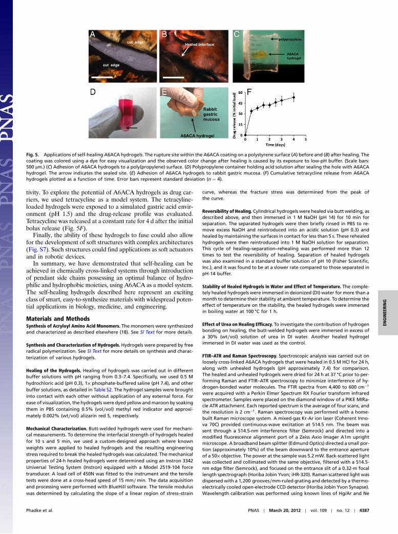
tivity. To explore the potential of A6ACA hydrogels as drug car-riers, we used tetracycline as a model system. The tetracyline-loaded hydrogels were exposed to a simulated gastric acid envir-onment (pH 1.5) and the drug-release profile was evaluated.Tetracycline was released at a constant rate for 4 d after the initialbolus release (Fig. 5F).
Finally, the ability of these hydrogels to fuse could also allowfor the development of soft structures with complex architectures(Fig. S7). Such structures could find applications as soft actuatorsand in robotic devices.
In summary, we have demonstrated that self-healing can beachieved in chemically cross-linked systems through introductionof pendant side chains possessing an optimal balance of hydro-philic and hydrophobic moieties, using A6ACA as a model system.The self-healing hydrogels described here represent an excitingclass of smart, easy-to-synthesize materials with widespread poten-tial applications in biology, medicine, and engineering.
Materials and MethodsSynthesis of Acryloyl Amino Acid Monomers. The monomers were synthesizedand characterized as described elsewhere (18). See SI Text for more details.
Synthesis and Characterization of Hydrogels. Hydrogels were prepared by freeradical polymerization. See SI Text for more details on synthesis and charac-terization of various hydrogels.
Healing of the Hydrogels. Healing of hydrogels was carried out in differentbuffer solutions with pH ranging from 0.3–7.4. Specifically, we used 0.5 Mhydrochloric acid (pH 0.3), 1× phosphate-buffered saline (pH 7.4), and otherbuffer solutions, as detailed in Table S2. The hydrogel samples were broughtinto contact with each other without application of any external force. Forease of visualization, the hydrogels were dyed yellow andmaroon by soakingthem in PBS containing 0.5% (vol∕vol) methyl red indicator and approxi-mately 0.002% (wt∕vol) alizarin red S, respectively.
Mechanical Characterization. Butt-welded hydrogels were used for mechani-cal measurements. To determine the interfacial strength of hydrogels healedfor 10 s and 5 min, we used a custom-designed approach where knownweights were applied to healed hydrogels and the resulting engineeringstress required to break the healed hydrogels was calculated. The mechanicalproperties of 24-h healed hydrogels were determined using an Instron 3342Universal Testing System (Instron) equipped with a Model 2519-104 forcetransducer. A load cell of 450N was fitted to the instrument and the tensiletests were done at a cross-head speed of 15 mm∕min. The data acquisitionand processing were performed with BlueHill software. The tensile moduluswas determined by calculating the slope of a linear region of stress–strain
curve, whereas the fracture stress was determined from the peak ofthe curve.
Reversibility of Healing. Cylindrical hydrogels were healed via butt welding, asdescribed above, and then immersed in 1 M NaOH (pH 14) for 10 min forseparation. The separated hydrogels were then briefly rinsed in PBS to re-move excess NaOH and reintroduced into an acidic solution (pH 0.3) andhealed bymaintaining the surfaces in contact for less than 5 s. These rehealedhydrogels were then reintroduced into 1 M NaOH solution for separation.This cycle of healing–separation–rehealing was performed more than 12times to test the reversibility of healing. Separation of healed hydrogelswas also examined in a standard buffer solution of pH 10 (Fisher Scientific,Inc.), and it was found to be at a slower rate compared to those separated inpH-14 buffer.
Stability of Healed Hydrogels in Water and Effect of Temperature. The comple-tely healed hydrogels were immersed in deionized (DI) water for more than amonth to determine their stability at ambient temperature. To determine theeffect of temperature on the stability, the healed hydrogels were immersedin boiling water at 100 °C for 1 h.
Effect of Urea on Healing Efficacy. To investigate the contribution of hydrogenbonding on healing, the butt-welded hydrogels were immersed in excess ofa 30% (wt∕vol) solution of urea in DI water. Another healed hydrogelimmersed in DI water was used as the control.
FTIR–ATR and Raman Spectroscopy. Spectroscopic analysis was carried out onloosely cross-linked A6ACA hydrogels that were healed in 0.5 M HCl for 24 h,along with unhealed hydrogels (pH approximately 7.4) for comparison.The healed and unhealed hydrogels were dried for 24 h at 37 °C prior to per-forming Raman and FTIR–ATR spectroscopy to minimize interference of hy-drogen-bonded water molecules. The FTIR spectra from 4,400 to 600 cm−1
were acquired with a Perkin Elmer Spectrum RX Fourier transform infraredspectrometer. Samples were placed on the diamond window of a PIKE MIRa-cle ATR attachment. Each reported spectrum is the average of four scans, andthe resolution is 2 cm−1. Raman spectroscopy was performed with a home-built Raman microscope system. A mixed-gas Kr-Ar ion laser (Coherent Inno-va 70C) provided continuous-wave excitation at 514.5 nm. The beam wassent through a 514.5-nm interference filter (Semrock) and directed into amodified fluorescence alignment port of a Zeiss Axio Imager A1m uprightmicroscope. A broadband beam splitter (Edmund Optics) directed a small por-tion (approximately 10%) of the beam downward to the entrance apertureof a 50× objective. The power at the sample was 5.2 mW. Back-scattered lightwas collected and collimated with the same objective, filtered with a 514.5-nm edge filter (Semrock), and focused on the entrance slit of a 0.32-m focallength spectrograph (Horiba Jobin Yvon; iHR-320). Raman scattered light wasdispersed with a 1;200 grooves∕mm-ruled grating and detected by a thermo-electrically cooled open-electrode CCD detector (Horiba Jobin Yvon Synapse).Wavelength calibration was performed using known lines of Hg/Ar and Ne
Fig. 5. Applications of self-healing A6ACA hydrogels. The rupture site within the A6ACA coating on a polystyrene surface (A) before and (B) after healing. Thecoating was colored using a dye for easy visualization and the observed color change after healing is caused by its exposure to low-pH buffer. (Scale bars:500 μm.) (C) Adhesion of A6ACA hydrogels to a poly(propylene) surface. (D) Polypropylene container holding acid solution after sealing the hole with A6ACAhydrogel. The arrow indicates the sealed site. (E) Adhesion of A6ACA hydrogels to rabbit gastric mucosa. (F) Cumulative tetracycline release from A6ACAhydrogels plotted as a function of time. Error bars represent standard deviation (n ¼ 4).
Phadke et al. PNAS ∣ March 20, 2012 ∣ vol. 109 ∣ no. 12 ∣ 4387
ENGINEE
RING

lamps for windows centered at 550 and 610 nm, respectively. See SI Text formore details.
Determination of Steric Feasibility of Configurations. We sought to determinewhether the face-on and interleaved configurations were sterically feasible.As a model system, we used five-unit oligomers of A6ACA (Fig. S4A). Twosuch oligomers were brought together and an energy minimization was per-formed using ChemBio3D Ultra 12.0 (CambridgeSoft) (Fig. S4B). Both face-onand interleaved species were observed in the resultant configurations(Fig. S4B), indicating that both types of configurations are sterically feasible.
Molecular Dynamics Simulations of Hydrogel Networks. To investigate theeffect of side-chain length on healing efficiency, we performed moleculardynamics simulations of hydrogel networks built from A6ACA, A8ACA, andA11AUA monomers having side chains of lengths 5, 7, and 10 CH2 groups,respectively. A nine-arm hydrogel motif was placed inside the simulationbox along with water molecules (Fig. 4B, Left) and replicated via periodicboundary conditions to yield the desired hydrogel network (Fig. 4B, Right).A gap in the �x direction prevented continuity of the network along the xdirection and allowed the creation of a hydrogel–water interface in betweenperiodic images of the network. The accessibility of the amide and carboxylgroups was quantified in terms of the number of hydrogen bonds they formwith the water molecules. We hypothesize that the accessibility of the func-tional groups for interacting with water molecules is a good measure fortheir accessibility for interacting with functional groups from the apposinghydrogel surface. See SI Text for more details.
A6ACA Hydrogels as Self-Healing Coating. A6ACA hydrogels were swollen in a0.01% solution of methyl red in PBS, to gain contrast between the coatingand the surface. Polystyrene surfaces were coated with the hydrogels by dry-ing at 37 °C for 12 h. A 300-μm-wide scratch was made in the coating surfaceusing a surgical scalpel and imaged using bright field microscopy (AxioObserver A1; Carl Zeiss). The scratch site was briefly hydrated for 60 s with50 μL DI water after which the excess water was removed and the site wastreated with 100 μL of 0.1 M HCl. The cut edges facing each other were thenreimaged after 5 min.
Adhesion of A6ACA Hydrogels to Plastics. A6ACA hydrogels were swollen inPBS for 4 h. The swollen hydrogel was found to adhere to polypropyleneand polystyrene surfaces within 15–20 s upon spraying with pH-0.3 solutionat the hydrogel-plastic interface.
A6ACA Hydrogels for Sealing Acid Leakages. The conical bottom portion of a2-mL centrifuge tube (Eppendorf) was cut out to create a hole, measuringapproximately 1 cm in diameter. The hole was plugged using PBS-swollenA6ACA hydrogels. This sealed conical tube was then filled with 1 mL of
0.5 M HCl (with 0.5% added methyl red, to make the solution pink for easeof visualization) and photographed to show lack of any leak.
A6ACA Hydrogels as Mucoadhesive Polymer. Stomach tissues were resectedfrom freshly killed New Zealand white rabbits and carefully rinsed withPBS to remove residual food material. After cleaning, the tissues were main-tained in PBS and used for the experiments the same day. To investigate mu-coadhesiveness of A6ACA hydrogels, hydrogels were first maintained incontact with inner gastric lining under immersion in simulated gastric acid[HCl-KCl buffer of pH 1.5 containing 54.7% (by volume) 0.2 M KCl and45.3% 0.2 M HCl] for 20 min and then photographed.
A6ACA Hydrogel as a Drug Carrier. A solution containing 0.5 mg∕mL tetracy-cline (50×) in PBS was prepared from a stock solution of tetracycline (1,000×,10 mg∕mL in 70% ethanol). As-synthesized A6ACA hydrogels (n ¼ 4) wereloaded with tetracycline by placing them in this solution for 24 h. Basedon the known swelling ratio of A6ACA hydrogels in PBS, the total tetracyclineload was calculated for each hydrogel. The hydrogels were then immersed in40 mL of simulated gastric fluid (pH 1.5) and placed on a shaker at 150 rpm.Every 12 h, 4 mL of the immersion solutions were collected and replaced with4 mL of fresh buffer. The released tetracycline in the collected solutions wasmeasured spectrophotometrically at 270 nm. The total tetracycline release(expressed as percentage of total tetracycline load, calculated from swellingratio of hydrogels) was calculated for each time point and averaged acrossthe replicates.
Synthesis of Complex Structures Using Healing Ability of A6ACA Hydrogels.Cylindrical A6ACA hydrogels were swollen in PBS containing 0.5% methylred or approximately 0.002% alizarin red S, respectively. Using the yellowpieces (swollen in methyl red), hydrogels were healed to form the letter“U” with 0.5 mL HCl. Following this, the healed U was separated into thedifferent pieces using 1 N NaOH. These pieces were then rehealed to formthe letter “S.” Using the pieces swollen in the alizarin red S (appearing mar-oon in color), a similar procedure was carried to form the letters “C” and “D.”A combination of yellow- and maroon-dyed pieces was also healed to form ahumanoid figure.
ACKNOWLEDGMENTS. The authors gratefully acknowledge Dr. Robert Pomer-oy for assistance with FTIR–ATR analysis; Dr. Mark A Meyers, Chung-Ting Wei,Marco Maruggi, and Rui Yang for their help with mechanical measurements;Dr. Srinivas Alla for discussions on molecular modeling; Dr. Koichi Masuda forproviding the rabbit stomachs; and Mr. Anthony Mrse for his assistance with13C NMR data acquisition. A.P. acknowledges financial assistance from theJacobs Fellowship at the University of California, San Diego. We also thankthe California Institute of Regenerative Medicine (RN2-00945) for financialsupport.
1. Varghese S, Lele AK, Mashelkar RA (2000) Designing new thermoreversible gels by mo-lecular tailoring of hydrophilic-hydrophobic interactions. J Chem Phys 112:3063–3070.
2. Varghese S, Lele AK, Srinivas D, Sastry M, Mashelkar RA (2001) Novel macroscopicself-organization in polymer gels. Adv Mater 13:1544–1548.
3. Varghese S, Lele AK, Mashelkar R (2006) Metal-ion-medaited healing of gels. J PolymSci A1 44:666–670.
4. Wojtecki RJ, Meador MA, Rowan SJ (2011) Using the dynamic bond to accessmacroscopically responsive structurally dynamic polymers. Nat Mater 10:14–27.
5. Hager MD, Greil P, Leyens C, van der Zwaag S, Schubert US (2010) Self-healing materi-als. Adv Mater 22:5424–5430.
6. Messersmith PB, Lee H, Dellatore SM, Miller WM (2007) Mussel-inspired surfacechemistry for multifunctional coatings. Science 318:426–430.
7. de Gennes PG (1971) Reptation of polymer chain in the presence of fixed obstacles.J Chem Phys 55:572–579.
8. Cordier P, Tournilhac F, Soulie-Ziakovic C, Leibler L (2008) Self-healing and thermore-versible rubber from supramolecular assembly. Nature 451:977–980.
9. Burnworth M, et al. (2011) Optically healable supramolecular polymers. Nature472:334–337.
10. Wang Q, et al. (2010) High-water-content mouldable hydrogels by mixing clay and adendritic molecular binder. Nature 463:339–343.
11. Lee BP, Messersmith PB, Israelachvili JN, Waite JH (2011) Mussel inspired wet adhesivesand coatings. Annu Rev Mater Res 41:99–132.
12. Holten-Andersen N, et al. (2011) pH-induced metal-ligand cross-links inspired bymussel yield self-healing polymer networks with near-covalent elastic moduli. ProcNatl Acad Sci USA 108:2651–2655.
13. Cho SH, Braun PV, White SR (2009) Self-healing polymer coatings. Adv Mater21:645–649.
14. Toohey KS, Sottos NR, Lewis JA, Moore JS, White SR (2007) Self-healing materials withmicrovascular networks. Nat Mater 6:581–585.
15. Balazs AC, et al. (2010) Using nanoparticle-filled microcapsules for site-specific healingof damaged substrates: Creating a “repair-and-go” system. ACS Nano 4:1115–1123.
16. Chen X, et al. (2002) A thermally re-mendable cross-linked polymeric material. Science295:1698–1702.
17. Ghosh B, Urban MW (2009) Self-repairing oxetane-substituted chitosan polyurethanenetworks. Science 323:1458–1460.
18. Ayala R, et al. (2011) Engineering the cell-material interface for controlling stem celladhesion, migration, and differentiation. Biomaterials 32:3700–3711.
19. McQueen-Mason S, Cosgrove DJ (1994) Disruption of hydrogen-bonding betweenplant-cell wall polymers by proteins that induce wall extension. Proc Natl Acad SciUSA 91:6574–6578.
20. Colthup NBDL, Wiberley SE (1975) Introduction to Infrared and Raman Spectroscopy(Academic, New York), pp 289–325.
21. Barth A, Zscherp C (2002) What vibrations tell us about proteins. Q Rev Biophys35:369–430.
22. Dong J, Ozaki Y, Nakashima K (1997) Infrared, Raman, and near-infrared spectroscopicevidence for the coexistence of various hydrogen-bond forms in poly(acrylic acid).Macromolecules 30:1111–1117.
23. Phadke A, Zhang C, Hwang Y, Vecchio K, Varghese S (2010) Templated mineralizationof synthetic hydrogels for bone-like composite materials: Role of matrix hydro-phobicity. Biomacromolecules 11:2060–2068.
24. Lide DR (2009) Handbook of Chemistry and Physics (CRC, Boca Raton, FL).
4388 ∣ www.pnas.org/cgi/doi/10.1073/pnas.1201122109 Phadke et al.