Rapid assay of stem cell functionality and potency using ...
11
METHOD Open Access Rapid assay of stem cell functionality and potency using electric cell-substrate impedance sensing Michael J. Rutten 1* , Bryan Laraway 1 , Cynthia R. Gregory 1,2,3 , Hua Xie 1 , Christian Renken 4 , Charles Keese 4 and Kenton W. Gregory 1,5 Abstract Regenerative medicine studies using autologous bone marrow mononuclear cells (BM-MNCs) have shown improved clinical outcomes that correlate to in vitro BM-MNC invasive capacity. The current Boyden-chamber assay for testing invasive capacity is labor-intensive, provides only a single time point, and takes 36 hours to collect data and results, which is not practical from a clinical cell delivery perspective. To develop a rapid, sensitive and reproducible invasion assay, we employed Electric Cell-substrate Impedance Sensing (ECIS) technology. Chemokine-directed BM-MNC cell invasion across a Matrigel-coated Transwell filter was measurable within minutes using the ECIS system we developed. This ECIS-Transwell chamber system provides a rapid and sensitive test of stem and progenitor cell invasive capacity for evaluation of stem cell functionality to provide timely clinical data for selection of patients likely to realize clinical benefit in regenerative medicine treatments. This device could also supply robust unambiguous, reproducible and cost effective data as a potency assay for cell product release and regulatory strategies. Introduction Measurement of a stem, progenitor, or stromal cell prepa- ration’ s potency or functionality is important to the characterization of a potential cell therapy product [1]. Ideally, the assessment of a cell product’ s potency is based on a relevant cell function for the desired clinical outcome [2]. While valuable, assessments of cell phenotype (i.e., surface marker expression), viability, and colony growth are not considered adequate functionality tests for cells being studied in clinical applications because they do not reliably predict clinical responses to cell treatments [1–4]. For regenerative therapies, the therapeutic cell’ s ability to invade injured tissue in response to a chemo- tactic gradient is considered to be a critical cell func- tion for the desired clinical outcome [5–8]. To assess the potential in vivo invasive capacity of a stem-cell preparation, an in vitro Transwell invasion assay is typ- ically performed [9–12]. This assay is based upon the Boyden chamber, which is separated into upper and lower chambers by a Matrigel matrix-coated porous filter. The progenitor or stem cells are added to the top chamber and a chemoattractant agent is added to the bottom chamber to induce the cells to invade the Matrigel matrix and migrate through the porous filter to the bottom chamber. Eighteen to 24 hours later, the number of cells that have migrated to the underside of the filter or to the floor of the bottom chamber is quantified by 4′,6-diamidino-2- phenylindole (DAPI) staining and then counting the migrated cells’ nuclei [13]. Transwell assay measurement of bone marrow mononuclear cell (BM-MNC) invasion in response to stromal cell-derived factor-1 (SDF-1) was found to be the only in vitro assessment of BM-MNC preparations that demonstrated a positive correlation to the clinical outcome of patients treated with BM- MNCs for heart repair [14, 15]. The SDF-1 Transwell invasion assay has also been used for testing the invasive function of other progenitor cell types such as mesenchy- mal stromal cells (MSCs) [16–18], endothelial progenitor cells (EPCs) [19–21], and peripheral blood mononuclear cells (PB-MNCs) [22–24]. While the standard Transwell * Correspondence: [email protected] 1 Center for Regenerative Medicine, Oregon Health & Science University, 3181 SW Sam Jackson Park Road, 97239 Portland, OR, USA Full list of author information is available at the end of the article © 2015 Rutten et al. Open Access This article is distributed under the terms of the Creative Commons Attribution 4.0 International License (http://creativecommons.org/licenses/by/4.0/), which permits unrestricted use, distribution, and reproduction in any medium, provided you give appropriate credit to the original author(s) and the source, provide a link to the Creative Commons license, and indicate if changes were made. The Creative Commons Public Domain Dedication waiver (http://creativecommons.org/publicdomain/zero/1.0/) applies to the data made available in this article, unless otherwise stated. Rutten et al. Stem Cell Research & Therapy (2015) 6:192 DOI 10.1186/s13287-015-0182-2
Transcript of Rapid assay of stem cell functionality and potency using ...

METHOD Open Access
Rapid assay of stem cell functionality andpotency using electric cell-substrateimpedance sensingMichael J. Rutten1*, Bryan Laraway1, Cynthia R. Gregory1,2,3, Hua Xie1, Christian Renken4, Charles Keese4
and Kenton W. Gregory1,5
Abstract
Regenerative medicine studies using autologous bone marrow mononuclear cells (BM-MNCs) have shownimproved clinical outcomes that correlate to in vitro BM-MNC invasive capacity. The current Boyden-chamber assayfor testing invasive capacity is labor-intensive, provides only a single time point, and takes 36 hours to collect dataand results, which is not practical from a clinical cell delivery perspective. To develop a rapid, sensitive andreproducible invasion assay, we employed Electric Cell-substrate Impedance Sensing (ECIS) technology.Chemokine-directed BM-MNC cell invasion across a Matrigel-coated Transwell filter was measurable withinminutes using the ECIS system we developed. This ECIS-Transwell chamber system provides a rapid and sensitivetest of stem and progenitor cell invasive capacity for evaluation of stem cell functionality to provide timelyclinical data for selection of patients likely to realize clinical benefit in regenerative medicine treatments. Thisdevice could also supply robust unambiguous, reproducible and cost effective data as a potency assay for cellproduct release and regulatory strategies.
IntroductionMeasurement of a stem, progenitor, or stromal cell prepa-ration’s potency or functionality is important to thecharacterization of a potential cell therapy product [1].Ideally, the assessment of a cell product’s potency is basedon a relevant cell function for the desired clinical outcome[2]. While valuable, assessments of cell phenotype (i.e.,surface marker expression), viability, and colony growthare not considered adequate functionality tests for cellsbeing studied in clinical applications because they donot reliably predict clinical responses to cell treatments[1–4]. For regenerative therapies, the therapeutic cell’sability to invade injured tissue in response to a chemo-tactic gradient is considered to be a critical cell func-tion for the desired clinical outcome [5–8]. To assessthe potential in vivo invasive capacity of a stem-cellpreparation, an in vitro Transwell invasion assay is typ-ically performed [9–12]. This assay is based upon the
Boyden chamber, which is separated into upper and lowerchambers by a Matrigel matrix-coated porous filter. Theprogenitor or stem cells are added to the top chamber anda chemoattractant agent is added to the bottom chamberto induce the cells to invade the Matrigel matrix andmigrate through the porous filter to the bottom chamber.Eighteen to 24 hours later, the number of cells that havemigrated to the underside of the filter or to the floor ofthe bottom chamber is quantified by 4′,6-diamidino-2-phenylindole (DAPI) staining and then counting themigrated cells’ nuclei [13]. Transwell assay measurementof bone marrow mononuclear cell (BM-MNC) invasion inresponse to stromal cell-derived factor-1 (SDF-1) wasfound to be the only in vitro assessment of BM-MNCpreparations that demonstrated a positive correlation tothe clinical outcome of patients treated with BM-MNCs for heart repair [14, 15]. The SDF-1 Transwellinvasion assay has also been used for testing the invasivefunction of other progenitor cell types such as mesenchy-mal stromal cells (MSCs) [16–18], endothelial progenitorcells (EPCs) [19–21], and peripheral blood mononuclearcells (PB-MNCs) [22–24]. While the standard Transwell
* Correspondence: [email protected] for Regenerative Medicine, Oregon Health & Science University, 3181SW Sam Jackson Park Road, 97239 Portland, OR, USAFull list of author information is available at the end of the article
© 2015 Rutten et al. Open Access This article is distributed under the terms of the Creative Commons Attribution 4.0International License (http://creativecommons.org/licenses/by/4.0/), which permits unrestricted use, distribution, andreproduction in any medium, provided you give appropriate credit to the original author(s) and the source, provide a link tothe Creative Commons license, and indicate if changes were made. The Creative Commons Public Domain Dedication waiver(http://creativecommons.org/publicdomain/zero/1.0/) applies to the data made available in this article, unless otherwise stated.
Rutten et al. Stem Cell Research & Therapy (2015) 6:192 DOI 10.1186/s13287-015-0182-2

invasion assay has been found to provide clinically im-portant data on the functional capacity of stem cellpreparations, limitations to the assay include the timerequired for measurable migration of cells, labor-intensive methods required for quantifying the invasivecells, investigator inter-assay variability, and measure-ment of migration (a dynamic process) at only a single(for example, 18–24 hour) time point [25, 26]. Forautologous bone marrow cell therapy, the largest limi-tation of present cell function assays is that the resultsare not available until about 36 hours after the bonemarrow harvest. Since many clinical applications ofautologous bone marrow stem and progenitor cells in-volve the cells being administered within a few hours ofthe bone marrow harvest, it is not then possible toidentify, prospectively, stem cell preparations with poorfunctional capacity. For clinical trials designed to deter-mine the therapeutic potential of a stem cell therapy,the inclusion of suboptimal cell preparations reducesthe statistical power of the study, obscuring the poten-tial benefit of the therapy under assessment. Import-antly, whether as part of a clinical trial or an acceptedtreatment protocol, administration of suboptimal cellpreparations can result in patients being treated without ahigh likelihood of clinical benefit. This assay also ad-dresses the need of the Food and Drug Administration(FDA) and other regulatory organizations for a reliable,low-cost, rapid assay of cell functionality as a cell potencytest.Many patients have preexisting clinical conditions
that can impact the functionality of their stem cells. Forexample, it is well documented that diabetes can impairBM-MNC functionality [27–30], but whether such anexisting clinical condition has impacted a patient’s stemcell functionality to a degree that the patient shouldnot undergo cell administration is presently difficult toassess in the hours between autologous stem cell har-vest and administration. Another circumstance where aquick and sensitive cell migration assay for measuringcell functionality would be helpful is in the testing ofstem cells from patient blood or bone marrow before andafter radiotherapy or chemotherapy treatment [31–33].Some of the undesired side effects from radiation therapy,chemotherapy, or treatment with bone marrow suppres-sive drugs are the reduction of peripheral blood stem cellviability and function [34]. In this regard, a cell potency in-vasion assay to measure the functionality of peripheralblood cells would be important in assessing the potentialtoxic effects of radiation therapy and chemotherapy.With the continued development of cell biosensor de-
tection methods, traditional methods, such as the Boy-den chamber for studying cell invasion, are beingupdated with newer analytical tools [35–38]. A cell inva-sion assay involves the cells first degrading an extracellular
matrix barrier or cell monolayer, followed by the movementof the cells through the porous filter in response to the che-mokine gradient [25, 39, 40]. In these studies, electric cell-substrate impedance sensing (ECIS), previously used to de-tect the invasion of cells through a cell monolayer growndirectly on an electrode array [41] or on a porous filter [42],is used to detect the invasion of cells through an extracellu-lar matrix barrier on a porous filter.The goal of this study was to adapt the standard
Transwell assay to a stem cell invasion assay using ECIStechnology for use as a rapid, reliable stem cell func-tionality or potency assay. The objective of the study wasto automate the measurement of cell invasion using resist-ance and impedance measurements that could detect SDF-1-directed cell invasion in minutes rather than 36 hours.We also sought to demonstrate early proof-of-principle re-sults showing that sublethal, deleterious effects of cell func-tionality could be detected as a consequence of exposureof stem cells to common doses of radiation cancer therapyregimens. Here, we show that by translating the standardTranswell assay to an assay using ECIS technology, onecan measure within minutes BM-MNC invasion in re-sponse to SDF-1. The BM-MNC invasion is dependent onspecific signaling by SDF-1; BM-MNCs pretreated withSDF-1 or AMD3100 (a SDF-1 receptor blocker) do not in-vade the Matrigel matrix. We also demonstrate that ECISmeasurement of SDF-1 stimulated PB-MNC invasion andthat radiation-exposure damage of the PB-MNCs reducestheir invasion. The results from our experiments demon-strate that the ECIS Transwell device and chamber pro-vides a rapid, sensitive, and reproducible test of BM-MNCand PB-MNC invasion capacity, making it a potential diag-nostic tool for testing stem cell functionality in regenerativemedicine studies.
MethodsBone-marrow harvest and purificationpt?>All bone marrow samples were collected from 12-month-old male domestic Sinclair miniswine (SinclairBio-resources, Columbia, MO, USA). All animal hand-ling and care procedures were performed strictly inaccordance with the 2004 National Research Council“Guide for the Care and Use of Laboratory Animals” fol-lowing protocol approval by the Institutional AnimalCare and Use Committee (IACUC) of the Legacy Clin-ical Research and Technology Center, Legacy HealthSystem, Portland, OR, USA. Under local anesthesia,40 ml porcine bone marrow was aspirated from eitherthe donor’s tibia or sternum into a syringe containing5 ml heparin (1000 USP units/ml). The bone marrowwas transferred into a 150 ml transfer bag (Baxter, Deer-field, IL, USA) containing 8 ml citrate–phosphate dextran(Sigma, St. Louis, MO, USA), and the bone marrow trans-fer bag was connected through a 40 μm Pall blood
Rutten et al. Stem Cell Research & Therapy (2015) 6:192 Page 2 of 11
transfusion filter (Fisher Sci., 300 Industry Drive, Pitts-burgh, PA, USA) to a SEPAX cartridge kit (#CS-900; Bio-safe America, Houston, TX, USA). This kit contained awash-buffer bag that was filled with Hanks’ balanced saltsolution containing cations (HBSS; Invitrogen, 3175 StaleyRoad, Grand Island, NY, USA) and a density gradient so-lution/waste bag that was filled with 100 ml Ficoll-PaquePremium-1077 (GE Health Care, Pittsburg, PA, USA). A150 ml transfer bag (Baxter Health Care, One BaxterParkway, Deerfield, IL, USA) was connected to receive thepurified BM-MNCs. The completed kit was then placedinto a SEPAX-2 (Biosafe America) automated cell-processing device to process the bone marrow [29]. Thefinal purified BM-MNC product was collected in HBSSand the BM-MNCs were counted with a Beckman Z2-Coulter Counter (Beckman Coulter, Brea, CA, USA).
Isolation of porcine PB-MNCsPorcine peripheral blood was collected from the fem-oral artery of domestic Yorkshire swine and diluted 1:2with HBSS (#14025-092; Gibco/Invitrogen, 3175 StaleyRoad, Grand Island, NY, USA). PB-MNCs were isolatedby density gradient centrifugation. Specifically, a 4 mlaliquot of diluted blood was layered on top of 3 mlFicoll-Paque Premium, density 1.077 (#17-5442-02; GEHealthcare, Pittsburgh, PA, USA) in a 15 ml centrifugetube, and the tubes were centrifuged (400 × g, 40 mi-nutes, room temperature, no brake). The recovered PB-MNCs were washed twice with HBSS (300 × g, 10 mi-nutes, room temperature). After washing, the cells wereresuspended in X-VIVO 15 media (#04-744Q; Lonza,Walkersville, MD USA), and an aliquot was taken for cellcounting and viability assessment.
Jurkat cell cultureHuman Jurkat T-cell line (JJK subclone) cells weregrown in RPMI-1640 media (#15-10.1186/s13287-015-0182-2040-CV; Corning CellGro, Mediatech, Inc., Ma-nassas, VA) supplemented with penicillin–streptomycin(#15140-122; Gibco/Invitrogen, 3175 Staley Road, GrandIsland, NY, USA), l-glutamine (#25030-081; Gibco/Invi-trogen, 3175 Staley Road, Grand Island, NY, USA), and10 % fetal bovine serum (Premium FBS, #14-501 F;Lonza, Walkersville, MD USA). The Jurkat cells werecultured in a 37°C carbon dioxide (CO2) incubator, andwere split 1:5 when the cells reached a concentration of1 × 106 cells/ml. The cells for this study were split nomore than 25 times relative to the original stock of cells.
Radiation of PB-MNCsTo show that our stem cell functionality assay could rap-idly detect nonlethal deleterious changes in cell functionin previously functioning cells, we exposed the cells to aradiation dose comparable with commonly prescribed
doses for cancer radiotherapy in humans. The PB-MNCsin X-VIVO 15 media received 0 Gy or 2.15 Gy of X-ray ir-radiation at room temperature (1.365 Gy/minute, RS2000Biological Research Irradiator; Rad Source, Suwanee, GA,USA). The dose of 2.15 Gy was chosen for the swine cellssince it is comparable with a moderate radiation exposureof human cells [43].After irradiation of the PB-MNCS the cells were not
washed, because it has been shown previously withother cell types that there is no difference in the rate ofapoptosis if the cells are kept in the original irradiatedmedium or switched to fresh medium [44]. Also, anadditional wash (centrifugation) step has the potentialto produce additional damage to cells [45], whichwould complicate the interpretation of the effects ofradiation alone on the invasion assay results. Both theirradiated cells and the nonirradiated cells were then cul-tured in hydrophobic dishes (Nunc Hydrocell, #174912;ThermoFisher Sci., Waltham, MA, USA) for 24 hoursin X-VIVO 15 media (37 °C, 5 % CO2), after which theywere centrifuged (300 × g, 1 minute, room temperature)and resuspended in fresh X-VIVO 15 media. The finalcell concentration was adjusted to 1.2 × 10⁶ cells/300 μlX-VIVO 15 media for the ECIS Transwell invasionassay.
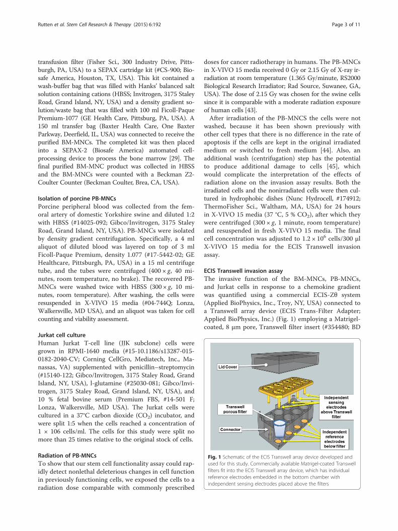
ECIS Transwell invasion assayThe invasive function of the BM-MNCs, PB-MNCs,and Jurkat cells in response to a chemokine gradientwas quantified using a commercial ECIS-Zθ system(Applied BioPhysics, Inc., Troy, NY, USA) connected toa Transwell array device (ECIS Trans-Filter Adapter;Applied BioPhysics, Inc.) (Fig. 1) employing a Matrigel-coated, 8 μm pore, Transwell filter insert (#354480; BD
Fig. 1 Schematic of the ECIS Transwell array device developed andused for this study. Commercially available Matrigel-coated Transwellfilters fit into the ECIS Transwell array device, which has individualreference electrodes embedded in the bottom chamber withindependent sensing electrodes placed above the filters
Rutten et al. Stem Cell Research & Therapy (2015) 6:192 Page 3 of 11

Biocoat, Fisher, Pittsburg, PA, USA). To minimize anyinter-assay variation, the Transwell array station andtransfilter adaptor were always prewarmed in a humidi-fied 5 % CO2 incubator for 2 hours, and then warm(37 °C) media were always used for resuspending thecells and chemokines. BM-MNCs, Jurkat cells, or PB-MNCs (1.2 × 10⁶ cells) were brought to a final volume of300 μl in X-VIVO 15 media and added to the chamberabove the Matrigel-coated Transwell filter insert. For ex-periments measuring BM-MNC or Jurkat cell invasion,SDF-1 (#350-NS; R&D Systems, Minneapolis, MN, USA)in X-VIVO 15 media (100 ng/ml in 625 μl medium) wasadded to the chamber below the Matrigel-coated Trans-well filter. For the PB-MNC ECIS Transwell studies, SDF-1 (100 ng/ml) and MIP-1 (100ng/ml; PeproTech, RockyHill, NJ, USA) were used in combination because bothchemoattractants have been shown to be involved in PB-MNC migration [46, 47]. After the addition of the re-spective cells and chemokines, the ECIS Transwell de-vice was placed in an incubator (37 °C, 5 % CO2) for2 hours, during which time the impedance changes wererecorded.The migratory action of SDF-1 is the result of SDF-1
binding to the receptor C-X-C chemokine receptor type4 (CXCR4) [48, 49]. For some control experiments, theBM-MNCs were pretreated for 30 minutes with 5 μg/mlAMD3100 (an inhibitor of the SDF-1 receptor, CXCR4[50, 51]), and then the cells were added to the top of theECIS Transwell chamber and 100 ng/ml SDF-1 wasadded to the bottom chamber. In other control experi-ments, to distinguish between a directed chemotaxis ver-sus a random chemokinesis response, 100 ng/ml SDF-1was added with the cells to the top of the ECIS Transwellchamber and medium alone was added to the bottomchamber.
ECIS data analysis and statisticsInitial data analysis was performed using ECIS Software(version 1.2.123; Applied BioPhysics, Inc.). The actualfilter resistance of each test or control well was calcu-lated by subtracting from each the resistance of a blankTranswell filter without cells:
actual filter resistance¼ test well resistanceð Þ – blank filter resistanceð Þ
The actual filter resistance values for replicate wellswere then averaged and plotted as the mean ± standarderror of the mean (SEM) over the 2-hour time course.Additional analysis was done by transferring the data toan Excel 2011 spreadsheet (Microsoft, Redmond, WA,USA) where the absolute relative change in resistance wascalculated at specific times by subtracting the initial
baseline resistance at time zero from its respective actualfilter resistance value:
absolute relative resistance¼ actual filter resistanceð Þ – baseline resistanceð Þ
The absolute relative resistance was plotted as the mean± SEM [52]. Significant differences between the ECIS re-sistance changes of control and chemokine-treated groupswere calculated using a one-way analysis of variance testand a probability of p <0.05. Graphs were plotted as themean ± SEM using SigmaPlot-11 (Systat Software, Inc.,Chicago, IL, USA). The use of “n” in our study was equalto the number of individual animals used for the isolationof the BM-MNCs and PB-MNCs, which was 4 and 2, re-spectively. For the Jurkat cells, “n” in our study was equalto the number of separate individual cell platings.
ResultsECIS measurement of chemotactic cell invasionSince the invasion of human cells from the Jurkat T-cell line to SDF-1 has been well characterized in stand-ard Boyden chamber migration assays [26, 53–55], weused Jurkat T cells to characterize our ECIS invasionassay. The assays employed an ECIS Transwell devicedeveloped for this study (Fig. 1). The ECIS Transwelldevice holds a standard Matrigel-coated Transwell filter,typically used for invasion assays. Jurkat T cells wereplaced in the Transwell top chambers and the chemokineSDF-1 was added to the bottom chambers. We found asignificant increase in SDF-1-stimulated Jurkat cell inva-sion of the Matrigel matrix measured by ECIS as increasedresistance (Fig. 2). Jurkat cells placed in the top half of achamber without SDF-1 in the bottom half of the chamberproduced only a slight increase in filter resistance that sta-bilized within 45–60 minutes and did not increase furtherover the remaining 2-hour time course (Fig. 2a). The ECISsystem continuously measures the resistance across theTranswell membrane over time. When the absolute rela-tive changes in filter resistance from Jurkat invasion inchambers with and without SDF-1 in the bottom half ofthe chamber (test and control chambers, respectively)were plotted over time, we found that the change in filterresistance for chambers with SDF-1 versus without SDF-1became significantly different (p <0.05) within 10 minutesafter starting the assay (i.e., after the addition of SDF-1),and that the difference increased over the 2-hour observa-tion period (Fig. 2b).For traditional Boyden chamber assays, cell migration or
invasion across porous filters can be classified as either arandom motion event in the absence of a chemokine gra-dient (i.e., chemokinesis) or directed migration in responseto a chemokine gradient (i.e., chemotaxis) [26, 56–58]. Forour assay we wanted to determine whether the SDF-1
Rutten et al. Stem Cell Research & Therapy (2015) 6:192 Page 4 of 11

change in filter resistance correlated to a directed chemo-tactic response. SDF-1 was added along with the Jurkat Tcells to the top chamber and not to the bottom chamberof Transwells. As would be anticipated for a chemotacticresponse, there was no measurable increase in Transwellfilter resistance when SDF-1 was added to the same cham-ber as the Jurkat T cells (i.e., the upper chamber) (Fig. 3),which is in contrast to the increased resistance measured
when Jurkat T cells are added to the top chamber andSDF-1 is added to the bottom chamber of the Transwell(Fig. 2).SDF-1 stimulates cell invasion and migration as a result
of it binding to the receptor CXCR4 [48, 49]. For somecontrol experiments, the BM-MNCs were pretreated for30 minutes with 5 μg/ml AMD3100 (an inhibitor of theSDF-1 receptor, CXCR4 [50, 51]), and then the cells were
Fig. 2 ECIS measurement of increased Transwell filter resistance resulting from SDF-1 chemokine-dependent Jurkat T-cell invasion of Matrigel-coatedTranswell filters. Jurkat T cells were placed in the upper chambers of Matrigel-coated Transwells with or without addition of 100 ng/ml SDF-1 to thelower chambers. a Transwell filter resistance is measured continuously over time by ECIS. Combined resistance tracings of six separate experiments areshown for Transwells with and without the addition of SDF-1 to the lower chamber, demonstrating the increased resistance in Transwells where theJurkat T cells invade the Matrigel in response to SDF-1. Arrow: time point of SDF-1 addition to test lower chambers. Mean ± SEM. b The absolutechange in ECIS Transwell filter resistance, relative to the resistance measured at the zero time point, increases over 2 hours due to Jurkat T-cell Matrigelinvasion in response to SDF-1. Each bar represents the mean ± SEM of six separate experiments (n = 6) performed in duplicate. *p <0.05. SDF-1 stromalcell derived factor-1
Fig. 3 Increased ECIS Transwell filter resistance is due to chemotaxis, not chemokinesis, and is specific to the chemokine (SDF-1). In these experiments,Jurkat T-cells were used without pretreatment or were pretreated with either 100 ng/ml SDF-1 or AMD3100 (an inhibitor of the SDF-1 receptor). Resistancewas measured in wells with Jurkat T cells (without or with pretreatment) added to top chambers with and without SDF-1 in the bottom chambers. Arrow:time point of SDF-1 addition to test lower chambers. Each tracing represents the mean ± SEM of two separate experiments (n= 2) performed in duplicate.SDF-1 stromal cell derived factor-1
Rutten et al. Stem Cell Research & Therapy (2015) 6:192 Page 5 of 11
added to the top of the ECIS Transwell chamber and100 ng/ml SDF-1 were added to the bottom chamber. In aconventional Boyden chamber assay, AMD3100 treatmentof the test cells inhibits their SDF-1-directed chemotaxis[54, 56, 59–61]. As shown in Fig. 3, there was no increasein filter resistance when the AMD3100 pretreated cellswere added to the top chamber of the Transwell withSDF-1 in the lower chamber, demonstrating the SDF-1specificity of the ECIS-measured chemotactic invasiveJurkat T-cell function. These results demonstrate that theprimary mechanism for the ECIS-measured change in theTranswell filter resistance is an SDF-1-specific chemotac-tic cell invasion response.
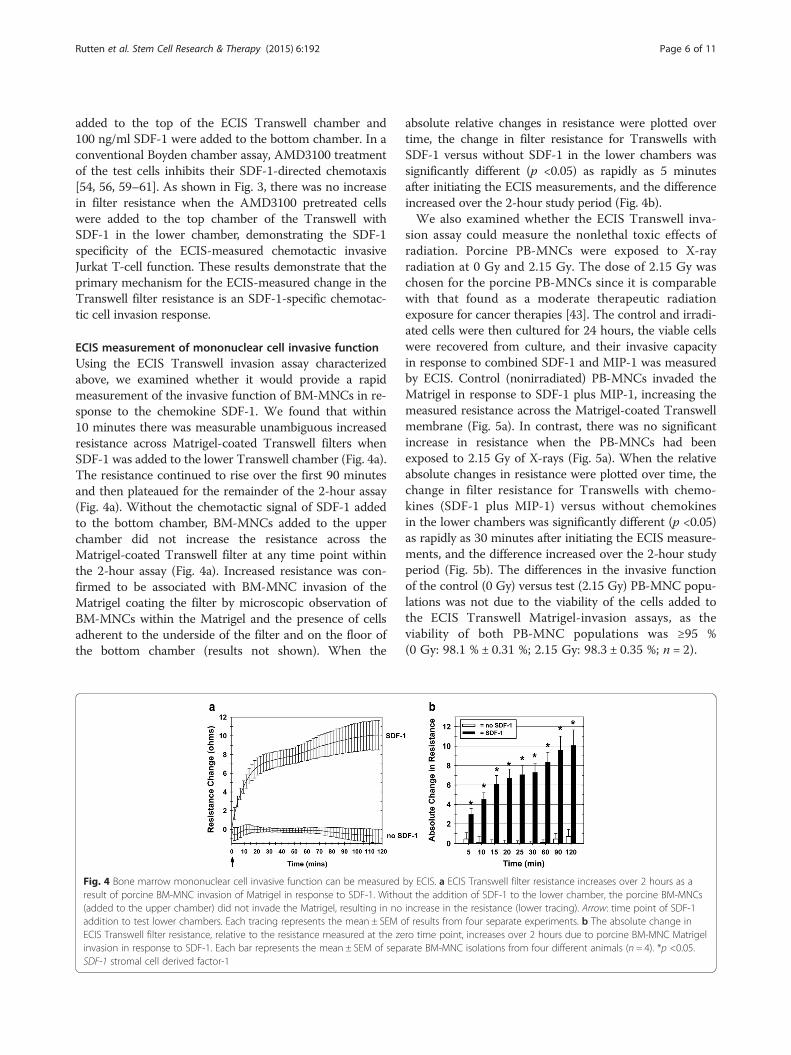
ECIS measurement of mononuclear cell invasive functionUsing the ECIS Transwell invasion assay characterizedabove, we examined whether it would provide a rapidmeasurement of the invasive function of BM-MNCs in re-sponse to the chemokine SDF-1. We found that within10 minutes there was measurable unambiguous increasedresistance across Matrigel-coated Transwell filters whenSDF-1 was added to the lower Transwell chamber (Fig. 4a).The resistance continued to rise over the first 90 minutesand then plateaued for the remainder of the 2-hour assay(Fig. 4a). Without the chemotactic signal of SDF-1 addedto the bottom chamber, BM-MNCs added to the upperchamber did not increase the resistance across theMatrigel-coated Transwell filter at any time point withinthe 2-hour assay (Fig. 4a). Increased resistance was con-firmed to be associated with BM-MNC invasion of theMatrigel coating the filter by microscopic observation ofBM-MNCs within the Matrigel and the presence of cellsadherent to the underside of the filter and on the floor ofthe bottom chamber (results not shown). When the
absolute relative changes in resistance were plotted overtime, the change in filter resistance for Transwells withSDF-1 versus without SDF-1 in the lower chambers wassignificantly different (p <0.05) as rapidly as 5 minutesafter initiating the ECIS measurements, and the differenceincreased over the 2-hour study period (Fig. 4b).We also examined whether the ECIS Transwell inva-
sion assay could measure the nonlethal toxic effects ofradiation. Porcine PB-MNCs were exposed to X-rayradiation at 0 Gy and 2.15 Gy. The dose of 2.15 Gy waschosen for the porcine PB-MNCs since it is comparablewith that found as a moderate therapeutic radiationexposure for cancer therapies [43]. The control and irradi-ated cells were then cultured for 24 hours, the viable cellswere recovered from culture, and their invasive capacityin response to combined SDF-1 and MIP-1 was measuredby ECIS. Control (nonirradiated) PB-MNCs invaded theMatrigel in response to SDF-1 plus MIP-1, increasing themeasured resistance across the Matrigel-coated Transwellmembrane (Fig. 5a). In contrast, there was no significantincrease in resistance when the PB-MNCs had beenexposed to 2.15 Gy of X-rays (Fig. 5a). When the relativeabsolute changes in resistance were plotted over time, thechange in filter resistance for Transwells with chemo-kines (SDF-1 plus MIP-1) versus without chemokinesin the lower chambers was significantly different (p <0.05)as rapidly as 30 minutes after initiating the ECIS measure-ments, and the difference increased over the 2-hour studyperiod (Fig. 5b). The differences in the invasive functionof the control (0 Gy) versus test (2.15 Gy) PB-MNC popu-lations was not due to the viability of the cells added tothe ECIS Transwell Matrigel-invasion assays, as theviability of both PB-MNC populations was ≥95 %(0 Gy: 98.1 % ± 0.31 %; 2.15 Gy: 98.3 ± 0.35 %; n = 2).
Fig. 4 Bone marrow mononuclear cell invasive function can be measured by ECIS. a ECIS Transwell filter resistance increases over 2 hours as aresult of porcine BM-MNC invasion of Matrigel in response to SDF-1. Without the addition of SDF-1 to the lower chamber, the porcine BM-MNCs(added to the upper chamber) did not invade the Matrigel, resulting in no increase in the resistance (lower tracing). Arrow: time point of SDF-1addition to test lower chambers. Each tracing represents the mean ± SEM of results from four separate experiments. b The absolute change inECIS Transwell filter resistance, relative to the resistance measured at the zero time point, increases over 2 hours due to porcine BM-MNC Matrigelinvasion in response to SDF-1. Each bar represents the mean ± SEM of separate BM-MNC isolations from four different animals (n = 4). *p <0.05.SDF-1 stromal cell derived factor-1
Rutten et al. Stem Cell Research & Therapy (2015) 6:192 Page 6 of 11

DiscussionIt is now being recognized that for stem and progeni-tor cells to be an effective regenerative therapy,characterization of their cell-surface markers and colony-forming unit (CFU) capacity might not be sufficientrelease criteria for establishing their cellular biologicalactivity. Recent retrospective studies of results from clin-ical trials in cardiac regenerative cell therapy have shownthat a measurement of therapeutic cell functional capacityis essential [62]. One of the principle assays for assessingprogenitor or stem cell functionality is the in vitro cell in-vasion assay [1, 63, 64]. The standard in vitro cell invasionassay typically has a single endpoint measurement, mea-sured 18–24 hours after the initiation of the assay. Sincemost autologous stem and progenitor cell therapy regi-mens administer cells within a few hours of their harvest,prospective evaluation of stem cell potency prior to celltreatments using this assay has not been possible. Retro-spective evaluations using these assays have accuratelyidentified patients who have a favorable benefit from celltherapy as well as patients who do not realize a clinicalbenefit. Thus, many patients have been treated withcells that were unlikely to provide clinical benefit, ex-posing patients to risks of the procedure with littlebenefit and reducing the statistical power of the study.To develop a rapid assessment of cell invasive capacity,
this study investigated a new approach for the dynamicmonitoring of cell invasion using ECIS technology and amodified Transwell chamber device. Using human JurkatT cells, a cell line well characterized for its transmigra-tion capacity in standard invasion assays, we found thatthere was a significant invasive response, as reflected bya change in ECIS Transwell resistance, that could easilybe detected within 10 minutes after starting the assay
(i.e., after the addition of SDF-1 to the lower Transwellchamber). This ECIS Transwell chemotactic response toSDF-1 was found to be specific to the chemokine in thatthe addition of AMD3100, a blocker of the SDF-1 recep-tor, abolished any change in the SDF-1-induced changein Transwell resistance. We also demonstrated that bothbone marrow and PB-MNC invasion could be measuredusing this ECIS Transwell assay. Importantly, measure-ment by ECIS allows cell invasion to be quantifiedwithin minutes, making it possible to include a cell prep-aration’s invasion and migration functional capacity aspart of the release criteria for cell administration. TheECIS Transwell assay can thus be used to quantify rap-idly the invasive and migratory function of cells, facilitat-ing timely acquisition of cell function data.The ECIS Transwell invasion assay described provides
quantitative results rapidly and continuously over timefor the invasion and migration of cells through aMatrigel-coated Transwell filter. Table 1 compares theECIS Transwell invasion assay with the traditional Boy-den chamber Transwell assay. The traditional Boydenchamber Transwell assay has a single endpoint for eachwell, typically 18–24 hours following the addition of thecells to the well. For example, in a traditional Boyden
Fig. 5 ECIS measurement of decreased PB-MNC invasive function resulting from radiation exposure. PB-MNCs were irradiated (2.15 Gy) or notirradiated, cultured for 24 hours, and then added to the upper chambers of ECIS Transwells, with 100 ng/ml SDF-1 and 100 ng/ml MIP-1 chemokinesadded to the appropriate bottom chambers at time zero (arrow). a Resistance measurement tracings over time demonstrating the loss of invasivefunction by irradiated PB-MNCs. Mean ± SEM. b The absolute change in ECIS Transwell filter resistance, relative to the resistance measured at the zerotime point, is decreased by irradiation of PB-MNCs. Each bar represents the mean ± SEM of separate PB-MNC isolations from two differentanimals (n = 2). *p <0.05. ECIS electric cell-substrate impedance sensing
Table 1 Comparison of ECIS invasion assay versus standardinvasion assay
ECIS method Standard Boyden method
Personnel time to runassay
Total run time:2 hours
Total run time: 36 hours(24 hours to perform assay,post-assay fixing of filters,histological staining, andimaging or CyQuant assay)
Data acquisition Continuous One time point (24 hours)
ECIS electric cell-substrate impedance sensing
Rutten et al. Stem Cell Research & Therapy (2015) 6:192 Page 7 of 11
chamber assay there was a significant delay within thefirst 90 minutes after the addition of SDF-1 before anysignificant Jurkat transmigration could be detected [26].In contrast, with ECIS we measured significant BM-MNCand Jurkat invasion and transmigration by 5 and 10 mi-nutes, respectively, after the addition of SDF-1.ECIS is a real-time, label-free, impedance-based method
used to study the activities of cells in tissue culture [35].The ECIS technique is highly sensitive to the changes inthe electrical resistance of the porous Transwell filter,making this method a valuable tool for quickly assessingchanges in cell transmigration. The rapid changes ob-served in the ECIS signal are probably due to thechemokine-stimulated cells at the top of the filter mov-ing to and reaching the 8 μm diameter pores andattempting to transmigrate through the pores. As morestimulated cells on the filter surface crawl to the poresites and attempt to transmigrate, the effective pore diam-eters are reduced, which increases the resistance acrossthe filter. This phenomenon is described by Coulter’s re-sistive pulse measurement theory [65].We also found for both BM-MNCs and Jurkat T cells
that the SDF-1-induced changes in ECIS filter resistancebegan to plateau between 1 and 2 hours after the initi-ation of the experiment. A partial explanation for thisobservation is that the chemotactic gradient starts tobecome dissipated within a few hours, resulting in thepores becoming occluded due to slow or nontransmi-grating cells. Additional experiments are now underwayto further clarify the relationship between changes in theECIS signal and the corresponding physical transmigra-tion cell morphology (CR, personal communication).Cell migration can continue to occur in the absence of a
concentration gradient due to the random walk of cells orchemokinesis [26]. In fact, when the SDF-1 chemokine isplaced with cells in the top portion of the Transwell filter,a random migratory cell movement walk can occur withsome of the cells going through the filter to the bottomchamber [26, 57, 66]. In our studies, however, no changein filter resistance was detected when SDF-1 was addedalong with the cells to the top chamber over the 2-hourperiod. This indicates that when SDF-1 is added to thebottom chamber, cell chemotaxis, and not chemokinesis,is responsible for the increased resistance measurement ofthe cell invasion response. This is similar to the data fromprevious studies using the traditional Transwell assay withJurkat T cells where only chemotaxis, and not chemokin-esis, was found to be involved in SDF-1-stimulated Jurkatcell migration [56, 67].While the speed by which ECIS can be used to quantify
cell invasion and migration makes it particularly desirablefor assessing the functional capacity of therapeutic cellproducts, it may also be applicable for monitoring thefunction of a patient’s cells in relation to a disease process
or therapy. For example, radiotherapy is an importanttherapeutic treatment for a variety of cancers, but as theradiotherapy dosage increases it has major side effectsincluding decreased function of the patient’s PB-MNCs[34, 68, 69]. This radiation effect on cells can be criticalsince many tissue-committed stem/progenitor cells circu-late in the peripheral blood and migrate to tissue-specificniches for healing and repair [70]. The optimal maximalradiotherapy dose with minimal side effects on PB-MNCsmay also vary between patients. Monitoring the invasiveand migratory function of PB-MNCs may help determinethe optimal maximum radiation dosage for any givenpatient. As an example of how ECIS may be a more sensi-tive measurement of cell health and potential individualsusceptibility to radiation toxicity than a standard viabilityassay, we show here that 24 hours after exposure of PB-MNCS to either no radiation (control cells) or 2.15 Gy ofradiation the viability measurements for the two PB-MNCpopulations were similar, but the ECIS invasion measure-ments are significantly different for the nonirradiated PB-MNCs versus those irradiated with 2.15 Gy. Other studieshave shown that radiation of mouse macrophages up to2 Gy had no effect on cell viability using an Alamar Bluemetabolic conversion assay [71]. However, both theTrypan blue and Alamar Blue dye assays have theirlimits regarding sensitivity and accuracy in detectingchanges in cell viability, which could account for whywe found no differences in viability between the un-treated and irradiated PB-MNCs [72–74]. It is also pos-sible that the low-dose X-ray radiation of the PB-MNCscould affect the cell signal transduction mechanisms[75], or the ability of the cells to properly adhere to thematrix coating on the filter [76], both of which couldaffect the cells’ ability to transmigrate through the filter.It should be noted that the effects of radiation on ratPB-MNC migration have been reported using a conven-tional Transwell assay [77], but again this manualTranswell assay is laborious and time consuming, andcan only measure one experimental time point at theend of several hours.As discussed, studies have shown that a BM-MNC prod-
uct’s invasive functional capacity correlates to its myocar-dial regenerative capacity, although the cell product’smigratory and invasive function was assessed by standardtechniques making the results available only after the cellproduct was delivered. ECIS measurement of BM-MNCmigration and invasion could make that information avail-able as part of the cell product’s release criteria, potentiallypreventing the invasive delivery of cells in instances wherethey will not have regenerative efficacy. Future studies willconfirm the utility of the method described here for ECISMatrigel-coated Transwell invasion assays for assessingBM-MNC product functional capacity and PB-MNCfunction in association with disease and therapies.
Rutten et al. Stem Cell Research & Therapy (2015) 6:192 Page 8 of 11

ConclusionsThe ECIS Transwell filter system developed and testedin this study was found to be rapid, accurate, and sensitivefor measuring the functional invasion activity of BM-MNCs and PB-MNCs. Given the marked variability instem and progenitor cell functionality, especially in olderpatients considered for autologous bone marrow stem andprogenitor regenerative medicine treatments, this assaycould be used to identify prospectively poorly functioningharvested and prepared stem cells whose use in patients isunlikely to result in significant benefit. Prospectively iden-tifying functional stem cell preparations would reduce pa-tient risks for procedures unlikely to provide clinicalbenefit as well as decrease the numbers of patients neededfor clinical trials in regenerative medicine, This ECISTranswell filter system has the potential to be used as analternative diagnostic platform for any cell type where cellmigration or invasion is being studied in conventionalBoyden or Transwell filter assays using custom or com-mercially available filters. Finally, this ECIS Transwell de-vice could also supply robust, unambiguous, reproducible,and cost-effective data as a potency assay for cell productrelease and FDA regulatory strategies.
AbbreviationsBM-MNC: Bone marrow mononuclear cell; CFU: Colony-forming unit;CO2: Carbon dioxide; CXCR4: C-X-C chemokine receptor type 4; DAPI: 4′,6-Diamidino-2-phenylindole; ECIS: Electric cell-substrate impedance sensing;EPC: Endothelial progenitor cell; FDA: Food and Drug Administration;HBSS: Hanks balanced salt solution; IACUC: Institutional Animal Care and UseCommittee; MSC: Mesenchymal stromal cell; PB-MNC: Peripheral bloodmononuclear cell; SDF-1: Stromal cell-derived factor-1; SEM: Standard error ofthe mean.
Competing interestsCK and CR are employees and owners of Applied BioPhysics Inc., thecompany that develops and commercializes the ECIS technology. As suchthey stand to benefit financially from any successful applications of thetechnology. KWG, CRG, and MR declare a competing interest in that apatent application based upon the methodology of this study is beingprepared for submission to the US Patent Office and will list KentonGregory and Michael Rutten as the inventors. The remaining authorsdeclare that they have no competing interests.
Authors’ contributionsMR designed and performed experiments and data acquisition, interpreteddata, and prepared the manuscript. BL was involved in the preparation ofthe bone marrow mononuclear samples and discussion of experiments. CRGwas involved in the preparation of the PB-MNC samples, discussion ofexperimental design, review of the data, and drafting and review of themanuscript. HX was involved in the preparation and analysis of the bonemarrow mononuclear cell samples. CR was involved in the design of thestudy, data analysis, and drafting and review of the manuscript. CK wasinvolved in the design of the experiments, contributed reagents andanalytical tools, data analysis, and drafting and review of the manuscriptthe manuscript. KWG was involved in the overall conception and design ofthe study, review and interpretation of the data, and drafting and reviewof the manuscript. All authors read and approved the final manuscript.
AcknowledgementsThe authors thank Annabeth Rose and Rebecca Sarao for their help in thecollection and processing of the bone marrow. They thank Teresa Malarkey,James Hunt, and Carrie Charlton for their excellent veterinary technicalsupport. Also, they thank Dr Martin Schreiber and Dr Belinda McCully (OHSU
Trauma Research Institute) for providing the porcine blood, and Dr AshleeMoses (OHSU Vaccine and Gene Therapy Institute) for providing the humanJurkat T-cell line (JJK subclone). Support for this study was from Departmentof Defense Army Grants #W81XWH-09-1-0688 and AFIRM #W81XWH-08-2-0032, and from Jerry Inskeep, Lynne and Jack Loacker, and James and ShirleyRippey family foundation philanthropic donations. The content of theinformation does not necessarily reflect the position of the Federal Governmentand no official endorsement should be inferred.
Author details1Center for Regenerative Medicine, Oregon Health & Science University, 3181SW Sam Jackson Park Road, 97239 Portland, OR, USA. 2VA Portland HealthCare System, 3710 SW US Veterans Hospital Road, 97239 Portland, OR, USA.3Department of Molecular Microbiology and Immunology, Oregon Health &Science University, 3181 SW Sam Jackson Park Road, 97239 Portland, OR,USA. 4Applied BioPhysics, Inc., 185 Jordan Road, 12180 Troy, NY, USA.5Department of Biomedical Engineering, Oregon Health & Science University,3181 SW Sam Jackson Park Road, 97239 Portland, OR, USA.
Received: 30 July 2015 Revised: 30 July 2015Accepted: 10 September 2015
References1. Bravery CA, Carmen J, Fong T, Oprea W, Hoogendoorn KH, Woda J, et al. Potency
assay development for cellular therapy products: an ISCT review of therequirements and experiences in the industry. Cytotherapy. 2013;15:9–19:e19.
2. Salmikangas P, Menezes-Ferreira M, Reischl I, Tsiftsoglou A, Kyselovic J, BorgJJ, et al. Manufacturing, characterization and control of cell-based medicinalproducts: challenging paradigms toward commercial use. Regen Med.2015;10:65–78.
3. Stroncek DF, Jin P, Wang E, Jett B. Potency analysis of cellular therapies: theemerging role of molecular assays. J Transl Med. 2007;5:24.
4. Lo Surdo JL, Millis BA, Bauer SR. Automated microscopy as a quantitativemethod to measure differences in adipogenic differentiation in preparationsof human mesenchymal stromal cells. Cytotherapy. 2013;15:1527–40.
5. Urbich C, Rossig L, Dimmeler S. Restoration of cardiac function with progenitorcells. Novartis Found Symp. 2006;274:214–23. discussion 223–7, 272–6.
6. Schachinger V, Assmus B, Britten MB, Honold J, Lehmann R, Teupe C, et al.Transplantation of progenitor cells and regeneration enhancement in acutemyocardial infarction: final one-year results of the TOPCARE-AMI Trial. J AmColl Cardiol. 2004;44:1690–9.
7. Schachinger V, Assmus B, Erbs S, Elsasser A, Haberbosch W, Hambrecht R, etal. Intracoronary infusion of bone marrow-derived mononuclear cellsabrogates adverse left ventricular remodelling post-acute myocardialinfarction: insights from the reinfusion of enriched progenitor cells andinfarct remodelling in acute myocardial infarction (REPAIR-AMI) trial. Eur JHeart Fail. 2009;11:973–9.
8. Schachinger V, Erbs S, Elsasser A, Haberbosch W, Hambrecht R,Holschermann H, et al. Improved clinical outcome after intracoronaryadministration of bone-marrow-derived progenitor cells in acute myocardialinfarction: final 1-year results of the REPAIR-AMI trial. Eur Heart J.2006;27:2775–83.
9. Franz RW, Parks A, Shah KJ, Hankins T, Hartman JF, Wright ML. Use ofautologous bone marrow mononuclear cell implantation therapy as a limbsalvage procedure in patients with severe peripheral arterial disease. J VascSurg. 2009;50:1378–90.
10. Jain P, Perakath B, Jesudason MR, Nayak S. The effect of autologous bonemarrow-derived cells on healing chronic lower extremity wounds: results ofa randomized controlled study. Ostomy Wound Manage. 2011;57:38–44.
11. Bozdag-Turan I, Turan RG, Paranskaya L, Arsoy NS, Turan CH, Akin I, et al.Correlation between the functional impairment of bone marrow-derivedcirculating progenitor cells and the extend of coronary artery disease. J TranslMed. 2012;10:143.
12. Turan RG, Bozdag TI, Ortak J, Kische S, Akin I, Schneider H, et al. Improvedfunctional activity of bone marrow derived circulating progenitor cells afterintra coronary freshly isolated bone marrow cells transplantation in patientswith ischemic heart disease. Stem Cell Rev. 2011;7:646–56.
13. Chen H-C. Boyden chamber assay. Cell Migration Springer. 2005;294:15–22.14. Heeschen C, Lehmann R, Honold J, Assmus B, Aicher A, Walter DH, et al.
Profoundly reduced neovascularization capacity of bone marrow
Rutten et al. Stem Cell Research & Therapy (2015) 6:192 Page 9 of 11

mononuclear cells derived from patients with chronic ischemic heartdisease. Circulation. 2004;109:1615–22.
15. Assmus B, Leistner DM, Schachinger V, Erbs S, Elsasser A, Haberbosch W,et al. Long-term clinical outcome after intracoronary application of bonemarrow-derived mononuclear cells for acute myocardial infarction:migratory capacity of administered cells determines event-free survival.Eur Heart J. 2014;35:1275–83.
16. Ponte AL, Marais E, Gallay N, Langonne A, Delorme B, Herault O, et al. Thein vitro migration capacity of human bone marrow mesenchymal stem cells:comparison of chemokine and growth factor chemotactic activities. StemCells. 2007;25:1737–45.
17. Liu X, Duan B, Cheng Z, Jia X, Mao L, Fu H, et al. SDF-1/CXCR4 axismodulates bone marrow mesenchymal stem cell apoptosis, migration andcytokine secretion. Protein Cell. 2011;2:845–54.
18. Eseonu OI, De Bari C. Homing of mesenchymal stem cells: mechanistic orstochastic? Implications for targeted delivery in arthritis. Rheumatology.2015;54:210–8.
19. Vasa M, Fichtlscherer S, Adler K, Aicher A, Martin H, Zeiher AM, et al.Increase in circulating endothelial progenitor cells by statin therapy inpatients with stable coronary artery disease. Circulation. 2001;103:2885–90.
20. Ceradini DJ, Kulkarni AR, Callaghan MJ, Tepper OM, Bastidas N, KleinmanME, et al. Progenitor cell trafficking is regulated by hypoxic gradientsthrough HIF-1 induction of SDF-1. Nat Med. 2004;10:858–64.
21. Prokoph S, Chavakis E, Levental KR, Zieris A, Freudenberg U, Dimmeler S, etal. Sustained delivery of SDF-1alpha from heparin-based hydrogels to attractcirculating pro-angiogenic cells. Biomaterials. 2012;33:4792–800.
22. Möhle R, Bautz F, Rafii S, Moore MA, Brugger W, Kanz L. The chemokinereceptor CXCR-4 is expressed on CD34+ hematopoietic progenitors andleukemic cells and mediates transendothelial migration induced by stromalcell-derived factor-1. Blood. 1998;91:4523–30.
23. Larochelle A, Krouse A, Metzger M, Orlic D, Donahue RE, Fricker S, et al.AMD3100 mobilizes hematopoietic stem cells with long-term repopulatingcapacity in nonhuman primates. Blood. 2006;107:3772–8.
24. Zheng H, Fu G, Dai T, Huang H. Migration of endothelial progenitor cellsmediated by stromal cell-derived factor-1α/CXCR4 via PI3K/Akt/eNOS signaltransduction pathway. J Cardiovasc Pharmacol. 2007;50:274–80.
25. Marshall J. Transwell invasion assays. In: Cell migration. New York, Dordrecht,Heidelberg, London: Springer Science; 2011. p.97–110.
26. Frow EK, Reckless J, Grainger DJ. Tools for anti-inflammatory drug design: invitro models of leukocyte migration. Med Res Rev. 2004;24:276–98.
27. Urbich C, Dernbach E, Rossig L, Zeiher AM, Dimmeler S. High glucosereduces cathepsin L activity and impairs invasion of circulating progenitorcells. J Mol Cell Cardiol. 2008;45:429–36.
28. Vasa M, Fichtlscherer S, Aicher A, Adler K, Urbich C, Martin H, et al.Number and migratory activity of circulating endothelial progenitor cellsinversely correlate with risk factors for coronary artery disease. Circ Res.2001;89:E1–7.
29. Fadini GP, Boscaro E, de Kreutzenberg S, Agostini C, Seeger F, Dimmeler S,et al. Time course and mechanisms of circulating progenitor cell reductionin the natural history of type 2 diabetes. Diabetes Care. 2010;33:1097–102.
30. Fadini GP, Albiero M, Seeger F, Poncina N, Menegazzo L, Angelini A, et al.Stem cell compartmentalization in diabetes and high cardiovascular riskreveals the role of DPP-4 in diabetic stem cell mobilopathy. Basic ResCardiol. 2013;108:313.
31. Mauch P, Constine L, Greenberger J, Knospe W, Sullivan J, Liesveld JL, etal. Hematopoietic stem cell compartment: acute and late effects ofradiation therapy and chemotherapy. Int J Radiat Oncol Biol Phys.1995;31:1319–39.
32. Dainiak N, Ricks RC. The evolving role of haematopoietic cell transplantation inradiation injury: potentials and limitations. Br J Radiol Suppl. 2005;27:169–74.
33. Heazlewood SY, Oteiza A, Cao H, Nilsson SK. Analyzing hematopoietic stemcell homing, lodgment, and engraftment to better understand the bonemarrow niche. Ann N Y Acad Sci. 2014;1310:119–28.
34. Cassidy J, Bissett D, Payne M, Morris-Stiff G. Oxford handbook of oncology.Oxford: OUP; 2015.
35. Giaever I, Keese CR. Electric cell-substrate impedance sensing and cancermetastasis. In: Electric cell-substrate impedance sensing concept tocommercialization. New York, Dordrecht, Heidelberg, London: SpringerScience; 2012. p.1–19.
36. Primiceri E, Chiriaco MS, Rinaldi R, Maruccio G. Cell chips as new tools for cellbiology—results, perspectives and opportunities. Lab Chip. 2013;13:3789–802.
37. Ertl P, Sticker D, Charwat V, Kasper C, Lepperdinger G. Lab-on-a-chiptechnologies for stem cell analysis. Trends Biotechnol. 2014;32:245–53.
38. Sackmann EK, Fulton AL, Beebe DJ. The present and future role ofmicrofluidics in biomedical research. Nature. 2014;507:181–9.
39. Valster A, Tran NL, Nakada M, Berens ME, Chan AY, Symons M. Cellmigration and invasion assays. Methods. 2005;37:208–15.
40. Kramer N, Walzl A, Unger C, Rosner M, Krupitza G, Hengstschlager M, et al.In vitro cell migration and invasion assays. Mutat Res. 2013;752:10–24.
41. Keese CR, Bhawe K, Wegener J, Giaever I. Real-time impedance assay tofollow the invasive activities of metastatic cells in culture. Biotechniques.2002;33:842–4. 846, 848–50.
42. Primiceri E, Chiriaco MS, Dioguardi F, Monteduro AG, D’Amone E, Rinaldi R,et al. Automatic transwell assay by an EIS cell chip to monitor cellmigration. Lab Chip. 2011;11:4081–6.
43. Waselenko JK, MacVittie TJ, Blakely WF, Pesik N, Wiley AL, Dickerson WE, et al.Medical management of the acute radiation syndrome: recommendations ofthe Strategic National Stockpile Radiation Working Group. Ann Intern Med.2004;140:1037–51.
44. Nevoie A, Pascariu M, Constantinescu D, Jitaru D, Ivanov I, Carasevici E, et al.X-ray irradiation of culture medium with or without cells. Dig J NanomaterBiostruct. 2011;6:761.
45. Katkov II, Mazur P. Factors affecting yield and survival of cells when suspensionsare subjected to centrifugation. Cell Biochem Biophys. 1999;31:231–45.
46. Wright DE, Bowman EP, Wagers AJ, Butcher EC, Weissman IL. Hematopoieticstem cells are uniquely selective in their migratory response to chemokines.J Exp Med. 2002;195:1145–54.
47. Rollins BJ. Chemokines Blood. 1997;90:909–28.48. Chavakis E, Urbich C, Dimmeler S. Homing and engraftment of progenitor
cells: a prerequisite for cell therapy. J Mol Cell Cardiol. 2008;45:514–22.49. Penn MS. SDF-1:CXCR4 axis is fundamental for tissue preservation and
repair. Am J Pathol. 2010;177:2166–8.50. Rosenkilde MM, Gerlach LO, Jakobsen JS, Skerlj RT, Bridger GJ, Schwartz TW.
Molecular mechanism of AMD3100 antagonism in the CXCR4 receptor:transfer of binding site to the CXCR3 receptor. J Biol Chem. 2004;279:3033–41.
51. Hatse S, Princen K, Bridger G, De Clercq E, Schols D. Chemokine receptorinhibition by AMD3100 is strictly confined to CXCR4. FEBS Lett.2002;527:255–62.
52. Yúfera A, Olmo A, Daza P, Cañete D. Cell biometrics based on bio-impedancemeasurements. Advanced Biometric Technologies. 2011;17:343–66.
53. Sekine Y, Ikeda O, Tsuji S, Yamamoto C, Muromoto R, Nanbo A,et al. Signal-transducing adaptor protein-2 regulates stromalcell-derived factor-1 alpha-induced chemotaxis in T cells. J Immunol.2009;183:7966–74.
54. Zepeda-Moreno A, Saffrich R, Walenda T, Hoang VT, Wuchter P,Sanchez-Enriquez S, et al. Modeling SDF-1-induced mobilization inleukemia cell lines. Exp Hematol. 2012;40:666–74.
55. Okabe S, Tauchi T, Ohyashiki K, Broxmeyer HE. Stromal-cell-derivedfactor-1/CXCL12-induced chemotaxis of a T cell line involvesintracellular signaling through Cbl and Cbl-b and their regulation bySrc kinases and CD45. Blood Cells Mol Dis. 2006;36:308–14.
56. Hesselgesser J, Liang M, Hoxie J, Greenberg M, Brass LF, Orsini MJ, et al.Identification and characterization of the CXCR4 chemokine receptor in humanT cell lines: ligand binding, biological activity, and HIV-1 infectivity. J Immunol.1998;160:877–83.
57. Eccles SA, Box C, Court W. Cell migration/invasion assays and their applicationin cancer drug discovery. Biotechnol Annu Rev. 2005;11:391–421.
58. Butler JT, Samantaray S, Beeson CC, Ray SK, Banik NL. Involvement ofcalpain in the process of Jurkat T cell chemotaxis. J Neurosci Res.2009;87:626–35.
59. Tan W, Martin D, Gutkind JS. The Galpha13-Rho signaling axis is required forSDF-1-induced migration through CXCR4. J Biol Chem. 2006;281:39542–9.
60. Kucia M, Reca R, Jala VR, Dawn B, Ratajczak J, Ratajczak MZ. Bone marrowas a home of heterogenous populations of nonhematopoietic stem cells.Leukemia. 2005;19:1118–27.
61. Vagima Y, Lapid K, Kollet O, Goichberg P, Alon R, Lapidot T. Pathwaysimplicated in stem cell migration: the SDF-1/CXCR4 axis. Methods Mol Biol.2011;750:277–89.
62. Behfar A, Crespo-Diaz R, Terzic A, Gersh BJ. Cell therapy for cardiacrepair—lessons from clinical trials. Nat Rev Cardiol. 2014;11:232–46.
63. Basu J, Ludlow JW. Cell-based therapeutic products: potency assaydevelopment and application. Regen Med. 2014;9:497–512.
Rutten et al. Stem Cell Research & Therapy (2015) 6:192 Page 10 of 11

64. Guthrie K, Bruce A, Sangha N, Rivera E, Basu J. Potency evaluation of tissueengineered and regenerative medicine products. Trends Biotechnol.2013;31:505–14.
65. Coulter W. Means for counting particles suspended in a fluid. US Patent No.2656508; 1953.
66. Okumura N, Tsuji K, Ebihara Y, Tanaka I, Sawai N, Koike K, et al. Chemotactic andchemokinetic activities of stem cell factor on murine hematopoietic progenitorcells. Blood. 1996;87:4100–8.
67. Pelletier AJ, van der Laan LJ, Hildbrand P, Siani MA, Thompson DA, Dawson PE,et al. Presentation of chemokine SDF-1 alpha by fibronectin mediates directedmigration of T cells. Blood. 2000;96:2682–90.
68. Mothersill C, Seymour C. Low-dose radiation effects: experimentalhematology and the changing paradigm. Exp Hematol. 2003;31:437–45.
69. Dainiak N. Hematologic consequences of exposure to ionizing radiation.Exp Hematol. 2002;30:513–28.
70. Kucia M, Ratajczak J, Reca R, Janowska-Wieczorek A, Ratajczak MZ. Tissue-specificmuscle, neural and liver stem/progenitor cells reside in the bone marrow,respond to an SDF-1 gradient and are mobilized into peripheral blood duringstress and tissue injury. Blood Cell Mol Dis. 2004;32:52–7.
71. Wunderlich R, Ernst A, Rödel F, Fietkau R, Ott O, Lauber K, et al. Low andmoderate doses of ionizing radiation up to 2 Gy modulate transmigrationand chemotaxis of activated macrophages, provoke an anti‐inflammatorycytokine milieu, but do not impact upon viability and phagocytic function.Clin Exp Immunol. 2015;179:50–61.
72. O’Brien J, Wilson I, Orton T, Pognan F. Investigation of the Alamar Blue(resazurin) fluorescent dye for the assessment of mammalian cell cytotoxicity.Eur J Biochem. 2000;267:5421–6.
73. Mascotti K, McCullough J, Burger S. HPC viability measurement: trypan blueversus acridine orange and propidium iodide. Transfusion. 2000;40:693–6.
74. Altman SA, Randers L, Rao G. Comparison of trypan blue dye exclusion andfluorometric assays for mammalian cell viability determinations. BiotechnolProg. 1993;9:671–4.
75. Schmidt-Ullrich RK, Dent P, Grant S, Mikkelsen RB, Valerie K. Signaltransduction and cellular radiation responses. Radiat Res. 2000;153:245–57.
76. Kern PM, Keilholz L, Forster C, Hallmann R, Herrmann M, SeegenschmiedtMH. Low-dose radiotherapy selectively reduces adhesion of peripheralblood mononuclear cells to endothelium in vitro. Radiother Oncol.2000;54:273–82.
77. Li MJ, Cui FM, Cheng Y, Sun D, Zhou PK, Min R. Changes in the adhesionand migration ability of peripheral blood cells: potential biomarkersindicating exposure dose. Health Phys. 2014;107:242–7.
Submit your next manuscript to BioMed Centraland take full advantage of:
• Convenient online submission
• Thorough peer review
• No space constraints or color figure charges
• Immediate publication on acceptance
• Inclusion in PubMed, CAS, Scopus and Google Scholar
• Research which is freely available for redistribution
Submit your manuscript at www.biomedcentral.com/submit
Rutten et al. Stem Cell Research & Therapy (2015) 6:192 Page 11 of 11