Rapid and sensitive detection of Venezuelan equine...
42
Rapid and sensitive detection of Venezuelan equine encephalitis virus by electrochemiluminescence immunoassay Defence R&D Canada X. Dai, R.E. Fulton, R. Hilsen, W.G. Hu, and J. Ranches Technical Report DRDC Suffield TR 2008-050 February 2008 Defence Research and Recherche et développement Development Canada pour la défense Canada
Transcript of Rapid and sensitive detection of Venezuelan equine...
Rapid and sensitive detection of Venezuelan equine encephalitis virus by electrochemiluminescence immunoassay
Defence R&D Canada
X. Dai, R.E. Fulton, R. Hilsen, W.G. Hu, and J. Ranches
Technical Report
DRDC Suffield TR 2008-050
February 2008
Defence Research and Recherche et développement Development Canada pour la défense Canada

Rapid and sensitive detection of Venezuelan equine encephalitis virus by electrochemiluminescence immunoassay
X. Dai, R.E. Fulton, R. Hilsen, W.G. Hu, and J. Ranches DRDC Suffield
Defence R&D Canada – Suffield Technical Report DRDC Suffield TR 2008-050 February 2008
Principal Author
Original signed by Xiaojiang Dai
Xiaojiang Dai
Molecular Biologist
Approved by
Original signed by C.A. Weickert
C.A. Weickert
Head, Detection and Identification Section
Approved for release by
Original signed by P.A. D'Agostino
P.A. D'Agostino
DRP Chair
© Her Majesty the Queen in Right of Canada, as represented by the Minister of National Defence, 2008
© Sa Majesté la Reine (en droit du Canada), telle que représentée par le ministre de la Défense nationale, 2008
Abstract
A sensitive and rapid electrochemiluminescence (ECL) immunoassay to detect and identify Venezuelan equine encephalitis virus (VEEV) was developed using the BioVeris M1MR system. VEEV strain TC-83 and recombinant VEEV envelope E2 protein were used as target antigens in the ECL assay. Under optimized conditions, the assay limit of detection (LOD) was 103 pfu/mL for TC-83 whole virus and 1 ng/mL for recombinant E2. Comparison of three antibody pairs against the different antigen targets demonstrated that some antibody pairs maintained high reactivity with both whole virus and recombinant E2, while others had reactivity with either whole virus or recombinant E2. Testing of samples over time (month to month) and by different operators (person to person) indicated that the assay was reproducible with the coefficient of variation ranging from 4.7% to 18.5%. In addition, a genetically biotinylated recombinant antibody, MA116SBP, was tested and the assay results were compared to those obtained using chemically biotinylated antibodies. Results indicated that MA116SBP was functional in the ECL assay but resulted in a higher LOD to that obtained using chemically biotinylated antibodies (100 ng/mL vs 1 ng/mL). The ECL assay was specific to VEEV as evidenced by the absence of cross-reactivity with two closely related alphaviruses and twenty-one other bacterial, viral, and toxin agents. The ECL assay was found to be better in terms of sensitivity, assay time, and automation than the standard microplate enzyme-linked immunosorbent assay.
Résumé
Un immunoessai par électrochimiluminescence (ECL) sensible et rapide qui détecte et identifie le virus encéphalitique équin du Venezuela (VEEV) a été développé au moyen du système M1MR de la corporation BioVeris. On a utilisé le TC-83 de la souche du VEEV et la protéine d’enveloppe E2 du VEEV recombinant comme antigènes cibles des épreuves par électrochimiluminescence. Dans les conditions optimisées, le seuil de détection des épreuves était de 103 ufp/ml pour le virus TC-83 entier et 1 ng/ml pour l’E2 recombinant. La comparaison entre de trois paires d’anticorps et les différentes cibles d’antigènes démontre que certaines paires d’anticorps maintiennent une haute réactivité contre le virus entier et le E2 recombinant alors que d’autres réagissent soit avec le virus entier soit avec le E2 recombinant. Les essais sur les échantillons périodiques (d’un mois à l’autre) effectués par différents opérateurs (d’une personne à une autre) indiquent que l’épreuve est reproductible avec un coefficient variant de 4,7% à 18,5%. De plus, on a testé un anticorps recombinant génétiquement biotinylé, MA116SBP, et on a comparé les résultats des épreuves à ceux obtenus au moyen des anticorps chimiquement biotinylés. Les résultats ont indiqué que MA116SBP était fonctionnel dans l’épreuve ECL mais résultait en un seuil de détection plus haut que celui obtenu au moyen des anticorps chiquement biotinylés (100 ng/ml vs 1 ng/l. L’épreuve ECL était spécifique au VEEV comme le prouvait l’absence de réactivité croisée entre deux arbovirus étroitement reliés et vingt et un autres agents bactériens, viraux et toxines. On a trouvé que l’épreuve ECL était supérieure en termes de sensitivité, de durée du dosage et de l’automation comparée à l’épreuve par la méthode immuno-enzymatique sur microplaque standard.
DRDC Suffield TR 2008-050 i

This page intentionally left blank.
ii DRDC Suffield TR 2008-050
Executive summary
Rapid and sensitive detection of Venezuelan equine encephalitis virus by electrochemiluminescence immunoassay
Xiaojiang Dai; R. Elaine Fulton; Rayanne Hilsen; Wei-Gang Hu; Jeffrey Ranches; DRDC Suffield TR 2008-050; Defence R&D Canada – Suffield; February 2008.
Background: The availability of rapid and sensitive diagnostic assays for biothreat agents is critical for the safety and protection of military and civilian populations who may be exposed to such agents. Although the microplate enzyme-linked immunosorbant assay (ELISA) has become the “gold standard” immunological assay, against which all other types of immunoassay are compared, it has deficiencies with respect to speed, sensitivity, throughput, ease-of-use, and robustness, especially for use in the field. Defence researchers in a number of countries are evaluating a newer immunoassay, electrochemiluminescence (ECL), as a possible replacement for ELISA. An ECL system, the M-series platform marketed by BioVeris Corp., has been deployed in theatre by the US Army and US Air Force following extensive testing by the Department of Defense; this platform has recently also been acquired by the Canadian Forces. In order for it to be effective, ECL assays need to be available for all biothreat agents of concern. Development of such assays is underway at DRDC Suffield. Results: An ECL assay for Venezuelan equine encephalitis virus (VEEV), which is more sensitive and requires less time and labour than the standard ELISA, has been developed using the BioVeris M1MR platform. The assay is specific for VEEV, as evidenced by a lack of cross-reactivity with two closely related alphaviruses and twenty-one other bacterial, viral, and toxin agents; it also gives highly reproducible results. Significance: ECL detection systems are inherently more robust than ELISA systems and require minimal training for their operators. These qualities, when coupled with the development of reliable, rapid, and sensitive ECL immunoassays, have the potential to dramatically improve the ability of the Canadian Forces and civilan first responders to detect and identify biothreat agents in the field. Future plans: ECL immunoassays for additional biothreat agents are currently under development and will be made available for use on the BioVeris M1M platform fielded by the Canadian Forces.
DRDC Suffield TR 2008-050 iii
Sommaire
Rapid and sensitive detection of Venezuelan equine encephalitis virus by electrochemiluminescence immunoassay
Xiaojiang Dai; R. Elaine Fulton; Rayanne Hilsen; Wei-Gang Hu; Jeffrey Ranches; DRDC Suffield TR 2008-050; R & D pour la défense Canada – Suffield; février 2008.
Contexte : La disponibilité de méthodes diagnostiques rapides et sensibles pour détecter les menaces bioterroristes est critique à la sécurité et la protection de l’armée et des populations civiles qui pourraient être exposées à ces agents. La méthode immuno-enzymatique sur microplaque (ELISA) est devenue « l’étalon » du dosage immunologique, à laquelle on compare tous les autres types d’immunoessais mais elle a des déficiences en ce qui concerne sa vitesse, sa sensibilité, son débit, sa facilité d’utilisation et sa robustesse, surtout lors de son utilisation sur le terrain. Les chercheurs de la défense d’un certain nombre de pays évaluent un immunoessai plus récent, par électrochimiluminescence, potentiellement capable de remplacer la méthode ELISA. Un système par électrochimiluminescence, la plateforme de série M commercialisée par BioVeris Corp. a été déployé en théâtre par l’armée américaine et sa force aérienne après que le département de la défense ait effectué des essais extensifs ; cette plateforme a aussi été récemment acquise par les Forces canadiennes. Pour qu’elle soit efficace, la méthode par électrochimiluminescence doit être disponible pour des les agents de bioterrorisme concernés et la mise au point de telles méthodes est en cours à RDDC Suffield.
Résultats : On a mis au point la méthode par électrochimiluminescence appliquée au virus encéphalitique équin du Venezuela (VEEV) qui est plus sensible et demande moins de temps et de travail que la norme ELISA, au moyen de la plateforme M1Mr de BioVeris. Cette méthode est spécifique au VEEV, comme l’a prouvé l’absence de réactivité croisée entre deux arbovirus étroitement reliés et vingt et un autres agents bactériens, viraux et toxines ; elle donne aussi des résultats hautement reproductibles.
Portée des résultats : Les systèmes de détection par électrochimiluminescence possède une robustesse inhérente supérieure à celle des systèmes ELISA et ne nécessitent qu’une formation minimale de ses opérateurs. Ces qualités doublées de la mise au point d’immunoessais fiables, rapides et sensibles ont le potentiel d’améliorer énormément la capacité des Forces canadiennes et des premiers intervenants civils à détecter et identifier les agents de bioterrorisme sur le terrain.
Perspectives d’avenir : Les immunoessais par électrochimiluminescence applicables à des agents de bioterrorisme supplémentaires sont actuellement en voie de mise au point et seront mis à la disposition des utilisateurs sur la plateforme MIM de BioVeris mise en service par les Forces canadiennes.
iv DRDC Suffield TR 2008-050
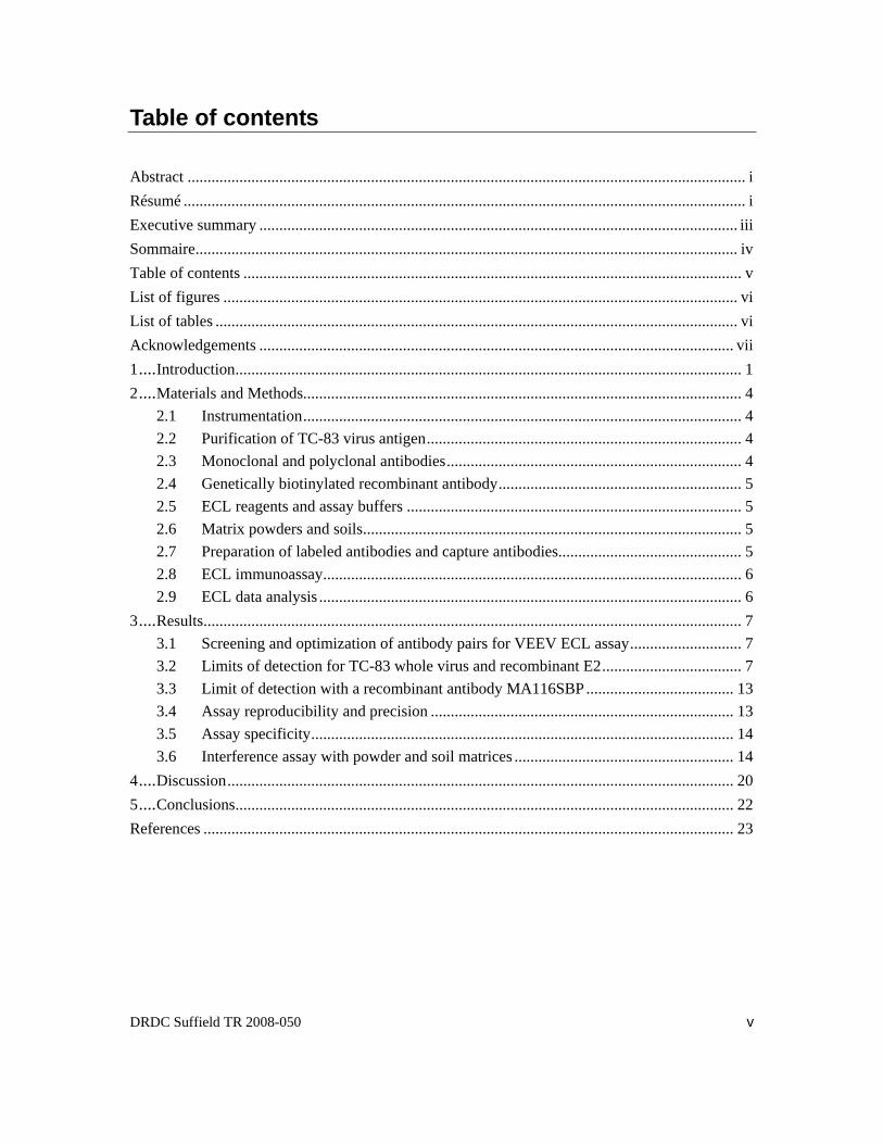
Table of contents
Abstract ............................................................................................................................................ i Résumé ............................................................................................................................................. i Executive summary ........................................................................................................................ iii Sommaire........................................................................................................................................ iv Table of contents ............................................................................................................................. v List of figures ................................................................................................................................. vi List of tables ................................................................................................................................... vi Acknowledgements ....................................................................................................................... vii 1....Introduction............................................................................................................................... 1 2....Materials and Methods.............................................................................................................. 4
2.1 Instrumentation.............................................................................................................. 4 2.2 Purification of TC-83 virus antigen............................................................................... 4 2.3 Monoclonal and polyclonal antibodies.......................................................................... 4 2.4 Genetically biotinylated recombinant antibody............................................................. 5 2.5 ECL reagents and assay buffers .................................................................................... 5 2.6 Matrix powders and soils............................................................................................... 5 2.7 Preparation of labeled antibodies and capture antibodies.............................................. 5 2.8 ECL immunoassay......................................................................................................... 6 2.9 ECL data analysis .......................................................................................................... 6
3....Results....................................................................................................................................... 7 3.1 Screening and optimization of antibody pairs for VEEV ECL assay............................ 7 3.2 Limits of detection for TC-83 whole virus and recombinant E2................................... 7 3.3 Limit of detection with a recombinant antibody MA116SBP..................................... 13 3.4 Assay reproducibility and precision ............................................................................ 13 3.5 Assay specificity.......................................................................................................... 14 3.6 Interference assay with powder and soil matrices ....................................................... 14
4....Discussion............................................................................................................................... 20 5....Conclusions............................................................................................................................. 22 References ..................................................................................................................................... 23
DRDC Suffield TR 2008-050 v
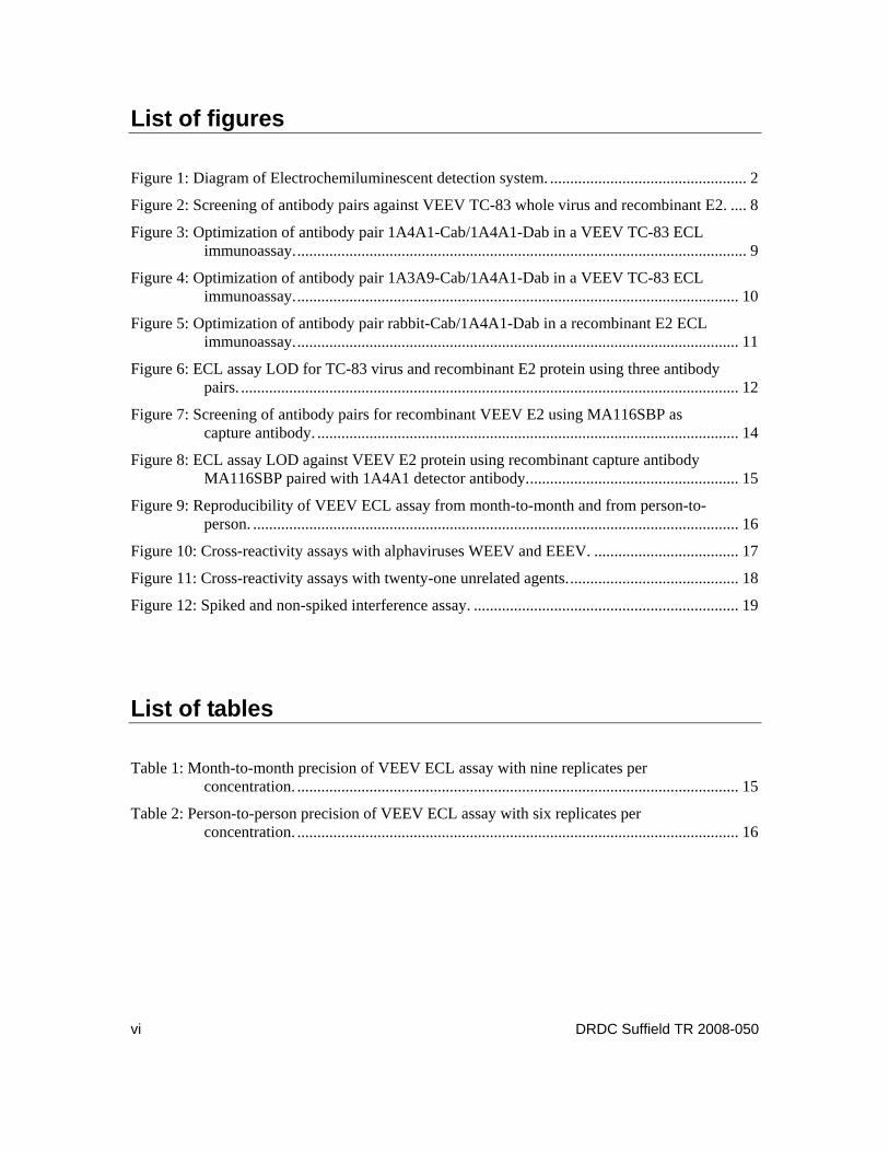
List of figures
Figure 1: Diagram of Electrochemiluminescent detection system. ................................................. 2 Figure 2: Screening of antibody pairs against VEEV TC-83 whole virus and recombinant E2. .... 8 Figure 3: Optimization of antibody pair 1A4A1-Cab/1A4A1-Dab in a VEEV TC-83 ECL
immunoassay................................................................................................................. 9 Figure 4: Optimization of antibody pair 1A3A9-Cab/1A4A1-Dab in a VEEV TC-83 ECL
immunoassay............................................................................................................... 10 Figure 5: Optimization of antibody pair rabbit-Cab/1A4A1-Dab in a recombinant E2 ECL
immunoassay............................................................................................................... 11 Figure 6: ECL assay LOD for TC-83 virus and recombinant E2 protein using three antibody
pairs. ............................................................................................................................ 12 Figure 7: Screening of antibody pairs for recombinant VEEV E2 using MA116SBP as
capture antibody. ......................................................................................................... 14 Figure 8: ECL assay LOD against VEEV E2 protein using recombinant capture antibody
MA116SBP paired with 1A4A1 detector antibody..................................................... 15 Figure 9: Reproducibility of VEEV ECL assay from month-to-month and from person-to-
person. ......................................................................................................................... 16 Figure 10: Cross-reactivity assays with alphaviruses WEEV and EEEV. .................................... 17 Figure 11: Cross-reactivity assays with twenty-one unrelated agents........................................... 18 Figure 12: Spiked and non-spiked interference assay. .................................................................. 19
List of tables
Table 1: Month-to-month precision of VEEV ECL assay with nine replicates per concentration. .............................................................................................................. 15
Table 2: Person-to-person precision of VEEV ECL assay with six replicates per concentration. .............................................................................................................. 16
vi DRDC Suffield TR 2008-050
Acknowledgements
We thank Josh Wu for providing the TC-83 virus, Gail Thompson and Sarah Hayward for critical reading of the manuscript. This study was supported by CBRN Research and Technology Initiative, CRTI 03-0021TD, ADAPT (Assay Development and Production Team for the Development, Validation, Production, and Distribution of Assays for the Identification of Bioterrorist Agents).
DRDC Suffield TR 2008-050 vii

viii DRDC Suffield TR 2008-050
This page intentionally left blank.
1 Introduction
Venezuelan equine encephalitis virus (VEEV) is a member of the alphavirus genus, which includes Sindbis virus and Semliki Forest virus as prototypes and has a positive-sense single-stranded RNA genome (Strauss and Strauss, 1994). The host range of VEEV includes both vertebrates (rodents, equines and humans) and invertebrates (a variety of mosquito species) (Strauss and Strauss, 1994). VEEV has affected humans and equines in many parts of the Americas throughout the 20th century (Work, 1964; Grayson and Galindo, 1968; Chamberlain et al., 1969; Gorelkin and Jahrling, 1974; Bowen and Calisher, 1976; Wang et al., 2001). Epizootic VEEV infections cause debilitating disease with a high fatality rate in equines. In humans, a potentially life threatening severe febrile illness occasionally develops, although human death only occurs in <1% of infections. VEEV is environmentally stable and highly infectious by aerosol inhalation, and thus is a potential biological weapons threat; it is crucial that rapid and sensitive immunoassays be developed for detection of VEEV in both environmental and clinical samples. A number of immunoassay techniques are currently available for the detection of VEEV, including enzyme-linked immunosorbent assays (ELISA), radioimmunosorbent assays (RIA), light addressable potentiometric assays (LAP), and dissociation-enhanced lanthanide fluorescent immunoassays (DELFIA) (Roehrig et al., 1982; Smith et al., 2001; Hu et al., 2002; Hu et al., 2004). Typically, these assays require multiple operational steps and long incubation periods, and acquisition and analysis of assay results may take many hours.
Electrochemiluminescence (ECL) is a relatively new technology for conducting immunoassays, with improved assay performance (Blackburn et al., 1991; Yang et al., 1994; Deaver, 1995). In contrast to conventional chemiluminescence, the chemiluminescent species in ECL are generated at the surface of an electrode as shown in Figure 1. There are three components in an ECL immunoassay: (i) a biotinylated capture antibody, pre-bound to streptavidin-coated magnetic beads, (ii) a detector antibody, labeled with ruthenium-trisbripyridal, for the emission of light when electrochemically stimulated, and (iii) an analyte which reacts with the capture and detector antibodies resulting in an antigen-antibody sandwich. In ECL reactions, a precursor molecule (tripropylamine) is activated on an electrode surface resulting in an electron transfer reaction. This transfer in turn initiates the excitation of ruthenium-trisbripyridal which ultimately results in the emission of a photon at 620 nm (Blackburn et al., 1991; Yang et al., 1994; Deaver, 1995) (Figure 1).
In comparison with conventional ELISA and RIA, the ECL technique has a number of distinct advantages. Firstly, detection sensitivity is increased due to high luminescent signal-noise ratios (Yang et al., 1994; Gatto-Menking et al., 1995; Yu et al., 1995; Yu et al., 2000; Shelton et al, 2001); it has been reported that the ECL detection limits for most toxins are at the femtogram to picogram per mL level and for most viruses at approximately 105pfu/mL (Kijek et al., 2000; Yu et al., 2000; Merrill et al., 2006; Rivera et al., 2006). Secondly, the dynamic range for analyte detection extends over five orders of magnitude (Yang et al., 1994; Kijek et al., 2000; Yu et al., 2000). Thirdly, the time and labour required to complete ECL assays are reduced compared with conventional immunoassays. ECL assays are stable and robust, and tolerant of analyte detection in the presence of a variety of sample matrices (unpublished data). The ECL assay is a nonseparation technique, hence there are no plate coating, washing, or aspiration procedures as are typically required for ELISA and RIA techniques. An average of one minute
DRDC Suffield TR 2008-050 1
Figure 1: Diagram of Electrochemiluminescent detection system.
per sample is required for sample analysis in ECL assays using the M1M and M1MR series instrumentation developed by BioVeris Corp. (Gaithersberg, MD). The use of ECL techniques eliminates the necessity of radioisotopes and the labeled ECL reagents are exceptionally stable. These features make the ECL detection system an attractive alternative to conventional ELISA and RIA techniques. In addition, the M-series instrumentation is available in a military hardened version, making it especially useful for military field use.
In the last several years, ECL assays have been successfully developed for over 300 analytes, including many biological threat agents such as staphylococcus enterotoxin B (SEB), ricin, Clostridium botulinum neurotoxins, Clostridium perfringens alpha toxin, Bacillus anthracis spore, Bacillus subtilis spore, E. coli O157, and MS2 (Bruno and Yu, 1996; Kijek et al., 2000; Yu et al., 2000; Shelton and Karns, 2001; Cote et al., 2005; Guglielmo-Viret et al., 2005; Merrill et al., 2006; Rivera et al., 2006; Guglielmo-Viret and Thullier, 2007). ECL technology is in use in defence research laboratories in Canada, the United States, the United Kingdom, and Australia. It has also been fielded by various theater laboratories of the US Army and US Air Force, as well as the US National Guard. The technology has been acquired recently by the Canadian Forces (CF).
2 DRDC Suffield TR 2008-050

As part of a collaborative research project with the Public Health Agency Canada (PHAC) and the Canadian Food Inspection Agency (CFIA), funded through the CBRN Research and Technology Initiative (CRTI) (Assay Development and Production Team for the Development, Validation, Production, and Distribution of Assays for the Identification of Bioterrorist Agents: CRTI 03-0021TD, ADAPT), DRDC Suffield is developing ECL assays on the M1MR platform for use by both military personnel and civilian first responders. This study describes the development of an ECL assay for VEEV. In this study, VEEV antibodies were screened for optimal performance using VEEV strain TC83 (a line attenuated VEEV vaccine) and VEEV recombinant envelope E2 protein as target antigens. The assay LOD was 103 pfu/mL for TC-83 whole virus and 1 ng/mL for recombinant E2. The ECL assay was reproducible with coefficients of variation ranging from 4.7% to 18.5%. The ECL assay was specific to VEEV as evidenced by the absence of cross-reactivity with two closely related alphaviruses and twenty-one other bacterial, viral, and toxin agents. This study demonstrated a sensitive, rapid and reproducible ECL immunoassay for detection and identification of VEEV.
DRDC Suffield TR 2008-050 3
2 Materials and Methods
2.1 Instrumentation
ECL measurements were performed using the M1MR analyzer (BioVeris Corp., Gaithersburg, MD).
2.2 Purification of TC-83 virus antigen
Purified VEE TC-83 whole virus was kindly provided by Dr. J. Wu, DRDC Suffield. Purification of TC-83 whole virus was performed as follows. Vero monkey kidney cells were cultured in Dulbecco’s modified Eagle medium (DMEM) supplemented with 10% fetal bovine serum (FBS), 1 mM sodium pyruvate, and 100 units /mL antibiotic–antimycotic. Supernatants from TC-83-infected vero cells were cleared of cell debris at 10,000×g for 30 min at 4°C in a Sorval GSA rotor. After addition of 7% PEG 8000 and 2.3% NaCl, the supernatant was incubated for 3 days at 4°C and then subjected to centrifugation at 10,000×g for 30 min at 4°C in a Sorval GSA rotor. Virus pellets were resuspended in 4 mL of sterile PBS and loaded onto a 20% continuous sucrose gradient, followed by centrifugation at 100,000×g for 3.5 hours at 4°C in a SW-28 rotor. Virus containing fractions, which were verified by sequencing analysis, were pooled and diluted at 1:3 in sterile PBS. The virus preparation was concentrated at 100,000×g for 1 hour at 4°C in a SW-28 rotor and resuspended in 2.5 mL of sterile PBS. Titers of the virus stocks were determined by a standard plaque assay using Vero cells.
2.3 Monoclonal and polyclonal antibodies
Hybridomas 1A4A1 and 1A3A9 were kindly provided by Dr. J.T. Roehrig (Mathews and Roehrig, 1982; Roehrig et al., 1982; Roehrig and Mathews, 1985). The hybridomas were grown in BD CellTM MAb Basal Medium (BD-Biosciences, Mississauga, ON) supplemented with 10% FBS, 2mM L-glutamine and 1% HT Media Supplement Hybri-Max (Sigma, Oakville, ON). The hybridomas were then weaned off of FBS and transferred to a CELLineTM 1000 flask (BD-Biosciences, Mississauga, ON) and incubated at 37°C under 5% CO2 for two weeks. Supernatant was harvested from the CELLineTM 1000 flask and the monoclonal antibody (mAb) was then purified using a MelonTM Gel Monoclonal IgG Purification Kit (Pierce, Ottawa, ON) according to the manufacturer’s instructions.
Rabbit and goat anti-VEEV polyclonal antibodies (pAb) were previously developed and purified by SciLab Consulting Incorporated (Redcliff, AB, Canada) under a DRDC Suffield contract. Both the rabbit and the goat IgG were purified on a Bio-GelR Protein G Fast Flow Gel column (Bio-Rad Laboratories, Mississauga, ON) using a high-performance liquid chromatography system (Spectral Physics, San Jose, CA) (Di Ninno, 1998).
4 DRDC Suffield TR 2008-050
2.4 Genetically biotinylated recombinant antibody
A genetically biotinylated recombinant VEEV single-chain variable fragment antibody, MA116SBP, was previously generated at DRDC Suffield (Hu et al., 2002).
2.5 ECL reagents and assay buffers
Reagents and assay buffers were purchased from BioVeris Corp.: Biotin-LC-Sulfo-NHS ester, Ruthenium (II) tris-bipyridine-NHS ester, Streptavidin-coated Dynabeads M-280, M-SERIES Positive Calibrator, M-SERIES Negative Calibrator, Stabilicoat Immunoassay Stabilizer, BV-Clean Plus solution, BV-Glo Plus solution, BV-Diluent solution, BV-Store solution, and BV-Sanitize solution.
2.6 Matrix powders and soils
Flour (white, enriched, all purpose, Safeway brand), cornstarch (Safeway brand), baking powder (Safeway brand), baking soda (Safeway brand), laundry detergent (Tide, original, Procter and Gamble), coffee creamer (Coffee-MateR, Carnation, Nestlé), skim milk, and powdered sugar were all purchased from Safeway Canada Ltd. (Medicine Hat, AB, Canada). Talcum powder, powdered cleanser, and spackling powder were purchased from the US CRP. The four soil samples including sand, sand loam, loamy sand, and clay loam were from the DRDC Suffield Experimental Proving Ground (EPG) and had been previously characterized by the Alberta Environmental Centre (Mr. P. Yeung, Vegreville, Alberta, Canada).
2.7 Preparation of labeled antibodies and capture antibodies
Prior to labeling, all antibodies were desalted using NAP-5 columns (Amersham Biosciences, Baie d’Urfé, QC) according to the manufacturer’s instructions. The four antibodies (1A4A1 mAb, 1A3A9 mAb, rabbit pAb and goat pAb) were each biotinylated with biotin-LC-Sulfo-NHS ester at a molar ratio of 10:1 (Tag:Ab) and ruthenylated with ruthenium (II) tris-bipyridine-NHS ester at 7.5:1 using a labeling procedure recommended by BioVeris with some minor modifications. In brief, the antibodies and tag solutions were incubated together using an end-over-end shaker with gentle rotation for 60 min at room temperature. The labeling reaction was quenched by addition of 20 μL of 2 M glycine and then incubated at room temperature for an additional 10 min. Uncoupled biotin and ruthenium esters were removed by dialysis in four changes of sterile PBS (pH 7.4) at 4°C over two days. The protein concentrations of labeled antibodies were determined using a MicroBCA assay (Pierce, Ottawa, ON) and a ND-1000 Spectrophotometer (LabX, Midland, ON) according to the manufacturer’s instructions with bovine serum albumin as the standard. Aliquots of each antibody were stored at 4°C until use.
For the preparation of capture antibody, biotinylated antibodies were pre-bound to streptavidin-coated Dynabeads at a concentration of 100 μg antibody per mL of Dynabeads. Biotinylated antibodies and beads were incubated together with an end-over-end rotation for 60 min at room temperature. Unbound antibody was removed by washing the beads three times with sterile PBS-0.1% Tween 20 using a magnetic particle concentrator, Dynal MPC®-S (Dynal Biotech ASA, Oslo, Norway), followed by three washes with sterile PBS (pH 7.4).
DRDC Suffield TR 2008-050 5
2.8 ECL immunoassay
Capture antibody (biotinylated antibody pre-bound to streptavidin-coated Dynabeads) (Cab) was diluted in Stabilicoat Immunoassay Stabilizer and the detector antibody (ruthenylated antibody) (Dab) was diluted in PBS-2% BSA-0.1% Tween 20. Antigen was diluted in PBS-0.3% Tween 20. ECL reaction solutions were prepared in 0.75 mL round-bottom tubes (Matrix Technologies Corp., Mississauga, ON) by adding 25 μL of Cab, 25 μL of Dab, and 50 μL of antigen solution or 50 μL of PBS-0.3% Tween 20 for the antigen negative control. The 96-well assay plate was loaded into the M-Series M1MR analyzer, incubated with shaking for 15 minutes at room temperature and analyzed.
2.9 ECL data analysis
A limit of detection was determined by calculating a cutoff value that was equal to 20% above the mean ECL reading for the negative control. Therefore, a signal was considered positive if the ECL ratio of the sample signal reading to background reading (S/B) was 1.2 or greater.
6 DRDC Suffield TR 2008-050
3 Results
3.1 Screening and optimization of antibody pairs for VEEV ECL assay
Four Abs (1A4A1 mAb, 1A3A9 mAb, rabbit pAb and goat pAb) were each labeled with biotin-LC-sulfo-NHS ester and ruthenium (II) tris-bipyridine-NHS ester. A total of sixteen combinations of antibody pairs were tested for utility in the ECL assays at a Cab dilution of 1:25, Dab concentration of 4 μg/mL, TC-83 virus concentration of 105 pfu/mL, and recombinant VEEV E2 concentration of 100 ng/mL. Amongst the 16 antibody pairs, only three antibody pairs (1A4A1-Cab/1A4A1-Dab, 1A4A1-Cab/1A3A9-Dab, 1A3A9-Cab/1A4A1-Dab) exhibited high reactivity with TC-83 virus (Figure 2A). Five antibody pairs (1A4A1-Cab/rabbit-Dab, 1A4A1-Cab/goat-Dab, 1A3A9-Cab/1A3A9-Dab, rabbit-Cab/1A4A1-DAb, goat-Cab/1A3A9-Dab) exhibited moderate reactivity with TC-83 virus (Figure 2A). Eight other antibody pairs produced background ECL signals (Figure 2A). Using recombinant VEEV E2 as the antigen, four antibody pairs (rabbit-Cab/1A4A1-Dab, goat-Cab/1A4A1-Dab, 1A4A1-Cab/1A4A1-DAb, 1A4A1-Cab/rabbit-Dab) exhibited high reactivity with this antigen (Figure 2B); five antibody pairs exhibited moderate reactivity, while the seven other antibody pairs produced only background ECL signals (Figure 2B). Approximately 50% of the antibody pairs tested had little or no reactivity with either TC-83 virus or recombinant E2 in the ECL assay. Based on these results, three antibody pairs (one pair 1A3A9-Cab/1A4A1-Dab with high reactivity to TC-83 whole virus, one pair rabbit-Cab/1A4A1-Dab with high reactivity to recombinant E2, and one pair 1A4A1-Cab/1A4A1-Dab with high reactivity to both TC-83 virus and E2) were chosen for further optimization in the ECL assay.
Initial experiments were performed to optimize the concentrations of both the Cab and Dab. Concentrations of Cab and Dab that yielded the highest signal-to-background (S/B) values and lowest limits of detection (LOD) were determined to be optimal. If two or more concentrations were comparable, the concentration requiring the least amount of antibody was used. Capture antibodies were first optimized at 1:16, 1:25, 1:50 dilution against 4 μg/mL of detector antibody; and then, the detector antibodies were optimized at 4, 10, and 20 μg/mL against the optimal dilution of capture antibody. As a result of optimization, the antibody pair 1A4A1-Cab/1A4A1-Dab at a Cab dilution of 1:35 and Dab concentration of 10 μg/mL produced the highest S/B values and the lowest LOD with TC-83 virus (Figure 3A & B). 1A3A9-Cab/1A4A1-Dab and rabbit-Cab/1A4A1-Dab pairs at Cab dilutions of 1:25 and 1:16 and Dab concentrations of 10 μg/mL and 4 μg/mL, respectively, produced the lowest LOD (Fig 4A, 4B, 5A & 5B). Hence, these concentrations of Cab and Dab were used in subsequent experiments.
3.2 Limits of detection for TC-83 whole virus and recombinant E2
Experiments were performed to determine the assay LOD and dynamic range for each optimized antibody pair. The results showed that amongst the three antibody pairs 1A4A1-Cab/1A4A1-Dab pair worked best for both TC-83 whole virus and recombinant E2 protein (Figure 6A & B). With this antibody pair, the LOD was approximately 103 pfu/mL for TC-83 and 1 ng/mL for
DRDC Suffield TR 2008-050 7
Figure 2: Screening of antibody pairs against VEEV TC-83 whole virus and recombinant E2.
A. The ECL assay was performed against 105 pfu/mL VEEV TC-83 whole virus at Cab dilution of 1:25 and Dab concentration of 4 μg/mL. B. The ECL assay was performed against 100
ng/mL VEEV recombinant E2 at Cab dilution of 1:25 and Dab concentration of 4 μg/mL. The data represents two separate experiments, each with two replicates. Neg Ctl: PBS-0.3%
Tween 20 as the antigen negative control. Signal to Background ECL Values: average ECL reading/average ECL reading of negative control values.
8 DRDC Suffield TR 2008-050
Figure 3: Optimization of antibody pair 1A4A1-Cab/1A4A1-Dab in a VEEV TC-83 ECL immunoassay.
A. Optimization of capture antibody. The 1A4A1-Cab was optimized at the dilutions of 1:25, 1:35 and 1:50 paired with 4 μg/mL of 1A4A1-Dab against three different concentrations of VEEV TC-83 virus (103, 104 and 105 pfu/mL). B. Optimization of detector antibody. The 1A4A1-Dab was optimized at the concentrations of 4, 10 and 20 μg/mL paired with 1:35
dilution of 1A4A1-Cab against four different concentrations of VEEV TC-83 virus (102, 103, 104 and 105 pfu/mL). The data represents two separate experiments, each with two replicates of each concentration of antigen. Neg Ctl: PBS-0.3% Tween 20 as negative control. Signal to
Background ECL Values: average ECL reading/average ECL reading of negative control values.
DRDC Suffield TR 2008-050 9
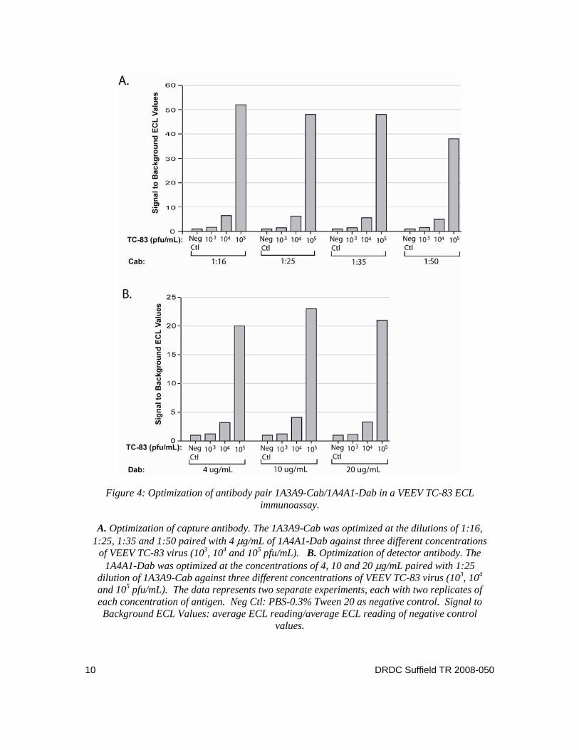
Figure 4: Optimization of antibody pair 1A3A9-Cab/1A4A1-Dab in a VEEV TC-83 ECL immunoassay.
A. Optimization of capture antibody. The 1A3A9-Cab was optimized at the dilutions of 1:16, 1:25, 1:35 and 1:50 paired with 4 μg/mL of 1A4A1-Dab against three different concentrations
of VEEV TC-83 virus (103, 104 and 105 pfu/mL). B. Optimization of detector antibody. The 1A4A1-Dab was optimized at the concentrations of 4, 10 and 20 μg/mL paired with 1:25
dilution of 1A3A9-Cab against three different concentrations of VEEV TC-83 virus (103, 104 and 105 pfu/mL). The data represents two separate experiments, each with two replicates of each concentration of antigen. Neg Ctl: PBS-0.3% Tween 20 as negative control. Signal to Background ECL Values: average ECL reading/average ECL reading of negative control
values.
10 DRDC Suffield TR 2008-050
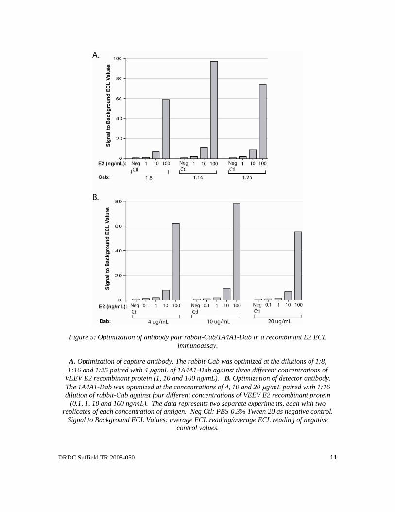
Figure 5: Optimization of antibody pair rabbit-Cab/1A4A1-Dab in a recombinant E2 ECL immunoassay.
A. Optimization of capture antibody. The rabbit-Cab was optimized at the dilutions of 1:8, 1:16 and 1:25 paired with 4 μg/mL of 1A4A1-Dab against three different concentrations of
VEEV E2 recombinant protein (1, 10 and 100 ng/mL). B. Optimization of detector antibody. The 1A4A1-Dab was optimized at the concentrations of 4, 10 and 20 μg/mL paired with 1:16 dilution of rabbit-Cab against four different concentrations of VEEV E2 recombinant protein
(0.1, 1, 10 and 100 ng/mL). The data represents two separate experiments, each with two replicates of each concentration of antigen. Neg Ctl: PBS-0.3% Tween 20 as negative control.
Signal to Background ECL Values: average ECL reading/average ECL reading of negative control values.
DRDC Suffield TR 2008-050 11
Figure 6: ECL assay LOD for TC-83 virus and recombinant E2 protein using three antibody pairs.
A. Titration of 10-fold dilutions of TC-83 whole virus (101−106 pfu/mL) against three antibody pairs, 1A4A1-Cab/1A4A1-Dab, 1A3A9-Cab/1A4A1-Dab, and rabbit-Cab/1A4A1-
Dab. B. Titration of 10-fold dilutions of recombinant E2 (0.01 −10,000 ng/mL) against three antibody pairs. The data was from three separate experiments, each with three replicates of each concentration of antigen (n=9). Standard error bars are indicated. Neg Ctl: PBS-0.3%
Tween 20 as negative control, n=18. Signal to Background ECL Values: average ECL reading/average ECL reading of negative control values.
recombinant E2 (Figure 6A & B). The S/B ratio was 608 for whole virus at a titer of 106 pfu/mL and as high as 3084 for E2 at a concentration of 10 μg/mL (Figure 6A & B). The second
12 DRDC Suffield TR 2008-050
antibody pair 1A3A9-Cab/1A4A1-Dab produced satisfactory ECL signals for TC-83 virus with a LOD of approximately 103 pfu/mL (Figure 6A), but performed poorly for recombinant E2 with a LOD of 100 ng/mL (Figure 6B). The third antibody pair rabbit-Cab/1A4A1-Dab produced very poor ECL signals for TC-83 whole virus with a LOD of approximately 105 pfu/mL (Figure 6A), but detected recombinant E2 with a LOD of 1 ng/mL (Figure 6B). These results demonstrated that different antibody pairs have different reactivity with whole virus and recombinant antigen with some antibody pairs having reactivity only with either whole virus or recombinant antigen.
The results showed that the assay dynamic range was 4−5 orders of magnitude, from 102 to 106
pfu/mL for TC-83 whole virus and from 0.1 to 10,000 ng/mL for recombinant E2 (Figure 6A & B).
3.3 Limit of detection with a recombinant antibody MA116SBP
To eliminate the need for chemical biotinylation of antibody, a genetically biotinylated recombinant antibody, MA116SBP, was tested for utility in the ECL assay. MA116SBP was pre-bound to streptavidin-coated Dynabeads by following the same procedures as described in the Materials and Methods. Subsequently, the MA116SBP Cab was paired with each of four Dabs, 1A4A1-Dab, 1A3A9-Dab, rabbit-Dab, and goat-DAb and evaluated in the ECL assay. Assay LODs obtained using genetically biotinylated Cab (MA116SBP) were compared with those obtained using chemically biotinylated Cab. Among the four antibody pairs, MA116SBP/1A4A1-Dab pair produced the highest S/B values for recombinant E2 antigen (Figure 7). The LOD obtained using this pair was 100 ng/mL for recombinant E2 (Figure 8). This value was two logs higher than that observed when antibody pairs 1A4A1-Cab/1A4A1-Dab and rabbit-Cab/1A4A1-Dab were used to detect recombinant E2 (Figure 6B). Even E2 antigen at a concentration as high as 10,000 ng/mL, the MA116SBP/1A4A1-DAb antibody pair produced a low S/B ECL value of 32 (Figure 8); by comparison antibody pairs 1A4A1-Cab/1A4A1-Dab and rabbit-Cab/1A4A1-DAb produced S/B values of 3084 and 314, respectively (Figure 6B). Hence, although the genetically biotinylated recombinant antibody MA116SBP was functional as Cab by ECL, the LOD obtained by its use was higher that that obtained with chemically biotinylated Cab.
3.4 Assay reproducibility and precision
The assay reproducibility and precision were evaluated by titrating samples of VEEV whole virus from month-to-month and from person-to-person (Tables 1 & 2 and Figure 9). For month-to-month reproducibility, LOD assays were performed on three separate occasions over a period of five months, from May 2007 to September 2007, with antibody pair 1A4A1-biotin/1A4A1-ru against 10-fold dilutions of whole virus. For person-to-person reproducibility, two different persons conducted standard LOD assays using the same antibody pairs and antigen on the 6th and 18th of September 2007. The assay was reproducible as shown in Figure 9. The coefficient of variation for the month-to-month assay precision determinations ranged from 4.7% to 18.5% (Table 1). The coefficient of variation for the person-to-person assay precision determinations ranged from 3.3% to 8.8% (Table 2).
DRDC Suffield TR 2008-050 13
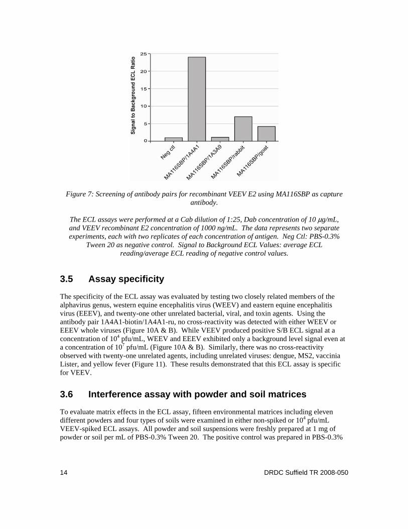
Figure 7: Screening of antibody pairs for recombinant VEEV E2 using MA116SBP as capture antibody.
The ECL assays were performed at a Cab dilution of 1:25, Dab concentration of 10 μg/mL, and VEEV recombinant E2 concentration of 1000 ng/mL. The data represents two separate experiments, each with two replicates of each concentration of antigen. Neg Ctl: PBS-0.3%
Tween 20 as negative control. Signal to Background ECL Values: average ECL reading/average ECL reading of negative control values.
3.5 Assay specificity
The specificity of the ECL assay was evaluated by testing two closely related members of the alphavirus genus, western equine encephalitis virus (WEEV) and eastern equine encephalitis virus (EEEV), and twenty-one other unrelated bacterial, viral, and toxin agents. Using the antibody pair 1A4A1-biotin/1A4A1-ru, no cross-reactivity was detected with either WEEV or EEEV whole viruses (Figure 10A & B). While VEEV produced positive S/B ECL signal at a concentration of 104 pfu/mL, WEEV and EEEV exhibited only a background level signal even at a concentration of 107 pfu/mL (Figure 10A & B). Similarly, there was no cross-reactivity observed with twenty-one unrelated agents, including unrelated viruses: dengue, MS2, vaccinia Lister, and yellow fever (Figure 11). These results demonstrated that this ECL assay is specific for VEEV.
3.6 Interference assay with powder and soil matrices
To evaluate matrix effects in the ECL assay, fifteen environmental matrices including eleven different powders and four types of soils were examined in either non-spiked or 104 pfu/mL VEEV-spiked ECL assays. All powder and soil suspensions were freshly prepared at 1 mg of powder or soil per mL of PBS-0.3% Tween 20. The positive control was prepared in PBS-0.3%
14 DRDC Suffield TR 2008-050
Figure 8: ECL assay LOD against VEEV E2 protein using recombinant capture antibody MA116SBP paired with 1A4A1 detector antibody.
Titration of 10-fold dilutions of recombinant E2 (0.1−100,000 ng/mL) against the antibody pair MA116SBP/1A4A1-Dab. The data was from three separate experiments, each with three replicates of each concentration of antigen (n=9). Neg Ctl: PBS-0.3% Tween 20 as negative
control, n=18. Signal to Background ECL Values: average ECL reading/average ECL reading of negative control values.
Table 1: Month-to-month precision of VEEV ECL assay with nine replicates per concentration.
The three separate assays were performed over a period of two months (May to June 2007).using the
antibody pair 1A4A1-Cab/1A4A1-Dab against 10-fold dilutions of VEEV TC-83 whole virus (101−106 pfu/mL) Each assay with three replicates of each concentration of antigen (n=9). S/B: average ECL
reading/average ECL reading of negative control values.
DRDC Suffield TR 2008-050 15
Figure 9: Reproducibility of VEEV ECL assay from month-to-month and from person-to-person.
Assays 1, 2 and 3 represent three separate titrations of 10-fold dilutions of TC-83 whole virus (101−106 pfu/mL) against the antibody pair 1A4A1-Cab/1A4A1-Dab, performed by person 1,
over a period of five months (May 2007, July 2007, and September 2007), each with three replicates of each concentration of antigen. Assay 4 represents a single titration of virus
using the same antibody pair, performed by person 2, in September 2007. Signal to Background ECL Values: average ECL reading/average ECL reading of negative control
values.
Table 2: Person-to-person precision of VEEV ECL assay with six replicates per concentration.
The two separate assays were performed by two different persons using the antibody pair 1A4A1-
Cab/1A4A1-Dab against 10-fold dilutions of VEEV TC-83 whole virus (101−106 pfu/mL) in September 2007. Each assay had three replicates of each concentration of antigen. S/B: average ECL reading/average
ECL reading of negative control values.
16 DRDC Suffield TR 2008-050
Figure 10: Cross-reactivity assays with alphaviruses WEEV and EEEV.
The assay was performed with the antibody pair 1A4A1-Cab/1A4A1-Dab. The data was from two separate assays, each with three replicates of each concentration of agent (n=6). Neg Ctl: PBS-0.3% Tween 20 as negative control, n=12. Signal to Background Values: average ECL
reading/average ECL reading of negative control values.
Tween 20 containing a final concentration of 104 pfu/mL TC-83 whole virus. The negative control was PBS-0.3% Tween 20 only. A signal was considered positive with interference effect if the S/B ratio was 20% above or below the negative control (for non-spiked) or positive control (for VEEV-spiked). As shown in Figure 12, talcum powder, powdered cleanser, sand, sand loam, loamy sand and clay loam tested positive by increasing background in the non-spiked ECL assay. In the VEEV-spiked assay, sand and loamy sand exhibited significant interference by quenching the ECL signal by 31%, relative to the positive control. There was no significant interference detected with thirteen other powders and soils in the VEEV-spiked assay. Thus, the results indicated that two powders and four soils interfered with the ECL assays by increasing background or quenching the ECL signals.
DRDC Suffield TR 2008-050 17
Figure 11: Cross-reactivity assays with twenty-one unrelated agents.
The assay was performed with the antibody pair 1A4A1-Cab/1A4A1-Dab. The data was from two separate assays, each with three replicates of each concentration of agent (n=6). A
concentration of 1 μg/mL was used for most of the 21 reagents unless indicated. Vaccinia, Lister: 106 pfu/mL. Aspergillus niger: 104 spores/mL. SEB: 2 ng/mL. Ricin: 3 ng/mL. Yellow
fever: 109 pfu/mL. Dengue: 104 pfu/mL. Negative control: PBS-0.3% Tween 20, n=12. Signal to Background ECL Values: average ECL reading/average ECL reading of negative control
values.
18 DRDC Suffield TR 2008-050
Figure 12: Spiked and non-spiked interference assay.
The assay was performed with the antibody pair 1A4A1-Cab/1A4A1-Dab. The data was from two separate assays, each with two replicates of each concentration (n=4). Negative Control:
PBS-0.3% Tween 20, n=8. Positive control: 104 pfu/mL TC-83 whole virus in PBS-0.3% Tween 20, n=8. Signal to Background Values: average ECL reading/average ECL reading of
negative control values.
DRDC Suffield TR 2008-050 19
4 Discussion
In this study, we have developed a sensitive and rapid ECL immunoassay to detect VEEV TC-83 whole virus and recombinant E2 protein using the BioVeris M1MR system. There are several advantages with the ECL assay compared to other immunoassays for VEEV. Firstly, assay LOD is significantly improved. The ECL assay can detect TC-83 whole virus at concentrations as low as 103 pfu/mL and E2 protein at 1 ng/mL. Previous studies reported LODs of 1.25×10⁷ pfu/mL by ELISA and 3.13×10⁶ pfu/mL by DELFIA using VEEV Trinidad donkey strain as antigen (Smith et al., 2001). Another previous study reported LODs of 250 ng/mL by ELISA and 300 ng/mL by immunofiltration enzyme assay with a light addressable potentiometric sensor (IFA/LAPS) using purified TC-83 whole virus as antigen (Hu et al., 2004). The ECL assay described in this paper is the most sensitive immunoassay reported to date for the detection of VEEV. Secondly, the ECL is a one-step assay with rapid generation of results. There is no plate coating, washing, or aspiration procedures as are typically required for ELISA and other immunoassays; compared to a 6-8 hour period for performance of ELISA, the ECL assay can be completed in 30-60 min, including a 15 min incubation period. Thirdly, we have observed in this study that the biotin and ruthenium labeled VEEV mAb and pAb capture and detector antibodies were stable and could be stored at 4°C for at least 6 months. It has been previously reported that biotin and ruthenium labeled antibodies could be stored at 4°C for up to one year (Kijek et al., 2000).
Three antibody pairs were analyzed against two different antigens in this study. These antibody pairs demonstrated differential reactivity with TC-83 whole virus and VEEV recombinant E2 protein. Eight epitopes (E2a-h) on the VEEV E2 glycoprotein have been previously identified (Roehrig et al., 1982; Roehrig and Mathews, 1985) and the mAbs 1A4A1 and 1A3A9 have been well characterized and found to be specific for E2c and E2g epitopes, respectively (Roehrig et al., 1982; Roehrig and Mathews, 1985). Furthermore, studies on the spatial arrangement of epitopes have indicated that the E2c and E2g epitopes are closely linked on VEEV TC-83 and that an anti-E2c antibody (1A4A1) and an anti-E2g antibody (1A3A9) actually compete with each other due to steric hindrance (Roehrig and Mathews, 1985). In VEEV, two envelope glycoproteins E1 and E2 form a stable heterodimer that remains intact upon dissociation of the virus with mild detergents. Three E1-E2 heterodimers interact to form the spike that is found on the virus surface; there are 240 heterodimers on the surface of the virus, assembled into 80 spikes (Strauss and Strauss, 1994). In our study, the 1A4A1-Cab/1A4A1-Dab pair and the 1A3A9-Cab/1A4A1-Dab pair exhibited high reactivity with TC-83 whole virus, suggesting that 1A4A1 and 1A3A9 did not compete with each other and that the competition of 1A4A1 with itself did not affect its binding against VEEV whole virus in the ECL assay. This is not unexpected as there are multiple copies of E2 on the surface of the VEEV whole virus due to the existence of multiple spikes. Furthermore, the observation that the 1A3A9-Cab/1A4A1-Dab pair produced poor reactivity with recombinant E2, suggests that 1A3A9 may require conformation to have good reactivity with the E2 epitope, or 1A4A1 and 1A3A9 may be sterically hindered by each other as Roehrig and Mathews previously reported (Roehrig and Mathews, 1985). Unlike 1A3A9-Cab/1A4A1-Dab, antibody pair 1A4A1-Cab/1A4A1-Dab exhibited high reactivity with recombinant E2; the reason for this is largely unknown. One possibility might be that the recombinant protein expressed in prokaryotic cells is misfolded and multiple misfolded E2 proteins coaggregate with each other to form discrete foci. The antibody pair rabbit-Cab/1A4A1-Dab exhibited good reactivity with
20 DRDC Suffield TR 2008-050
recombinant E2 but poor reactivity with TC-83 whole virus. Overall, our data suggest that, in the context of developing an ECL assay, some antibody pairs have reactivity with both whole virus and recombinant antigen with some antibody pairs having reactivity only with either whole virus or recombinant antigen.
During the labeling of antibody with biotin-LC-Sulfo-NHS ester or ruthenium (II) tris-bipyridine-NHS ester, the N-hydroxysulfo-succinimide ester of biotin or ruthenium combines with ε–amide group of lysine to form a stable amide bond (Miralles et al., 1991; Yang et al., 1994; Deaver, 1995). The results by ELISA showed that there was no significant loss of antigen binding ability for biotinylated or ruthenylated antibodies in comparison with unlabeled antibodies (data not shown); this agrees with a previous report by Kijek et al. (Kijek et al., 2000). However, our results indicated that approximately 50% of the antibody pairs tested had little or no reactivity with either TC-83 virus or recombinant E2 in the ECL assay. This could be a reflection of the sandwich format in which two antibodies may have competed for the same epitope on the antigen or have been sterically hindered by each other. We typically observed in development of the ECL assay for VEEV and other agents that the same antibody, when labeled with biotin and ruthenium, respectively, didn’t produce good ECL signals in most cases. For example, antibody pairs 1A3A9-Cab/1A3A9-Dab, rabbit-Cab/rabbit-Dab, goat-Cab/goat-Dab produced poor ECL signals. However, 1A4A1-Cab/1A4A1-Dab pair was an exception, exhibiting excellent ECL signals for both TC-83 whole virus and recombinant E2 protein. The reason for this is still unknown, though the data suggest that 1A4A1 has a strong reactivity with the epitope of E2.
ECL is a streptavidin-coated magnetic bead-based immunoassay that involves the preparation of biotinylated antibody. Chemical biotinylation of antibody followed by removal of unbound biotin is quite time-consuming. The procedure followed in this study typically takes 3-4 days to complete a process for chemical biotinylation of antibody. Previous studies have reported that the degree of conjugation with biotin could be different from batch to batch (Miralles et al., 1991). To eliminate the need for chemical biotinylation of antibodies, we tested a genetically biotinylated recombinant antibody, MA116SBP, for utility in the ECL assay. Our findings indicated that MA116SBP was functional as capture antibody in the ECL assay but produced a higher LOD in comparison with chemically biotinylated capture antibodies (100 ng/mL vs 1 ng/mL). This could be due to (i) expression of MA116SBP in E coli., which may have resulted in improperly folded protein and lack of spatial conformation, (ii) the recombinant protein being 11 residues of the streptavidin-binding peptide rather than the true biotin itself, (iii) the antibody being a single-chain variable fragment rather than the full length antibody. Function of the genetically biotinylated recombinant antibody might be improved by constructing new expression vectors containing full length biotin and antibody. Moreover, the recombinant antibody could be expressed in eukaryotic expression systems, for example in baculovirus-insect cell or mammalian cell expression systems, to gain proper posttranslational modification and spatial conformations. Further studies are required to address these issues.
DRDC Suffield TR 2008-050 21
5 Conclusions
This study described a sensitive and rapid ECL immunoassay to detect VEEV TC-83 whole virus and recombinant E2 protein using the BioVeris M1MR system. The ECL assay can detect TC-83 whole virus at concentrations as low as 103 pfu/mL and E2 protein at 1 ng/mL. The ECL assay did not cross-react with either alphaviruses EEEV or WEEV nor any of the other twenty-one unrelated agents tested. Interference assay with eleven powder and four soil matrices indicated that two powders (talcum powder and powdered cleanser) and four soils (sand, sand loam, loamy sand and clay loam ) interfered with the ECL assays by increasing background or quenching the ECL signals. Testing of samples over time and by different operators indicated that the assay was reproducible with the month-to-month coefficient of variation ranging from 4.7% to 18.5% and person-to-person coefficient of variation ranging from 3.3% to 8.8%. In addition, a genetically biotinylated recombinant antibody, MA116SBP, was applied to the ECL assay and results indicated that MA116SBP was functional in the ECL assay but resulted in a higher LOD to that obtained using chemically biotinylated antibodies (100 ng/mL vs 1 ng/mL).
22 DRDC Suffield TR 2008-050
References
[1] Strauss, J.H. and Strauss, E.G. (1994), The alphaviruses: gene expression, replication, and evolution, Microbiol. Rev., 58, 491–562.
[2] Work, T.H. (1964), Serological Evidence of Arbovirus Infection in the Seminole Indians of Southern Florida, Science, 145, 270–272.
[3] Grayson, M.A. and Galindo, P. (1968), Epidemiologic studies of Venezuelan equine encephalitis virus in Almirante, Panama, Am. J. Epidemiol., 88, 80–96.
[4] Chamberlain, R.W.; Sudia, W.D.; Work, T.H.; Coleman, P.H.; Newhouse, V.F.; and Johnston, J.G., Jr. (1969), Arbovirus studies in south Florida, with emphasis on Venezuelan equine encephalomyelitis virus, Am. J. Epidemiol., 89, 197–210.
[5] Gorelkin, L. and Jahrling, P.B. (1974), Pancreatic involvement by Venezuelan equine encephalomyelitis virus in the hamster, Am. J. Pathol., 75, 349–362. [14] Deaver, D.R. (1995), A new non-isotopic detection system for immunoassays, Nature, 377, 758–760.
[6] Bowen, G.S. and Calisher, C.H. (1976), Virological and serological studies of Venezuelan equine encephalomyelitis in humans, J. Clin. Microbiol., 4, 22–27.
[7] Wang, E.; Bowen, R.A.; Medina, G.; Powers, A.M.; Kang, W.; Chandler, L.M.; Shope, R.E.; and Weaver, S.C. (2001), Virulence and viremia characteristics of 1992 epizootic subtype IC Venezuelan equine encephalitis viruses and closely related enzootic subtype ID strains, Am. J. Trop. Med. Hyg., 65, 64–69.
[8] Roehrig, J.T.; Day, J.W.; and Kinney, R.M. (1982), Antigenic analysis of the surface glycoproteins of a Venezuelan equine encephalomyelitis virus (TC-83) using monoclonal antibodies, Virology, 118, 269–278. [10] Hu, W.G.; Alvi, A.Z.; Fulton, R.E.; Suresh, M.R.; and Nagata, L.P. (2002), Genetic engineering of streptavidin-binding peptide tagged single-chain variable fragment antibody to Venezuelan equine encephalitis virus, Hybrid. Hybridomics, 21, 415–420.
[9] Smith, D.R.; Rossi, C.A.; Kijek, T.M.; Henchal, E.A.; and Ludwig, G.V. (2001), Comparison of dissociation-enhanced lanthanide fluorescent immunoassays to enzyme-linked immunosorbent assays for detection of staphylococcal enterotoxin B, Yersinia pestis-specific F1 antigen, and Venezuelan equine encephalitis virus, Clin. Diagn. Lab. Immunol., 8, 1070–1075.
[10] Hu, W.G.; Alvi, A.Z.; Fulton, R.E.; Suresh, M.R.; and Nagata, L.P. (2002), Genetic engineering of streptavidin-binding peptide tagged single-chain variable fragment antibody to Venezuelan equine encephalitis virus, Hybrid. Hybridomics, 21, 415–420.
[11] Hu, W.G.; Thompson, H.G.; Alvi, A.Z.; Nagata, L.P.; Suresh, M.R.; and Fulton, R.E. (2004), Development of immunofiltration assay by light addressable potentiometric sensor with genetically biotinylated recombinant antibody for rapid identification of Venezuelan equine encephalitis virus, J. Immunol. Methods, 289, 27–35.
DRDC Suffield TR 2008-050 23
[12] Blackburn, G.F.; Shah, H.P.; Kenten, J.H.; Leland, J.; Kamin, R.A.; Link, J.; Peterman, J.; Powell, M.J.; Shah, A.; and Talley, D.B. (1991), Electrochemiluminescence detection for development of immunoassays and DNA probe assays for clinical diagnostics, Clin. Chem., 37, 1534–1539.
[13] Yang, H.; Leland, J.K.; Yost, D.; and Massey, R.J. (1994), Electrochemiluminescence: a new diagnostic and research tool. ECL detection technology promises scientists new "yardsticks" for quantification, Biotechnology (N.Y.), 12, 193–194.
[14] Deaver, D.R. (1995), A new non-isotopic detection system for immunoassays, Nature, 377, 758–760.
[15] Gatto-Menking, D.L.; Yu, H; Bruno, J.G. (1995), Sensitive detection of biotoxoids and bacterial spores using an immunomagnetic electrochemiluminescence sensor, Biosensors & Bioelectronics, 10, 501-507.
[16] Yu, H.; Raymonda, J.W.; McMahon, T.M.; and Campagnari, A.A. (2000), Detection of biological threat agents by immunomagnetic microsphere-based solid phase fluorogenic- and electro-chemiluminescence, Biosens. Bioelectron., 14, 829–840.
[17] Shelton, D.R. and Karns, J.S. (2001), Quantitative detection of Escherichia coli O157 in surface waters by using immunomagnetic electrochemiluminescence, Appl. Environ. Microbiol., 67, 2908–2915.
[18] Kijek, T.M.; Rossi, C.A.; Moss, D.; Parker, R.W.; and Henchal, E.A. (2000), Rapid and sensitive immunomagnetic-electrochemiluminescent detection of staphyloccocal enterotoxin B, J. Immunol. Methods, 236, 9–17. [21] Bruno, J.G. and Yu, H. (1996), Immunomagnetic-Electrochemiluminescent Detection of Bacillus anthracis Spores in Soil Matrices, Appl. Environ. Microbiol., 62, 3474–3476.
[19] Merrill, G.A.; Rivera, V.R.; Neal, D.D.; Young, C.; and Poli, M.A. (2006), A quantitative electrochemiluminescence assay for Clostridium perfringens alpha toxin, Anal. Biochem., 357, 181–187.
[20] Rivera, V.R.; Gamez, F.J.; Keener, W.K.; White, J.A.; and Poli, M.A. (2006), Rapid detection of Clostridium botulinum toxins A, B, E, and F in clinical samples, selected food matrices, and buffer using paramagnetic bead-based electrochemiluminescence detection, Anal. Biochem., 353, 248–256.
[21] Bruno, J.G. and Yu, H. (1996), Immunomagnetic-Electrochemiluminescent Detection of Bacillus anthracis Spores in Soil Matrices, Appl. Environ. Microbiol., 62, 3474–3476.
[22] Cote, C.K.; Rossi, C.A.; Kang, A.S.; Morrow, P.R.; Lee, J.S.; and Welkos, S.L. (2005), The detection of protective antigen (PA) associated with spores of Bacillus anthracis and the effects of anti-PA antibodies on spore germination and macrophage interactions, Microb. Pathog., 38, 209–225.
24 DRDC Suffield TR 2008-050
[23] Guglielmo-Viret, V.; Attree, O.; Blanco-Gros, V.; and Thullier, P. (2005), Comparison of electrochemiluminescence assay and ELISA for the detection of Clostridium botulinum type B neurotoxin, J. Immunol. Methods, 301, 164–172.
[24] Guglielmo-Viret, V. and Thullier, P. (2007), Comparison of an electrochemiluminescence assay in plate format over a colorimetric ELISA, for the detection of ricin B chain (RCA-B), J Immunol Methods, 328, 70–78. [27] Di Ninno, V. (1998), Production of goat and rabbit polyclonal antibodies, (DRES CR 98-26) Defence Research Establishment Suffield.
[25] Mathews, J.H. and Roehrig, J.T. (1982), Determination of the protective epitopes on the glycoproteins of Venezuelan equine encephalomyelitis virus by passive transfer of monoclonal antibodies, J. Immunol., 129, 2763–2767.
[26] Roehrig, J.T. and Mathews, J.H. (1985), The neutralization site on the E2 glycoprotein of Venezuelan equine encephalomyelitis (TC-83) virus is composed of multiple conformationally stable epitopes, Virology, 142, 347–356.
[27] Di Ninno, V. (1998), Production of goat and rabbit polyclonal antibodies, (DRES CR 98-26) Defence Research Establishment Suffield.
[28] Miralles, F.; Takeda, Y.; and Escribano, M.J. (1991), Comparison of carbohydrate and peptide biotinylation on the immunological activity of IgG1 murine monoclonal antibodies, J. Immunol. Methods, 140, 191–196.
DRDC Suffield TR 2008-050 25

26 DRDC Suffield TR 2008-050
This page intentionally left blank.
DOCUMENT CONTROL DATA (Security classification of title, body of abstract and indexing annotation must be entered when the overall document is classified) 1. ORIGINATOR (The name and address of the organization preparing the document.
Organizations for whom the document was prepared, e.g. Centre sponsoring a contractor's report, or tasking agency, are entered in section 8.) Defence R&D Canada – Suffield P.O. Box 4000, Station Main Medicine Hat, Alberta T1A 8K6
2. SECURITY CLASSIFICATION (Overall security classification of the document including special warning terms if applicable.)
UNCLASSIFIED
3. TITLE (The complete document title as indicated on the title page. Its classification should be indicated by the appropriate abbreviation (S, C or U) in parentheses after the title.) Rapid and sensitive detection of Venezuelan equine encephalitis virus by electrochemiluminescence immunoassay
4. AUTHORS (last name, followed by initials – ranks, titles, etc. not to be used) Xiaojiang Dai; R. Elaine Fulton; Rayanne Hilsen; Wei-Gang Hu; Jeffrey Ranches
5. DATE OF PUBLICATION (Month and year of publication of document.) February 2008
6a. NO. OF PAGES (Total containing information, including Annexes, Appendices, etc.)
38
6b. NO. OF REFS (Total cited in document.)
28 7. DESCRIPTIVE NOTES (The category of the document, e.g. technical report, technical note or memorandum. If appropriate, enter the type of report,
e.g. interim, progress, summary, annual or final. Give the inclusive dates when a specific reporting period is covered.) Technical Report
8. SPONSORING ACTIVITY (The name of the department project office or laboratory sponsoring the research and development – include address.) Defence R&D Canada – Suffield P.O. Box 4000, Station Main Medicine Hat, Alberta T1A 8K6
9a. PROJECT OR GRANT NO. (If appropriate, the applicable research and development project or grant number under which the document was written. Please specify whether project or grant.)
CRTI 03-0021TD, ADAPT
9b. CONTRACT NO. (If appropriate, the applicable number under which the document was written.)
10a. ORIGINATOR'S DOCUMENT NUMBER (The official document number by which the document is identified by the originating activity. This number must be unique to this document.) DRDC Suffield TR 2008-050
10b. OTHER DOCUMENT NO(s). (Any other numbers which may be assigned this document either by the originator or by the sponsor.)
11. DOCUMENT AVAILABILITY (Any limitations on further dissemination of the document, other than those imposed by security classification.)
Unlimited
12. DOCUMENT ANNOUNCEMENT (Any limitation to the bibliographic announcement of this document. This will normally correspond to the Document Availability (11). However, where further distribution (beyond the audience specified in (11) is possible, a wider announcement audience may be selected.)) Unlimited
13. ABSTRACT (A brief and factual summary of the document. It may also appear elsewhere in the body of the document itself. It is highly desirable that the abstract of classified documents be unclassified. Each paragraph of the abstract shall begin with an indication of the security classification of the information in the paragraph (unless the document itself is unclassified) represented as (S), (C), (R), or (U). It is not necessary to include here abstracts in both official languages unless the text is bilingual.)
A sensitive and rapid electrochemiluminescence (ECL) immunoassay to detect and identify Venezuelan equine encephalitis virus (VEEV) was developed using the BioVeris M1MR system. VEEV strain TC-83 and recombinant VEEV envelope E2 protein were used as target antigens in the ECL assay. Under optimized conditions, the assay limit of detection (LOD) was 103 pfu/mL for TC-83 whole virus and 1 ng/mL for recombinant E2. Comparison of three antibody pairs against the different antigen targets demonstrated that some antibody pairs maintained high reactivity with both whole virus and recombinant E2, while others had reactivity with either whole virus or recombinant E2. Testing of samples over time (month to month) and by different operators (person to person) indicated that the assay was reproducible with the coefficient of variation ranging from 4.7% to 18.5%. In addition, a genetically biotinylated recombinant antibody, MA116SBP, was tested and the assay results were compared to those obtained using chemically biotinylated antibodies. Results indicated that MA116SBP was functional in the ECL assay but resulted in a higher LOD to that obtained using chemically biotinylated antibodies (100 ng/mL vs 1 ng/mL). The ECL assay was specific to VEEV as evidenced by the absence of cross-reactivity with two closely related alphaviruses and twenty-one other bacterial, viral, and toxin agents. The ECL assay was found to be better in terms of sensitivity, assay time, and automation than the standard microplate enzyme-linked immunosorbent assay.
14. KEYWORDS, DESCRIPTORS or IDENTIFIERS (Technically meaningful terms or short phrases that characterize a document and could be
helpful in cataloguing the document. They should be selected so that no security classification is required. Identifiers, such as equipment model designation, trade name, military project code name, geographic location may also be included. If possible keywords should be selected from a published thesaurus, e.g. Thesaurus of Engineering and Scientific Terms (TEST) and that thesaurus identified. If it is not possible to select indexing terms which are Unclassified, the classification of each should be indicated as with the title.) Electrochemiluminescence immunoassay; ECL; M1MR; Venezuelan equine encephalitis virus; Recombinant E2 protein

Defence R&D Canada R & D pour la défense Canada Canada's Leader in Defence
and National Security Science and Technology
Chef de file au Canada en matière de science et de technologie pour la défense et la sécurité nationale
www.drdc-rddc.gc.ca