Radiotherapy in benign disease. II: Ophthalmic … · confined to the treatment of malignant...
4
British Journal of Ophthalmology, 1988, 72, 289-292 Radiotherapy in benign orbital disease. II: Ophthalmic Graves' disease and orbital histiocytosis X A N HARNETT, D DOUGHTY, A HIRST, AND P N PLOWMAN From the Department of Radiotherapy, St Bartholomew's Hospital, London EC] SUMMARY Ophthalmic Graves' disease and histiocytosis X involving the orbit are occasionally refractory to treatment, so that vision may be threatened. In these situations megavoltage external beam radiotherapy should be employed, and the indications for this treatment are discussed. A highly accurate technique is described, using precise planning with information obtained from high definition CT scans, a complete patient head shell for immobilisation, and modern megavoltage radiotherapy treatment machines. As a result the dose to the lens is minimised (to a maximum of 10% of the prescribed dose), and late morbidity will be small. Two cases are described to illustrate this procedure and the response to treatment. It is accepted practice that radiotherapy should be confined to the treatment of malignant disorders apart from exceptional circumstances. There exist benign orbital disorders which prove refractory to other treatments and, when imminently threatening vision, are well treated by low-dose external beam radiotherapy to the orbits. In such cases the radio- therapist is under a strict obligation to minimise any possible late tissue morbidity. Orbital Graves' disease is a poorly understood condition that responds to low-dose orbital radio- therapy. The orbit and extraocular muscles are infiltrated by inflammatory cells, notably lympho- cytes, and it is the exquisite sensitivity of the lympho- cyte to steroids and radiation that is the rationale for this immunosuppressive treatment. The initial studies employed radiotherapy directed at the pituitary orbital regions using conventional ortho- voltage equipment.'2 The poorly collimated ortho- voltage beams, with their considerable penumbra together with crude treatment planning, led to effect- ively large treatment portals being used. With modern radiotherapy for ophthalmic Graves' disease it is the tight collimation of the beam and the extreme accuracy of beam position that minimise late morbidity. Megavoltage radiotherapy was first employed for ophthalmic Graves' disease in 1973,3 Correspondence to Dr P N Plowman, Department of Radiotherapy, St Bartholomew's Hospital, West Smithfield, London ECIA 7BE. and it is this Stanford technique that has laid the foundations of the modern techniques, one develop- ment of which is described here. Another aetiologically obscure condition is histio- cytosis X, which is no longer considered to be a malignant tumour.45 The bones of the skull are a site of predilection, and occasional cases of complicated orbital disease occur. A considerable body of litera- ture has now recorded the efficacy of low-dose radiotherapy in such cases."7 Orbital radiotherapy is indicated where steroids have proved unsuccessful in arresting progressive orbital histiocytosis X and probably before administering systemic mutagenic chemotherapy for a local problem. We here describe the technique used at St Bartholomew's Hospital to treat patients with orbital Graves' disease and histiocytosis X refractory to other forms of therapy and illustrate the technique with clinical examples of both conditions. Material and methods TECHNIQUE Orbital radiotherapy is applied with a similar tech- nique for both ophthalmic Graves' disease and histiocytosis X. A high-definition computer tomography (CT) scan of the orbits is obtained before planning the radio- therapy. Cursor measurements from the lids to the 289 on 6 September 2018 by guest. Protected by copyright. http://bjo.bmj.com/ Br J Ophthalmol: first published as 10.1136/bjo.72.4.289 on 1 April 1988. Downloaded from
Transcript of Radiotherapy in benign disease. II: Ophthalmic … · confined to the treatment of malignant...

British Journal of Ophthalmology, 1988, 72, 289-292
Radiotherapy in benign orbital disease.II: Ophthalmic Graves' disease and orbitalhistiocytosis XA N HARNETT, D DOUGHTY, A HIRST, AND P N PLOWMAN
From the Department of Radiotherapy, St Bartholomew's Hospital, London EC]
SUMMARY Ophthalmic Graves' disease and histiocytosis X involving the orbit are occasionallyrefractory to treatment, so that vision may be threatened. In these situations megavoltage externalbeam radiotherapy should be employed, and the indications for this treatment are discussed. Ahighly accurate technique is described, using precise planning with information obtained from highdefinition CT scans, a complete patient head shell for immobilisation, and modern megavoltageradiotherapy treatment machines. As a result the dose to the lens is minimised (to a maximum of10% of the prescribed dose), and late morbidity will be small. Two cases are described to illustratethis procedure and the response to treatment.
It is accepted practice that radiotherapy should beconfined to the treatment of malignant disordersapart from exceptional circumstances. There existbenign orbital disorders which prove refractory toother treatments and, when imminently threateningvision, are well treated by low-dose external beamradiotherapy to the orbits. In such cases the radio-therapist is under a strict obligation to minimise anypossible late tissue morbidity.
Orbital Graves' disease is a poorly understoodcondition that responds to low-dose orbital radio-therapy. The orbit and extraocular muscles areinfiltrated by inflammatory cells, notably lympho-cytes, and it is the exquisite sensitivity of the lympho-cyte to steroids and radiation that is the rationale forthis immunosuppressive treatment. The initialstudies employed radiotherapy directed at thepituitary orbital regions using conventional ortho-voltage equipment.'2 The poorly collimated ortho-voltage beams, with their considerable penumbratogether with crude treatment planning, led to effect-ively large treatment portals being used. Withmodern radiotherapy for ophthalmic Graves' diseaseit is the tight collimation of the beam and the extremeaccuracy of beam position that minimise latemorbidity. Megavoltage radiotherapy was firstemployed for ophthalmic Graves' disease in 1973,3Correspondence to Dr P N Plowman, Department of Radiotherapy,St Bartholomew's Hospital, West Smithfield, London ECIA 7BE.
and it is this Stanford technique that has laid thefoundations of the modern techniques, one develop-ment of which is described here.Another aetiologically obscure condition is histio-
cytosis X, which is no longer considered to be amalignant tumour.45 The bones of the skull are a siteof predilection, and occasional cases of complicatedorbital disease occur. A considerable body of litera-ture has now recorded the efficacy of low-doseradiotherapy in such cases."7 Orbital radiotherapy isindicated where steroids have proved unsuccessful inarresting progressive orbital histiocytosis X andprobably before administering systemic mutagenicchemotherapy for a local problem.We here describe the technique used at St
Bartholomew's Hospital to treat patients with orbitalGraves' disease and histiocytosis X refractory toother forms of therapy and illustrate the techniquewith clinical examples of both conditions.
Material and methods
TECHNIQUEOrbital radiotherapy is applied with a similar tech-nique for both ophthalmic Graves' disease andhistiocytosis X.A high-definition computer tomography (CT) scan
of the orbits is obtained before planning the radio-therapy. Cursor measurements from the lids to the
289
on 6 Septem
ber 2018 by guest. Protected by copyright.
http://bjo.bmj.com
/B
r J Ophthalm
ol: first published as 10.1136/bjo.72.4.289 on 1 April 1988. D
ownloaded from
A N Harnett, D Doughty, A Hirst, and P N Plowman
back of the lens and the back of the globe aidplanning. Standard tables of ocular dimensionssupplement these data.'A supine plastic immobilization head shell is made
for each patient. This two-part device provides rigidconfinement of the patient's head throughout thetherapy session. Bilateral opposed temporal beamportals are planned on the treatment simulator. Thesuperior and inferior field borders are defined by theorbital bony margins, and the posterior volume is setat the back of the orbital cone. The anterior fieldborder is ascertained retrospectively by the planningconstraint that the 10% isodose must be the maxi-mum dose received at the back of the lens. Atransverse section of the patient's head is taken at thelevel of the canthi with the lids closed; the positions ofthe orbits are marked in accord with CT data. Dosedistribution (isodose) from the radiation portals issuperimposed on this section, see Figs. 2 and 4. Thelateral 6 MV photon beams are angled posteriorly bytheir divergent angle so that the anterior field bordersare parallel to the back of both lenses. In addition, toaid dose homogeneity, the fields are 'wedged',accommodating the change in patient contouranteriorly.
It is recognised that in order to spare the lensanteriorly sited swellings (notably the oedematousupper lid so common in ophthalmic Graves' disease)will not be encompassed within the primary beam. InGraves' disease placement of the posterior fieldborder is influenced further by the requirement toretain pituitary dose level below 50% of the pre-scribed dose. Routine thermoluminescent dosi-meters are employed on the lids of both eyes to checkthe dose is minimal in these regions.
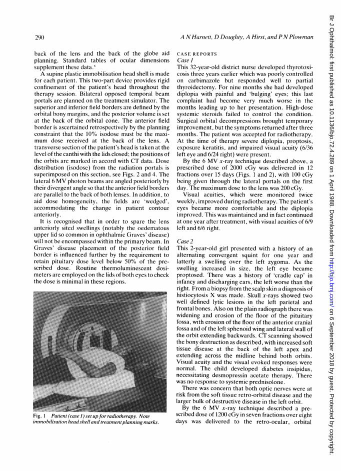
Fig. 1 Patient (case I) set upfor radiotherapy. Noteimmobilization head shell and treatmentplanning marks.
CASE REPORTS
Case IThis 32-year-old district nurse developed thyrotoxi-cosis three years earlier which was poorly controlledon carbimazole but responded well to partialthyroidectomy. For nine months she had developeddiplopia with painful and 'bulging' eyes; this lastcomplaint had become very much worse in themonths leading up to her presentation. High-dosesystemic steroids failed to control the condition.Surgical orbital decompressions brought temporaryimprovement, but the symptoms returned after threemonths. The patient was accepted for radiotherapy.At the time of therapy severe diplopia, proptosis,exposure keratitis, and impaired visual acuity (6/36left eye and 6/24 right) were present.By the 6 MV x-ray technique described above, a
prescribed dose of 2000 cGy was delivered in 12fractions over 15 days (Figs. I and 2), with 100 cGybeing given through the lateral portals on the firstday. The maximum dose to the lens was 200 cGy.
Visual acuities, which were monitored twiceweekly, improved during radiotherapy. The patient'seyes became more comfortable and the diplopiaimproved. This was maintained and in fact continuedat one year after treatment, with visual acuities of 6/9left and 6/6 right.
Case 2This 2-year-old girl presented with a history of analternating convergent squint for one year andlatterly a swelling over the left zygoma. As theswelling increased in size, the left eye becameproptosed. There was a history of 'cradle cap' ininfancy and discharging ears, the left worse than theright. From a biopsy from the scalp skin a diagnosis ofhistiocytosis X was made. Skull x-rays showed twowell defined lytic lesions in the left parietal andfrontal bones. Also on the plain radiograph there waswidening and erosion of the floor of the pituitaryfossa, with erosion of the floor of the anterior cranialfossa and of the left sphenoid wing and lateral wall ofthe orbit extending backwards. CT scanning showedthe bony destruction as described, with increased softtissue disease at the back of the left apex andextending across the midline behind both orbits.Visual acuity and the visual evoked responses werenormal. The child developed diabetes insipidus,necessitating desmopressin acetate therapy. Therewas no response to systemic prednisolone.There was concern that both optic nerves were at
risk from the soft tissue retro-orbital disease and thelarger bulk of destructive disease in the left orbit.By the 6 MV x-ray technique described a pre-
scribed dose of 1200 cGy in seven fractions over eightdays was delivered to the retro-ocular, orbital
290
on 6 Septem
ber 2018 by guest. Protected by copyright.
http://bjo.bmj.com
/B
r J Ophthalm
ol: first published as 10.1136/bjo.72.4.289 on 1 April 1988. D
ownloaded from

Radiotherapy in benign orbital disease. II: Ophthalmic Graves' disease and orbital histiocytosisX
Fig. 2 CTscan ofthe orbits (case1). Radiation isodoses aresuperimposed showing 10%isodose at back oflens and 100%encompassing the back oftheorbital cone. Note also the greatlyenlarged medial rectus muscles.
regions, the treatment volume extending furtherposteriorly than in case 1 to encompass the retro-orbital soft tissue extension, hypothalamus, andpituitary (Figs. 3 and 4). However, a lower dose wasemployed.The diabetes insipidus did not respond. The swell-
ing in the left zygoma region and the proptosisregressed over a few months and remained so at oneyear. Magnetic resonance imaging at nine monthsafter therapy confirmed less soft tissue disease. Thechild remains well.
Discussion
Traditional reluctance to employ radiotherapy forbenign orbital disease has been based on anxietyconcerning late morbidity, notably cataractogenesis.However, with modern radiotherapeutic techniques,well collimated x-ray beams can treat the orbit whilekeeping the lens below its cataract threshold. Thetechnique described here relies on modern treatmentequipment and on known depths of critical ocularpoints by high definition CT scanning with cursormeasurements, supplemented by standard tables ofocular measurements. In addition, and critically, ahead fixed by a full head shell allows reproducibilityof positioning. These factors allow the dosimetricadvance over previous treatment methods whichhistorically used orthovoltage x-rays or telecobaltbeams, the lateral skin canthus as the set-up mark forthe anterior field edge or a bite block only to supportthe head.
In 1951 Jones9 described a series of 29 patients withGraves' ophthalmopathy treated by radiotherapy atSt Bartholomew's Hospital. He showed that it is thepatients with fast, progressive, and serious diseasethat respond best to radiotherapy, an observationthat has been confirmed by others.3 Glaser"' con-sidered that the indications for orbital radiotherapyin Graves' ophthalmopathy were only two: these aresteroid refractory patients with 'documented opticneuropathy with visual acuity less than 20/40 oran acute painful inflammatory phase of Graves'disease'. Our own indications also include pro-
Fig. 3 Patient (case2) set upfor radiotherapyfor orbitalhistiocytosis X. The immobilisation headshell is againimportantforprecision treatment.
291
on 6 Septem
ber 2018 by guest. Protected by copyright.
http://bjo.bmj.com
/B
r J Ophthalm
ol: first published as 10.1136/bjo.72.4.289 on 1 April 1988. D
ownloaded from
A N Harnett, D Doughty, A Hirst, and PN Plowman
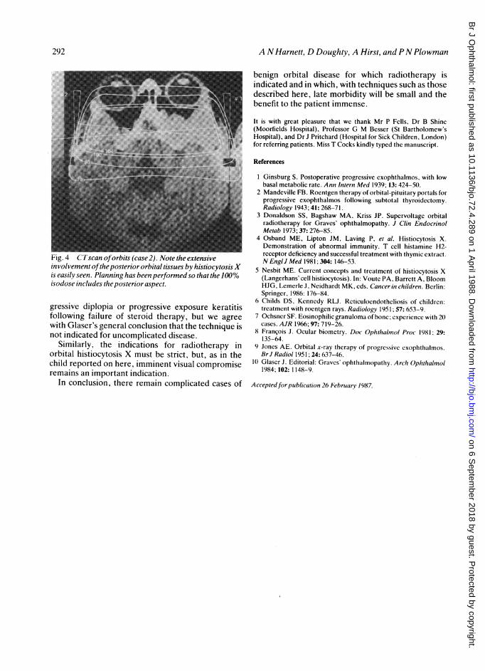
Fig. 4 C'Tscan oforbits (case2). Note the extensiveinvolvement oftheposterior orbital tissues by histiocytosisXis easily seen. Planning has been performed so that the 100%isodose includes the posterior aspect.
gressive diplopia or progressive exposure keratitisfollowing failure of steroid therapy, but we agreewith Glaser's general conclusion that the technique isnot indicated for uncomplicated disease.
Similarly, the indications for radiotherapy inorbital histiocytosis X must be strict, but, as in thechild reported on here, imminent visual compromiseremains an important indication.
In conclusion, there remain complicated cases of
benign orbital disease for which radiotherapy isindicated and in which, with techniques such as thosedescribed here, late morbidity will be small and thebenefit to the patient immense.
It is with great pleasure that we thank Mr P Fells, Dr B Shine(Moorfields Hospital), Professor G M Besser (St Bartholomew'sHospital), and Dr J Pritchard (Hospital for Sick Children, London)for referring patients. Miss T Cocks kindly typed the manuscript.
References
1 Ginsburg S. Postoperative progressive exophthalmos, with lowbasal metabolic rate. Ann Intern Med 1939; 13: 424-50.
2 Mandeville FB. Roentgen therapy of orbital-pituitary portals forprogressive exophthalmos following subtotal thyroidectomy.Radiology 1943; 41: 268-71.
3 Donaldson SS, Bagshaw MA, Kriss JP. Supervoltage orbitalradiotherapy for Graves' ophthalmopathy. J Clin EndocrinolMetab 1973; 37: 276-85.
4 Osband ME, Lipton JM, Laving P. et al. Histiocytosis X.Demonstration of abnormal immunity. T cell histamine H2-receptor deficiency and successful treatment with thymic extract.N EnglJ Med 1981; 304:146-53.
5 Nesbit ME. Current concepts and treatment of histiocytosis X(Langerhans' cell histiocytosis). In: Voute PA, Barrett A, BloomHJG, Lemerle J, Neidhardt MK, eds. Cancer in children. Berlin:Springer, 1986: 176-84.
6 Childs DS, Kennedy RLJ. Reticuloendotheliosis of children:treatment with roentgen rays. Radiology 1951; 57: 653-9.
7 Ochsner SF. Eosinophilic granuloma of bone; experience with 20cases. AJR 1966; 97: 719-26.
8 Franqois J. Ocular biometry. Doc Ophthalmol Proc 1981; 29:135-64.
9 Jones AE. Orbital x-ray therapy of progressive exophthalmos.Br J Radiol 1951; 24: 637-46.
10 Glaser J. Editorial: Graves' ophthalmopathy. Arch Ophthalmol1984; 102: 1148-9.
Acceptedfor publication 26 February 1987.
292
on 6 Septem
ber 2018 by guest. Protected by copyright.
http://bjo.bmj.com
/B
r J Ophthalm
ol: first published as 10.1136/bjo.72.4.289 on 1 April 1988. D
ownloaded from