Radiologico-pathologic Correlation of the Pulmonary ... · cases of bronchopneumonia associated...
8
3 pp . 477 - 484, 1988 Journal of Korean Radiologi cal Society, 24(3) 477- 484, 1988 Radiologico-pathologic Correlation of the Pulmonary Disease in the Autopsied Infants Young Seok Lee, M.D. , Dae Chul Suh , M. D.' , Sung Sik Lee , M.D. ** Soon 11 Lee , M.D." , Je Geun Chi , M.D. De partm ent of Radi ol ogy , So wha Children ’ 's H ospital • ••• 1. 2. (27% ), ), 3. 50% , 77 . 8% , 88 . 9% , 4. • Introduction pulmonary hemorrhage are also most frequently encountered 1 -2). It has been known that the major causes of The authors analyzed radiological and patholo- death in infancy are many conditions originating in gical findings of perinatal pulmonary disease trying perinatal period. Pulmonary disease is the most to elucidate radiological clues for the diagnosis , common cause of morbidity and mortality in the treatment and prognosis of pulmonary diseases in newbron infan t. Pathologically , hyaline membrane the newborn infants. disease , fetal aspiration syndrome , pneumonia and • • Department o[ R adiol ogy, Chung Ang Gil Genera l Hospi ta l •• • Department o[ Pediatrics, So wh a Children ’ 's Hos. pital … .. Department o[ Pa th ology, S eoul National U niversi ty Children ’s Hos pital 24 Materials and Methods The materials for this study were sixty two in- fants who expired in Neonatal Intensive Care Unit at Sowha Children’ s Hospital from Nov. 1984 to Mar , 1986. The postmortem examinations were performed at department of pathology , Seoul National University Children ’ s Hospital. We retrospectively reviewed radiologic and - 477-
Transcript of Radiologico-pathologic Correlation of the Pulmonary ... · cases of bronchopneumonia associated...

大햄}jj( íl、u흉뽑횡용註‘ 第 24 卷 第 3 號 pp. 477 - 484, 1988 Journal of Korean Radiological Society, 24(3) 477-484, 1988
Radiologico-pathologic Correlation of the
Pulmonary Disease in the Autopsied Infants
Young Seok Lee, M.D. , Dae Chul Suh, M.D.' , Sung Sik Lee , M.D. **
Soon 11 Lee, M.D. " , Je Geun Chi , M.D.
D epartment o f R adiology , S o wha Children ’'s Hospital
〈국문초록〉
부검을 시행한 신생아에 있어서 폐질환의 방사선학적 병리학적 고찰
소화아동병 원 방사선과
이영석 • 서대철 이성식 이순일 지제근 •••
1 984년 11월 부터 1986년 3월 까지 소화아동뱅원 신생아 집중치료실에서 사망하여 서울대학교 의파대
학 뱅러학교살에서 부검을 시행한 62영의 폐 질환에 대한 방사션학석 , 영리학석 고찰을 하여 마음과 같
은 결론을 얻었마.
1. 총 62명의 신생아중 미 숙아 33예 (53 . 2%) , 저출생체중아 44예 ( 7 1. 0%)였다.
2. 발생 반도에 따른 폐질환의 분포는 페램이 33예 (53% )로 가장 않았A여 그외 유리질막질환 1 7예
(27 % ) , 폐영변이 없는 경우 9예 ( 15% ), 폐 형성부선증이 3예 (5%) , 폐 출혈이 1예 ( 2 %)였 마.
3. 양수흡인성 퍼l 령의 50% , 선천성 폐령의 77. 8% , 후천성 폐령의 88. 9% , 유리 질악질환의 88 . 2 %에
서 방사선학척 소견이 병리 학적 소견과 일치 하였다.
4. 선천성 폐 렴 9예 의 방사선학적 소견은 폐 문주위 칭 윤을 보인 경 우가 4여1. 미 만성 파립 성 • 앙상결절
성 첨윤이 3예 , 페 침윤이 보이지 않는 경우가 2예였마.
Introduction pulmonary hemorrhage are also most frequently encountered 1-2) .
It has been known that the major causes of The authors analyzed radiological and patholo-
death in infancy are many conditions originating in gical findings of perinatal pulmonary disease trying
perinatal period. Pulmonary disease is the most to elucidate radiological clues for the diagnosis , common cause of morbidity and mortality in the treatment and prognosis of pulmonary diseases in
newbron infant. Pathologically , hyaline membrane the newborn infants.
disease , fetal aspiration syndrome , pneumonia and
• 중앙걸뱅원 방사선파 • Department o[ R adiology, Chung Ang Gil General
Hospi tal •• 소화 아동병 원 소아파 • Department o[ Pedia trics, Sowha Children ’'s Hos.
pital … 서울대학교 의파대 학 뱅 리학교실 .. Department o[ Pathology, S eo ul National Universi ty
Children ’s Hospital 이 논문은 1988년 4월 30일에 캡 수하여 1 988년 6월 24 일에 채택되었음
Materials and Methods
The materials for this study were sixty two in
fants who expired in Neonatal Intensive Care Unit
at Sowha Children’s Hospital from Nov. 1984 to
Mar, 1986. The postmortem examinations were
performed at department of pathology, Seoul
National University Children ’s Hospital.
We retrospectively reviewed radiologic and
- 477-

1988 -第 3 號第 24 卷- 大韓放射線醫學會註 :
Gestational Age and Birth Weight Distribution of Autopsied Infants Table 1.
만쁘Weight(gm )
Gestational Age(wks) Total
?l
qι 。ι
nU
5
8
7
2
---i
qι
2,500 <
o o n l
1,500-2 ,500
8
얘 8
1
< 1,500
-32
32-37 37-42 42-
62 18 33 11 Total
Distribution of Pulmonary Disease Table 2. pathologic findings of lungs in sixty two autopsied
intants. We also evaluated distribution and pat
terns of p비monary diseases and the radio
logico-pathologic correlation. M
9
9
N。
32
n 3
1
9
Pneumonia Aspiration Congenital Acquired
Hyaline Membrane Disease Pulmonary Hypoplasia Pulmonary Hemorrhage No pulmonary Lesion
Diseases
62
nia . The amniotic fluid aspiration pneumonia was
associated with 5 cases of meconium and hemorr
hage, 4 of meconium , 2 of meconium and bron
chopneumonia and one case of broncho
pneumonia(Fig. 1) .
Findings of chest roentgenograms were diagnos
tic for only in 7 cases among all amniotic fluid
aspiration penumonia. The other half showed nor
mal findings in 5 cases and hyperinflation without
p비monic infiltrations in 2 cases(Table 3) .
Total
There were 33 cases(53 .2%) of prematurity
among the sixty two infants and 44 cases(71.1 % )
of low birth weight including 11 cases of the very
low birth weight less than 1,500 gm(Table 1) . The
male to female ratio was l.3: l.
The pu!monary diseases were confirmed patho
logically through complete postmortem examina
tion. They consisted of 33 cases of pneumonia, 17
of hyaline membrane diseases , 3 of pulmonary
hypoplasia , one of pulmonary hemorrhage and 9
of no demonstrable pulmonic lesions(Table 2)
Pneumonia was the most common pulmonary dis
ease in the autopsied infants.
Among 33 cases of pneumonia, there were 14
cases of amniotic fluid aspiration pneumonia , 9 of
congenital pneumonia and 9 of acquired pneumo-
Results
Radiologico-Pathologic Correlation of Amniotic Fluid Aspiration Pneumonia Table 3.
Chest Roentgenogram Diagnostic Not diagnostic
Total
N 0 Associate Lesion c Meconium c Meconium & Hemorrhage c Meconium & Bronchopneumonia c Bronchopneumonia
?ι A*
FD
9“ 14
2
4
1
?‘ ?“ 1i
14
1i
Pathological Diagnosis
14
- 478-
7 7 Total

- Y oung Seok Lee , et al.: Radiologico-patholog ic Correlat ion oÍ the Pulmonary .. -
A
Fig. 1. Amniotic fluid aspiration pneumonia and superimposed bacterial pneumonia
A. Non-uniform , coarse infiltrations and hyperinflati on of the lung on the first day 。f life
B. Progressive hyperinflation and patchy infiltrations of the lung on the seventh day of life
C. Gross specimen of the lung Cut surface of the right and left lungs show diffuse and patchy di scoloration in the background of pulmonary congestion
Fig. 2. 2 Cases of congenital pneumonia A. P erihilar streaky infiltrations of both lungs
and decreased thymic shadow B. Diffuse granul ar densities of both lungs
and prominent thymic shadow C. Microscopic picture showing interstitial
thickening and small round cell infiltration The alveolar spaces are re latively preserved
- 479 -
A
B
C
- 大韓放射線醫學會誌 : 第 24 卷 第 3 號 1988
Radiological findings of congenital pneumonia
(9 cases) presented perihilar streakl) , diffuse
granular or reticulonodular3) and no demonstrable
p비monic infiltrations2). In addition , thymic sha
dows were invisible or decreased in 8 cases and
prominent in one case on the chest roentgeno
grams(Table 4)(Fig. 2).
Nine cases of acquired pneumonia consisted of 6
cases of bronchopneumonia associated with
hemorrhage2) , and 3 cases of pneumocystis carinii
pneumonia with 2 of cytomegalic inclusion dis
eases(Fig. 3) and one candidiasis. One case of
pneumocystis carinii pneumonia showed atypical
findings of pneumatocele. There were radiologi
co-pathologic good correlations with 8 cases of
acquired pneumonia except only one case of nor
mal chest roentgenographic finding(Table 3).
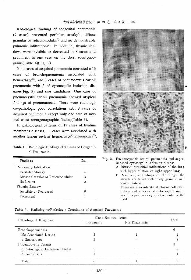
In pathological patterns of 17 cases of hyaline
membrans diseases , 11 cases were associated with
another lesions such as hemorrhage4) , pneumonia2
),
Table 4. Radiologic Findings of 9 Cases of Congenit. al Pneumonia
Findings N。
Pulmonary Infiltration Perihilar Streaky 4 Diffuse Granular or Reticulonodular 3 No Lesion 2
Thymic Shadow Invisible or Decreased 8 Prominent
A
B
Fig. 3. Pneumocystitis cann l1 pneumonia and super. imposed cytomegalic inclusion disease A. Dìffuse interstitial infiltrations of the lung
with hyperinflation of right upper lung B. Microscopic findiilgs of the lungs. the
alveoli are filled with finely granular and foamy material There are also interstitial plasma cell infiltration and a focus of cytomegalic inclu. sion.in a pneumonocyte in the center of the field
Table 5. Radiologico-Pathologic Correlation of Acquired Pneumonia
Pathological Diagnosis Diagnostic Not Diagnostic
Chest Roentgenogram Total
Bronchopneumonia N 0 Associated Lesion c Hemorrhage
Pneumocystis Carinii c Cytomegalic Inclusion Disease c Candidiasis
6
3 2
4·?ι
q니
2 2
Total 9 8
- 480-
A
B
- Y oung Seok Lee , et al.: Radiologico'pathologic Correlation 01 the Pulmonary" 'I-
hemorrhage and pneumonia3l(Fig. 4) , hemorrhage
and bronchopulmorary dysplåsia ll(Fig . 5) and
meconium aspiration1l, six cases were not associ
ated with other lesions.
The findings of chest roentgenogram was di
agnostic for hyaline membrane diseases and its
associated lesions in 15 cases. Two cases of hyaline
membrane diseases showed normal finding and
patchy infiltrations on chest roentgenogram re
spectively(Table 6) .
Distribution of misceIlaneous cases was one case
of pulmonary hemorrhage and 3 cases of p비mon
ary hypoplasia related to two of Potter’s syn
dromes and one of 18 trisomy.
Discussion
Mortality rate of newborn and neonate has been
decreased by the development of neonatology and
other supporting systems. Nevertheless, respira-
C
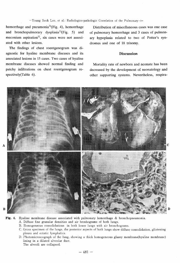
Fig. 4. Hyaline membrane disease associated with pulmonary hemorrhage & bronchopneumonia A. Diffuse fine granular densities and ai r bronhograms of both lungs B. Homogeneous consolidations in both lower lungs with air bronchograms C. Gross specimen of the lungs. the posterior aspects of both lungs show di ffus딩 consolidation, glistening
pleura and ectatic lymphatics D. Photomicroscograph of the lung, showing a thick homogeneous glassy membrane(hyaline membrane)
lining in a dilated alveolar duct The alveoli are collapsed
- 481-
D

- 大韓放射線훌훌學會誌 : 第 24 卷 第 3 號 1988 -
A
B
C
Fig. 5. Hyaline membrane disease associated with bronchopulmonary dysplasia A. Diffuse granular densities and fine air
bronchograms of both lungs on the third day of life
B. Irregular linear increased densities and uneven aeration of the lungs on the twenty day of life
c. Photomicrograph of the lung, showing obliterated alveolar spaces and replacement of fibrous connective tissue. Focal proliferation of alveolar penumonocytes is also note
tory disorders have been the major causes of mor
tality and morbidity in infancy until now.
According to Schaffer’s report3) , pulmonary in
flammation of some degree was found in 27(35%)
of 76 autopsies on stillborn and newborn infants ,
was considered a contributory cause of death in
7(9%) , and was the sole causε of death in 6(8%).
In our series, pneumonia also was the most
common cause of the p비monary diseases in the
autosied infants . Florman et a14) observed en
hancement of bacterial growth in amniotic fluid by
meconium and they suggested that the fetus who
remained in meconium stained amniotic fluid
might be exposed to far greater numbers of bac
teria than one whose amniotic fluid does not con
tam mecomum _
According to previous reports , radi이ogical fea
tures of meconium aspiration pneumonia consisted
of non-uniform , coarse , patchy infiltrates radiating
from hilum :ïnto the peripheral lung fields and une
ven aeration of the lung2,5,6). 50% of amniotic and
mecomum asplrahon pneumonía m our senes were
compatible with the radiologic features. Gooding
and Gregory7) found that 60 per cent of the infants
born through meconium stained amniotic fluid had
normal chest roentgenograms. 35.7% of all amnio
tic aspiration pneumonia in our series showed nor
mal chest roentgenograms.
Diffuse , though not necessarily uniform , in
flammation has been the prominent pattern associ
ated with congenital pneumonia. The inflamma
tory infiltrate was largely of lymphocytes , plasma
cells and neutrophils almost in equal composition , and its distribution was more interstitial than
alveolar. Although there were some exudates in
the alveoli they were mostly cellular and fibrin
exudation was minimaL In most of the cases prem
ature rupture of membrane was noted in the his
tory of the mothers.
The inflammatory cell reaction has been vari
ably described as acute , mononuclear and intersti
tial but diffl.lse pne l.lmonia in the peripheral por-
- 482-

- Young Seok Lee , et al.: Radiologico'pathologic Correlation of the Pulmonary ... -
Table 6. Radiologico'Pathologic Correlation of Hyaline Membrane Disease
Pathological Diagnosis Diagnostic Not diagnostic
Chest Roentgenogram Total
No Associated Lesion c Hemorrhage c Pneumonia c Hemorrhage & Pneumonia c Hemorrhage & Bronchopulmonary
Dysplasia c Meconium Aspiration
ζU A%
1i
。J
ζU A*
ηι 。、υ
Total 2 15 17
tion of the lung has been conversely taken as
characteristic of antenatal infection용11) The au
thors reviewed radiological findings of pathologi
cally proven congenital penumonia. 77.8% of con
genital pneumonia showed characteristic patterns
of interstitial pneumonia which were consistent
with pathológical findings. The other 22.2 per cent
showed normal chest roentgenograms probably
due to being masked interstitial densities by over
aeration of the lung.
Diagnostic accuracy of chest roentgenograms
was 88.9% for 뽕quired pneumonia in this series.
One case of pneumnocystis carir피 pneumonia
showed penumatocele, one of unusual manifesta
tions including pleural effusion , empyema, pneumothorax, pneumomediastinum, lobar con
solidation and nodular densities12).
The name “ hyaline membrane disease" is de
rived from the histopathologic findings. At necrop
sy , the lung are of uniform dark purple color and
sink when placed in water. Microscopically the
alveoli are a characteristically atelectatic, and the
frequently overdistended alveolar ducts are lined
with rather amorphous eosinophilic materials13 ,14).
The radiologic patterns of hyaline membrane dis
ease can be described as finely granular or even
reticulonodular densities and air bronchograms ex
tending far peripherally into the lung. 88.2 per cent
of the author ’s cases showed the characteristic
findings for hyaline membrane disease.
Ellis et a114) reported that acquired pulmonary
disease other than hyaline membrane disease such
as hemorrhage, pneumonia, atelectasis, or aspira
tion of amniotic fluid , singly or in comtination , was a major finding at necropsy. The authors also
experienced hyaline membrane disease associated
with hemórrhage , pneumonia , bronchopulmonaπ dysplasia and aspiration of meconium.
Summary
Authors retrospectively analysed chest roent
genographic and pathologic findings of the sixty
two autQpsied infants under the clinical informa
tions. We also evaluated distribution and patterns
of P비monary diseases and the radiologicc• patho
logic correlation. The results were as follow:
1. There were 33 cases(53.2%) of prematurity
among Sixty-two infants and 44 cases(71.0%) of
low birth weight.
2. The distribution of the pulmonary diseases , in
order of frequency , was 33 cases(53%) of pneumo
nia , 17(27%) of hyaline membrane diseases , 9(15% ) of no demonstrable pulmonic lesion, 3(5%) of pulmonary hypoplasia and 1(2%) of pul
monary hemorrhage.
3. The radiological findings were consistent with
the pathological findings in 50% of amniotic fluid
- 483-
- 大韓放射線훌훌學會誌 : 第 24 卷 第 3 號 1988 -
aspiration pneumonia 88 .9% of acquired and 88.2
% of hyaline membrane diseases.
4. Radiological findings of 9 cases of congenital
pneumonia were perihilar streaky infiltrations in 4
cases, diffuse granular or reticulonodular infiltra
tions in 3 cases , and no demonstrable pulmonic
infiltrations in 2 cases .
REFERENCES
l. Hoffman RR , Campbell RE , Decker ]P: Fetal
Aspiration syndrome Clinical, roentgenologic and
pathologic features. A]R 122(1): 90 , 96, 1974
2. Peterson HG , pendleton ME: Contrasting Roentge.
nographic pulmonary patterns of the hyaline mem
brane and fetal Aspiration syndrome. A]R
74(5):800-817, 1955
3. Schaffer AJ, Markowitz M, Perlman A: Pneumonia
in Newborn Infants. ].A. M. A. 159(η 663-668, 1955
4. Florman AL , Teubner D: Enhancement of Bacterial
Growth in amniotic fluid by Meconium. ]. Pediatr
74(1): 111 -114, 1969
5. Bacsik RD: Meconium Aspiration syndrome Pedia-
tric Clinics of North America 24(3): 463-079, 1977
6 이영석, 이 성식, 이홍규 둥 : 신생아에 있어서 태변
흉안층의 방사선학적 고창. 대한방사선의학회지 21
(1): 76-83, 1985
7. Gooding CA , Gregory GA: Roentgenographic analy
sis of meconium Aspiration. Radiology 100:131 ,
1971
8. Bernstein ] , Wang ]: Thepathology of Neonatal
pneumonia. Am ] dis of child 101:104-116, 1961
9. Adams ]M ‘ Congenital pneumonitis in Newborn
infants Am ] dis of child 75:544-554, 1948
10. Anderson GS , Green CA , Neligon BA et al: Con
genital Bacterial Pneumonia. Lan cet 2:585-587,
1962
11 이성식, 이 순일, 이영석 둥 : 선천성 폐램에 대한 임
상. 영리학적 고찰. 소아과 29(6) : 653-660, 1986
12. Luddy RE , Champion LA , Schwartz AD et al
Pneumocystitis carinii pneumonia with pn euma
tocele formation. Am ] dis of child 131 :4 70, 1977
13. Wohlfeld GM: Hyaline Membran e Disease. A]R
93:425-427, 1965
14. Ellis K, N adelhaft ]: Roentgenographic Findings in
Hyalin e Membrane disease in infants weighing
2,000 Grams and over, A]R 28(3):444 -45α 1957
- 484