Radiological distribution of pulmonary emphysema · Radiological distribution of pulmonary...
9
Thorax (1974), 29, 81. Radiological distribution of pulmonary emphysema Clinical and physiological features of patients with emphysema of upper or lower zones of lungs N. A. MARTELLI1, D. C. S. HUTCHISON, and C. E. BARTER2 Chest Unit, King's College Hospital, London SE5 Martelli, N. A., Hutchison, D. C. S., and Barter, C. E. (1974). Thorax, 29, 81-89. Radiologi- cal distribution of pulmonary emphysema: clinical and physiological features of patients with emphysema of upper or lower zones of lungs. Pulmonary emphysema exists in two main pathological forms, centrilobular and panlobular (panacinar) emphysema, the lesions predominantly affecting the upper and lower zones of the lungs respectively. There is disagreement among authors as to the clinical and physiological differences between these two forms, and direct evidence of the pathological type is seldom available during life. Patients with emphysema can, however, be divided on radiological criteria into an 'upper zone' and a 'lower zone' group, and it can be argued that these groups relate respectively to the centrilobular and panlobular forms of the disease. The evidence is far from conclusive but it was thought that a comparison of the two radiological groups would be of value. Patients in whom there was no obvious zonal preponderance were not included in the study. Fifty patients with definite radiological evidence of pulmonary emphysema have been studied, those with a1-antitrypsin deficiency being excluded. Thirty-one patients (62%) had emphysema which predominantly affected the upper zones of the lungs; the lower zones were the more severely affected in the remainder. Bullae were found in approximately equal proportions in each group. All the patients were, or had been, cigarette smokers. There was no significant difference between the mean ages of the two groups; only seven patients were free from exertional dyspnoea, all being in the upper zone group. Chronic bronchitis occurred with equal frequency in the two groups but started on average about 10 years earlier in those with lower zone disease; the latter patients had rather more severe aiiflow obstruction and more severe blood-gas abnormalities. The presence or absence of chronic bronchitis per se, however, did not appear to have any significant effect upon the common respiratory function tests. No data emerged from this study which suggested that there were differing aetiological factors in the two groups. Pulmonary emphysema is generally considered to occur in two main pathological forms (Gough, 1952; McLean, 1956; Leopold and Gough, 1957) now commonly known as centrilobular emphysema and panlobular (or panacinar) emphysema; the former type is found mainly in the upper zones of the lungs and the latter in the lower zones, particularly in the advanced case (Thurlbeck, 1963 ; Wyatt, Fischer, and Sweet, lPresent address: Centro de Rehabilitaci6n Respiratoria 'Maria Ferrer', E. Finochietto 849, Buenos Aires, Argentina 2Present address: Repatriation General Hospital, Heidelberg West, 3077 Victoria, Australia Requests for reprints: Dr. D. C. S. Hutchison, Chest Unit, King's College Hospital, London SE5 1964). A precise classification is, however, difficult in many cases (Thurlbeck et al., 1969; Mitchell et al., 1970), and not all authors agree about the clinical, radiological, and physiological features, particularly of centrilobular emphysema (Sweet, Wyatt, Fritsch, and Kinsella, 1961 ; Snider, Brody, and Doctor, 1962; Reid, 1967; Thurlbeck, Henderson, Fraser, and Bates, 1970). In one type of emphysema, however, that associated with al-antitrypsin deficiency, there seems little doubt that the lower zones are predominantly affected (Eriksson, 1965; Guenter et al., 1968; Hutchison et al., 1971). Direct evidence of pathological type is seldom 81 G on February 28, 2020 by guest. Protected by copyright. http://thorax.bmj.com/ Thorax: first published as 10.1136/thx.29.1.81 on 1 January 1974. Downloaded from
Transcript of Radiological distribution of pulmonary emphysema · Radiological distribution of pulmonary...
Thorax (1974), 29, 81.
Radiological distribution of pulmonary emphysemaClinical and physiological features of patients with emphysema of upper or
lower zones of lungs
N. A. MARTELLI1, D. C. S. HUTCHISON, and C. E. BARTER2
Chest Unit, King's College Hospital, London SE5
Martelli, N. A., Hutchison, D. C. S., and Barter, C. E. (1974). Thorax, 29, 81-89. Radiologi-cal distribution of pulmonary emphysema: clinical and physiological features of patients withemphysema of upper or lower zones of lungs. Pulmonary emphysema exists in two mainpathological forms, centrilobular and panlobular (panacinar) emphysema, the lesionspredominantly affecting the upper and lower zones of the lungs respectively. There isdisagreement among authors as to the clinical and physiological differences between thesetwo forms, and direct evidence of the pathological type is seldom available during life.Patients with emphysema can, however, be divided on radiological criteria into an 'upperzone' and a 'lower zone' group, and it can be argued that these groups relate respectivelyto the centrilobular and panlobular forms of the disease. The evidence is far from conclusivebut it was thought that a comparison of the two radiological groups would be of value.Patients in whom there was no obvious zonal preponderance were not included in the study.
Fifty patients with definite radiological evidence of pulmonary emphysema have beenstudied, those with a1-antitrypsin deficiency being excluded. Thirty-one patients (62%)had emphysema which predominantly affected the upper zones of the lungs; the lowerzones were the more severely affected in the remainder. Bullae were found in approximatelyequal proportions in each group. All the patients were, or had been, cigarette smokers.There was no significant difference between the mean ages of the two groups; only sevenpatients were free from exertional dyspnoea, all being in the upper zone group. Chronicbronchitis occurred with equal frequency in the two groups but started on average about10 years earlier in those with lower zone disease; the latter patients had rather more severeaiiflow obstruction and more severe blood-gas abnormalities. The presence or absence ofchronic bronchitis per se, however, did not appear to have any significant effect upon thecommon respiratory function tests. No data emerged from this study which suggested thatthere were differing aetiological factors in the two groups.
Pulmonary emphysema is generally considered tooccur in two main pathological forms (Gough,1952; McLean, 1956; Leopold and Gough,1957) now commonly known as centrilobularemphysema and panlobular (or panacinar)emphysema; the former type is found mainlyin the upper zones of the lungs and the latter inthe lower zones, particularly in the advancedcase (Thurlbeck, 1963 ; Wyatt, Fischer, and Sweet,lPresent address: Centro de Rehabilitaci6n Respiratoria 'MariaFerrer', E. Finochietto 849, Buenos Aires, Argentina2Present address: Repatriation General Hospital, Heidelberg West,3077 Victoria, AustraliaRequests for reprints: Dr. D. C. S. Hutchison, Chest Unit, King'sCollege Hospital, London SE5
1964). A precise classification is, however, difficultin many cases (Thurlbeck et al., 1969; Mitchellet al., 1970), and not all authors agree aboutthe clinical, radiological, and physiologicalfeatures, particularly of centrilobular emphysema(Sweet, Wyatt, Fritsch, and Kinsella, 1961 ; Snider,Brody, and Doctor, 1962; Reid, 1967; Thurlbeck,Henderson, Fraser, and Bates, 1970). In one typeof emphysema, however, that associated withal-antitrypsin deficiency, there seems little doubtthat the lower zones are predominantly affected(Eriksson, 1965; Guenter et al., 1968; Hutchisonet al., 1971).
Direct evidence of pathological type is seldom
81G
on February 28, 2020 by guest. P
rotected by copyright.http://thorax.bm
j.com/
Thorax: first published as 10.1136/thx.29.1.81 on 1 January 1974. D
ownloaded from
N. A. Martelli, D. C. S. Hutchison, and C. E. Barter
available during life, and in view of differingopinions which have been expressed, we feltthat there would be reasonable grounds for study-ing the relationship between clinical and physio-logical findings and radiological distribution ofthe lesions. We therefore selected two groups ofpatients for special study, one group having radio-logical evidence of emphysema predominantlyaffecting the upper zones and the other group(omitting those with a1-antitrypsin deficiency)with evidence of lower zone disease. The argu-ments for and against this approach will bediscussed later in this report.
PATIENTS AND METHODS
The patients included in this study were referredto the Chest Unit from chest clinics, and othermedical or surgical units, in or outside the King'sCollege Hospital Group. Seventy-eight patientsattending for the first time during the period1964 to mid-1971 had definite radiological evi-dence of pulmonary emphysema (Laws andHeard, 1962) which occurred either alone or inassociation with bronchitis. Patients with asthma
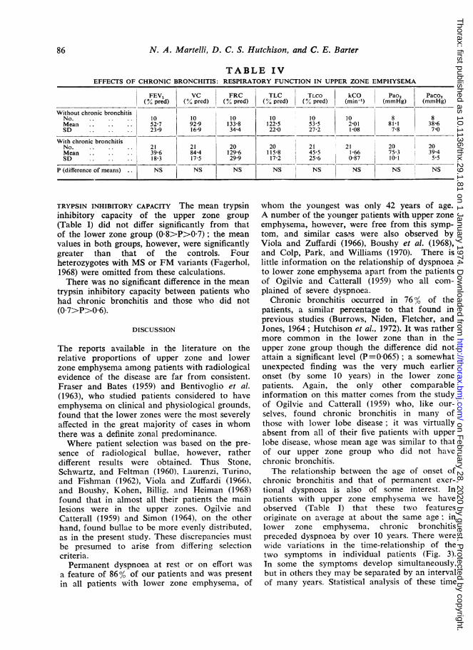
or severe pulmonary fibrosis and those who hadundergone previous thoracic surgery were notconsidered. No patients were included unlessobvious and extensive pulmonary abnormalitieswere seen (Figs 1 and 2).
In the 50 patients who form the main study,emphysema predominantly affected either theupper half or the lower half of the lung fieldsas seen on the postero-anterior chest radiograph.A detailed case history was obtained by theauthors and special emphasis was placed on thepresence and age of onset of permanent un-remitting exertional dyspnoea and of chronicbronchitis as defined by the Medical ResearchCouncil (1965). The total lifetime cigarette con-sumption was expressed in grams, as previouslydescribed (Hutchison et al., 1971). The bodyweight was expressed as a percentage of the meanvalue for subjects of equivalent height and age(Society of Actuaries, 1959).Data from 18 severely affected patients have
been included in a previous study (Hutchisonet al., 1972); 28 of the original 78 patients wereexcluded from the main study:
(a) eight patients with al-antitrypsin defi-
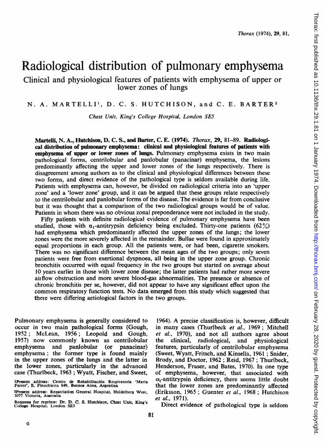
FIG. 1. Upper zone emphysema: 45-year-old man with normal exercisetolerance. FEV1 53% predicted value; TLco 113% predicted value.
82
on February 28, 2020 by guest. P
rotected by copyright.http://thorax.bm
j.com/
Thorax: first published as 10.1136/thx.29.1.81 on 1 January 1974. D
ownloaded from
Radiological distribution of pulmonary emphysema
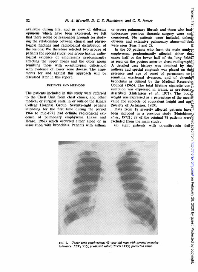
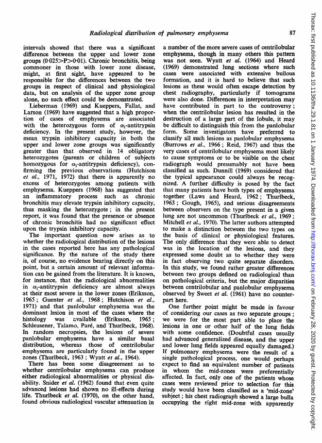
FIG. 2. Lower zone emphysema: a severely disabled 63-year-old man.FEy1 24% predicted value. TLCO 37% predicted value.
ciency previously described (Hutchison et al.,1971);
(b) eight patients in whom there was no definitepredominance of emphysema in either the upperor the lower half of the lung fields. In one patienta large bulla occupied the central part of theright lung field;
(c) 12 patients for whom data were incomplete;eight had upper zone emphysema and four hadlower zone emphysema.
RADIOLOGY Standard postero-anterior and lateralchest radiographs were obtained in all patients,and bilateral whole-lung tomograms in all buttwo. The tomograms were taken with the patientsupine, at intervals of 3-4 cm, from approximately6 to 20 cm from the table top. Emphysema wasrecorded as present when the peripheral vascula-ture was attenuated or destroyed (Laws and Heard,1962). The localization of bullae, if present, wasalso noted. A decision was made as to whetheremphysema was more severe in the upper halfof the lung fields ('upper zone emphysema') orin the lower half ('lower zone emphysema').The abnormalities were obvious in all cases, but
no attempt was made to match the two groupsin respect of the apparent severity of the radio-logical lesions, nor were these lesions requiredto be distributed symmetrically between the twolungs.BIOCHEMICAL METHODS Starch gel electrophoresisand assay of trypsin inhibitory capacity werecarried out as previously described, the values fortrypsin inhibitory capacity in 279 healthy unrelatedsubjects being used as controls (Hutchison et al.,1971). The nomenclature of the al-antitrypsinvariants is given by Fagerhol (1968).PULMONARY FUNCTION Forced expiratory volumein one second (FEV1), vital capacity (VC), func-tional residual capacity (FRC), carbon monoxidetransfer factor (TLCO), permeability constant(kCO), and arterial oxygen and carbon dioxidetensions (Pao,, Paco2) were measured by methodspreviously described (Hutchison et al., 1971).Normal values for FEV1, VC, FRC, and TLCOwere obtained from Cotes (1968) and the resultswere expressed as a percentage of the predictedvalue. Normal values for Pao2 were obtainedfrom the equation of Raine and Bishop (1963).
83
on February 28, 2020 by guest. P
rotected by copyright.http://thorax.bm
j.com/
Thorax: first published as 10.1136/thx.29.1.81 on 1 January 1974. D
ownloaded from
N. A. Martelli, D. C. S. Hutchison, and C. E. Barter
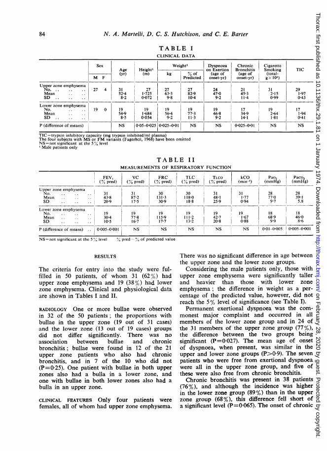
TABLE ICLINICAL DATA
Sex Weight' Dyspnoea Chronic CigaretteAge Height' on Exertion Bronchitis Smoking TIC(yr) (m) kg % of (age of (age of (total-
M F Predicted onset-yr) onset-yr) g x 105)
Upper zone emphysemaNo. 27 4 31 27 27 27 24 21 31 29Mean 52-4 1-725 63-3 82-9 47 0 45 3 2-15 1-97SD 8-2 0-072 9-8 10-4 9-2 11*4 0 99 0 43
Lower zone emphysema I-No. 19 0 19 19 19 19 19 17 19 17Mean 54-9 1-684 564 77-3 46-8 34 9 2-64 1P94SD 8-5 0-054 9-2 11-3 9-2 14-1 1-81 0-41
P (difference of means) NS 0.05-0.025 0.025-001 NS NS 0.025-0-01 NS NS
TIC =trypsin inhibitory capacity (mg trypsin inhibited/ml plasma)The four subjects with MS or FM variants (Fagerhol, 1968) have been omittedNS =not significant at the 5° level'Male patients only
TABLE ILMEASUREMENTS OF RESPIRATORY FUNCTION
FEV, VC FRC TLC TLCO kCO Pao Paco2(% pred) (% pred) (% pred) (% pred) (% pred) (min-') (mmHg) (mmHg)
Upper zone emphysemaNo. 31 31 30 30 31 31 28 28Mean .43-8 87-2 131-3 118-0 48-1 1 77 77 0 39-1SD 20-9 17-5 30 9 18-8 25 9 0 94 9.7 5.8
Lower zone emphysemaNo. 19 19 19 19 19 19 18 18Mean.30 4 77-8 115 9 111 2 42-7 1-67 68-9 46-0SD. 105 16-7 17-7 13-2 20-8 0-88 9 9 8-6
P (difference of means) .. 0-005-0-001 NS NS NS NS NS 001 0-005 0005-0 001
NS=not significant at the 5% level % pred -- %* of predicted value
RESULTS
The criteria for entry into the study were ful-filled in 50 patients, of whom 31 (62 %) hadupper zone emphysema and 19 (38%) had lowerzone emphysema. Clinical and physiological dataare shown in Tables I and II.
RADIOLOGY One or more bullae were observedin 32 of the 50 patients; the proportions withbullae in the upper zone (19 out of 31 cases)and the lower zone (13 out of 19 cases) groupsdid not differ significantly. There was no
association between bullae and chronicbronchitis; bullae were found in 12 of the 21upper zone patients who also had chronicbronchitis, and in 7 of the 10 who did not(P=0-25). One patient with bullae in both upperzones also had a bulla in a lower zone, andone with bullae in both lower zones also had a
bulla in an upper zone.
CLINICAL FEATURES Only four patients were
females, all of whom had upper zone emphysema.
There was no significant difference in age betweenthe upper zone and the lower zone groups.
Considering the male patients only, those withupper zone emphysema were significantly tallerand heavier than those with lower zoneemphysema; the difference in weight as a per-centage of the predicted value, however, did notreach the 5% level of significance (see Table I).Permanent exertional dyspnoea was the com-
monest major complaint and occurred in allmembers of the lower zone group and in 24 ofthe 31 members of the upper zone group (77%),the difference between the two groups beingsignificant (P=0 027). The mean age of onsetof dyspnoea, when present, was similar in theupper and lower zone groups (P>0 9). The sevenpatients who were free from exertional dyspnoeawere all in the upper zone group, and five ofthese were also free from chronic bronchitis.Chronic bronchitis was present in 38 patients
(76%), and although the incidence was higherin the lower zone group (89%) than in the upperzone group (68%), this difference fell short ofa significant level (P=0 065). The onset of chronic
84
on February 28, 2020 by guest. P
rotected by copyright.http://thorax.bm
j.com/
Thorax: first published as 10.1136/thx.29.1.81 on 1 January 1974. D
ownloaded from
Radiological distribution of pulmonary emphysema
No ChronicERronchitis
40r
DyspnoaeebeforeChronic
.Bronchitis
Years
ChronicBronchitisbefore
Dyspnoea
30
20-
10t
o
10
0000@0000 00
0
0
0
0
00
00
0
0 0*000
0
20-
0
0
0
0
0
0
30-
40 LUZE
0
0
LZE
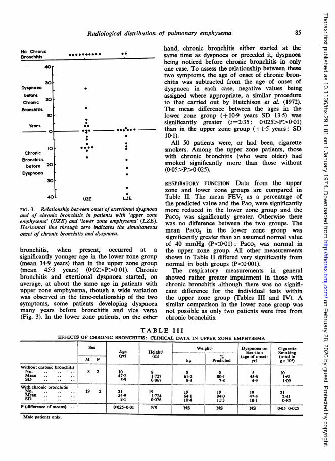
FIG. 3. Relationship between onset ofexertional dyspnoeaand of chronic bronchitis in patients with 'upper zone
emphysema' (UZE) and 'lower zone emphysema' (LZE).Horizontal line through zero indicates the simultaneousonset of chronic bronchitis and dyspnoea.
bronchitis, when present, occurred at a
significantly younger age in the lower zone group(mean 34-9 years) than in the upper zone group(mean 45-3 years) (0-02>P>0 01). Chronicbronchitis and exertional dyspnoea started, onaverage, at about the same age in patients withupper zone emphysema, though a wide variationwas observed in the time-relationship of the twosymptoms, some patients developing dyspnoeamany years before bronchitis and vice versa
(Fig. 3). In the lower zone patients, on the other
hand, chronic bronchitis either started at thesame time as dyspnoea or preceded it, dyspnoeabeing noticed before chronic bronchitis in onlyone case. To assess the relationship between thesetwo symptoms, the age of onset of chronic bron-chitis was subtracted from the age of onset ofdyspnoea in each case, negative values beingassigned where appropriate, a similar procedureto that carried out by Hutchison et al. (1972).The mean difference between the ages in thelower zone group (+ 10-9 years SD 13-5) wassignificantly greater (t=2 35: 0O025>P>0 01)than in the upper zone group (+1-5 years: SD10 1).
All 50 patients were, or had been, cigarettesmokers. Among the upper zone patients, thosewith chronic bronchitis (who were older) hadsmoked significantly more than those without(0 05>P>0 025).
RESPIRATORY FUNCrION Data from the upperzone and lower zone groups are compared inTable II. The mean FEV1 as a percentage ofthe predicted value and the Pao2 were significantlymore reduced in the lower zone group and thePaCO2 was significantly greater. Otherwise therewas no difference between the two groups. Themean PaCo2 in the lower zone group wassignificantly greater than an assumed normal valueof 40 mmHg (P<0O01); Paco2 was normal inthe upper zone group. All other measurementsshown in Table II differed very significantly fromnormal in both groups (P<0001).The respiratory measurements in general
showed rather greater impairment in those withchronic bronchitis although there was no signifi-cant difference for the individual tests withinthe upper zone group (Tables III and IV). Asimilar comparison in the lower zone group was
not possible as only two patients were free fromchronic bronchitis.
TABLE IIIEFFECTS OF CHRONIC BRONCHITIS: CLINICAL DATA IN UPPER ZONE EMPHYSEMA
AgSeHegh1Weight1 Dyspnoea on CigaretteTSex4Age | Heightl_L_______ Exertion Smoking(yr) (m) °/0 (age of onset- (total in
M F kg Predicted yr) g x 10')
Without chronic bronchitisNo.. 8 2 10 8 8 8 5 10Mean 47-2 1-727 61-2 80-1 456 1-61SD 5-9 0-067 8-3 7-8 4-9 1-09
With chronic bronchitisNo. 19 2 21 19 19 19 19 21Mean 54.9 1-724 64-1 84-0 47 4 2141SD 8-1 0-076 10-4 11-3 10-1 0-85
P (difference of means) .. 0-025-0-01 NS NS NS NS 0-05-0-025
Male patients only.
Cp I %AI w- III 1.1 a
85
on February 28, 2020 by guest. P
rotected by copyright.http://thorax.bm
j.com/
Thorax: first published as 10.1136/thx.29.1.81 on 1 January 1974. D
ownloaded from
N. A. Martelli, D. C. S. Hutchison, and C. E. Barter
TABLE IVEFFECTS OF CHRONIC BRONCHITIS: RESPIRATORY FUNCTION IN UPPER ZONE EMPHYSEMA
FEV1 VC FRC TLC TLCO kCO Pao2 Paco2(% pred) (% pred) (% pred) (% pred) (% pred) (min-') (mmHg) (mmHg)
Without chronic bronchitisNo. .. .. .. 10 10 10 10 10 10 8 8Mean.52 7 92-9 133-8 122-5 53 5 2-01 81-1 38-6SD 23-9 16-9 34-4 22-0 27-2 1-08 7-8 710
With chronic bronchitisNo. 21 21 20 20 21 21 20 20Mean .39-6 84-4 129-6 115 8 45 5 1-66 75 3 39.4SD.18-3 17-5 29-9 17-2 25 6 0-87 10-1 5 5
P (difference of means) . . NS NS NS NS NS NS NS
TRYPSIN INHIBITORY CAPACITY The mean trypsininhibitory capacity of the upper zone group(Table I) did not differ significantly from thatof the lower zone group (0-8>P>07); the meanvalues in both groups, however, were significantlygreater than that of the controls. Fourheterozygotes with MS or FM variants (Fagerhol,1968) were omitted from these calculations.There was no significant difference in the mean
trypsin inhibitory capacity between patients whohad chronic bronchitis and those who did not(0 7>P>0 6).
DISCUSSION
The reports available in the literature on therelative proportions of upper zone and lowerzone emphysema among patients with radiologicalevidence of the disease are far from consistent.Fraser and Bates (1959) and Bentivoglio et al.(1963), who studied patients considered to haveemphysema on clinical and physiological grounds,found that the lower zones were the most severelyaffected in the great majority of cases in whomthere was a definite zonal predominance.Where patient selection was based on the pre-
sence of radiological bullae, however, ratherdifferent results were obtained. Thus Stone,Schwartz, and Feltman (1960), Laurenzi, Turino,and Fishman (1962), Viola and Zuffardi (1966),and Boushy, Kohen, Billig, and Heiman (1968)found that in almost all their patients the mainlesions were in the upper zones. Ogilvie andCatterall (1959) and Simon (1964), on the otherhand, found bullae to be more evenly distributed,as in the present study. These discrepancies mustbe presumed to arise from differing selectioncriteria.Permanent dyspnoea at rest or on effort was
a feature of 86% of our patients and was presentin all patients with lower zone emphysema, of
whom the youngest was only 42 years of age.A number of the younger patients with upper zoneemphysema, however, were free from this symp-tom, and similar cases were also observed byViola and Zuffardi (1966), Boushy et al. (1968),and Colp, Park, and Williams (1970). There islittle information on the relationship of dyspnoeato lower zone emphysema apart from the patientsof Ogilvie and Catterall (1959) who all com-plained of severe dyspnoea.
Chronic bronchitis occurred in 76% of thepatients, a similar percentage to that found inprevious studies (Burrows, Niden, Fletcher, andJones, 1964; Hutchison et al., 1972). It was rathermore common in the lower zone than in theupper zone group though the difference did notattain a significant level (P=0065); a somewhatunexpected finding was the very much earlieronset (by some 10 years) in the lower zonepatients. Again, the only other comparableinformation on this matter comes from the studyof Ogilvie and Catterall (1959) who, like our-selves, found chronic bronchitis in many ofthose with lower lobe disease ; it was virtuallyabsent from all of their five patients with upperlobe disease, whose mean age was similar to thatof our upper zone group who did not havechronic bronchitis.The relationship between the age of onset of
chronic bronchitis and that of permanent exer-tional dyspnoea is also of some interest. Inpatients with upper zone emphysema we haveobserved (Table I) that these two featuresoriginate on average at about the same age; inlower zone emphysema, chronic bronchitispreceded dyspnoea by over 10 years. There werewide variations in the time-relationship of thetwo symptoms in individual patients (Fig. 3).In some the symptoms develop simultaneously,but in others they may be separated by an intervalof many years. Statistical analysis of these time
86
on February 28, 2020 by guest. P
rotected by copyright.http://thorax.bm
j.com/
Thorax: first published as 10.1136/thx.29.1.81 on 1 January 1974. D
ownloaded from
Radiological distribution of pulmonary emphysema
intervals showed that there was a significantdifference between the upper and lower zonegroups (0O025>P>001). Chronic bronchitis, beingcommoner in those with lower zone disease,might, at first sight, have appeared to beresponsible for the differences between the twogroups in respect of clinical and physiologicaldata, but on analysis of the upper zone groupalone, no such effect could be demonstrated.Lieberman (1969) and Kueppers, Fallat, and
Larson (1969) have suggested that a high propor-tion of cases of emphysema are associatedwith the heterozygous form of a1-antitrypsindeficiency. In the present study, however, themean trypsin inhibitory capacity in both theupper and lower zone groups was significantlygreater than that observed in 14 obligatoryheterozygotes (parents or children of subjectshomozygous for a1-antitrypsin deficiency), con-firming the previous observations (Hutchisonet al., 1971, 1972) that there is apparently noexcess of heterozygotes among patients withemphysema. Kueppers (1968) has suggested thatan inflammatory process such as chronicbronchitis may elevate trypsin inhibitory capacity,thus masking the heterozygote; in the presentreport, it was found that the presence or absenceof chronic bronchitis had no significant effectupon the trypsin inhibitory capacity.The important question now arises as to
whether the radiological distribution of the lesionsin the cases reported here has any pathologicalsignificance. By the nature of the study thereis, of course, no evidence bearing directly on thispoint, but a certain amount of relevant informa-tion can be gained from the literature. It is known,for instance, that the radiological abnormalitiesin al-antitrypsin deficiency are almost alwaysat their most severe in the lower zones (Eriksson,1965; Guenter et al., 1968; Hutchison et al.,1971) and that panlobular emphysema was thedominant lesion in most of the cases where thehistology was available (Eriksson, 1965;Schleusener, Talamo, Pare, and Thurlbeck, 1968).In random necropsies, the lesions of severepanlobular emphysema have a similar basaldistribution, whereas those of centrilobularemphysema are particularly found in the upperzones (Thurlbeck, 1963; Wyatt et al., 1964).There has been some disagreement as to
whether centrilobular emphysema can produceeither radiological abnormalities or physical dis-ability. Snider et al. (1962) found that even quiteadvanced lesions had shown no ill-effects duringlife. Thurlbeck et al. (1970), on the other hand,found obvious radiological vascular attenuation in
a number of the more severe cases of centrilobularemphysema, though in many others this patternwas not seen. Wyatt et al. (1964) and Heard(1969) demonstrated lung sections where suchcases were associated with extensive bullousformation, and it is hard to believe that suchlesions as these would often escape detection bychest radiography, particularly if tomogramswere also done. Differences in interpretation mayhave contributed in part to the controversy;when the centrilobular lesion has resulted in thedestruction of a large part of the lobule, it maybe difficult to distinguish this from the panlobularform. Some investigators have preferred toclassify all such lesions as panlobular emphysema(Burrows et al., 1966; Reid, 1967) and thus thevery cases of centrilobular emphysema most likelyto cause symptoms or to be visible on the chestradiograph would presumably not have beenclassified as such. Dunnill (1969) considered thatthe typical appearance could always be recog-nized. A further difficulty is posed by the factthat many patients have both types of emphysematogether (Laws and Heard, 1962; Thurlbeck,1963; Gough, 1965), and serious disagreementsbetween observers on the type present in a givenlung are not uncommon (Thurlbeck et al., 1969;Mitchell et al., 1970). The latter authors attemptedto make a distinction between the two types onthe basis of clinical or physiological features.The only difference that they were able to detectwas in the location of the lesions, and theyexpressed some doubt as to whether they werein fact observing two quite separate disorders.In this study, we found rather greater differencesbetween two groups defined on radiological thanon pathological criteria, but the major disparitiesbetween centrilobular and panlobular emphysemaobserved by Sweet et al. (1961) have no counter-part here.One further point might be made in favour
of considering our cases as two separate groups;we were for the most part able to place thelesions in one or other half of the lung fieldswith some confidence. (Doubtful cases usuallyhad advanced generalized disease, and the upperand lower lung fields appeared equally damaged.)If pulmonary emphysema were the result of asingle pathological process, one would perhapsexpect to find an equivalent number of patientsin whom the mid-zones were preferentiallyaffected. In fact, only one of the patients whosecases were reviewed prior to selection for thisstudy would have been classified as a 'mid-zone'subject; his chest radiograph showed a large bullaoccupying the right mid-zone with apparently
87
on February 28, 2020 by guest. P
rotected by copyright.http://thorax.bm
j.com/
Thorax: first published as 10.1136/thx.29.1.81 on 1 January 1974. D
ownloaded from
N. A. Martelli, D. C. S. Hutchison, and C. E. Barter
normal lung above and below it. It is unfortunatethat a pathological assessment of the type ofemphysema is seldom available during life, sinceadequate biopsy specimens can be obtained onlyat open thoracotomy.
In spite of the conflicts of evidence which havealready been mentioned, it appears to us thatthere are still some grounds for believing in theexistence of the two separate pathological typesand that these types may correspond to the tworadiological groups considered in this report.
We acknowledge with thanks the expert help of Mr.L. A. Smith, Miss M. Rusbridge, Staff Nurse A.Swaine, and Miss C. Cridland of the Chest Unittechnical staff, of Miss Mary Howell, who gave
valuable typing and secretarial assistance, and of theradiographic staff of King's College Hospital.We are most grateful to Dr. P. J. L. Cook of
the Galton Laboratory, University College, London,for the al-antitrypsin determinations, and to themany colleagues who have referred patients to thisUnit.
N. A. M. was supported by a grant from theUniversity of Buenos Aires and C. E. B. by a grantfrom the Medical Research Council.
REFERENCES
Bentivoglio, L. G., Beerel, F., Stewart, P. B., Bryan, A. C.,Ball, W. C., and Bates, D. V. (1963). Studies of regionalventilation and perfusion in pulmonary emphysemausing Xenon133. American Review of RespiratoryDiseases, 88, 315.
Boushy, S. F., Kohen, R., Billig, D. M., and Heiman, M. J.(1968). Bullous emphysema: clinical, roentgenologicand physiologic study of 49 patients. Diseases of theChest, 54, 327.
Burrows, B., Fletcher, C. M., Heard, B. E., Jones, N. L.,and Wootliff, J. S. (1966). The emphysematous andbronchial types of chronic airways obstruction: Aclinicopathological study of patients in London andChicago. Lancet, 1, 830.
, Niden, A. H., Fletcher, C. M., and Jones, N. L. (1964).Clinical types of chronic obstructive lung disease inLondon and in Chicago. American Review of RespitatotyDiseases, 90, 14.
Colp, C., Park, S. S., and Williams, M. H. (1970). Emphysemawith little airway obstruction. American Review ofRespiratory Diseases, 101, 615.
Cotes, J. E. (1968). Lung Function, 2nd ed., Blackwell,Oxford.
Dunnill, M. S. (1969). The classification and quantificationof emphysema. Proceedings of the Royal Society ofMedicine, 62, 1024.
Eriksson, S. (1965). Studies in alpha,-antitrypsin deficiency.Acta Medica Scandinavica, 177, Suppl. 432.
Fagerhol, M. K. (1968). The Pi-system: genetic variants ofserum alpha,-antitrypsin. Series Haematologica, 1, 153.
Fraser, R. G. and Bates, D. V. (1959). Body section ro-entgenography in the evaluation and differentiation ofchronic hypertrophic emphysema and asthma. AmericanJournal of Roentgenology, 82, 39.
Gough, J. (1952). The pathological diagnosis of emphysema.Proceedings of the Royal Society of Medicine, 45, 576., (1965). The pathology of emphysema. PostgraduateMedical Journal, 41, 392.
Guenter, C. A., Welch, M. H., Russell, T. R., Hyde, R. M.,and Hammarsten, J. F. (1968). The pattern of lungdisease associated with alpha1-antitrypsin deficiency.Aschives of Internal Medicine, 122, 254.
Heard, B. E. (1969). Pathology of Chronic Bronchitis andEmphysema, pp. 61-73. Churchill, London.
Hutchison, D. C. S., Barter, C. E., Cook, P. J. L., Laws,J. W., Martelli, N. A., and Hugh-Jones, P. (1972).Severe pulmonary emphysema: a comparison of patientswith and without alpha1-antitrypsin deficiency. QuarterlyJournal of Medicine, 41, 301., Cook, P. J. L., Barter, C. E., Harris, H., and Hugh-Jones, P. (1971). Pulmonary emphysema and alpha,-antitrypsin deficiency. British Medical Journal, 1, 689.
Kueppers, F. (1968). Genetically determined differences inthe response of alpha,-antitrypsin levels in humanserum to typhoid vaccine. Human Genetics, 6, 207.
Fallat, R., and Larson, R. K. (1969). Obstructivelung disease and alpha,-antitrypsin deficiency geneheterozygosity. Science, 165, 899.
Laurenzi, G. A., Turino, G. M., and Fishman, A. P. (1962).Bullous disease of the lung. American Journal of Medi-cine, 32, 361.
Laws, J. W. and Heard, B. E. (1962). Emphysema and thechest film: a retrospective radiological and pathologicalstudy. British Journal of Radiology, 35, 750.
Leopold, J. G. and Gough, J. (1957). The centrilobularform of hypertrophic emphysema and its relation tochronic bronchitis. Thorax, 12, 219.
Lieberman, J. (1969). Heterozygous and homozygousalpha1-antitrypsin deficiency in patients with pulmonaryemphysema. New England Journal of Medicine, 281, 279.
McLean, K. H. (1956). The macroscopic anatomy of pul-monary emphysema. Austialasian Annals of Medicine,5, 73.
Medical Research Council (1965). Definition and classifi-cation of chronic bronchitis for clinical and epidemio-logical purposes. Lancet, 1, 775.
Mitchell, R. S., Silvers, G. W., Goodman, N., Dart, G.,and Maisel, J. C. (1970). Are centrilobular emphysemaand panlobular emphysema two different diseases?Human Pathology, 1, 433.
Ogilvie, C. and Catterall, M. (1959). Patterns of disturbedlung function in patients with emphysematous bullae.Thorax, 14, 216.
Raine, J. M. and Bishop, J. M. (1963). A-a difference in 02tension and physiological dead space in normal man.
Journal of Applied Physiology, 18, 284.Reid, L. (1967). In The Pathology of Emphysema, chap. 5.
Lloyd-Luke (Medical Books), London.Schleusener, A., Talamo, R. C., Pare, J. A. P., and Thurlbeck,
W. M. (1968). Familial emphysema. American Reviewof Respiratory Diseases. 98, 692.
88
on February 28, 2020 by guest. P
rotected by copyright.http://thorax.bm
j.com/
Thorax: first published as 10.1136/thx.29.1.81 on 1 January 1974. D
ownloaded from
Radiological distribution of pulmonary emphysema
Simon, G. (1964). Radiology and emphysema. ClinicalRadiology, 15, 293.
Snider, G. L., Brody, J. S., and Doctor, L. (1962). Sub-clinical puilmonary emphysema: Incidence and anatomicpatterns. American Review of Respiratory Diseases, 85,666.
Society of Actuaries (1959). In Documenta Geigy ScientificTables, 6th ed., p. 623. Geigy Pharmaceutical Co. Ltd.Manchester.
Stone, D. J., Schwartz, A., and Feltman, J. A. (1960).Bullous emphysema: a long-term study of the naturalhistory and the effects of therapy. Ametican Review ofRespiratoty Diseases, 82, 493.
Sweet, H. C., Wyatt, J. P., Fritsch, A. J., and Kinsella, P. W.(1961). Panlobular and centrilobular emphysema:correlation of clinical findings with pathologic patterns.Annals of Internal Medicine, 55, 565.
Thurlbeck, W. M. (1963). The incidence of pulmonaryemphysema: with observations on the relative incidenceand spatial distribution of various types of emphysema.American Review of Respiratory Diseases, 87, 206., Henderson, J. A., Fraser, R. G., and Bates, D. V.(1970). Chronic obstructive lung disease: a comparisonbetween clinical, roentgenologic, functional and mor-phologic criteria in chronic bronchitis, emphysema,asthma and bronchiectasis. Medicine, 49, 81., Horowitz, I., Siemiatycki, J., Dunnill, M. S., Maisel,J. C., Pratt, P., and Ryder, R. (1969). Intra- and inter-observer variations in the assessment of emphysema.Archives of Environmental Health, 18, 646.
Viola, A. R., and Zuffardi, E. A. (1966). Physiologic andclinical aspects of pulmonary bullous disease. AmeticanReview of Respi,atory Diseases, 94, 574.
Wyatt, J. P., Fischer, V. W., and Sweet, H. C. (1964). Thepathomorphology of the emphysema complex. Part I.
American Review of Respiratory Diseases, 89, 533.
89
on February 28, 2020 by guest. P
rotected by copyright.http://thorax.bm
j.com/
Thorax: first published as 10.1136/thx.29.1.81 on 1 January 1974. D
ownloaded from