
Radiological changes hepatic amoebiasispmj.bmj.com/content/postgradmedj/47/551/615.full.pdf ·...
7
Postgraduate Medical Journal (September 1971) 47, 615-621. CLINICAL REVIEW Radiological changes in hepatic amoebiasis S. RAMACHANDRAN M.D., M.R.C.P., M.R.C.P.E. Physician D. L. N. JAYAWARDENA M.B.B.S., D.M.R.D.(Lond.) Visiting Radiologist J. R. A. PERUMAL M.B.B.S. Medical Officer The Government Hospital, Negombo, Ceylon Summary Radiological abnormalities were observed in 88-3% of patients with the syndrome of hepatic amoebiasis. While this figure is higher than those reported in previous studies, it appears that it is due to the large number of patients who showed an elevation of the diaphragm in the right lateral views of the chest. It was observed that a proportion of the patients with an elevation of the right dome of the diaphragm in the lateral views showed no significant elevation in the postero-anterior views, these patients constituting a radiologically distinct group. It is thus important that lateral views of the chest should be performed routinely in all cases of suspected hepatic amoebiasis. Cases of proved hepatic abscesses also conform to one or other of the radiological groups described. Abscesses situated inferiorly or anteriorly in the liver could be associated with an elevation of the right dome of the diaphragm. While chest radiology is of importance in the diagnosis of the syndrome of hepatic amoebiasis, it is not peculiar to this syndrome alone and is of only relative diagnostic value in clinically differentiating the cases where pus may be demonstrated from the cases where pus may not be demonstrable. Introduction The importance of radiology in the diagnosis of the syndrome of hepatic amoebiasis has been recog- nized since the descriptions of Munk (1944) and sub- sequently, radiological abnormalities were included as one of the criteria for the diagnosis of the syndrome (Lamont & Pooler, 1958). The frequency of the abnormal findings in chest X-rays varied markedly in different series, the figures ranging from 64-8% to as much as 83-5%. The commonest abnormality seen was an elevation of the right dome of the diaphragm, the frequency of which ranged from 27-5 to 74-3%y in the larger studies reported (Table 1) The cause for the wide discrepancy in these figures, while obscure, may in part be due to the geographical distribution and varying patterns in the manifesta- tions of the syndrome. The purpose of this study was to evaluate the frequency and the type of abnormalities seen in chest radiology in cases of uncomplicated hepatic amoe- biasis as it occurs in Ceylon and to detail any changes or patterns that may hitherto have not been sufficiently emphasized. Patients, materials, and methods One hundred and twenty cases of uncomplicated hepatic amoebiasis admitted to the medical unit of the Government Hospital, Negombo, Ceylon, were subjected to routine chest radiology. Both standard postero-anterior (PA) and right lateral teleradio- grams of the chest were done in all the cases on admission to hospital. The criteria for the diagnosis of the syndrome were a modification of those laid down by Kean (1955), TABLE 1. Radiological Elevation of Authors Cases abnormalities the diaphragm studied (%) (%) Lamont & Pooler (1958) 242 83-5 69-4 Islam et al. (1960) 48 71-8 56-1 Rajasuriya & Nagaratnam (1962) 91 64-8 27-5 Amarjit Singh & Jolly (1962) 35 74-3 74.3 Turrill & Burnham (1966) 100 66-6 No details Present series (1970) 120 88-3 83*3 by copyright. on 22 June 2018 by guest. Protected http://pmj.bmj.com/ Postgrad Med J: first published as 10.1136/pgmj.47.551.615 on 1 September 1971. Downloaded from
-
Upload
vuongkhanh -
Category
Documents
-
view
224 -
download
2
Transcript of Radiological changes hepatic amoebiasispmj.bmj.com/content/postgradmedj/47/551/615.full.pdf ·...
Postgraduate Medical Journal (September 1971) 47, 615-621.
CLINICAL REVIEW
Radiological changes in hepatic amoebiasis
S. RAMACHANDRANM.D., M.R.C.P., M.R.C.P.E.
Physician
D. L. N. JAYAWARDENAM.B.B.S., D.M.R.D.(Lond.)
Visiting RadiologistJ. R. A. PERUMAL
M.B.B.S.Medical Officer
The Government Hospital, Negombo, Ceylon
SummaryRadiological abnormalities were observed in 88-3%of patients with the syndrome of hepatic amoebiasis.While this figure is higher than those reported inprevious studies, it appears that it is due to the largenumber of patients who showed an elevation of thediaphragm in the right lateral views ofthe chest. It wasobserved that a proportion of the patients with anelevation of the right dome of the diaphragm in thelateral views showed no significant elevation in thepostero-anterior views, these patients constituting a
radiologically distinct group. It is thus important thatlateral views of the chest should be performed routinelyin all cases of suspected hepatic amoebiasis.
Cases of proved hepatic abscesses also conform toone or other of the radiological groups described.Abscesses situated inferiorly or anteriorly in the livercould be associated with an elevation of the rightdome of the diaphragm.While chest radiology is of importance in the
diagnosis of the syndrome of hepatic amoebiasis, it isnot peculiar to this syndrome alone and is of onlyrelative diagnostic value in clinically differentiating thecases where pus may be demonstrated from the caseswhere pus may not be demonstrable.
IntroductionThe importance of radiology in the diagnosis of
the syndrome of hepatic amoebiasis has been recog-nized since the descriptions ofMunk (1944) and sub-
sequently, radiological abnormalities were includedas one ofthe criteria for the diagnosis ofthe syndrome(Lamont & Pooler, 1958). The frequency of theabnormal findings in chest X-rays varied markedlyin different series, the figures ranging from 64-8% toas much as 83-5%. The commonest abnormalityseen was an elevation of the right dome of thediaphragm, the frequency of which ranged from27-5 to 74-3%y in the larger studies reported (Table 1)The cause for the wide discrepancy in these figures,while obscure, may in part be due to the geographicaldistribution and varying patterns in the manifesta-tions of the syndrome.The purpose of this study was to evaluate the
frequency and the type of abnormalities seen in chestradiology in cases of uncomplicated hepatic amoe-biasis as it occurs in Ceylon and to detail anychanges or patterns that may hitherto have not beensufficiently emphasized.
Patients, materials, and methodsOne hundred and twenty cases of uncomplicated
hepatic amoebiasis admitted to the medical unit ofthe Government Hospital, Negombo, Ceylon, were
subjected to routine chest radiology. Both standardpostero-anterior (PA) and right lateral teleradio-grams of the chest were done in all the cases onadmission to hospital.The criteria for the diagnosis of the syndrome were
a modification of those laid down by Kean (1955),TABLE 1.
Radiological Elevation ofAuthors Cases abnormalities the diaphragm
studied (%) (%)Lamont & Pooler (1958) 242 83-5 69-4Islam et al. (1960) 48 71-8 56-1Rajasuriya & Nagaratnam (1962) 91 64-8 27-5Amarjit Singh & Jolly (1962) 35 74-3 74.3Turrill & Burnham (1966) 100 66-6 No detailsPresent series (1970) 120 88-3 83*3
by copyright. on 22 June 2018 by guest. P
rotectedhttp://pm
j.bmj.com
/P
ostgrad Med J: first published as 10.1136/pgm
j.47.551.615 on 1 Septem
ber 1971. Dow
nloaded from
Clinical review
they were: (1) an enlarged tender liver, (2) a previoushistory of diarrhoea with blood and mucus or anillness suggestive of hepatic amoebiasis, (3) sug-gestive haematological changes, (4) suggestive radio-logical changes and (5) a response to specific amoe-bicidal therapy. All the cases included in the studysatisfied at least three of the above five criteria. Noattempt was made during the study to separate thecases where pus was demonstrated from the caseswhere no pus was demonstrated.
ResultsThe individual radiological abnormalities seen in
our cases are summarized in Table 2. They were:
(1) Elevation of the right dome of the diaphragm inPA views
Elevation of the right dome of the diaphragm inthe PA view was seen in 55-8%y of cases. This eleva-tion was of a moderate or severe degree. By moderateis meant an elevation of between 1 and 2 in. abovethe level of the left dome of the diaphragm providedthere is no elevation of the latter by gaseous disten-tion of the stomach. 'Severe degrees of elevation'denotes an upward doming of the right diaphragm ofover 2 in. above the left side. In a small proportionof cases (5-8%) elevation of the diaphragm wasassociated with visible pleural reactions.
(2) Elevation of the right dome of the diaphragm inright lateral views
Elevation of the diaphragm either localized or
involving the whole of the diaphragm was moststriking in the lateral views, it being noted in 83-3%of the cases. Once again it varied in its degree, beingeither moderate or severe. When the diaphragmaticelevation was localized it always involved theanterior or the middle third of the dome. Elevationsof the posterior part of the diaphragm were notencountered. It was an interesting observation thatabout 30% of these patients who showed an eleva-tion in the right diaphragm either localized or
generalized in the lateral views did not show thischange in the postero-anterior views.
(3) Pleural reactionsEvidence of minor pleural reactions were seen in
7-5% of the cases, manifesting as small effusions or
pleural thickening. In 5-8% of the cases it was
associated with varying degrees of diaphragmaticelevation.
(4) Pulmonary changesChanges in the lung parenchymawere notedinonly
5% of the cases, they were consolidation with orwithout effusion or changes suggestive of bronchiec-tasis.
(5) Absence of radiological changesNo radiological abnormality was seen in 11 7%
of the cases. Thus 88-3% of the cases in this studyshowed some radiological abnormality in the chestX-rays as a consequence of hepatic amoebiasis.
Further observations revealed that these casescould be grouped into one of five fairly clear-cutradiologically distinct patterns depending on thecombination of the various abnormalities noted(Table 3). They were:
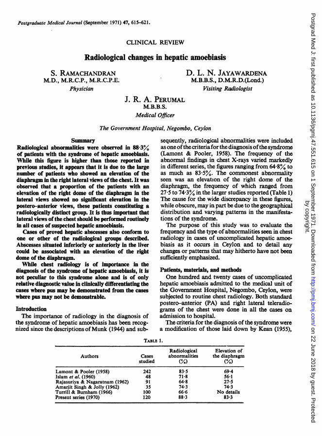
Group 1. This included a proportion of cases, 50%,who showed a significant elevation of the right domeof the diaphragm in both the PA and the rightlateral views, with no evidence of either pleural or
pulmonary changes. The elevation seen in the lateralviews were often more striking than that seen in thePA views (Fig. la and b).
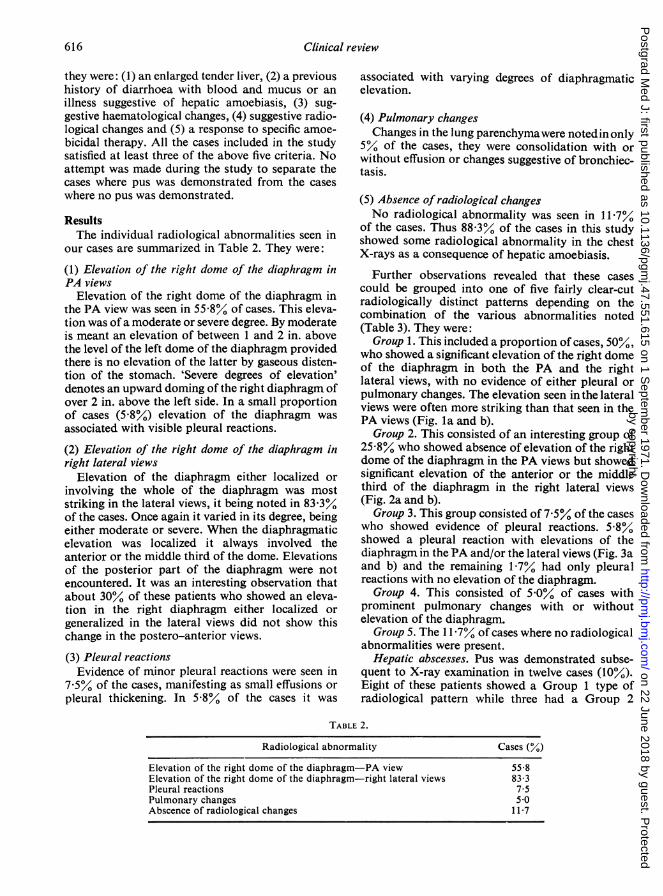
Group 2. This consisted of an interesting group of25-8% who showed absence of elevation of the rightdome of the diaphragm in the PA views but showedsignificant elevation of the anterior or the middlethird of the diaphragm in the right lateral views(Fig. 2a and b).
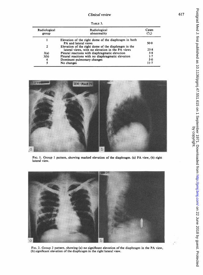
Group 3. This group consisted of 7-5%y of the caseswho showed evidence of pleural reactions. 5-8%showed a pleural reaction with elevations of thediaphragm in the PA and/or the lateral views (Fig. 3aand b) and the remaining 1-7% had only pleuralreactions with no elevation of the diaphragm.
Group 4. This consisted of 5-0%y of cases withprominent pulmonary changes with or withoutelevation of the diaphragm.
Group 5. The 11 -7% of cases where no radiologicalabnormalities were present.
Hepatic abscesses. Pus was demonstrated subse-quent to X-ray examination in twelve cases (10%).Eight of these patients showed a Group 1 type ofradiological pattern while three had a Group 2
TABLE 2.
Radiological abnormality Cases (%)Elevation of the right dome of the diaphragm-PA view 55-8Elevation of the right dome of the diaphragm-right lateral views 83-3Pleural reactions 7-5Pulmonary changes 5-0Abscence of radiological changes 11-7
616by copyright.
on 22 June 2018 by guest. Protected
http://pmj.bm
j.com/
Postgrad M
ed J: first published as 10.1136/pgmj.47.551.615 on 1 S
eptember 1971. D
ownloaded from
Clinical review 617
TABLE 3.
Radiological Radiological Casesgroup abnormality (%)
1 Elevation of the right dome of the diaphragm in bothPA and lateral views 50-0
2 Elevation of the right dome of the diaphragm in thelateral views, with no elevation in the PA views 25-8
3(a) Pleural reactions with diaphragmatic elevation 5-83(b) Pleural reactions with no diaphragmatic elevation 1-74 Dominant pulmonary changes 5-05 No changes 11-7
i:
*;; Izk·,ii.
i;i·t:·
··l*i.· -·I i.l.P.'m.:%
·c:.
Y:.···;)::?,,
BiP
.:rr.l.RI= a
;*,·
iii .i..
·r :iiiii(·l;[-*,i,aPiii: ····:'8 1.
FIG. 1. Group 1 pattern, showing marked elevation of the diaphragm. (a) PA view, (b) rightlateral view.
.r."
B:: :'
.i: .,·ii28 ·i ·. ::·
I. 'Ga';"
''':
ii.·is.ikFI.I.P.!i''':: "ii:raiiii.ii.ji:- 'i;i'i:iiii
':f: :d·:·..?
a IYtt..
FIG. 2. Group 2 pattern, showing (a) no significant elevation of the diaphragm in the PA view,(b) significant elevation of the diaphragm in the right lateral view.
by copyright. on 22 June 2018 by guest. P
rotectedhttp://pm
j.bmj.com
/P
ostgrad Med J: first published as 10.1136/pgm
j.47.551.615 on 1 Septem
ber 1971. Dow
nloaded from

618 Clinical review
/ ~ ~ ~ ~ ~ ~~ ~ ~ ~ ~ ~ ~ ~ ~ ~ ~ ~ ~ ~~~~~~~~~~4
0~~~~~~~~~~~~~~~~~~~~~~~~~~~~~~~~~~~~~~~~:'· :· ···..: ·i .j~T
FIG. 3. Group 3 pattern, showing pleural reaction with an elevation of the diaphragm. (a) PAview, (b) right lateral view.
ci~~~~~~~~~~~~
FIG. 4. Patient M.A., showing (a) no significant elevation of the diaphragm in the PA view, (b) significant elevationin the diaphragm in the right lateral view (Group 2). (c) Dye injected into the abscess cavity after aspiration, show-ing the position of the abscess.
pattern, the remaining patient having a Group 4pattern. It thus appears that the cases of provenabscesses also conformed to the same radiologicaltypes as the cases where no pus was demonstrated.The following case reports draw attention to some
of the radiological features of hepatic abscesseswhich will be discussed.
(a) Patient M.A. had a large epigastric lump whichon aspiration yielded two pints of chocolate pus.He showed a Group 2 radiological pattern, with noelevation of the diaphragm in the PA view and
significant elevation of the diaphragm anteriorly inthe right lateral view (Fig. 4a, b, and c).
(b) Patient M.A.I. had an abscess pointing into theright loin. His radiological pattern was of a Group 1type with moderate elevation of the diaphragm in thePA view and more prominent elevation in the lateralview (Fig. 5a). Dye injected into the abscess cavityafter aspiration showed that it was placed postero-inferiorly in the liver (Fig. 5b).
(c) Patient V had a prominent right subcostalabscess pointing externally which was aspirated.
by copyright. on 22 June 2018 by guest. P
rotectedhttp://pm
j.bmj.com
/P
ostgrad Med J: first published as 10.1136/pgm
j.47.551.615 on 1 Septem
ber 1971. Dow
nloaded from

Clinical review 619
··i':::;:·' 4
a~~~~~~~~~~~~~~~~~~~~~~~~ii
FIG. 5. (a) Patient M.A.I, showing an elevation of the diaphragm in'the right lateral'view. (b) Dyein the abscess cavity, confirming its position in the inferior aspect of the liver. Note elevation ofdiaphragm in the PA view (Group 1 pattern).
-:!
w~~~-:::S
"~~~~~2 ~~~~bFIG. 6. Patient V. (a) Elevation of the diaphragm in PA and (b) right lateral view of a Group 1pattern.
Radio-opaque dye was injected into the abscesscavity which revealed its situation in the liver. Thispatient showed a typical Group 1 radiologicalpattern with prominent elevation of the diaphragmin both views (Fig. 6a and b).
It thus appears that elevations of the right domeof the diaphragm could be associated with hepaticabscesses situated inferiorly in the liver or in theepigastric region.Discussion
Elevation of the right dome of the diaphragm, aradiological feature of hepatic amoebiasis docu-
mented in studies reported earlier was seen in a higherpercentage of cases in this study. This in turnnecessarily gave rise to a larger proportion ofpatients (88Y%) who showed radiological abnormali-ties in their chest X-rays. This higher incidence isprobably due to one or other of two factors: (i)that the manifestations of the syndrome may bedependent on the geographical distribution or (ii)that lateral X-ray views were performed routinely inall the cases included in this study. No mention ismade in the larger series reported earlier regardingthe views of the chest taken. By doing lateral X-raysof the chest in all cases of hepatic amoebiasis, apart
by copyright. on 22 June 2018 by guest. P
rotectedhttp://pm
j.bmj.com
/P
ostgrad Med J: first published as 10.1136/pgm
j.47.551.615 on 1 Septem
ber 1971. Dow
nloaded from

620 Clinical review
from increasing the incidence of the percentage ofpatients with abnormal X-ray findings, the presenceof the interesting Group 2 patients was noted.The cause of this Group 2 radiological pattern withan absence of elevation of the right dome of thediaphragm in the PA views and significant elevationin the lateral views is obscure. DeBakey & Ochsner(1951) and later Turrill & Burnham (1966) havedocumented that the diaphragmatic elevation ofteninvolves the medial and anterior portions of theright hemi-diaphragm. It may well be that the eleva-tion of the diaphragm in these Group 2 cases, bybeing placed more medially is obscured by a super-imposed heart shadow.
It was a striking feature that even in the Group 1patients, the elevations of the diaphragm were moreprominently seen in the right lateral views. It ispossible that the radiological Group 1 described, ispreceded by the radiological Group 2 where theearliest abnormality of a significant elevation of thediaphragm in the right lateral view either localizedor generalized is not accompanied by any elevationin the PA view.
Localized humping rather than a generalizedelevation, a feature seen in this study, particularly inthe Group 1 patients has also been documented bySchorr & Schwartz (1951). It is not possible to saywith any certainty whether this is due to an abscesssituated in the superior aspect of the liver, as it wasfrequently seen in the patients with a good responseto specific therapy and where pus was not demon-strated (Fig. 7a and b).The observation that the cases with proved abscess
also conformed to the radiologically distinct patternsseen in the cases where no pus was demonstrated wasof interest. This is at variance with the views docu-
mented earlier by Sepulveda et al. (1959) that thereis a moderate elevation of the diaphragm in acuteamoebic hepatitis while marked elevations orlocalized prominences are seen in acute amoebicabscesses. We observed that such clear-cut distinc-tions are incorrect, this observation being supportedby case reports of hepatic abscesses with no signi-ficant radiological changes (Rowland, 1963). Thecase reports from this study also illustrate somepoints of interest: (1) that abscesses could be associ-ated with any of the radiological patterns describedand (2) that abscesses even placed inferiorly in theliver or epigastrically could produce significantelevations in the right dome of the diaphragm, eitherlocalized or generalized. This observation lends nosupport to the belief that an elevated diaphragm isoften associated with an abscess placed in the rightsuperior aspect of the liver, the commonest site for aliver abscess. The fact that the radiological patternsseen in this study are common to both groups,whether pus was demonstrated or not, and theobservation that the Group 1 radiological type maybe preceded by a Group 2 radiological pattern,supports our contention that all cases of hepaticamoebiasis probably belong to some part of a con-tinuous spectrum of the syndrome culminating in theformation of large hepatic abscesses, single ormultiple. This would in turn also indicate that chestradiology while being of importance in the diagnosisof the syndrome, is of only relative value in differenti-ating clinically the cases where pus is demonstrable(hepatic abscesses) from the cases where no pus maybe demonstrated.
Elevation in the right dome of the diaphragmhowever is not peculiar to the syndrome of hepaticamoebiasis. Rowland (1963) has described this
ci~~~~~~~~~~~~~~~~~~~~~RFIG. 7. Localized humping of the diaphragm. (a) No pus demonstrated, (b) pus demonstrated.
by copyright. on 22 June 2018 by guest. P
rotectedhttp://pm
j.bmj.com
/P
ostgrad Med J: first published as 10.1136/pgm
j.47.551.615 on 1 Septem
ber 1971. Dow
nloaded from

Clinical review 621
abnormality in cases of primary carcinoma of theliver, while we have observed it in cases of hepaticcirrhosis usually with ascites, where the elevation ofthe diaphragm in the lateral views is often morestriking. Thus radiological abnormalities shouldbe assessed concurrently with the other criteria in thediagnosis of the syndrome of hepatic amoebiasis.
ReferencesAMARJIT SINGH & JOLLY, S.S. (1962) Hepato-pulmonary
amoebiasis in India. Journal of Tropical Medicine andHygiene, 63, 71.
DEBAKEY. M.E. & OCHSNER, A (1951) Collective review-hepatic amoebiasis, 20 year experience and analysis of 263cases. International Abstracts of Surgery, 92, 209.
ISLAM, N., ALLAM, K.S. & QUADERI, M.A. (1960) Hepaticamoebiasis. The Journal of Tropical Medicine and Hygiene,63, 131.
KEAN, B.H. (1955) Amoebic hepatitis. Archives of InternalMedicine, 96, 667.
LAMONT, N. McE. & POOLER, N.R. (1958) Hepatic amoe-biasis. Quarterly Journal of Medicine, 27, 390.
MUNK, J. (1944) X-ray appearance in amoebic hepatitis.British Journal of Radiology, 17, 48.
RAJASURIYA, K. & NAGARATNAM, N. (1962) Hepatic amoe-biasis in Ceylon. The Journal of Tropical Medicine andHygiene, 65, 165.
ROWLAND, H.A.K. (1963) Radiology of amoebic liverabscess. The Journal of Tropical Medicine and Hygiene, 66,113.
SCHORR, S. & SCHWARTZ, A. (1951) Roentgenologic mani-festations of amoebiasis of the liver with concomitantfindings in the chest. American Journal of Roentgenology,66, 546.
SEPULVEDA, B., JINCH, H., BASSOLS, F. & MUNOZ, R. (1959)Amoebiasis of the liver. American Journal of DigestiveDisorders, 4, 43.
TURRILL, F.L. & BURNHAM, J.R. (1966) Hepatic amoebiasis.American Journal of Surgery, 111, 424.
by copyright. on 22 June 2018 by guest. P
rotectedhttp://pm
j.bmj.com
/P
ostgrad Med J: first published as 10.1136/pgm
j.47.551.615 on 1 Septem
ber 1971. Dow
nloaded from


