Radiofrequency denervation for sacroiliac and facet joint paineprints.aihta.at/1096/1/DSD_99.pdf ·...
77
Radiofrequency denervation for sacroiliac and facet joint pain Systematic Review Decision Support Document No. 99 ISSN online: 1998-0469
Transcript of Radiofrequency denervation for sacroiliac and facet joint paineprints.aihta.at/1096/1/DSD_99.pdf ·...
Radiofrequency denervation for sacroiliac and facet joint pain
Systematic Review
Decision Support Document No. 99 ISSN online: 1998-0469


Radiofrequency denervation for sacroiliac and facet joint pain
Systematic Review
Vienna, March 2016

Project Team
Project leader: Dr.in rer. nat. Agnes Kisser
Authors: Dr.in med. Brigitte Piso, MPH Mag.a rer. nat. Inanna Reinsperger, MPH Katharina Rosian, BSc, MSc
Project Support
Literature search: Tarquin Mittermayr, BA
Calculations: Mag. rer. nat. Robert Emprechtinger
External Review: Univ. Prof. Dr. Martin Krismer Universitätsklinik für Orthopädie, Medizinische Universität Innsbruck
Internal Review: Priv.-Doz.in Dr.in Claudia Wild
Correspondence
Brigitte Piso, [email protected]
This report should be referenced as follows:
Piso B, Reinsperger I, Rosian K. Radiofrequency denervation for sacroiliac and facet joint pain. Decision Support Document No. 99; 2016. Vienna: Ludwig Boltzmann Institute for Health Technology Assessment.
Conflict of interest
All authors and the reviewers involved in the production of this report have declared they have no conflicts of interest in relation to the technology assessed according to the Uniform Requirements of Manuscripts Statement of Medical Journal Editors (www.imce.org).
Disclaimer
The external reviewers did not co-author the scientific report and do not necessarily all agree with its content. Only the LBI-HTA is responsible for errors or omissions that could persist. The final version and the policy recommendations are under the full responsibility of the LBI-HTA.
The HTA Core Model ®, developed within EUnetHTA (www.eunethta.eu), has been utilised when producing the contents and/or structure of this work. The following version of the Model was used: [HTA Core Model. Version 3.0]. Use of the HTA Core Model does not guarantee the accuracy, completeness, quality or usefulness of any information or service produced or provided by using the Model.
Commissioned by the Austrian Ministry of Health, this report systematically assessed the intervention described herein as decision support for the inclusion in the catalogue of benefits.
CONTENT INFORMATION
Publisher:
Ludwig Boltzmann Gesellschaft GmbH Nußdorferstr. 64, 6 Stock, A-1090 Wien http://hta.lbg.ac.at/page/imprint
Responsible for content:
Ludwig Boltzmann Institute for Health Technology Assessment (LBI-HTA) Garnisongasse 7/20, A-1090 Vienna http://hta.lbg.ac.at/
Decision support documents of the LBI-HTA do not appear on a regular basis and serve to publicize the research results of the Ludwig Boltzmann Institute of Health Technology Assessments.
Decision support documents of the LBI-HTA are only available to the public via the Internet at http://eprints.hta.lbg.ac.at
Decision Support Document No.: 99
ISSN-online: 1998-0469
© 2016 LBI-HTA – All rights reserved

LBI-HTA | 2016 3
Content
Summary ................................................................................................................................................................ 7
Zusammenfassung ............................................................................................................................................. 11
1 Scope .................................................................................................................................................................... 15 1.1 PICO question ............................................................................................................................................ 15 1.2 Inclusion criteria ........................................................................................................................................ 15
2 Methods ............................................................................................................................................................... 17 2.1 Research questions ..................................................................................................................................... 17 2.2 Sources ......................................................................................................................................................... 18 2.3 Systematic literature search ....................................................................................................................... 18
2.3.1 Additional searches ................................................................................................................................ 19 2.4 Flow chart of study selection ..................................................................................................................... 19 2.5 Analysis ....................................................................................................................................................... 21 2.6 Synthesis ...................................................................................................................................................... 21
3 Description and technical characteristics of technology ............................................................................. 23
4 Health Problem and Current Use.................................................................................................................... 27
5 Clinical effectiveness ......................................................................................................................................... 31 5.1 Outcomes ..................................................................................................................................................... 31 5.2 Included studies .......................................................................................................................................... 32 5.3 Results ......................................................................................................................................................... 33
6 Safety .................................................................................................................................................................... 37 6.1 Outcomes ..................................................................................................................................................... 37 6.2 Included Studies ......................................................................................................................................... 37 6.3 Results ......................................................................................................................................................... 38
7 Quality of evidence ............................................................................................................................................ 41
8 Discussion ........................................................................................................................................................... 51
9 Recommendation ............................................................................................................................................... 55
10 References ............................................................................................................................................................ 57
Appendix ............................................................................................................................................................. 61 Evidence tables of individual studies included for clinical effectiveness and safety ............................ 61 Risk of bias table ......................................................................................................................................... 71 Applicability tables..................................................................................................................................... 72 List of ongoing randomised controlled trials ........................................................................................... 73 Additional information: List of (registered) completed but unpublished
or terminated randomised controlled trials .............................................................................................. 74 Literature search strategies ........................................................................................................................ 75
Search strategy for MEDLINE ............................................................................................................. 75 Additional information: Search strategy for PubMed ........................................................................ 75

Radiofrequency denervation for sacroiliac and facet joint pain
4 LBI-HTA | 2016
List of figures
Figure 2-1: Flow chart of study selection (PRISMA Flow Diagram) .......................................................................... 20
List of tables
Table 1-1: Inclusion criteria ............................................................................................................................................ 15
Table 7-1: Evidence profile for RFD vs. placebo: efficacy in patients with facet joint pain ................................... 43
Table 7-2: Evidence profile for RFD vs. steroid injections: efficacy in patients with facet joint pain .................. 45
Table 7-3: Evidence profile for RFD vs. placebo, steroid injections or alternative RFD: safety in patients with facet joint pain ........................................................................................................ 47
Table 7-4: Evidence profile: efficacy of RFD vs. placebo in patients with sacroiliac joint pain ............................ 47
Table 7-5: Evidence profile for RFD vs. placebo: safety in patients with sacroiliac joint pain .............................. 49
Table 8-1: Overview of RFD techniques, used in included RCTs ............................................................................. 52
Table 9-1: Evidence based recommendation: ............................................................................................................... 55
Table A-1: Facet joint: Radiofrequency denervation vs. placebo treatment: Results from randomised controlled trials ................................................................................................. 61
Table A-2: Facet joint: Radiofrequency denervation vs. steroid injection: Results from randomised controlled trials ................................................................................................. 65
Table A-3: Facet joint: Radiofrequency denervation vs. other treatment (other RFD method): Results from randomised controlled trials ................................................................................................. 67
Table A-4: Sacroiliac joint Radiofrequency denervation vs. placebo: Results from randomised controlled trials ................................................................................................. 69
Table A-5: Risk of bias – study level (randomised studies) ......................................................................................... 71
Table A-6a: Summary table characterising the applicability of a body of studies (RFD for facet joint pain) ........................................................................................................................................ 72
Table A-6b: Summary table characterising the applicability of a body of studies (RFD for sacroiliac joint pain) ................................................................................................................................ 72
Table A-7: List of ongoing randomised controlled trials of RFD for facet joint pain .............................................. 73
Table A-8: List of ongoing randomised controlled trials of RFD for sacroiliac pain ............................................... 73
Table A-9a: List of completed but unpublished randomised controlled trials (RFD for facet joint pain) ............. 74
Table A-9b: List of terminated randomised controlled trials (RFD for sacroiliac pain) ........................................... 74

Content
LBI-HTA | 2016 5
List of abbreviations
AQoL ..................... Assessment of Quality of Life (generic instrument to measure health related quality of life)
CE marking .......... Conformité Européene Mark
CRF ....................... conventional RFD
DRG ...................... Diagnosis Related Groups (diagnosebezogene Fallgruppen)
EQ-5D ................... generic instrument to measure health related quality of life
FDA....................... Food and Drug Administration
FJ ........................... facet joint
FJI ......................... facet joint injection
GPE ....................... Global Perceived Effect
GRADE ................. Grading of Recommendations Assessment, Development and Evaluation
HTA ...................... Health Technology Assessment
LBI-HTA .............. Ludwig Boltzmann Institut for Health Technology Assessment
LBP ....................... low back pain
MD ........................ mean difference
NR ......................... not reported
NRS ....................... Numeric Rating Scale
n.s. ......................... not significant
ODI ....................... Oswestry Disability Index
PRF ....................... pulsed RFD
pts .......................... patient(s)
RCT ....................... Randomised Controlled Trial
RF .......................... radiofrequency
RFD ...................... radiofrequency denervation
SF-36 ..................... generic instrument to measure health related quality of life
SI ........................... sacroiliac
SIJ .......................... sacroiliac joint
s.s. .......................... statistically significant
VAS ....................... Visual Analogue Scale
VNS ....................... Visual Numeric Pain Scale
vs ............................ versus
yrs .......................... years

LBI-HTA | 2016 7
Summary
Introduction
Health Problem
In the scope of this assessment, chronic low back pain deriving from the fac-et or sacroiliac joints is the condition of interest. Low back pain is defined as pain and discomfort, localised between the costal margin and above the infe-rior gluteal folds, with or without referred leg pain that persists for at least 12 weeks. The cause for sacroiliac pain is a sacroiliac joint dysfunction (due to hypermobility/instability or hypomobility/fixation).
The lifetime prevalence of low back pain has been estimated up to 84%. With-in 12 months, a quarter of the Austrian population is affected by chronic low back pain/problems (2014). Reliable epidemiological data on the proportion of facet or sacroiliac joint pain is missing.
Description of Technology
Radiofrequency denervation (RFD) is a minimally invasive procedure. A ra-diofrequency generator produces an alternating electrical current through an insulated needle. At the tip of the needle, the electric field induces ionic move-ments in the tissue directly surrounding the tip. The heat from the tip of the device is used to produce a lesion in the nerves suspected of contributing to the pain.
Research question
Is radiofrequency denervation of the facet joints or sacroiliac joint in compar-ison to placebo or other treatments in patients with chronic facet joint pain or sacroiliac joint pain with a positive response to diagnostic block more effec-tive and safe concerning pain, functional status, global improvement, health-related quality of life, and complications?
Methods
Domain effectiveness and safety
During the scoping process, we identified a recently published Cochrane Re-view dealing with radiofrequency denervation for chronic low back pain. The Cochrane review’s literature search (from inception to May 2014) had been conducted in several databases. We decided to use the relevant parts of the Cochrane review (on facet and sacroiliac joint pain) as the primary source for this report and refrained from conducting a (redundant) systematic literature search by ourselves. To ensure completeness and up-to-dateness, we performed the following manual searches: contact with the Cochrane Review fist author, systematic search in Medline using the search strategy from the Cochrane re-view (complemented by the search term sacroiliac joint), hand-search in Pub-med, and contact of two manufacturers.
chronische Rückenschmerzen können ihren Ausgang u. a. von Facetten- oder den Iliosakralgelenken nehmen
Lebenszeitprävalenz von Rückenschmerzen bis zu 84 %
Radiofrequenz-denervierung (RFD) ist minimal invasive Methode, die durch Wärmeentwicklung gezielt schmerzleitende Nerven zerstört
Frage, ob RFD andere Interventionen überlegen
rezenter Cochrane Review identifiziert, ergänzt um: Kontaktaufnahme mit Cochrane Erstautor, (systematisches Update) Medline Suche ergänzt um den spezifischen Begriff der Iliosakralgelenke Gelenke, Handsuche in Pubmed

Radiofrequency denervation for sacroiliac and facet joint pain
8 LBI-HTA | 2016
Results
Available evidence
10 RCTs for facet joint pain and 2 RCTs for sacroiliac joint pain fulfilled the inclusion criteria for the effectiveness assessment. Further, 3 RCTs (compar-ing different RFD methods) were included for safety considerations.
6 of 10 facet joint pain RCTs were placebo controlled (Sham intervention), 4 used steroid injections as comparator. The studies were published between 1994 and 2014, and included 31-120 patients each (a total of 323 patients in the placebo-controlled and 356 in the steroid injection controlled trials). In-clusion criteria differed considerably between studies, e.g., patients had to suf-fer from back pain for more than 3 months up to at least 2 years prior to the intervention. The mean age of included patients ranged from 41 to 64 years. The percentage of female participants was more than 55% in all but one tri-al. Patient follow up ranged from 3 to 12 months. Loss to follow up ranged from 0% to 10%, 2 trials did not report drop-outs. Half of the trials have been judged to have a high risk of bias, e.g., due to unclear blinding, unclear or high number of drop-outs, or differing baseline characteristics.
Both RCTs evaluating radiofrequency denervation for sacroiliac joint pain were placebo controlled, published from 2008-2012 and included a total of 79 patients. Patients had to suffer from low back pain at least for 6 months. The mean age of patients was 52-64 years, 57-82% of the patients were female. Length of follow-up was up to 9 months, but patients of the control groups were given the opportunity to cross-over after 1-3 months. One RCT was judged to have a low risk of bias, whereas the second RCT might imply a high risk due to differing baseline characteristics.
Clinical effectiveness
Overall, the strength of evidence for the effectiveness of radiofrequency de-nervation for facet joint pain in comparison to placebo (sham intervention) is low. RFD compared to placebo might reduce pain in the short term (≤1 month), but not in the intermediate term (>1 to 12 months); it might not in-crease functional status <6 months, but between 6 to 12 months; it might not improve quality of life at 3 months, but might lead to a global improvement in the intermediate term (>1 up to 6 months).
Overall, the strength of evidence for the effectiveness of radiofrequency de-nervation for facet joint pain in comparison to steroid injections is low to very low. RFD compared to steroid injections might reduce pain up to 12 months post intervention; it might not improve the functional status in the interme-diate term (≥6 to <12 months); it might not improve quality of life up to 12 months. For the outcome of global improvement, no evidence is available.
Overall, the strength of evidence for the effectiveness of radiofrequency de-nervation for sacroiliac joint pain in comparison to placebo (sham inter-vention) is low to very low. RFD compared to placebo might not reduce pain or improve functional status in the short term (≤1 month), but thereafter up to 3 months; it might lead to a global improvement up to 3 months; it might not increase quality of life up to 1, but up to 3 months. No evidence is avail-able for all critical outcomes in an observation period of >3 months.
15 RCTs
für Facettengelenke: 6 Plazebo/4 Steroid-
injektionstherapie kontrolliert;
~680 PatienInnen, max. Beobachtungsdauer
12 Monate, die Hälfte der Studien mit hohem Biasrisiko
für Iliosakralgelenke: 2 Plazebo kontrolliert;
~80 PatienInnen, max. Beobachtungsdauer
3 Monate, 1 Studie mit hohem/
1 mit geringem Biasrisiko
Facettengelenks-RFD vs. Plazebo insgesamt
geringe Evidenz (z. B. für kurzfristige
Schmerzlinderung <1 Monat)
Facettengelenks-RFD vs. Steroidinjektion
insgesamt (sehr) geringe Evidenz (z. B. für Schmerzlinderung
bis zu 12 Monate)
RFD im Bereich der Iliosakralgelenke vs.
Plazebo insgesamt (sehr) geringe Evidenz
(z. B. für Schmerzlinderung >1 bis ≤3 Monate)

Summary
LBI-HTA | 2016 9
Safety
There is very low evidence that
facet-joint RFD compared to placebo might not increase complications.
facet-joint RFD compared to steroid injections does not lead to com-plications/major adverse events, infections or motor or sensory deficits, but might cause superficial burns after the intervention and an initial increase in back pain.
sacroiliac-joint RFD compared to placebo might not increase serious complications.
in the comparison of different RFD techniques, no complications/major adverse events, but mild localised pain or neuropathy like pain, can be observed.
Upcoming evidence
We identified 2 ongoing RCTs on lumbar facet joint pain comparing con-ventional and cooled RFD (NCT02478437) and RFD with 80 vs. 90°C (NCT-02148003), and 2 on sacroiliac joint pain that compare RFD vs. a sham in-tervention (NCT01726608) or two different RFD methods (NCT02382289).
Reimbursement
Currently, seven RF lesion probe devices (not ‘back-pain specific’) are ap-proved by the Food and Drug Administration (FDA). The cooled RFD sys-tem, which had been used in sacroiliac joint RFD studies so far, received FDA clearance and CE Marking. In Austria, radiofrequency denervation of the fac-et and sacroiliac joints can be reimbursed via the Austrian DRG-system (Leis-tungsorientierte Krankenanstaltenfinanzierung/LKF) using the code AJ140 “percutaneous destruction of peripheral nerves”.
Discussion
Overall, the level of evidence for the estimated effects is very low to low. This conclusion is primarily based on the high risk of bias of the majority of in-cluded studies (especially the potential effects of non-blinding of patients on exclusively patient-reported outcomes) and the small sample sizes.
For reliable statements on effectiveness, long-term data (based on e.g., regis-tries) would be essential, to determine the mean period of the effect, which is assumed to be not permanent due to nerve recovery, as well as alternative (more invasive) therapies prevented (e.g, SI joint fusion).
Different RFD techniques were used in the included trials (cooled RFD for sacroiliac joint pain and conventional or pulsed RFD methods for facet joint pain). Due to the observed heterogeneity (temperature, duration) and the cur-rent research focus (comparison of different techniques), it seems that the op-timal “dose” of RFD is still under examination. In addition, the patient pop-ulation varied between studies (different levels of ‘chronicity’), which could have had an impact on the study results.
Results from facet joint RFD studies cannot be adopted one-to-one to SI joint RFD. Differences between medial and lateral branch neurotomy result from differences in the sensory innervation. Variations in the sacroiliac joint in-nervation are addressed by a different RFD technique that aims at causing ‘bigger’ (and more) lesions than used in conventional facet joint RFD.
sehr geringe Evidenz für „keine schwerwiegenden Komplikationen”
3 von 4 laufenden RCTs vergleichen unterschiedliche RFD Methoden
diverse RFD Devices ‘FDA approved’ bzw. CE-zertifiziert Abrechnung aktuell in Ö über Code AJ140
insgesamt basieren die Effektschätzer auf einem geringen Evidenzlevel (v. a. durch Biasrisiko und geringe Stichprobengröße)
Übertragbarkeit der Ergebnisse potenziell eingeschränkt durch Unterschiede in den verwendeten RFD Methoden und eingeschlossenen Pat.
Ergebnisse der Facettengelenks-RFD nicht 1:1 auf Iliosakralgelenks-RFD übertragbar
Radiofrequency denervation for sacroiliac and facet joint pain
10 LBI-HTA | 2016
Patients with joint pain due to acute trauma, fracture, malignancy, and in-flammatory disease were excluded from this review. Therefore, no conclusions can be drawn on the effectiveness of RFD in these indications.
Neither for RFD in facet joints nor for RFD in sacroiliac joints (SI) are the long-term data (>12 months) available. Due to crossover, the situation is worst for RFD in SI joints, for which no group comparisons >3 months exist.
Limitations of our work comprise the bare confidence that the Cochrane Re-view’s authors have not missed relevant trials, our safety assessment is only based on RCTs and some of the meta-analyses are insufficiently sound due to study dependencies or small number of studies.
Conclusion
The current evidence is not sufficient to prove that the assessed technology radiofrequency denervation [in adult patients with chronic (>3 months, facet joint- or sacroiliac joint) low back pain who had a positive response to diag-nostic block] is more effective than, and as safe as, the comparator(s) (place-bo/sham intervention or conventional treatment). New study results will in-fluence the estimated effect considerably. The re-evaluation is recommended in 2019.
Identifizierte Evidenzlücken v. a.
keine Langzeitergebnisse
derzeit keine Empfehlung zur
Aufnahme in den Leistungskatalog

Zusammenfassung
LBI-HTA | 2016 11
Zusammenfassung
Einleitung
Indikation und therapeutisches Ziel
Im Fokus dieses Berichts stehen PatientInnen mit chronischen Rückenschmer-zen, die von den Facettengelenken oder den Iliosakralgelenken ausgehen. (Chronische) Schmerzen im Bereich des „unteren Rückens“ (Lumbalgien) sind Schmerzen zwischen Rippenbogen und Gesäßfalten, die in das entspre-chende Bein ausstrahlen können, und für mindestens 12 Wochen bestehen. Schmerzen aus dem Bereich der Iliosakralgelenke sind durch eine Gelenks-funktionsstörung bedingt (Hypermobilität/Instabilität bzw. Hypomobilität/ Fixierung).
Die Lebenszeitprävalenz von Rückenschmerzen beträgt bis zu 84 %. Inner-halb von 12 Monaten gibt ein Viertel der Österreichischen Bevölkerung an, von chronischen Rückenschmerzen/-problemen betroffen gewesen zu sein (2014). Verlässliche epidemiologische Daten zum Anteil von Facetten- bzw. Iliosakralgelenksschmerzen fehlen.
Beschreibung der Technologie
Radiofrequenzdenervierung (RFD) ist eine minimal invasive Behandlungs-methode. Ein Radiofrequenzgenerator erzeugt Wechselstrom. Das elektrische Feld induziert an der Spitze einer isolierten Nadel Ionenbewegungen im um-gebenden Gewebe. Diese Wärme wird verwendet, um eine Läsion (Verletzung) jenes Nervens hervorzurufen, welcher als Verursacher der Schmerzweiterlei-tung vermutet wird.
Forschungsfrage
Ist die Radiofrequenzdenervierung im Bereich der Facetten- bzw. Iliosakral-gelenke im Vergleich zu Plazebo(Schein-)interventionen oder anderen Be-handlungsmethoden bei PatientInnen mit chronischen Facetten- oder Iliosa-kralgelenksschmerzen (mit positivem Ansprechen auf eine diagnostische Ner-venblockade) im Hinblick auf Schmerzen, Funktionalität, Allgemeine Ver-besserung, gesundheitsbezogene Lebensqualität und Komplikationen wirksa-mer und sicherer?
Methoden
Klinische Wirksamkeit und Sicherheit
Während des Scopingprozesses identifizierten wir einen rezenten Cochrane Review zur Radiofrequenzdenervierung bei chronischen Rückenschmerzen. Die systematische Literatursuche war in mehrere Literaturdatenbaken (bis Mai 2014) durchgeführt worden. Wir entschieden, die relevanten Berichts-teile dieser Übersichtsarbeit (zu Facetten- und Iliosakralgelenken) als Grund-lage für den vorliegenden Bericht zu verwenden und verzichteten auf eine ei-gene (redundante) systematische Literatursuche. Um die Vollständigkeit und Aktualität zu gewährleisten führten wir zusätzlich folgende Handsuchen durch: Kontaktaufnahme mit dem Erstautor des Cochrane Reviews, systematische Suche in Medline unter Verwendung der Suchstrategie des Cochrane Reviews (ergänzt um den Suchbegriff ‚sacroiliac joint’), manuelle Suche in PubMed und Kontaktierung zweier Hersteller.
chronische Rückenschmerzen können ihren Ausgang u. a. von Facetten- oder den Iliosakralgelenken nehmen
Lebenszeitprävalenz von Rückenschmerzen bis zu 84 %
Radiofrequenz-denervierung (RFD) ist minimal invasive Methode, die durch Wärmeentwicklung gezielt schmerzleitende Nerven zerstört
Frage, ob RFD andere Interventionen überlegen
rezenter Cochrane Review identifiziert, ergänzt um: Kontaktaufnahme mit Cochrane Erstautor, (systematisches Update) Medline Suche ergänzt um den spezifischen Begriff der Iliosakralgelenke Gelenke, Handsuche in PubMed

Radiofrequency denervation for sacroiliac and facet joint pain
12 LBI-HTA | 2016
Ergebnisse
Verfügbare Evidenz
10 randomisiert kontrollierte Studien (RCTs) zu Facettengelenksschmerzen und 2 RCTs zu Iliosakralgelenksschmerzen erfüllten die Einschlusskriterien für die Wirksamkeitsanalyse. Weitere 3 RCTS (Vergleiche unterschiedlicher RFD Methoden) wurden für die Sicherheitsbeurteilung eingeschlossen.
6 der 10 Facettengelenksschmerz-RCTs waren Plazebo-kontrolliert (Schein-intervention), 4 verwendeten Steroidinjektionen als Kontrollintervention. Die Studien wurden zwischen 1994 und 2014 publiziert und inkludierten jeweils 31-120 PatientInnen (gesamt 323 Patientinnen in den Plazebo-kontrollierten und 356 in den Steroidinjektions-kontrollierten Studien). Die Einschlusskri-terien unterschieden sich beträchtlich zwischen den Studien: PatientInnen mussten beispielsweise mehr als 3 Monate bis zu mindestens 2 Jahre an Rü-ckenschmerzen leiden. Das mittlere Alter eingeschlossener PatientInnen reich-te von 41 bis 64 Jahren. Der Anteil weiblicher StudienteilnehmerInnen be-trug mit Ausnahme einer Studie mehr als 55 %. Die Nachbeobachtungszeit reichte von 3 bis 12 Monaten, zu 0-10 % der PatientInnen lagen zu diesem Zeitpunkt keine Daten vor (2 Studien machten keine Angaben hierzu). Die Hälfte der Studien weist ein Hohes Biasrisiko auf, beispielsweise aufgrund unklarer Verblindung, unklarer oder hoher Anzahl an Drop-outs oder Grup-penunterschieden in den Basischarakteristika.
Die beiden RCTs zu RFD bei Iliosakralgelenksschmerzen waren Plazebo-kontrolliert, wurden 2008-2012 publiziert und schlossen insgesamt 79 Pati-entInnen ein. PatientInnen mussten zuvor mindestens 6 Monate an Rücken-schmerzen gelitten haben. Das mittlere Alter der PatientInnen war 52-64 Jah-re, 57-82 % der PatientInnen waren weiblich. Die Nachbeobachtungsdauer be-trug 9 Monate, allerdings hatten die PatientInnen der Kontrollgruppe nach 1-3 Monaten die Möglichkeit, in die Interventionsgruppe zu wechseln. Ein RCT wies ein geringes, das zweite ein hohes Biasrisiko (aufgrund von Grup-penunterschieden in den Basischarakteristika) auf.
Klinische Wirksamkeit
Insgesamt ist die Stärke der Evidenz für die Wirksamkeit der RFD bei Fa-cettengelenksschmerzen im Vergleich zu einer Plazebointervention gering. Die RFD könnte im Plazebovergleich zu einer kurzfristigen (≤1 Monat), nicht jedoch mittelfristigen (>1 bis 12 Monate) Schmerzreduktion führen, die Funktionalität nicht im Zeitraum < 6 Monaten, jedoch zwischen 6 und 12 Monaten verbessern, keinen Einfluss auf die gesundheitsbezogene Lebens-qualität nach 3 Monaten haben, aber zu einer mittelfristigen (>1 bis zu 6 Monate) allgemeinen Verbesserung führen.
Insgesamt ist die Stärke der Evidenz für die Wirksamkeit der RFD bei Fa-cettengelenksschmerzen im Vergleich zur Steroidinjektion gering bis sehr gering. Die RFD könnte im Steroidinjektionsvergleich zu einer Schmerzre-duktion bis zu 12 Monate postinterventionell führen, die Funktionalität mit-telfristig (≥6 bis <12 Monate), und die gesundheitsbezogene Lebensqualität im Beobachtungszeitraum von 12 Monaten jedoch nicht verbessern. Für Aus-wirkungen auf die allgemeine Verbesserung ist keine Evidenz verfügbar.
15 RCTs
für Facettengelenke: 6 Plazebo-/4 Steroid-
injektionstherapie kontrolliert;
~680 PatienInnen, max. Beobachtungsdauer
12 Monate, die Hälfte der Studien mit hohem Biasrisiko
für Iliosakralgelenke: 2 Plazebo kontrolliert;
~80 PatienInnen, max. Beobachtungsdauer
3 Monate, 1 Studie mit hohem/
1 mit geringem Biasrisiko
Facettengelenks-RFD vs. Plazebo insgesamt
geringe Evidenz (z. B. für kurzfristige
Schmerzlinderung <1 Monat)
Facettengelenks-RFD vs. Steroidinjektion
insgesamt (sehr) geringe Evidenz (z. B. für Schmerzlinderung
bis zu 12 Monate)
Zusammenfassung
LBI-HTA | 2016 13
Insgesamt ist die Stärke der Evidenz für die Wirksamkeit der RFD bei Ilio-sakralgelenksschmerzen im Vergleich zu einer Plazebointervention gering bis sehr gering. Die RFD konnte kurzfristig (≤1 Monat) Schmerzen nicht reduzieren sowie die Funktionalität oder Lebensqualität nicht verbessern (je-doch >1 bis zu 3 Monate) und führte zu einer allgemeinen Verbesserung bis zu 3 Monate. Für den Beobachtungszeitraum >3 Monate fehlt jegliche Evi-denz zu den o.g. Ergebnisparametern.
Sicherheit
Es ist sehr geringe Evidenz verfügbar, dass
die RFD im Bereich der Facettengelenke im Plazebovergleich nicht mit mehr Komplikationen assoziiert ist.
die RFD im Bereich der Facettengelenke im Vergleich mit Steroid-injektionen nicht zu mehr Komplikationen/schwerwiegenden uner-wünschten Ereignissen, Infektionen oder motorischen bzw. sensori-schen Defiziten führt, aber zu oberflächlichem Brennen nach er In-tervention sowie initialer Verschlechterung der Rückenschmerzsymp-tomatik.
die RFD im Bereich der Iliosakralgelenke im Plazebovergleich nicht zu mehr schwerwiegenden Komplikationen führt
im Vergleich verschiedener RFD Techniken keine Komplikationen/ schwerwiegende unerwünschte Ereignisse beobachtet wurden, jedoch geringfügige lokalisierte bzw. Neuropathie-ähnliche Schmerzen.
Laufende Studien
Wir identifizierten 2 laufende RCTs, welche unterschiedliche RFD Techni-ken (NCT02478437: konventionelle vs. ‚cooled’ RFD; NCT02148003: RFD mit 80 vs. 90°C) bei lumbalen Facettengelenksschmerzen vergleichen sowie 2 RCTs zu RFD bei Schmerzen im Bereich der Iliosakralgelenke (NCT-01726608: RFD vs. Plazebo; NCT02382289: Vergleich zweier RFD Techni-ken).
Kostenerstattung
Derzeit sind sieben RFD Systeme (nicht Rückenschmerz-spezifisch) von der US Amerikanischen Food and Drug Administration (FDA) zugelassen. Das ‘cooled RFD-’System, welches in den Studien zu den Iliosakralschmerzen verwendet wurde, besitzt sowohl eine FDA Zulassung als auch eine CE Zerti-fizierung. In Österreich kann die RFD der Facetten- oder Iliosakralgelenke derzeit über den (unspezifischen) Code AJ140 („perkutane Destruktion peri-pherer Nerven“) des Leistungskatalogs für die leistungsorientierte Kranken-anstaltenfinanzierung abgerechnet werden.
Diskussion
Insgesamt basieren die Effektschätzer auf einem geringen bis sehr geringen Evidenzlevel. Diese Schlussfolgerung basiert primär auf dem hohen Biasri-siko des Großteils der inkludierten Studien (v. a. die potenziellen Auswirkun-gen der nicht-Verblindung von PatientInnen auf ausschließlich Patientinnen-berichtete Ergebnisparameter) und die geringen Stichprobenumfänge.
Für belastbare Aussagen zur Wirksamkeit wären Langzeitdaten (z. B. auf Re-gistern basierend) erforderlich, um die mittlere Wirkungsdauer zu bestim-
RFD im Bereich der Iliosakralgelenke vs. Plazebo insgesamt (sehr) geringe Evidenz (z. B. für Schmerzlinderung >1 bis <=3 Monate)
sehr geringe Evidenz für „keine schwerwiegenden Komplikationen”
3 von 4 laufenden RCTs vergleichen unterschiedliche RFD Methoden
diverse RFD Devices ‘FDA approved’ bzw. CE-zertifiziert Abrechnung aktuell in Ö über Code AJ140
insgesamt basieren die Effektschätzer auf einem geringen Evidenzlevel (v. a. durch Biasrisiko und geringe Stichprobengröße)

Radiofrequency denervation for sacroiliac and facet joint pain
14 LBI-HTA | 2016
men, welche aufgrund der Nervenregeneration als nicht permanent angenom-men wird, sowie um bestimmen zu können, ob/welche alternativen (invasi-veren) Behandlungen verhindert werden können (z. B. die Fusion des Iliosa-kralgelenks).
In den eingeschlossenen Studien wurden unterschiedliche RFD Techniken (‘cooled’ RFD für Iliosakralgelenksschmerzen und konventionelle oder ge-pulste RFD Methoden für Facettengelenksschmerzen). Aufgrund der beo-bachteten Heterogenität (Temperatur, Dauer) und dem aktuellen Forschungs-fokus (Vergleich unterschiedlicher RFD Techniken) scheint die ‚optimale Do-sis’ der RFD nach wie vor beforscht zu werden. Darüber hinaus unterschied sich die PatientInnenpopulation zwischen den Studien (Unterschiedliches Aus-maß der Chronizität der Schmerzen), was einen Einfluss auf die Studiener-gebnisse haben könnte.
Die Ergebnisse der Facettengelenks-RFD können nicht eins zu eins auf die Anwendung der RFD bei Iliosakralgelenksschmerzen übertragen werden. Die Unterschiede zwischen der medialen und lateralen Neurotomie basieren auf Unterschieden in der Gelenks-Innervation. Die anatomischen Variationen im Bereich der Iliosakralgelenke werden durch eine spezielle RFD Technik ad-ressiert, welche darauf abzielt größere (und auch mehr) Läsionen hervorzu-rufen als die konventionelle Technik.
PatientInnen mit Gelenksschmerzen aufgrund von Traumata, Frakturen, ma-lignen oder entzündlichen Erkrankungen wurden von diesem Bericht ausge-schlossen. Daher können keine Aussagen zur Wirksamkeit der RFD bei die-sen PatientInnenpopulationen getroffen werden.
Weder für die RFD im Bereich der Facettengelenke, noch für jene im Be-reich der Iliosakralgelenke sind Langzeitdaten (>12 Monate) verfügbar. Auf-grund des Cross-overs (der Möglichkeit aus der Plazebo- in die Interventi-onsgruppe zu wechseln) ist die Situation am schlechtesten für die RFD im Bereich der Iliosakralgelenke, für welche keine Gruppenvergleiche (RFD vs. Plazebo) nach mehr als 3 Monaten vorliegen.
Als Limitationen unserer Arbeite können folgende Faktoren betrachtet wer-den: unser Vertrauen, dass die AutorInnen des Cochrane Reviews keine re-levanten Studien übersehen hatten, die Basierung unserer Sicherheitsanaly-se ausschließlich auf RCTs und die teilweise beschränkte Aussagekraft der Metaanalysen (aufgrund von geringer Studienanzahl bzw. voneinander ab-hängigen Studienergebnissen).
Empfehlung
Die aktuelle Evidenz ist nicht ausreichend zu belegen, dass die untersuchte Technologie Radiofrequenzdenervierung [bei erwachsenen PatientInnen mit chronischen (>3 Monate, Facetten- oder Iliosakralgelenks-)Rückenschmer-zen, die auf eine diagnostische Nervenblockade angesprochen hatten] wirk-samer und vergleichbar sicher als/wie die Kontrollintervention(en) (Plazebo/ Scheinintervention oder konservative Behandlung) ist. Neue Studienergeb-nisse werden den geschätzten Effekt maßgeblich beeinflussen. Eine Reeva-luierung ist 2019 empfohlen.
Übertragbarkeit der Ergebnisse potenziell eingeschränkt durch Unterschiede in den
verwendeten RFD Methoden und
eingeschlossenen Pat.
Ergebnisse der Facettengelenks-RFD
nicht 1:1 auf Iliosakralgelenks-RFD
übertragbar
Identifizierte Evidenzlücken
v. a. keine Langzeitergebnisse
derzeit keine Empfehlung zur
Aufnahme in den Leistungskatalog
LBI-HTA | 2016 15
1 Scope
1.1 PICO question
Is radiofrequency denervation of the facet joints or sacroiliac joint in com-parison to placebo or other treatments in patients with chronic facet joint pain or sacroiliac joint pain with a positive response to diagnostic block more effective and safe concerning pain, functional status, global improvement, health-related quality of life, and complications?
1.2 Inclusion criteria
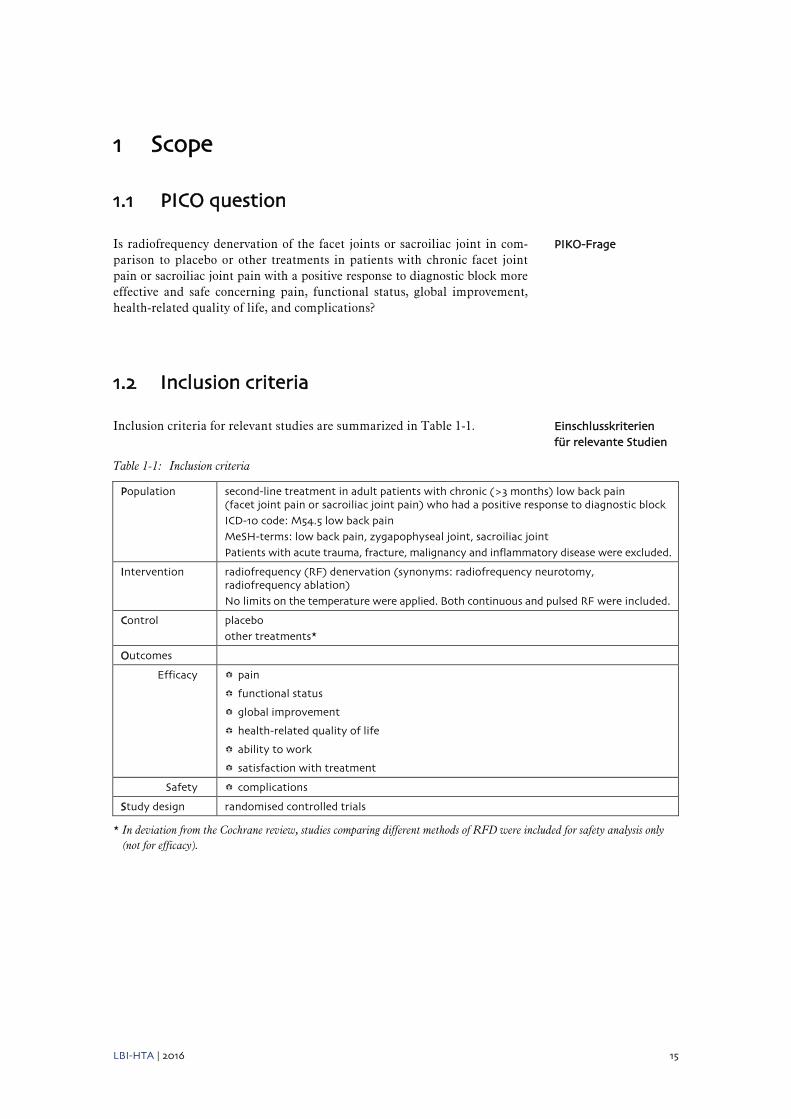
Inclusion criteria for relevant studies are summarized in Table 1-1.
Table 1-1: Inclusion criteria
Population second-line treatment in adult patients with chronic (>3 months) low back pain (facet joint pain or sacroiliac joint pain) who had a positive response to diagnostic block
ICD-10 code: M54.5 low back pain
MeSH-terms: low back pain, zygapophyseal joint, sacroiliac joint
Patients with acute trauma, fracture, malignancy and inflammatory disease were excluded.
Intervention radiofrequency (RF) denervation (synonyms: radiofrequency neurotomy, radiofrequency ablation)
No limits on the temperature were applied. Both continuous and pulsed RF were included.
Control placebo
other treatments*
Outcomes
Efficacy pain
functional status
global improvement
health-related quality of life
ability to work
satisfaction with treatment
Safety complications
Study design randomised controlled trials
* In deviation from the Cochrane review, studies comparing different methods of RFD were included for safety analysis only (not for efficacy).
PIKO-Frage
Einschlusskriterien für relevante Studien


LBI-HTA | 2016 17
2 Methods
2.1 Research questions
Description of the technology
Element ID Research question
B0001 What is radiofrequency denervation?
B0002 What is the claimed benefit of radiofrequency denervation in relation to the comparators?
B0004 Who administers radiofrequency denervation and in what context and level of care is it provided?
B0008 What kind of special premises are needed to use radiofrequency denervation?
B0009 What supplies are needed to use radiofrequency denervation?
A0020 For which indications has radiofrequency denervation received marketing authorisation or CE marking?
A0021 What is the reimbursement status of radiofrequency denervation?
Health problem and Current Use
Element ID Research question
A0001 For which health conditions and for what purposes is radiofrequency ablation/denervation used?
A0002 What is the disease or health condition in the scope of this assessment?
A0003 What are the known risk factors for chronic low back pain?
A0004 What is the natural course of the disease or health condition?
A0005 What is the burden of disease for patients with chronic low back pain?
A0006 What are the consequences of chronic low back pain for the society?
A0024 How is chronic low back pain currently diagnosed according to published guidelines and in practice?
A0025 How is chronic low back pain currently managed according to published guidelines and in practice?
A0007 What is the target population in this assessment?
A0023 How many people belong to the target population?
A0011 How much is radiofrequency denervation of the facet and sacroiliac joints utilised?
Clinical Effectiveness
Element ID Research question
D0001 What is the expected beneficial effect of radiofrequency denervation on mortality?
D0005 How does radiofrequency denervation affect symptoms and findings (severity, frequency) of chronic low back pain?
D0006 How does radiofrequency denervation affect progression (or recurrence) of chronic low back pain?
D0016 How does the use of radiofrequency denervation affect activities of daily living?
D0012 What is the effect of radiofrequency denervation on generic health-related quality of life?
D0013 What is the effect of radiofrequency denervation on disease-specific quality of life?
D0017 Was the use of radiofrequency denervation worthwhile?

Radiofrequency denervation for sacroiliac and facet joint pain
18 LBI-HTA | 2016
Safety
Element ID Research question
C0008 How safe is the technology in comparison to the comparator(s)?
C0002 Are there harms related to dosage or frequency of applying the technology?
C0004 How does the frequency or severity of harms change over time or in different settings?
C0005 What are the susceptible patient groups that are more likely to be harmed through the use of the technology?
C0007 Are the technology and the comparator(s) associated with user-dependent harms?
2.2 Sources
Description of the technology and Health Problem and current use
Publications identified by hand search
Questionnaire completed by the submitting hospital
2.3 Systematic literature search
During the scoping process we identified a recently published Cochrane Re-view [1] dealing with radiofrequency denervation for chronic low back pain. This Cochrane review had a broader scope than our assessment (including not only pain from facet joints’ and sacroiliac joints, but also from interver-tebral discs and the dorsal root ganglion). The Cochrane review’s literature search (from inception to May 2014) had been conducted in the following databases:
Cochrane Central Register of Controlled Trials (CENTRAL)
MEDLINE
MEDLINE In-Process & Other Non-Indexed Citations
EMBASE
Cumulative Index to Nursing and Allied Health Literature (CINAHL)
PsycINFO
ClinicalTrials.gov
World Health Organization (WHO) International Clinical Trials Registry Platform (ICTRP)
The complete search strategies can be found in the Appendix of the Cochrane review [1]. Additionally, the review authors mentioned an update of the lit-erature search in June 2015.
Therefore, we decided to use the relevant parts of the Cochrane review (deal-ing with the facet and sacroiliac joints) as primary source for this report and we refrained from conducting a (redundant) systematic literature search by ourselves.
Informationen aus Handsuche und
Einreicherangaben für Beschreibung des
Gesundheitsproblems und der Technologie
vorab Identifikation eines rezenten
Cochrane Reviews
Verzicht auf eigene systematische Literatursuche

Methods
LBI-HTA | 2016 19
2.3.1 Additional searches
In a first step, we contacted the first author of the Cochrane review via e-mail. She informed us that one additional relevant study [2] had been identified by the update search of 2015 and not yet been incorporated in the review.
Additionally, on December 9th 2015, we conducted a (systematic) search in Medline and Pubmed using the search strategy from the Cochrane review (complemented by the search term sacroiliac joint) without date restrictions. This search yielded 9 additional references. None met the inclusion criteria.
A hand-search in PubMed identified one article [3], which presented 12-month follow-up data of a trial [4] already included in the Cochrane Review. How-ever, the results were not relevant for this review because the patients of the control group were unblinded after 3 months and had the opportunity to “cross-over” (i.e., almost all of the control patients also received the interven-tion). The 3-month follow-up data had already been presented in the includ-ed article (Patel et al. 2012 [4]).
Manufacturers of two products for sacroiliac joint pain radiofrequency de-nervation (Halyard Health and Baylis Medical) were contacted on the 21st of December, 2015. However, they didn’t respond to our e-mail.
2.4 Flow chart of study selection
The Cochrane review identified a total of 748 records through database and reference searching. 36 records were screened in full text and 23 studies ful-filled the inclusion criteria of the Cochrane review. Of these, 12 RCTs for facet joint pain and 2 RCTs for sacroiliac joint pain were relevant for our as-sessment.
Through our additional search (see 2.3.1.), we identified further 11 refer-ences of which 1 additional RCT fulfilled the inclusion criteria. The selec-tion process is displayed in Figure 2-1.
dennoch ergänzt um: Kontaktaufnahme mit Cochrane Erstautorin systematisches Update der Medline Suche ergänzt um den spezifischen Begriff der Iliosakralgelenke Handsuche in Medline
und Kontaktaufnahme mit 2 Herstellern
Literaturauswahl im CR ursprünglich 12 RCTs für Facetten- und 2 für Iliosakralgelenke
ergänzt um ein weiteres RCT aus Handsuche

Radiofrequency denervation for sacroiliac and facet joint pain
20 LBI-HTA | 2016
* for effectiveness: n=12 (facet joint pain: 10; sacroiliac joint pain:2); for safety: additional 3 RCTs for facet joint pain
Figure 2-1: Flow chart of study selection (PRISMA Flow Diagram)
Cochrane Review: Records identified through
database and reference searching (n=748)
Scre
enin
g
Incl
ud
ed
Elig
ibili
ty
Iden
tifi
cati
on
LBI-HTA: Additional records identified
through hand search (n=11)
Records fully screened (n=36)
Studies ongoing and therefore excluded
(n=5)
Full-text articles assessed for eligibility
(n=31)
Studies included in the Cochrane review (n=23)
disc pain (n=5)
radicular chronic low back pain (n=2)
radiating chronic low back pain (n=1)
chronic low back pain with or without radiation (n=1)
facet joint pain (n=12)
sacroiliac joint pain (n=2)
Studies excluded for various reasons
(n=8)
Cochrane Review
Studies included in assessment (n=15)*
facet joint pain (n=13)
sacroiliac joint pain (n=2)
Records excluded (n=10)

Methods
LBI-HTA | 2016 21
2.5 Analysis
As the scope of the Cochrane review [1] was broader than our research ques-tion, this report only focuses on its facet and the sacroiliac joint results. We checked the data extraction tables of the Cochrane review for accuracy and completeness (based on the included primary studies), adapted them to our format, extracted them, and added further relevant information from the pri-mary studies. Data extraction was performed by one author (IR) and con-trolled by another author (KR).
We assessed the quality of included primary studies using the Cochrane Risk of Bias Tool (see Table A-5).
2.6 Synthesis
For the crucial outcomes pain (VAS) and functional status (ODI), we aimed at providing pooled data for effectiveness. We were able to extract some of the Cochrane Review’s meta-analyses to our evidence profiles (‘GRADE ta-bles’) and added own calculations if necessary. For these, we used the R pack-age “meta”1. Because of heterogeneity, we chose a random effects model for all of our calculations. The only exception was the meta-analysis for the ODI score at 12 months (RFD vs. placebo), which only included 2 results from 1 study [5]. These were highly similar and the results remained exactly the same, regardless of whether the random or fixed effects model was used (p. 43). Meta-analyses are provided as mean differences (MDs) with 95% confidence intervals (95% CIs). When necessary, VAS/NRS scores were converted to scales ranging from 0 to 10. The Cochrane Review’s authors solved the prob-lem of missing standard deviations (SDs) by calculating them using reported values of the CI (and, if CIs were not available, they used SDs of baseline scores, or estimations of SDs based on other studies with the same population, treatment and score). We decided, in one case of missing SDs, for the post-interventional observation data [6] to use the baseline SD (and to additionally provide the effect estimates with halved and doubled SD; see footnote p. 43).
The questions were answered in plain text format with reference to the evi-dence profiles (see Table 7-1 to Table 7-5).
1 Guido Schwarzer (2015). meta: General Package for Meta-Analysis. R package version
4.3-2., available at http://CRAN.R-project.org/package=meta
Daten des Cochrane Review auf Basis der Primärstudien auf Korrektheit und Vollständigkeit überprüft, extrahiert und ggfs. ergänzt
Biasrisiko der Primärstudien mit dem Cochrane RoB Tool beurteilt
Meta-Analysen für Schmerz und Funktionalität extrahiert bzw. durchgeführt

LBI-HTA | 2016 23
3 Description and technical characteristics of technology
Features of the technology and comparators
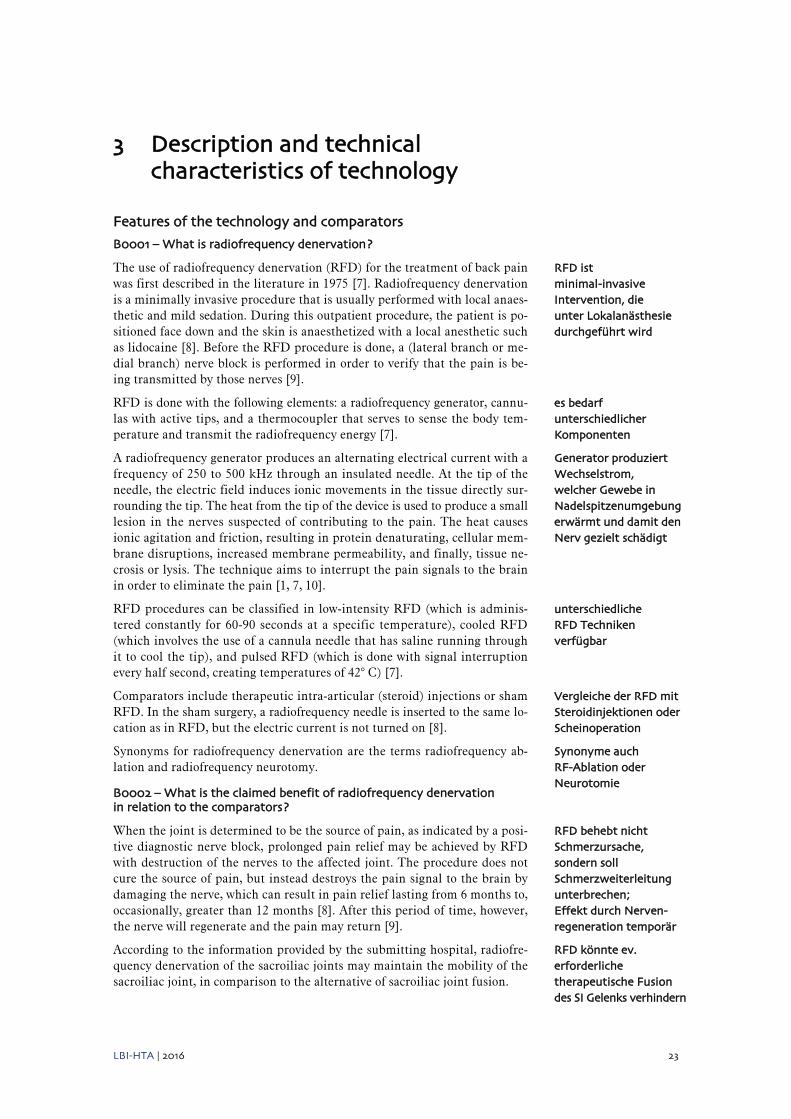
B0001 – What is radiofrequency denervation?
The use of radiofrequency denervation (RFD) for the treatment of back pain was first described in the literature in 1975 [7]. Radiofrequency denervation is a minimally invasive procedure that is usually performed with local anaes-thetic and mild sedation. During this outpatient procedure, the patient is po-sitioned face down and the skin is anaesthetized with a local anesthetic such as lidocaine [8]. Before the RFD procedure is done, a (lateral branch or me-dial branch) nerve block is performed in order to verify that the pain is be-ing transmitted by those nerves [9].
RFD is done with the following elements: a radiofrequency generator, cannu-las with active tips, and a thermocoupler that serves to sense the body tem-perature and transmit the radiofrequency energy [7].
A radiofrequency generator produces an alternating electrical current with a frequency of 250 to 500 kHz through an insulated needle. At the tip of the needle, the electric field induces ionic movements in the tissue directly sur-rounding the tip. The heat from the tip of the device is used to produce a small lesion in the nerves suspected of contributing to the pain. The heat causes ionic agitation and friction, resulting in protein denaturating, cellular mem-brane disruptions, increased membrane permeability, and finally, tissue ne-crosis or lysis. The technique aims to interrupt the pain signals to the brain in order to eliminate the pain [1, 7, 10].
RFD procedures can be classified in low-intensity RFD (which is adminis-tered constantly for 60-90 seconds at a specific temperature), cooled RFD (which involves the use of a cannula needle that has saline running through it to cool the tip), and pulsed RFD (which is done with signal interruption every half second, creating temperatures of 42° C) [7].
Comparators include therapeutic intra-articular (steroid) injections or sham RFD. In the sham surgery, a radiofrequency needle is inserted to the same lo-cation as in RFD, but the electric current is not turned on [8].
Synonyms for radiofrequency denervation are the terms radiofrequency ab-lation and radiofrequency neurotomy.
B0002 – What is the claimed benefit of radiofrequency denervation in relation to the comparators?
When the joint is determined to be the source of pain, as indicated by a posi-tive diagnostic nerve block, prolonged pain relief may be achieved by RFD with destruction of the nerves to the affected joint. The procedure does not cure the source of pain, but instead destroys the pain signal to the brain by damaging the nerve, which can result in pain relief lasting from 6 months to, occasionally, greater than 12 months [8]. After this period of time, however, the nerve will regenerate and the pain may return [9].
According to the information provided by the submitting hospital, radiofre-quency denervation of the sacroiliac joints may maintain the mobility of the sacroiliac joint, in comparison to the alternative of sacroiliac joint fusion.
RFD ist minimal-invasive Intervention, die unter Lokalanästhesie durchgeführt wird
es bedarf unterschiedlicher Komponenten
Generator produziert Wechselstrom, welcher Gewebe in Nadelspitzenumgebung erwärmt und damit den Nerv gezielt schädigt
unterschiedliche RFD Techniken verfügbar
Vergleiche der RFD mit Steroidinjektionen oder Scheinoperation
Synonyme auch RF-Ablation oder Neurotomie
RFD behebt nicht Schmerzursache, sondern soll Schmerzweiterleitung unterbrechen; Effekt durch Nerven-regeneration temporär
RFD könnte ev. erforderliche therapeutische Fusion des SI Gelenks verhindern
Radiofrequency denervation for sacroiliac and facet joint pain
24 LBI-HTA | 2016
Administration, Investments, personnel and tools required to use the technology and the comparator(s)
B0004 – Who administers radiofrequency denervation and in what context and level of care is it provided?
B0008 – What kind of special premises are needed to use radiofrequency denervation?
B0009 – What supplies are needed to use radiofrequency denervation?
According to the information received by the submitting hospital, the inter-vention is performed in specialised centres.
The intervention is performed by an experienced operation team, including an experienced orthopaedist, an assistant physician, a nurse, an anaesthetist (in the case of sedoanalgesia), and a radiological assistant for intraoperative X-ray monitoring.
An operating room with intraoperative X-ray monitoring is needed for the intervention.
Regulatory & reimbursement status
A0020 – For which indications has radiofrequency denervation received marketing authorisation or CE marking?
RFD for back pain is a procedure and is therefore not subject to regulation by the Food and Drug Administration (FDA). However, the FDA regulates RFD devices and hence, there are various devices listed in the FDA 510(k) Pre-market Notification database. The following generators received FDA clear-ance within the past 5 years:
Erase Pen and Erase Tip System for Nerve Ablation, Models HC-0O01 and CS-0001 (Cheng Medical Corp.; approved 2011),
NT 2000 Lesioning Generator (Neurotherm Inc.; approved 2011),
Diros OWL™ URF-3AP(ML) (Diros Technology Inc.; approved 2010),
Cosman G4 Radiofrequency Generator (Cosman Medical Inc.; approved 2008) [11].
There are currently seven RF lesion probe devices approved by the FDA:
Baylis Pain Management Probe (Baylis Medical Co., Ontario, Canada; approved 2000),
Baylis Pain Management Cooled Probe (Baylis Medical Co., Ontario, Canada; approved 2005; see also below),
Baylis Pain Management Single-Use Probe (Baylis Medical Co., Ontario, Canada; approved 2007),
Pajunk RFTL Radiofrequency Needle (Pajunk GmbH Medizintechnologie, Geisingen, Germany; approved 2006),
Smith & Nephew RF Denervation Probes & RF Cannulae (Smith & Nephew Inc., Andover, MA, USA; approved 2004),
Stryker RF Electrodes and Cannulae (Stryker Instruments Kalamazoo, MI, USA; approved 2004),
Radionics disposable RF Cannulae (Technomed Europe, The Netherlands; approved 2004) [8].
laut Einreichern erforderlich:
- spezialisiertes Zentrum
- erfahrenes OP-Team - intraoperatives
Röntgen
FDA reguliert RFD Produkte
(nicht spezifisch für die Indikation)
aktuell 7 Systeme von der FDA zugelassen
Description and technical characteristics of technology
LBI-HTA | 2016 25
The cooled RFD system Sinergy by Baylis Medical Co. Inc, used in the RFD studies for sacroiliac joint pain, was acquired by the Kimberly-Clark Corp. in 2009. SInergy comprises a pump, generator, and probe. The pump circu-lates sterile water through the probes. The various Baylis Pain Management probes are sterile, single-use devices that deliver RF energy while being cooled. The probes are used in conjunction with an RF generator to create RF le-sions in nerves. All components have received FDA clearance as substantial-ly equivalent to an approved predicate device.
The SInergy Pain Management System has also received CE Marking [12]. We were not able to identify a comprehensive list of other currently CE mark-ed RFD systems and we refrained from a manual search for the CE marking of various RFD components.
A0021 – What is the reimbursement status of radiofrequency denervation?
Currently, radiofrequency denervation of the facet and sacroiliac joints can be reimbursed via the Austrian DRG-system (Leistungsorientierte Krankenan-staltenfinanzierung/LKF) using the code AJ140 “percutaneous destruction of peripheral nerves”.
das SInergy System (dzt. in Verwendung für SI RFD) ‘ist zugelassen von der FDA und CE zertifiziert
RFD kann derzeit über Code AJ140 abgerechnet werden


LBI-HTA | 2016 27
4 Health Problem and Current Use
Overview of the disease or health condition
A0001 – For which health conditions and for what purposes is radiofrequency ablation/denervation used?
Radiofrequency ablation is one of several types of ablation therapy. There-fore, it can be used to treat a wide range of conditions. For example, RFA is sometimes used in oncology [13] to treat (bone, kidney, liver, lung or pros-tate) cancers or precancerous lesions in the esophagus (Barrett’s esophagus), in cardiology [14] to treat arrhythmias (e.g., supraventricular tachyarrhyth-mias), or in dermatology [15] to treat skin lesions. Finally, RFA is used in pain therapy, e.g., for the treatment of neck or low back pain (LBP).
A0002 – What is the disease or health condition in the scope of this assessment?
In the scope of this assessment, chronic low back pain deriving from the fac-et or sacroiliac joints is the condition of interest. Low back pain is defined as pain and discomfort, localised between the costal margin and above the infe-rior gluteal folds, with or without referred leg pain, that persists for at least 12 weeks (European Guidelines from 2004 [16]). The cause for sacroiliac pain is a sacroiliac joint dysfunction (due to hypermobility/instability or hypomo-bility/fixation).
A0003 – What are the known risk factors for chronic low back pain?
There are many possible causes for low back pain, e.g., infections, tumours, osteoporosis, fractures, or spinal disc herniation. The majority of patients (ap-proximately 85%) seen in the primary care, however, have non-specific low back pain, which is not attributable to a recognisable, known specific pathol-ogy or anatomical structure (e.g., infection, tumour, osteoporosis, fracture) [1, 17]. Suspected sources of back pain include lumbar facet (zygapophyseal) joints, sacroiliac joints, and degenerated intervertebral discs [1].
Risk factors associated with back pain include smoking, obesity, age, female gender, physically strenuous work, sedentary work, psychologically strenuous work, low educational attainment, job dissatisfaction, and psychologic factors such as somatization disorder, anxiety, and depression [17].
A0004 – What is the natural course of chronic low back pain?
Chronic low back pain is seen as recurring or persistent condition showing a fluctuating course over time. It is likely that patients who report LBP will continue to report LBP in the future [18]. After an initial episode of low back pain, 44-78% of the patients suffer relapses of pain [16].
RFD ist eine von zahlreichen Ablationstherapien, welche auch in anderen Indikationsbereichen eingesetzt wird
chronischer Rückenschmerz kann durch Veränderungen im Bereich der Facetten- oder Iliosakralgelenke bedingt sein
zahlreiche Schmerzursachen, in der Mehrzahl der PatientInnen jedoch unspezifisch/nicht eindeutig einer Ursache zuordenbar
Risikofaktoren für Rückenschmerzen u. a. Übergewicht, sitzende Tätigkeit, Stress etc.
Rückenschmerzen zeigen rezidivierenden oder persistierenden Verlauf

Radiofrequency denervation for sacroiliac and facet joint pain
28 LBI-HTA | 2016
Effects of the disease or health condition on the individual and society
A0005 – What is the burden of disease for patients with chronic low back pain?
Chronic low back pain is one of the most commonly reported pain conditions. It is often characterized by a long duration of illness and multiple recurrent episodes of pain. Patients with low back pain often report comorbidities such as osteoarthritis, cardiovascular and cerebrovascular diseases, as well as men-tal disorders, such as depression, anxiety disorders, and post-traumatic stress disorder. International data also show a positive correlation between low back pain and symptoms, such as migraine and headache, exhaustion, and respir-atory symptoms [19].
A0006 – What are the consequences of chronic low back pain for the society?
Low back pain is one of the most expensive diseases in industrialised coun-tries. It is one of the most frequent causes for inability to work and early re-tirement. In Germany, low back pain is estimated to cause direct costs of € 8.4 billion per year. According to international estimates, 85% of these costs are due to productivity losses because of inability to work and the remaining 15% are spent for medical treatment [19]. In the United States, costs of low back pain have been estimated to be more than 100 billion USD per year, primar-ily due to lost productivity [10].
Current clinical management of the disease or health condition
A0024 – How is chronic low back pain currently diagnosed according to published guidelines and in practice?
Chronic low back pain is diagnosed by a detailed medical history (anamnesis). The medical history should include asking for onset of symptoms, duration, localisation and causes of pain, correlation of pain with specific positions and movements, earlier pain episodes, problems in activities of daily living, as well as psychosocial risk factors. An important part of the medical history is ask-ing for red flag symptoms, which can indicate specific causes (e.g., fracture, tumour, infection, radiculopathy/neuropathy) with potentially urgent need for action [19, 20].
An additional physical examination (e.g., inspection, palpation, test of the mo-bility of the lumbar spine, examination of the sacroiliac joint [19, 20]) aims at distinguishing between non-specific vs. specific (physical or mental) causes (as the above mentioned red flag symptoms do). Medical imaging also aims at identifying specific causes for low back pain. However, study results ques-tion its use as single diagnostic modality, because degenerative changes (here: ostoartritis of facet joints detected by CT) are common in the general popu-lation and increasing with age [21]. A large population based study failed to find an association of this CT-verified presence of degenerative osteoarthri-tis and low back pain [21].
During the care process, the assessment of psychosocial and somatic risk fac-tors for pain chronification (‘yellow flags’; e.g., depressiveness, pain-related cognition, passive pain behaviour, workplace-related factors, iatrogenic fac-tors) is recommended [19].
PatientInnen weisen meist
Komorbiditäten auf
Rückenschmerzen sind eine der teuersten
Erkrankungen in Industrieländern
(v. a. durch Arbeitsunfähigkeit/ Frühpensionierung)
die Diagnose chronischer
Rückenschmerz erfolgt auf Basis der Anamnese
potentielle spezifische Ursachen sollen durch
klinische Untersuchung und Bildgebung
abgeklärt werden

Health Problem and Current Use
LBI-HTA | 2016 29
A0025 – How is chronic low back pain currently managed according to published guidelines and in practice?
Conservative treatment options for chronic low back pain may include pharmaceuticals (drug therapy [19]),
non-steroidal antirheumatics/antiphlogistics (tNSAR),
and, if applicable, opioid analgesics, muscle relaxants, or antidepressants,
or non-drug therapies [19],
physical activity and movement therapy,
therapeutic exercise,
patient education (information/training),
ergonomics,
multimodal, multi-, and interdisciplinary treatment/rehabilitation,
self-management programmes,
occupational therapy,
behavioural therapy, or
progressive muscle relaxation.
According to this guideline, invasive therapeutic procedures are not recom-mended for the treatment of patients with nonspecific low back pain [19], due to the lack of reliable data (despite of numerous studies) for percutane-ous procedures and the lack of studies on the use of surgical procedures.
The Austrian Guideline for the management of acute and chronic non-specific low back pain, published in 2011 [20], recommends multidisciplinary treat-ment programmes for chronic low back pain. Single interventions, such as phar-macological treatment only, are not considered to be sufficient to alleviating pain in the long term, as well as to prevent disability and work absences [20].
Target population
A0007 – What is the target population in this assessment?
The target population in this assessment are adult patients with chronic low back pain (longer than three months), who had a positive response to a dia-gnostic block in the sacroiliac or facet joints. Patients with acute trauma, frac-ture, malignancy, and inflammatory disease were excluded [1].
A0023 – How many people belong to the target population?
According to a health survey conducted by Statistik Austria in 2014, 23% of Austrian men and 26% of Austrian women were affected by chronic low back pain or other chronic back problems in the last 12 months. The prevalence increases with age and more women are affected than men [22]. The lifetime prevalence of low back pain (in total) is estimated to be up to 84% [16].
Prevalence-estimates of facet or sacroiliac joint pain are provided by the RFD studies’ authors as follows: thus, facet-joint pain accounts for 15% to 50% [6, 23, 24] and sacroiliac joint pain for 15-20% [4, 25] of low back pain. Against the backdrop of the high prevalence of degenerative changes detected in facet joints (63% in a sample of 188 men and women, at mean ~52 years old [21]) or sacroiliac joints (65% in a sample of 373 men and women, at mean ~58 years old [26]) of the general population, the provided (high) proportion of
konservative Behandlungsmethoden reichen von medikamentösen zu nicht-medikamentösen Therapien
invasive Maßnahmen werden nicht empfohlen
Einzelmaßnahmen werden als nicht ausreichend angesehen, langfristige Folgen (z. B. Krankenstände) zu verhindern
Zielpopulation dieses Berichts sind Erwachsene mit LBP >3 Monaten und entsprechender vor-angehender Diagnostik
¼ der ÖsterreicherInnen innerhalb eines Jahres betroffen; Lebenszeitprävalenz von Rückenschmerzen bis zu 84 % Anteil an Schmerzen im Bereich der Facetten- bzw. Iliosakralgelenke letztlich unklar

Radiofrequency denervation for sacroiliac and facet joint pain
30 LBI-HTA | 2016
‘facet or sacroiliac joint pain’ as causes of low back pain can be questioned. Due to non-specific imaging and clinical testing reliable epidemiological data seems to be missing.
A0011 – How much is radiofrequency denervation of the facet and sacroiliac joints utilised?
According to the information provided by one Austrian hospital, the annual frequency in this hospital is estimated to be 40 procedures of sacroiliac joint radiofrequency denervation. In 2014, 20 treatments were recorded in the sub-mitting hospital. No estimations were provided regarding the annual frequen-cy in Austria in total. The number of procedures of facet joint radiofrequency denervation in Austrian hospitals is unknown.
Frequenz der Iliosakral-RFD von einreichender
Institution auf ~ 40/Jahr geschätzt;
für Gesamtösterreich keine Schätzungen
(weder für Facetten- noch Iliosakral-Gelenke)

LBI-HTA | 2016 31
5 Clinical effectiveness
5.1 Outcomes
The following outcomes were defined as crucial to derive a recommendation:
pain
functional status
global improvement
health-related quality of life
Changes in pain intensity were measured in all included studies using the Visual Analogue Scale (VAS) or the Numeric Rating Scale (NRS). The VAS is a continuous scale comprised of a horizontal or vertical line, usually 10 cm in length. Numbers or verbal descriptors at intermediate points are not rec-ommended. The NRS (used in [2] and [4, 25]) is a segmented numeric ver-sion of the VAS. The most commonly used is the 11-item NRS, an 11-point numeric scale with 0 representing “no pain” and 10 representing “worst pain imaginable” [27]. One study used the Visual Numeric Pain Scale (VNS) for pain measurement [23].
The functional status was measured using the Oswestry disability index score (ODI) in 7 of the included studies. The ODI was published in 1980 and has been widely used as a condition-specific outcome measure for patients with spinal disorders. It is comprised of 10 items (including pain intensity, per-sonal care, walking, sleeping, social life, ...) with associated statements for the patient to select, which reflect the patient’s ability to manage their everyday life. A maximum score of 50 is possible. The score can also be expressed as a percentage score (0-20% means minimal disability, 81-100% means that the patient is bed-bound) [28].
As ‘global improvement’ is a non-specific outcome, study authors used differ-ent tools to determine potential effects. For assessing the ‘global perceived effect’ (GPE, used in two studies [4, 25]), three questions are asked (1. My pain has improved/worsened/stayed the same since my last visit; 2. The treatment I received improved/did not improve my ability to perform daily activities; 3. I am satisfied/not satisfied with the treatment I received and would recom-mend it to others.). Three studies used 4-7 point scales to determine a change in the ‘global effect on back pain’[29], the ‘subjective global assessment’ [30]/ the ‘global perceived effect’[6].
Health related quality of life (generic or disease-specific) can be assessed by various standardised instruments. Studies included in this review used EQ-5D [23], SF-36 [29], and AQoL [4].
The outcomes
patient satisfaction and
ability to work
were defined as important, but not crucial to derive a recommendation.
als entscheidende Ergebnisparameter wurden Schmerz, Funktionalität, allgemeine Verbesserung und die gesundheits-bezogene Lebensqualität definiert Schmerz wird meist mit der Visual Analogue Scale (VAS) bestimmt
zur Abbildung der Funktionalität ist der Oswestry disability index (ODI) weit verbreitet
die allgemeine Verbesserung ist unspezifisch und kann mit unterschiedlichen Instrumenten erhoben werden
zur Messung der HRQoL existieren zahlreiche standardisierte Instrumente
als wichtig (nicht entscheidend) wurden Patientenzufriedenheit und Arbeitsfähigkeit definiert

Radiofrequency denervation for sacroiliac and facet joint pain
32 LBI-HTA | 2016
5.2 Included studies
Facet joint pain: study and patient characteristics
Overall, we included 10 studies that evaluated radiofrequency denervation for facet joint pain [2, 5, 6, 23, 24, 29-33]. All studies were randomised controlled trials. 6 [5, 6, 29-32] compared radiofrequency denervation with a placebo/ sham treatment, the remaining 4 studies used steroid injections as the com-parator [2, 23, 24, 33].
3 trials were conducted in Turkey [5, 23, 33], 2 in the Netherlands [6, 29], and the remaining 5 studies in Canada, Germany, Iran, Sweden, and the UK. The two Dutch studies were nationally funded [6, 29], the Swedish, German and Canadian study stated no (industry) funding [24, 30]/academic research [32], the remaining 5 studies did not provide funding information. The placebo controlled trials were published between 1994 and 2008, the remaining 4 stud-ies between 2012 and 2014. Sample sizes of the placebo-controlled trials ranged from 31 to 81, those with the injection control groups included more patients by trend (80-120 each, except for one trial with only 56 patients). The total number of patients was 323 patients in the 6 placebo-controlled and 356 in the 4 steroid injection controlled trials.
Inclusion criteria differed considerably between studies. In the placebo-con-trolled trials, patients hat to suffer from back pain for more than 3 months [31, 32] up to at least 2 years [30] (>6 months [5, 29], >12 months [6]). In the trials with the steroid injection control, patients had to suffer from back pain for more than 6 months [2, 33] to >2 years in [24] (not dependant on the total duration of complaints, but no response to conservative treatment for up to 6 weeks in [23]).
The mean age of included patients ranged from 41 to 61 years in the placebo controlled trials, 50 to 64 years in the steroid injection controlled trials. Pa-tients in the latter studies were slightly older: none of these studies show mean ages below 50 years in comparison to 3 of the 6 placebo-controlled trials. The percentage of female participants was more than 55% in all but one (steroid comparison) trial (35-39%) [24] with a maximum of up to 75% in [29]. Pa-tient follow-up ranged from 3 [32] to most commonly 12 months (in half of the placebo-controlled [5, 6, 29] as well as steroid controlled [23, 33] trials).
Loss to follow-up ranged from 0% [5, 23, 29, 30] to 10% in [24], 2 trials did not report drop-outs [31, 33].
Facet joint pain: quality assessment
4 of 6 placebo-controlled and 1 of 4 steroid-injection controlled trials have been judged to have a low risk of bias on a single study level. The remaining 5 studies [2, 23, 30, 31, 33] involve a high bias risk, e.g., due to unclear blind-ing (where possible), unclear or high number of drop-outs, or differing base-line characteristics.
Characteristics of included studies are displayed in Table A-1 and Table A-2.
Schmerzen im Bereich der Facettengelenke:
10 RCTs
- 6 RFD vs. Plazebo (1994-2008,
323 PatientInnen)
- 4 RFD vs. Steroid-injektionen
(2012-2014, 356 PatIentinnen)
Einschlusskriterien unterschiedlich v. a.
hinsichtlich vorangehender Schmerzdauer
mittleres Alter 41-64 Jahre
(in den Injektionsstudien tendenziell etwas älter);
mit Ausnahme einer Studie Frauenanteil
>55 %; Beobachtungszeitraum
bis zu 12 Monate
die Hälfte der RCTs weist hohes Bisasrisiko auf

Clinical effectiveness
LBI-HTA | 2016 33
Sacroiliac joint pain: study and patient characteristics
For evaluating radiofrequency denervation for sacroiliac joint pain, 2 random-ised controlled trials fulfilled the inclusion criteria [4, 25]. Both RCTs com-pared radiofrequency denervation with placebo/sham treatment. They were published 2008-2012, conducted in the US and funded by Baylis Medical. The total number of patients was 79. Both RCTs included patients who had low back pain lasting for 6 months or longer and who had not achieved ade-quate improvement after conservative therapies.
The mean age of patients was 52-64 years. 57-82% of the patients were female. Length of follow-up was 6 [25] and 9 months [4]. However, patients of the con-trol group were given the opportunity to cross-over after 1 [25] or 3 [4] months.
No lossestofollow-up were observed up to 3 months.
Sacroiliac joint pain: quality assessment
One RCT was judged to have a low bias risk [4], whereas the other RCT [25] might imply a high risk due to differing baseline characteristics.
Characteristics of included studies are displayed in Table A-4.
5.3 Results
Mortality
D0001 – What is the expected beneficial effect of radiofrequency denervation on mortality? and
D0003 – What is the effect of radiofrequency denervation on the mortality due to causes other than low back pain?
Mortality is not a relevant outcome for assessing the clinical effectiveness of radiofrequency denervation, since neither the disease nor the intervention is life-threatening. Therefore, none of the included studies reported this outcome.
Morbidity (Pain)
D0005 – How does radiofrequency denervation affect symptoms and findings (severity, frequency) of chronic low back pain?
Facet joint pain
All 6 placebo-controlled trials report pain outcomes as changes in VAS score. Up to one month, one study reports statistically significant group differences in favour of the intervention at the post procedure observation point (conven-tional or pulsed RFD compared to placebo) [5]. One month after the inter-vention, statistically significant group differences are only observed in a sub-group comparison of one trial [31], whereas the third study doesn’t observe a statistically significant group difference [32] (The overall estimate up to/in-cluding one month post intervention is statistically significant in favour of the intervention: MD -1.47 [-2.28, -0,67]). In the time period up to 6 months, only one [6] trial (of the three studies that report VAS group differences in this observation period) finds a statistically significant group difference (the overall estimate is n.s.: MD -0.71 [-2.25, 0.84]) [6, 29, 32]. At 6 months, 2 [5, 31] of 3 trials report statistically significant group differences, but the over-
Schmerzen im Bereich der Iliosakralgelenke: 2 RCTs, beide Plazebo-kontrolliert (2008-2012, 79 PatientInnen)
mittleres Alter 52-64 Jahre, Frauenanteil 57-82 %; Beobachtungszeitraum (durch cross-over) 1-3 Monate
1 der 2 RCTs mit hohem Bisarisiko
Mortalität: für die vorliegende Fragestellung kein relevanter Endpunkt
Schmerz: Facettengelenke/RFD vs. Plazebo (6 RCTs):
bis zu 1 Monat: insgesamt signifikant zu Gunsten der Intervention
danach insgesamt nicht signifikante Gruppen-unterschiede (einzelne Studien kommen zu widersprüchlichen Ergebnissen)

Radiofrequency denervation for sacroiliac and facet joint pain
34 LBI-HTA | 2016
all estimate remains marginal, non-significant (MD -0.70 [-1.48, 0.08] [5, 30, 31]). A twelve months result is only provided by one trial, which reports a sta-tistically significant difference only between the placebo and the conventional RFD group (not in comparison with the pulsed RFD group; overall: MD -0.96 [-0.04, 0.12]) [5].
In comparison to steroid injections, both studies that report pain outcomes at ≤1 month find statistically significant group differences (overall estimate MD: -1.81 [-3.05, -0.58]) [23, 33]. Overall estimates for group differences at 6 and 12 months are also statistically significant (at 6 months [23, 24, 33]: MD -2.13 [-3.45, -0.81], only one trial reported a n.s. difference [24]); at 12 months [23, 33]: MD -2.65 [-3.43, -1.88]). The remaining trial [2] states significant group differences at 12 weeks and 6 months (not at 6 weeks), but doesn’t pro-vide detailed data.
Sacroiliac joint pain
At one month, one study [25] reported a significant reduction in pain, where-as the second study [4] was not able to detect a significant group difference (overall estimate MD -2.21 [-5.34, 0.92]). Meaningful results at 3 months can only be provided by one of the two trials [4] (which reports a significant group difference; MD -1.30 [-2.06, -0,54]). At this observation point, data of the sec-ond trial is without value for the comparison due to cross over (only three pa-tients of the original placebo group declined to cross over to the RFD group) [25].
D0006 – How does radiofrequency denervation affect progression (or recurrence) of chronic low back pain?
Due to the lack of long-term follow-up data (>12 months), this question can-not be answered.
Functional status
D0016 – How does the use of radiofrequency denervation affect activities of daily living?
Facet joint pain
3 of the 6 placebo-controlled trials report ODI outcomes [5, 6, 32]2. One study [5] observes significant group differences between the RFD and control groups post procedure, as well as at 6 and 12 months. A statistically significant group difference has also been reported in the second trial 8 weeks post interven-tion. On the contrary, the third study wasn’t able to detect statistically signif-icant group differences (neither at 4, nor at 12 weeks) [32]. Overall estimates at the different observation periods are non-significant within the first 6 months (≤1 month: MD -3.45 [-7.68, 0.77]; >1 to <6 months: MD -9.48 [-28.73, 9.76]; 2 studies each) and significant in favour of the intervention in the following 6 months (based on one trial). A forth study reported functional status as ‘mean change in physical activity’ [29] at 3 months (n.s. group difference), the remaining 2 studies did not report this outcome [30, 31].
2 in [18] a n.s. group difference (-3.13 vs. -1.62) was reported, when the functional sta-
tus was measured by the COOP/WONCA Functional Assessment Charts at 8 weeks (in contrary to the s. difference if measured by ODI)
Facettengelenke/RFD vs. Steroidinjektionen
(3 RCTs):
in allen Beobachtungs-zeiträumen signifikant
zu Gunsten der Intervention
Iliosakralgelenke/RFD vs. Plazebo (2 RCTs):
n.s. nach 1 Monat (widersprüchliche
Studienergebnisse); signifikant zu Gunsten der Intervention nach
3 Monaten (nur 1 Studie)
Auswirkungen auf Progression/
Wiederauftreten der Beschwerden ist
aufgrund fehlender Langzeitdaten
unbekannt
Funktionalität:
Facettengelenke/RFD vs. Plazebo (3 RCTs)
n.s. <6 Monate,
danach (≤12 Monate) signifikant zu Gunsten
der Intervention, jedoch basierend auf 1 Studie
Clinical effectiveness
LBI-HTA | 2016 35
In comparison to steroid injections, only one trial provides data [24] (for on-ly one observation point at 6 months) and does not find a statistically signifi-cant group difference (MD: -5.00 [-15.19, 5.19]). The most recent trial [2] re-ports statistically significant group differences at 12 weeks and 6 months (n.s. at 6 weeks), but doesn’t provide detailed data (results are shown in a figure only). The remaining 2 studies did not report this outcome [23, 33].
Sacroiliac joint pain
Both trials provide ODI data [4, 25] and both report statistically significant group differences at 1 month (if changes from baseline are compared; the over-all effect for the group comparisons at 1 month is n.s.: MD -14.06 [-30.42, 2.30]). Meaningful results at 3 months can only be provided by one of the two trials [4] (still significant difference; MD -11.00 [-17.91, -4.09]), whereas at this observation point, data of the second trial is without value for the com-parison due to cross over (only three patients of the original placebo group declined to cross over to the RFD group)[25].
Global improvement
D0005 – How does radiofrequency denervation affect symptoms and findings (severity, frequency) of chronic low back pain?
In addition to changes in pain or functional status, overall ‘global improve-ment’ has been assessed in 5 (placebo-controlled) trials with different instru-ments (no overall estimates provided).
Facet joint pain
In an observation period of 2-6 months post intervention, 3 studies reported significant global improvements in the RFD group compared to the placebo control [6, 29, 30].
Sacroiliac joint pain
In an observation period of 1-3 months post intervention, each study reports significant global improvements in the RFD group compared to the placebo control [4, 25].
Health-related quality of life
D0012 – What is the effect of radiofrequency denervation (RFD) on generic health-related quality of life?
Facet joint pain
One placebo-controlled study [29] on facet joint pain used the SF-36 to meas-ure quality of life at 3 months post intervention and did not observe statisti-cally significant group differences (except for the item vitality).
One study comparing RFD in facet joints with steroid injections didn’t find any statistically significant differences in quality of life assessed by the EQ-5D (at 1, 6 and 12 months) [23].
The remaining 8 studies did not report this outcome.
Facettengelenke/RFD vs. Steroidinjektionen (1 RCT) n.s. 6 Monate post Intervention (keine anderen Beobachtungs-zeitpunkte verfügbar)
Iliosakralgelenke/RFD vs. Plazebo (2 RCTs) insgesamt n.s. 1 Monat post Intervention (die beiden Einzelstudien berichten jedoch s. Verbesserungen), s. zu Gunsten der Intervention nach 3 Monaten (hier 1 Studie)
Allgemeine Verbesserung:
Facettengelenke/RFD vs. Plazebo (3 RCTs): s. Gruppenunterschiede nach 2-6 Monaten
Iliosakralgelenke/RFD vs. Plazebo: s. besser nach 1 bzw. 3 Monaten (je ein RCT)
Lebensqualität (generisch): Facettengelenke/RFD vs. Plazebo: 1 RCT, n.s. nach 3 Monaten; vs. Steroidinjektion (1 RCT): n.s. nach 1-12 Monaten

Radiofrequency denervation for sacroiliac and facet joint pain
36 LBI-HTA | 2016
Sacroiliac joint pain
One of the two studies on RFD in SI joints used the AQoL instrument and reported no difference at one, but a statistically significant difference at 3 months [4]. The second study did not report this outcome [25].
D0013 – What is the effect of radiofrequency denervation on disease-specific quality of life?
None of the studies reported on disease-specific quality of life.
Ability to work
D0016- How does the use of radiofrequency denervation affect activities of daily living?
In addition to the measurement of the functional status (see above), this ques-tion should have been answered by the outcome “ability to work”, but none of the RCTs reported this outcome.
Patient satisfaction
D0017 – Was the use of radiofrequency denervation worthwhile?
To answer this research question, the outcome “satisfaction with treatment” was used.
Facet joint pain
The outcome was reported in 1 of 6 placebo controlled RCTs on RFD in facet joints and 3 of 4 RCTs comparing RFD with steroid injections in facet joints.
Compared to the placebo intervention, one study reported significantly higher patient satisfaction (≥1 month) in the intervention group without providing detailed data [5].
Compared to steroid injections one study [23] reported no difference in pa-tient satisfaction at 1 and 6 months (whereas the 12 month result favoured RFD). The remaining two studies found significant group differences in fa-vour of RFD at all observation points (after 12 weeks and 6 months [2], at 1, 6 and 12 months [33]).
Sacroiliac joint pain
The two RCTs assessing RFD in the sacroiliac joint did not report on patient’s satisfaction with treatment [4, 25].
Iliosakralgelenke/RFD vs. Plazebo:
1 RCT, n.s. nach 1, s. nach 3 Monaten
Lebensqualität
(indikations-spezifisch): Effekt aufgrund fehlender Daten
unbekannt
Arbeitsfähigkeit: Effekt aufgrund fehlender Daten
unbekannt
PatientInnen-zufriedenheit
Facettengelenke/RFD vs. Plazebo (1 RCT):
„s. besser“ (ohne Daten)
vs. Steroidinjektion (3 RCTs): widersprüchliche
Studienergebnisse, tendenziell zu Gunsten
der Intervention
Iliosakralgelenke/RFD vs. Plazebo: Effekt
aufgrund fehlender Daten unbekannt

LBI-HTA | 2016 37
6 Safety
6.1 Outcomes
The following outcome was defined as crucial to derive a recommendation:
Complications
The outcome ‘complications’ is non-specific and includes direct intervention-related side-effects (e.g., during or immediately after the intervention), as well as any other negative consequences observed in the follow-up period.
6.2 Included Studies
Facet joint pain: study and patient characteristics
In addition to the 10 studies [2, 5, 6, 23, 24, 29-33] that we had already in-cluded for the clinical effectiveness assessment, we included 3 further RCTs [34-36] for the safety analysis. All of these studies compared different RFD methods (continuous vs. pulsed RFD [34], a distal vs. a tunnel vision approach for RFD [35] and an intra-articular vs. extra-articular RFD [36]).
The trials were conducted in the US [34], Korea [35] and the Netherlands, [36] and published between 1999 [36] and 2013 [35]. Sample sizes ranged from 34 [36] to 82 [35]. The total number of patients was 166. Inclusion criteria varied between studies. Patients had to suffer from back pain for more than 1 [34], 3 [35], or 6 months [36].
The mean age of included patients ranged from 57 to 69 years. The percent-age of female participants differed considerably between and within studies (24-29% [36], 46-62% [34] and 62-72% [35]). Patient follow-up ranged from 3 [34, 36] to 6 [35] months, loss to follow-up was from 12-15% [35] to 48% in [34] ([36]did not report drop-outs).
Facet joint pain: quality assessment
All 3 RCTs were judged to have a high risk of bias due to the unclear/high number of drop-outs, unclear blinding, and allocation concealment processes.
For the study and patient characteristics of the further 10 RCTs, see the de-scription of included studies in the section on clinical effectiveness.
Study characteristics and results of included studies are displayed in Table A-1, Table A-2, and Table A-3, and in the evidence profile in Table 7-3.
Sacroiliac joint pain: study and patient characteristics
The 12 month follow up [3] of Patel 2012 [4] (which could have been includ-ed for safety issues despite cross-over) didn’t provide any safety data. There-fore, we included the same 2 RCTs [4, 25] (that we had already included for the clinical effectiveness assessment) for evaluating the safety of RFD for sa-croiliac joint pain (see 5.2).
Study characteristics and results of included studies are displayed in Table A-4 and in the evidence profile in Table 7-5.
für den Bereich Sicherheit wurden ‚Komplikationen’ als entscheidender Ergebnisparameter definiert
Schmerzen im Bereich der Facettengelenke: 13 RCTs: 10 (siehe Wirksamkeit) + 3 RCTs, die unterschiedliche RFD Methoden verglichen (1999-2013, 166 PatientInnen)
Alter: 57-69 Jahre, Frauenanteil 24-72 %; Drop-out in einer Studie 48 %
alle 3 RCTs mit hohem Biasrisiko
Schmerzen im Bereich der Iliosakralgelenke: 2 RCTs (siehe Wirksamkeit)

Radiofrequency denervation for sacroiliac and facet joint pain
38 LBI-HTA | 2016
6.3 Results
Patient safety
C0008 – How safe is radiofrequency denervation in comparison to placebo or steroid injections?
RFD for facet joint pain (compared to placebo)
5 of the 6 placebo controlled studies reported no complications at all [5, 6, 30-32]. The remaining study observed no significant group differences ‘in the occurrence of treatment-related pain and subjective sensory or motor chang-es’ [29].
RFD for facet joint pain (compared to steroid injections)
2 studies observed no complications/adverse events [24, 33]. Study authors of one trial stated that no infections or new motor or sensory deficits occurred, rare complaints of small superficial burns after RFD were reported, and an increase in severity of low back pain had been observed in 2 RFD group pa-tients (of 50; 4%) in the early follow-up period [23]. The remaining trial did not report safety outcomes [2].
RFD for sacroiliac joint pain (compared to placebo)
None of the two studies reported any serious complications [4, 25].
Authors of one study state that ‘a majority of patients reported temporary worsening pain’ (procedure-related pain and/or temporary neuritis up to 5-10 days after the procedure). It can be assumed, but it is not clearly stated, that/if this observation was made for both groups. In addition, they report ‘one patient reported transient nonpainful buttock paresthesia that resolved without therapy’ (=1/25 pts. due to crossover (4%))[25].
The second study only reports a ‘small proportion of subjects with soreness or numbness at the introducer sites in the 2 weeks following treatment’ and that one patient developed shingles at the introducer site (seen as unrelated to the treatment) [4]. Again, a between group distinction cannot be made be-cause this information has not been provided.
C0002 – Are there harms related to dosage or frequency of applying radiofrequency denervation?
Facet joint pain
The trial comparing continuous RFD (performed at 80° C for 75 seconds) with pulsed RFD (at 42° C with pulse duration of 20 milliseconds and pulse rate of 2 Hz for 120 seconds) did not encounter any adverse event (in any group) during the intervention or up to 3 months post-procedure (this observation relies on the 26 of 50 patients (only 52%) who completed the 3 months fol-low-up) [34].
The trial comparing intra- with extra-articular RFD stated that with ‘no com-plications’ were observed (34 patients, 3 months observation period, loss to follow-up not reported)[36].
Komplikationen:
Facettengelenke/RFD vs. Plazebo (6 RCTs):
„keine Komplikationen/ Gruppenunterschiede“
vs. Steroidinjektionen (3 RCTs): „keine
Komplikationen“ bzw. selten oberflächliches
Brennen/stärkere postinterventionelle Schmerzen bei RFD
Iliosakralgelenke/RFD vs. Plazebo (6 RCTs):
„keine schwerwiegenden Komplikationen“
stärkere postinterventionelle
Schmerzen/ggfs. Taubheitsgefühl
Unterschiede bezüglich Dosis/Häufigkeit der
Anwendung:
Vergleiche von pulsed vs. continous RFD
sowie von intra- und extraartikulärer RFD
berichten von „keinen Komplikationen“

Safety
LBI-HTA | 2016 39
The direct comparison of two techniques (distal vs. tunnel vision approach) found one complication (2%) in the distal approach group (localised pain) and 5 complications (10%) in the tunnel vision approach group (2 patients with localised pain and 3 with new neuropathy-like pain lasting >3 months) during the 6-month follow-up [35]. However, this group difference was not statistically significant.
Sacroiliac joint pain
No evidence was identified to answer the research question.
C0004 – How does the frequency or severity of harms change over time or in different settings?
No evidence was identified to answer the research question.
C0005 – What are the susceptible patient groups that are more likely to be harmed through the use of radiofrequency denervation?
No evidence was identified to answer the research question.
C0007 – Are radiofrequency denervation, placebo interventions, or steroid injections associated with user-dependent harms?
No evidence was identified to answer the research question.
Vergleiche von distal vs. tunnel vision approach berichten von mehr (n.s.) Komplikationen mit letzterer Technik
Veränderung durch verschiedene Settings/im Zeitverlauf unbekannt
vulnerable Gruppen unbekannt
AnwenderInnen- bedingte Komplikationen unbekannt


LBI-HTA | 2016 41
7 Quality of evidence
The strength of evidence was rated according to GRADE (Grading of Recom-mendations Assessment, Development and Evaluation) scheme [37] for each endpoint individually. Each study was rated by two independent researchers. In case of disagreement a third researcher was involved to solve the differ-ence. A more detailed list of criteria applied can be found in the recommen-dations of the GRADE Working Group [37].
GRADE uses four categories to rank the strength of evidence:
High = We are very confident that the true effect lies close to that of the estimate of the effect;
Moderate = We are moderately confident in the effect estimate: the true effect is likely to be close to the estimate of the effect, but there is a possibility that it is substantially different;
Low = Our confidence in the effect estimate is limited: the true effect may be substantially different from the estimate of the effect;
Very low = Evidence either is unavailable or does not permit a conclusion.
The ranking according to the GRADE scheme for the research question can be found in Table 7-1 to Table 7-5.
RFD for facet joint pain
Overall, the strength of evidence for the effectiveness and safety of radiofre-quency denervation for facet joint pain in comparison to placebo (sham in-tervention) is low:
There is low evidence that RFD compared to placebo
might reduce pain in the short term (≤1 month), but not in the intermediate term (>1 to 12 months),
might not increase functional status <6 months, but between 6 to 12 months,
might not improve quality of life at 3 months,
might lead to a global improvement in the intermediate term (>1 up to 6 months).
There is very low evidence that RFD compared to placebo
might not increase complications.
For the outcome quality of life and global improvement (≤1 and >6 months), no evidence is available.
1. Overall, the strength of evidence for the effectiveness and safety of radio-frequency denervation for facet joint pain in comparison to steroid injec-tions is low to very low:
There is low evidence that RFD compared to steroid injections
might reduce pain up to 12 months post intervention.
Qualität der Evidenz nach GRADE
Facettengelenks-RFD vs. Plazebo
insgesamt geringe Evidenz, z. B.: kurzfistige Schmerzreduktion (<1 Monat), nicht längerfristig
sehr geringe Evidenz, dass Komplikationen nicht zunehmen
Facettengelenks-RFD vs. Steroidinjektionen
insgesamt (sehr) geringe Evidenz, z. B.: Schmerz-reduktion bis zu 1 Jahr, keine Funktionalitäts-verbesserung

Radiofrequency denervation for sacroiliac and facet joint pain
42 LBI-HTA | 2016
There is very low evidence that RFD compared to steroid injections
might not improve the functional status in the intermediate term (≥6 to <12 months),
might not improve quality of life up to 12 months,
does not lead to complications/major adverse events, infections or motor or sensory deficits, but might cause superficial burns after the intervention and an initial increase in back pain.
For the outcome global improvement, no evidence is available (as well as for: pain >1 to <6 months, functional status <6 and ≥12 months and health related quality of life >1 to <6 months).
2. Based on studies that compared different RFD techniques, there is very low evidence of no complications/major adverse events, but mild local-ised pain (<1 month) or neuropathy like pain (>3 months) had been ob-served in 2-12% of patients.
RFD for sacroiliac joint pain
1. Overall, the strength of evidence for effectiveness and safety of radiofre-quency denervation for sacroiliac joint pain in comparison to placebo (sham intervention) is low to very low:
There is low to very low evidence that RFD compared to placebo
might not reduce pain or improve functional status in the short term (≤1 month), but thereafter up to 3 months,
might lead to a global improvement up to 3 months,
might not increase quality of life up to 1, but up to 3 months.
There is very low evidence that RFD compared to placebo
might not increase serious complications.
No evidence is available for all critical outcomes (pain, functional status, global improvement, health related quality of life) in an observation pe-riod of >3 months.
sehr geringe Evidenz, dass keine
(schwerwiegenden) Komplikationen
auftreten
keine Evidenz zu allgemeiner
Verbesserung
Iliosakralgelenks-RFD vs. Plazebo
insgesamt (sehr) geringe Evidenz, z. B.:
Schmerzreduktion und Funktionalitäts-
verbesserung >1 bis < 3 Monate
sehr geringe Evidenz, dass keine (schwerwie-
genden) Komplikationen auftreten
keine Evidenz >3 Monate

Qu
ality of evid
ence
LBI-H
TA
| 2016
4
3
Table 7-1: Evidence profile for RFD vs. placebo: efficacy in patients with facet joint pain
No of studies/patients Study Design Estimate of effect
Study limitations Inconsistency Indirectness
Other modifying factors
Strength of evidence
Efficacy (RFD vs. placebo)
pain
pain intensity (VAS), ≤1 month
3/160 (94 vs. 66) [5, 31, 32] RCT MD -1.47 [-2.28, -0,67] 0 -1 0 -11 low
pain intensity (VAS), >1 to <6 months
3/182 (91 vs. 91) [6, 29, 32] RCT MD -0.71 [-2.25, 0.84] 0 -1 0 -11 low
pain intensity (VAS), ≥6 to <12 months
3/130 (78 vs. 52) [5, 30, 31] RCT MD -0.70 [-1.48, 0.08] 0 -1 0 -11 low
pain intensity (VAS), ≥12 months
1/60 (40 vs. 20) [5] RCT MD -0,96 [-0,04, 0.12]* 0 -1 (NA) 0 -11 low
functional status
functional status (ODI), ≤1 month
2/130 (76 vs. 54) [5, 32] RCT MD -3,45 [-7,68; 0,77]* 0 -1 0 -11 low
functional status (ODI), >1 to <6 months3
2/101 (51 vs. 50) [6, 32] RCT MD -9.48 [-28.73; 9.76]4* 0 -1 0 -11 low
functional status (ODI), ≥6 to < 12 months
1/60 (40 vs. 20) [5] RCT -3,7 [-6,94;-0,47]* 0 -1 (NA) 0 -11 low
functional status (ODI), ≥12 months
1/60 (40 vs. 20) [5] RCT -5,33 [-8.56; -2.11]* 0 -1 (NA) 0 -11 low
global improvement
Global improvement, ≤1 month
NR
global improvement, >1 to <6 months
2/112 (55 vs. 57) [6, 29] RCT at 8 weeks [6]: 1,33 vs. 0,37 (s.)
at 3 months [29]: 61.5% vs. 39.0% (s.)
0 -1 0 -11 low
3 In addition, Van Wijk [27] reports mean changes in physical activity (as functional outcome) and finds n.s. group differences at 3 months 4 VanKleef [20] didn’t provide standard deviations (SD) for the ODI scores at 8 weeks. The shown calculation is based on the given SDs for the baseline data.
The estimated effect remains comparable even if the SDs are halved (-9.95 [-29.22, 9.32]) or doubled (-7.64 [-26.38, 11.09]).
Rad
iofreq
uen
cy den
ervation
for sacro
iliac and
facet join
t pain
44
LB
I-HT
A| 20
16
No of studies/patients Study Design Estimate of effect
Study limitations Inconsistency Indirectness
Other modifying factors
Strength of evidence
global improvement, ≥6 to <12 months
1/40 (20 vs. 20) [30] RCT at 6 months: -1.1 vs. -0,3 (s.) 0 -1 (NA) 0 -11 low
global improvement, ≥12 months
NR
health related quality of life
health related quality of life, ≤1 month
NR
health related quality of life, >1 to <6 months
1/81 (40 vs. 41) [29] RCT (SF-36 general health) at 3 months: 1.8 (13.6) vs. -1.3 (17.5) (n.s.)
0 -1 (NA) 0 -11 low
global improvement, ≥6 to <12 months
NR
health related quality of life, ≥12 months
NR
ability to work
NR
satisfaction with treatment
1/60 (40 vs. 20) [5] RCT “higher in IGs than CGs” (s.) 0 -1 (NA) 0 -11 low
complications (RFD vs. placebo)
6/323 (175 vs. 148) [5, 6, 29-32] RCT 5 of 6 studies reported “no complications”, 1 study [29] reported “no significant differences between groups”
0 -1 0 -11 low
* own calculation; 1 downgraded due to imprecise data; bold font indicates statistically significant group difference(s.)
Nomenclature for GRADE table:
Limitations: 0: no limitations or no serious limitations; -1: serious limitations
Inconsistency: NA: Not applicable (only one trial); 0: no important inconsistency; -1: important inconsistency
Indirectness: 0: direct, no uncertainty, -1: some uncertainty, -2 major uncertainty
Other modifying factors: publication bias likely (-1), imprecise data (-1)1, strong or very strong association (+1 or +2), dose-response gradient (+1), Plausible confounding (+1)

Qu
ality of evid
ence
LBI-H
TA
| 2016
4
5
Table 7-2: Evidence profile for RFD vs. steroid injections: efficacy in patients with facet joint pain
No of studies/patients Study Design Estimate of effect
Study limitations Inconsistency Indirectness
Other modifying factors
Strength of evidence
Efficacy (RFD vs. steroid injections)
Pain
pain intensity (VAS), ≤1 month
2/180 (90 vs. 90) [23, 33] RCT MD -1.81 [-3.05, -0,58]*+ -1 0 0 -11 low
pain intensity (VAS), >1 to <6 months
NR**
pain intensity (VAS), ≥6 to <12 months
3/232 (116 vs. 116) [23, 24, 33] RCT MD -2.13 [-3.45, -0.81]** -1 0 0 -11 low
pain intensity (VAS), ≥12 months
2/180 (90 vs. 90) [23, 33] RCT MD -2.65 [-3.43, -1.88]+ -1 0 0 -11 low
Functional status
Functional status, ≤1 month
NR
Functional status, >1 to <6 months
NR**
functional status (ODI), ≥6 to <12 months
1/52 (26 vs. 26) [24] RCT MD -5.00 [-15.19, 5.19]** -1 -1 (NA) 0 -11 very low
functional status, ≥12 months
NR
Global improvenement
NR
Health related quality of life
Health related quality of life, ≤1 month
1/100 (50 vs. 50) [23] RCT -8.2 vs -8.7 (n.s.) -1 (-1) NA 0 -11 very low
Health related quality of life, >1 to <6 months
NR
Health related quality of life, ≥6 to <12 months
1/100 (50 vs. 50) [23] RCT -7.3 vs -7.5 (n.s.) -1 (-1) NA 0 -11 very low

Rad
iofreq
uen
cy den
ervation
for sacro
iliac and
facet join
t pain
46
LB
I-HT
A| 20
16
No of studies/patients Study Design Estimate of effect
Study limitations Inconsistency Indirectness
Other modifying factors
Strength of evidence
Health related quality of life, ≥12 months
1/100 (50 vs. 50) [23] RCT -7.1 vs -6.7 (n.s.) -1 (-1) NA 0 -11 very low
ability to work
NR
satisfaction with treatment***
satisfaction with treatment, ≤1 month
1/100 (50 vs. 50) [23]
1/120 (80 vs. 40) [33]
RCT at 1 month:
1.3 vs. 1.3 (n.s.) [23]
3.2 vs. 2.6 (s.) [33]
-1 -1 0 -11 very low
satisfaction with treatment, >1 to <6 months
1/80 (40 vs. 40) [2] RCT at 12 weeks: higher in IG than KG (s.) -1 (-1) NA 0 0 very low
satisfaction with treatment, ≥6 to <12 months
1/80 (40 vs. 40) [2]
1/100 (50 vs. 50) [23]
1/120 (80 vs. 40) [33]
RCT at 6 months:
higher in IG than KG (s.) [2]
1.4 vs. 1.7 (n.s.) [23]
3.2-3.6 vs. 2.7 (s.) [33]
-1 -1 0 -11 very low
satisfaction with treatment, ≥12 months
1/100 (50 vs. 50) [23]
1/120 (80 vs. 40) [33]
RCT at 12 months:
1.5 vs. 2.0 (s.) [23]
3.1-3.2 vs. 2.70 (s.) [33]
-1 0 0 -11 very low
Abbreviations: MD mean difference; ODI Oswestry Disability Index; RCT randomized controlled trial; VAS Visual Analogue Scale; 1 downgraded due to imprecise data; bold font indicates statistically significant group difference(s.)
* own calculation; + difference also significant between the RFD plus steroid injection and the injection only group (-4,2 vs. -1,7) [33]
** Hashemi et al [2] do not provide data for the between group differences for pain (VAS) and ODI at 6 weeks, 12 weeks and 6 months (only figure given). They state no significant group differences at 6 weeks (VAS p=0.75, ODI p=0,31), but significant group differences favoring the intervention at 12 weeks (VAS p=0.012; ODI p=0.022) and 6 months (VAS p=0.02; ODI p=0.03)
*** All three studies that reported patient satisfaction with treatment used a 4 point scale. In [2] and [33] a higher number indicates higher patient satisfaction (max. 4) whereas in [23] a lower NASS score (min. 1) reflects higher patient satisfaction.

Qu
ality of evid
ence
LBI-H
TA
| 2016
4
7
Table 7-3: Evidence profile for RFD vs. placebo, steroid injections or alternative RFD: safety in patients with facet joint pain
No of studies/patients Study Design Estimate of effect
Study limitations Inconsistency Indirectness
Other modifying factors
Strength of evidence
Safety
complications (RFD vs. placebo)
6/323 [5, 6, 29-32] RCT 5 of 6 studies reported “no complications”, 1 study [29] reported “no significant differences between groups”
-1 -1 0 -11 very low
complications (RFD vs. steroid injections)
3/276 [23, 24, 33] RCT no complications [33]; no major adverse events [24]; no infections or motor or sensory deficits [23];
rare complaints of superficial burns after RFD [23]
-1 -1 0 -11 very low
complications (RFD vs. other RFD)
3/142 [34-36] RCT no complications [36]/no major adverse events [34]; mild localised pain <1 month or neuropathy like pain
>3 months: 1/41 distal vs. 5/41 tunnel vision approach [35]
-1 -1 0 -11 very low
1 downgraded due to imprecise data
Table 7-4: Evidence profile: efficacy of RFD vs. placebo in patients with sacroiliac joint pain
No of studies/patients Study Design Estimate of effect
Study limitations Inconsistency Indirectness
Other modifying factors
Strength of evidence
Efficacy
Pain
Pain intensity (NRS), ≤1 month
2/79 (48 vs. 31) [4, 25] RCT MD -2,21 [-5.34, 0,92]* -1 -1 0 -11 very low
Pain intensity (NRS), >1 to <6 months
1/51 (34 vs. 17) [4] RCT MD -1.30 [-2.06, -0.54] 0 -1 (NA) 0 -11 low
pain intensity (NRS), ≥6 to <12 months
NR
pain intensity (NRS), ≥12 months
NR
Functionality
Functional status (ODI), ≤1 month
2/75 (46 vs. 29) [4, 25] RCT MD -14.06 [-30.42, 2.30] -1 -1 0 -11 very low

Rad
iofreq
uen
cy den
ervation
for sacro
iliac and
facet join
t pain
48
LB
I-HT
A| 20
16
No of studies/patients Study Design Estimate of effect
Study limitations Inconsistency Indirectness
Other modifying factors
Strength of evidence
Functional status (ODI), >1 to ≥6 months
1/49 (34 vs. 15) [4] RCT MD -11.00 [-17.91, -4.09] 0 -1 (NA) 0 -11 low
Functional status (ODI), ≥6 to <12 months
NR
Functional status (ODI), ≥12 months
NR
Global improvement
Global improvement (positive GPE % (95% CI)), ≤1 month
1/18 (14 vs. 14) [25] RCT 93 (78-100) vs. 21 (2-45) (s.) -1 -1 (NA) 0 -11 very low
Global improvement (positive GPE % (95% CI)), >1 to ≥6 months
1/51 (34 vs. 17) [4] RCT 47 (29-65) vs. 8 (0-36) (s.) 0 -1 (NA) 0 -11 low
Global improvement (positive GPE % (95% CI)), ≥6 to <12 months
NR
Global improvement (positive GPE % (95% CI)), ≥12 months
NR
Health related quality of life
Health related quality of life (AQoL), ≤1 month
1/51 (34 vs. 17) [4] RCT 0 (0.07 vs. 0.07 (n.s.)) 0 -1 (NA) 0 -11 low
Health related quality of life (AQoL), >1 to ≥6 months
1/51 (34 vs. 17) [4] RCT 0.09 vs. 0.02 (s.) 0 -1 (NA) 0 -11 low
Health related quality of life (AQoL), ≥6 to <12 months
NR
Health related quality of life (AQoL), ≥12 months
NR
Ability to work
NR
Satisfaction with treatment
NR
Abbreviations: MD mean difference; ODI Oswestry Disability Index; RCT randomized controlled trial; VAS Visual Analogue Scale; 1 downgraded due to imprecise data; bold font indicates statistically significant group difference (s.); *own calculation

Qu
ality of evid
ence
LBI-H
TA
| 2016
4
9
Table 7-5: Evidence profile for RFD vs. placebo: safety in patients with sacroiliac joint pain
No of studies/patients Study Design Estimate of effect
Study limitations Inconsistency Indirectness
Other modifying factors
Strength of evidence
Safety
complications (RFD vs. placebo)
2/79 (48 vs. 31) [4, 25] RCT “no serious complications” [4, 25];
“majority with temporary worsening pain” [25];
“small portion of pts. with soreness or numbness” [4];
1 (of 25 IG pts.) transient buttock paresthesia [25]
-1 -1 0 -11 very low
1 downgraded due to imprecise data
Nomenclature for GRADE table:
Limitations: 0: no limitations or no serious limitations; -1: serious limitations
Inconsistency: NA: Not applicable (only one trial); 0: no important inconsistency; -1: important inconsistency
Indirectness: 0: direct, no uncertainty, -1: some uncertainty, -2 major uncertainty
Other modifying factors: publication bias likely (-1), imprecise data (-1), strong or very strong association (+1 or +2), dose-response gradient (+1), Plausible confounding (+1)


LBI-HTA | 2016 51
8 Discussion
Interpretation of findings
Study quality, validity of endpoints, and overall level of evidence
Overall, the level of evidence for the estimated effects is very low to low. This conclusion is primarily based on the following observations:
Despite the chosen adequate study design (randomised controlled trials), the risk of bias of the majority of included single studies can be assumed to be high. While the placebo-controlled trials aimed at (and most likely succeeded in) blinding the patients and outcome assessors, the studies that compared RFD to steroid injection therapy inevitably carry this high bias risk because of the impossibility to conceal the group allocation from the patients. The relevance of this non-blinding is due to the implications for the validity of measured outcomes. All relevant, therefore chosen, and also reported efficacy outcome parameters (pain, functional status, overall improvement, health re-lated quality of life and patient satisfaction) rely on patient self-report, which is likely to be susceptible to the knowledge of having or having not received the “best available” treatment. This effect might have led to the better results (by trend) in the facet-joint RFD vs. steroid injection comparisons than in those compared to the sham intervention. Better results for pain reduction observed in the comparison with a therapeutic comparator (steroid injections) than with a sham intervention (which is not likely to provide more than a placebo ef-fect) seem otherwise hardly explainable, at least, as far as the population is comparable to a certain degree. Only a substantial change in the RFD tech-nique over time (the steroid injection controlled facet joint RFD trials were published more recently) could have additionally influenced the outcomes in favour of the intervention (1 of 6 placebo-controlled, but 2 of 4 steroid injec-tion controlled trials used a pulsed RFD technique).
The number of included patients in placebo controlled facet joint RFD trials was ≤80, in steroid injection controlled trials (except for one trial) 80-120. The sacroiliac RFD trials were even smaller (28/51 patients). The small sam-ple sizes might be adequately powered to detect considerable differences in the primary outcome (e.g., VAS differences for pain), but not for smaller ef-fects or rare events (e.g., complications).
Relevance of the outcomes assessed to the potential patient-relevant benefits
All chosen and reported outcomes are patient-relevant. From a single patient, but also social perspective implications of the ability to work would have been of high interest, but haven’t been reported in a single trial. (Costs and poten-tially adverse events-producing) Medication (analgetics) has been addressed in some of the studies, but has not been considered in this review (e.g., achieved reduction).
For reliable statements on effectiveness, long-term data would be essential to determine the mean period of the effect, which is assumed to be not perma-nent due to nerve recovery. These data should include outcomes like alterna-tive (more invasive) therapies prevented (e.g., SI joint fusion). RCTs might not be able to provide the proposed long-term data, therefore prospective reg-istries might be necessary to determine these effects.
insgesamt basieren die Effektschätzer auf einem geringen Evidenzlevel, da
- die eingeschlossenen Studien meist ein hohes Biasrisiko aufweisen (z. B. nicht mögliche Verblindung bei Injektionstherapie mit potentiellen Folgen für ausschließlich durch PatientInnenangaben erhobene Ergebnisse)
- die eingeschlossenen Studien geringe Stichprobenumfänge aufweisen
fehlende Daten zu Arbeitsfähigkeit; Medikation könnte auch von Gesundheitssystem-interesse sein
Wirkungsdauer oder auch Verhinderung anderer Interventionen nur in Langzeit-beobachtungen (z. B. Registern) möglich

Radiofrequency denervation for sacroiliac and facet joint pain
52 LBI-HTA | 2016
Factors that may influence the external validity
The two RCTs on RFD for sacroiliac joint pain used a “cooled radiofrequency denervation” method (with temperatures from 60 to 80° C and durations from 90 to 150 seconds). In the RCTs on RFD for facet joint pain, an even greater heterogeneity can be observed. Continuous or pulsed RFD methods (40-85° C, 60-120s) were applied (see Table 8-1). Different techniques result in different lesions and ongoing research focuses on these variable effects (see list of on-going trials Table A-7 and Table A-8). It seems that (like in dose-finding stud-ies of pharmaceuticals) the optimal “dose” of RFD is still under examination.
The patient population varied between studies, especially with regard to the duration of preceding pain (>3 months up to >2 years). These different lev-els of ‘chronicity’ could have an impact on the study results.
Table 8-1: Overview of RFD techniques, used in included RCTs
Author, year Gallagher, 1994 [31]
Leclaire, 2001 [32]
Nath, 2008 [30]
Tekin, 2007 [5]
Van Kleef, 1999 [6]
Van Wijk, 2005 [29]
comparison facet RFD vs. placebo
intervention NR (continuous)
NR (continuous)
NR (continuous)
continuous RFD/ pulsed RFD
NR (continuous)
NR (continuous)
temperature 80° C 80° C 85° C 80° C/42° C 80° C 80° C
duration 90 seconds 90 seconds 60 seconds 90 seconds/ 4 minutes
60 seconds 60 seconds
Author, year Civelek, 2012 [23] Duger, 2012 [33] Hashemi, 2014 [2] Lakemeier, 2013 [24]
comparison facet RFD vs. steroid injection
intervention NR (continuous) pulsed RFD pulsed RFD NR (continuous)
temperature 80° C 40° C <42° C 80° C
duration 120 seconds 6 minutes 120 seconds 90 seconds
Author, year Kroll, 2008 [34] Moon, 2013 [35] Sanders, 1999 [36]
comparison different RFD methods
intervention continuous RFD/pulsed RFD NR (continuous) NR (continuous)
temperature 80° C/42° C 80° C NR
duration 75 seconds/120 seconds 90 seconds 60 seconds
Author, year Cohen, 2008 [25] Patel, 2012 [4]
comparison SI RFD vs. placebo
intervention cooled RFD cooled RFD
temperature 80° C 60° C
duration 90 seconds 150 seconds
SI sacroiliac, RFD radiofrequency denervation, NR not reported
Übertragbarkeit der Ergebnisse potenziell eingeschränkt durch:
- Unterschiede in den
verwendeten RFD Methoden
- heterogene PatientInnenpopulation
(v. a. Schmerzdauer)
Discussion
LBI-HTA | 2016 53
Applicability of study results to the proposed clinical situation
Some of the study authors (e.g., [32]) clearly state that the careful patient se-lection is essential for the success of the intervention. Gallagher [31] reported back in 1994 that RFD should “be reserved for those reporting a clear tem-porary improvement from local anaesthetic” (based on the observation that 1/3 of the patients were not helped by facet joint injection). Based on availa-ble study results (and hypotheses from subgroup analyses), it could be pos-sible to define further selection criteria in the future.
Results from facet joint RFD studies cannot be adopted one-to-one to SI joint RFD. Facet joints are pairs of small joints that are situated at each vertebral level in the back of the spine. Sacroiliac joints are located in the pelvis (be-tween the sacrum and ilium). Each facet joint is connected to two medial branch nerves that may carry pain signals to the brain, whereas lateral branch nerves carry (pain) signals from SI joints to the brain [9]. Therefore, two dif-ferent types of RFD have to be distinguished: medial (facet joints) and lateral (sacroiliac joints) branch neurotomy. Lumbar facet joint innervation patterns are well-known, whereas the sensory innervation of the sacroiliac joint has not yet been as definitively defined. Variations of sensory innervation can occur [7]. To address this uncertainty, the cooled RFD technique aims at causing ‘bigger’ (and more) lesions5 than used in conventional facet joint RFD.
Patients with joint pain due to acute trauma, fracture, malignancy, and in-flammatory disease were excluded from this review. Therefore, no conclusions can be drawn on the effectiveness of RFD in these circumstances. An orient-ing search in PubMed (see Literature search strategies in the Appendix) for SI-RFD in patients with pseudoarthrosis, tumors/cancers, rheumatoid arthri-tis/spondyloarthropathies (as suggested as potential other indications by the submitting hospital) revealed 1 relevant trial (conducted in China, published in 2014) [38]. This RCT compared sacroiliac joint RFD (cooled RFD under CT-guidance) with a pharmaceutical therapy (celecoxib) in patients with an-kylosing spondylitis (M. Bechterew; 82 vs. 73 patients). In an observation pe-riod of up to 24 weeks, they observed a significant pain reduction in favour of the RFD intervention and significant less epigastric pain, nausea, diarrhea (etc.) than with the nonsteroidal antirheumatic therapy. No severe adverse events were reported, but haemorrhages and infections occurred in 6% and 4% of RFD patients at the treatment site. At least based on this single trial re-sult, it can be assumed that there might be selected patient populations which could benefit from the treatment in the future.
Evidence gaps
Neither for RFD in facet joints nor for RFD in sacroiliac joints, long-term data (>12 months) is available. Due to crossover, the situation is worst for RFD in SI joints, for which no group comparisons >3 months are available. Due to the natural course of the disease (chronic/recurrent) and the nature of the intervention (non-permanent due to the recovery of nerves) and the estimated high number of potentially benefitting patients (common disease), long-term data will be essential, not only to estimate the anticipated effect for single patients, but also to estimate the effect on the public health level. These data will not be obtained by placebo-controlled trials, as “doing noth-
5 Online information provided by the manufacturer:
http://www.halyardhealth.com/solutions/pain-management/chronic-pain-solutions/sinergy.aspx
aufgrund der Beobachtungen von „Non-Respondern” wird sorgfältige PatientInnenselektion seit den 1990er Jahren diskutiert
Ergebnisse der Facettengelenks-RFD nicht 1:1 auf Iliosakralgelenks-RFD übertragbar (andere anatomische Gegebenheiten, andere RFD Technik)
PatientInnen mit Schmerzen aufgrund von Tumoren, rheumatischen Erkrankungen etc. wurden in diesem Review nicht berücksichtigt; daher keine Aussagen über die Wirksamkeit möglich orientierende Suche nach potentiell relevanten RCTs identifizierte eine Studie mit Iliosakral-RFD bei M. Bechterew PatientInnen
Identifizierte Evidenzlücken: v. a. keine Langzeitergebnisse verfügbar (für Iliosakralgelenke lediglich nur bis 3 Monate)
Radiofrequency denervation for sacroiliac and facet joint pain
54 LBI-HTA | 2016
ing” will not be tolerated by pain-troubled patients. But as far as the superi-ority of RFD over the standard treatment (from conservative (steroid injec-tions, analgetics, ...) to surgical (joint fusion) interventions) is not finally prov-en, (further or in case of the SI RFD new) direct comparisons will be needed.
Upcoming evidence
We identified 2 ongoing RCTs on lumbar facet joint pain comparing conven-tional and cooled RFD (NCT02478437) and RFD with 80 vs. 90° C (NCT-02148003) that will be completed Feb 2016-June 2017. The two ‘ongoing’ RCTs on sacroiliac joint pain (estimated completion date Dec. 2013-March 2015) compare RFD vs. a sham intervention (NCT01726608) and two different RFD methods (NCT02382289).
Limitations
We did not repeat the whole literature search and the selection process of the Cochrane Review. We therefore have to be confident that no relevant trial (up to 2014) had been missed.
In line with the Cochrane Review, we did not include other study designs than randomised controlled trials. Therefore, safety outcomes should be han-dled with care. Observational data might have added complications that can-not be observed in small RCTs.
Some of the meta-analyses (conducted by Cochrane Review’s authors or based on own calculations) are insufficiently sound due to study dependencies, small number of studies, or even single studies. We aimed at providing them for the sake of completeness.
Ausblick: 3 der 4 laufenden RCTs
zu Vergleichen
unterschiedlicher RFD Techniken
mögliche Limitationen unserer Arbeit: - ‚Vertrauen’ in Literatursuche/
-auswahl der Cochrane Review AutorInnen
(bis 2014) - RCTs könnten in Sicherheitsfragen seltene Ereignisse
übersehen - z. T. fragwürdige
Metaanalysen
LBI-HTA | 2016 55
9 Recommendation
In Table 9-1 the scheme for recommendations is displayed and the accord-ing choice is highlighted.
Table 9-1: Evidence based recommendation:
The inclusion in the catalogue of benefits is recommended.
The inclusion in the catalogue of benefits is recommended with restrictions.
X The inclusion in the catalogue of benefits is currently not recommended.
The inclusion in the catalogue of benefits is not recommended.
Reasoning:
The current evidence is not sufficient to prove that the assessed technology, radiofrequency denervation [in adult patients with chronic (>3 months, fac-et or sacroiliac joint-) low back pain who had a positive response to diagnos-tic block], is more effective than, and as safe as, the comparator(s) (placebo/ sham intervention or conventional treatment). New study results will influ-ence the effect estimate considerably.
The re-evaluation is recommended in 2019.


LBI-HTA | 2016 57
10 References
[1] Maas ET, Ostelo RW, Niemisto L, Jousimaa J, Hurri H, Malmivaara A, et al. Radiofrequency denervation for chronic low back pain. The Cochrane database of systematic reviews. 2015;10:CD008572.
[2] Hashemi M, Hashemian M, Mohajerani SA, Sharifi G. Effect of pulsed radiofrequency in treatment of facet-joint origin back pain in patients with degenerative spondylolisthesis. European spine journal: official publication of the European Spine Society, the European Spinal Deformity Society, and the European Section of the Cervical Spine Research Society. 2014 Sep;23(9):1927-32.
[3] Patel N. Twelve-Month Follow-Up of a Randomized Trial Assessing Cooled Radiofrequency Denervation as a Treatment for Sacroiliac Region Pain. Pain Practice. 2015.
[4] Patel N, Gross A, Brown L, Gekht G. A Randomized, Placebo-Controlled Study to Assess the Efficacy of Lateral Branch Neurotomy for Chronic Sacroiliac Joint Pain. Pain Medicine. 2012;13(3):383-98.
[5] Tekin I, Mirzai H, Ok G, Erbuyun K, Vatansever D. A comparison of conventional and pulsed radiofrequency denervation in the treatment of chronic facet joint pain. Clinical Journal of Pain. 2007;23(6):524-9.
[6] Van Kleef M, Barendse GAM, Kessels A, Voets HM, Weber WEJ, De Lange S. Randomized trial of radiofrequency lumbar facet denervation for chronic low back pain. Spine. 1999;24(18):1937-42.
[7] Aydin SM, Gharibo CG, Mehnert M, Stitik TP. The role of radiofrequency ablation for sacroiliac joint pain: a meta-analysis. Pm R. 2010 Sep;2(9):842-51.
[8] Hashimoto R, Holmer H, Sherry N, Skelly A. Facet Neurotomy. 2014 [cited 11/02/2016]; Available from: http://www.hca.wa.gov/hta/Documents/facet_final_report_022114.pdf.
[9] Baker R. Radiofrequency Neurotomy for Facet and Sacroiliac Joint Pain. [cited 03/02/2016]; Available from: http://www.spine-health.com/treatment/injections/radiofrequency-neurotomy-facet-and-sacroiliac-joint-pain.
[10] Leggett LE, Soril LJ, Lorenzetti DL, Noseworthy T, Steadman R, Tiwana S, et al. Radiofrequency ablation for chronic low back pain: a systematic review of randomized controlled trials. Pain research & management: the journal of the Canadian Pain Society = journal de la societe canadienne pour le traitement de la douleur. 2014 Sep-Oct;19(5):e146-53.
[11] UCare Clinical & Quality Management. Radiofrequency ablation for chronic spinal pain. 2016 [cited 11/02/2016]; Available from: https://www.ucare.org/providers/Resources-Training/Medical-Policy/Documents/Radiofrequency%20Ablation%20for%20Chronic%20Spinal%20Pain.pdf.
[12] Hayes. Cooled radiofrequency denervation of the sacroiliac joint (pain management SInergy System, Baylis Medical Co. Inc.) for treatment of chronic low back pain. Hayes brief 2011 [cited 11/02/2016]; Available from: http://www.hayesinc.com/hayes/wp-content/uploads/2011/07/Cooled-Radiofrequency-Denervation-HTB-insights.pdf.
[13] Mayo Clinic. Radiofrequency ablation for cancer. [cited 2016 7.3.]; Available from: http://www.mayoclinic.org/tests-procedures/radiofrequency-ablation/basics/definition/prc-20013951.
[14] American Heart Association. Ablation for Arrhythmias. [cited 2016 7.3.]; Available from: http://www.heart.org/HEARTORG/Conditions/Arrhythmia/PreventionTreatmentofArrhythmia/ Ablation-for-Arrhythmias_UCM_301991_Article.jsp#.VsbFZGf2ZnI.
[15] Sachdeva S, Dogra A. Radiofrequency ablation in dermatology Indian Jounal of Dermatology. 2007;52(3):134-7.
[16] Airaksinen O, Brox JI, Cedraschi C, Hildebrandt J, Klaber-Moffett J, Kovacs F, et al. European Guidelines for the Management of Chronic Non-Specific Low Back Pain. 2006 [cited cited 05/01/2016]; Available from: http://www.ncbi.nlm.nih.gov/pmc/articles/PMC3454542/pdf/586_2006_Article_1072.pdf.

Radiofrequency denervation for sacroiliac and facet joint pain
58 LBI-HTA | 2016
[17] UpToDate. Evaluation of low back pain in adults. [cited cited 05/01/2016]; Available from: http://www.uptodate.com/contents/evaluation-of-low-back-pain-in-adults.
[18] Lemeunier N, Leboeuf-Yde C, Gagey O. The natural course of low back pain: a systematic critical literature review. Chiropr Man Therap. 2012;20:33.
[19] Bundesärztekammer (BÄK), Kassenärztliche Bundesvereinigung (KBV), Arbeitsgemeinschaft der Wissenschaftlichen Medizinischen Fachgesellschaften (AWMF). Nationale VersorgungsLeitlinie Kreuzschmerz – Langfassung, 1. Auflage. Version 5. 2010, zuletzt verändert: Oktober 2015 [cited cited 05/01/2016]; Available from: http://www.kreuzschmerz.versorgungsleitlinien.de.
[20] Update der evidenz- und konsensusbasierten österreichischen Leitlinien für das Management akuter und chronischer unspezifischer Kreuzschmerzen. 2011 [cited 09/02/2016]; Available from: http://www.aekwien.at/aekmedia/UpdateLeitlinienKreuzschmerz_2011_0212.pdf.
[21] Kalichman L, Li L, Kim D, Guermazi A, Berkin V, O’Donnell C, et al. Osteoarthritis and low back pain in the community-based population. Spine (Phila Pa 1976). 2008;33(23):2560-5.
[22] Statistik Austria. Chronische Krankheiten und Gesundheitsprobleme 2014. [cited cited 05/01/2016]; Available from: http://www.statistik.at/web_de/statistiken/menschen_und_gesellschaft/ gesundheit/gesundheitszustand/chronische_krankheiten/index.html.
[23] Civelek E, Cansever T, Kabatas S, Kircelli AK, Yilmaz C, Musluman M, et al. Comparison of effectiveness of facet joint injection and radiofrequency denervation in chronic low back pain. Turkish Neurosurgery. 2012;22(2):200-6.
[24] Lakemeier S, Lind M, Schultz W, Fuchs-Winkelmann S, Timmesfeld N, Foelsch C, et al. A comparison of intraarticular lumbar facet joint steroid injections and lumbar facet joint radiofrequency denervation in the treatment of low back pain: A randomized, controlled, double-blind trial. Anesthesia and Analgesia. 2013;117(1):228-35.
[25] Cohen SP, Hurley RW, Buckenmaier ICC, Kurihara C, Morlando B, Dragovich A. Randomized placebo-controlled study evaluating lateral branch radiofrequency denervation for sacroiliac joint pain. Anesthesiology. 2008;109(2):279-88.
[26] Eno J, Boone C, Bellino M, Bishop J. The prevalence of sacroiliac joint degeneration in asymptomatic adults. J Bone Joint Surg Am. 2015;97(11):932-6.
[27] Hawker GA, Mian S, Kendzerska T, French M. Measures of adult pain: Visual Analog Scale for Pain (VAS Pain), Numeric Rating Scale for Pain (NRS Pain), McGill Pain Questionnaire (MPQ), Short-Form McGill Pain Questionnaire (SF-MPQ), Chronic Pain Grade Scale (CPGS), Short Form-36 Bodily Pain Scale (SF-36 BPS), and Measure of Intermittent and Constant Osteoarthritis Pain (ICOAP). Arthritis care & research. 2011 Nov;63 Suppl 11:S240-52.
[28] National Council for Osteopathic Research. Oswestry Disability Index (ODI). [cited 09/02/2016]; Available from: http://www.ncor.org.uk/wp-content/uploads/2012/12/Oswestry-Disability-questionnairev2.pdf.
[29] Van Wijk RMAW, Geurts JWM, Wynne HJ, Hammink E, Buskens E, Lousberg R, et al. Radiofrequency denervation of lumbar facet joints in the treatment of chronic low back pain: A randomized, double-blind, sham lesion-controlled trial. Clinical Journal of Pain. 2005;21(4):335-44.
[30] Nath S, Nath CA, Pettersson K. Percutaneous lumbar zygapophysial (facet) joint neurotomy using radiofrequency current, in the management of chronic low back pain: A randomized double-blind trial. Spine. 2008;33(12):1291-7.
[31] Gallagher J, Petriccione Di Vadi PL, Wedley JR, Hamann W, Ryan P, Chikanza I, et al. Radiofrequency facet joint denervation in the treatment of low back pain: A prospective controlled double-blind study to assess its efficacy. Pain Clinic. 1994;7(3):193-8.
[32] Leclaire R, Fortin L, Lambert R, Bergeron YM, Rossignol M. Radiofrequency facet joint denervation in the treatment of low back pain: A placebo-controlled clinical trial to assess efficacy. Spine. 2001;26(13):1411-6.
References
LBI-HTA | 2016 59
[33] Duger C, Kol IO, Kaygusuz K, Gursoy S, Mimaroglu C. Effects of facet joint nerve block addition to radiofrequency in the treatment of low back pain. HealthMED. 2012;6(6):2052-6.
[34] Kroll HR, Kim D, Danic MJ, Sankey SS, Gariwala M, Brown M. A randomized, double-blind, prospective study comparing the efficacy of continuous versus pulsed radiofrequency in the treatment of lumbar facet syndrome. Journal of Clinical Anesthesia. 2008;20(7):534-7.
[35] Moon JY, Lee PB, Kim YC, Choi SP, Sim WS. An alternative distal approach for the lumbar medial branch radiofrequency denervation: A prospective randomized comparative study. Anesthesia and Analgesia. 2013;116(5):1133-40.
[36] Sanders M, Zuurmond WWA. Percutaneous intra-articular lumbar facet joint denervation in the treatment of low back pain: A comparison with percutaneous extra-articular lumbar facet denervation. The Pain Clinic. 1999;11(4):329-35.
[37] Guyatt G, Oxman A, Akl E, Kunz R, Vist G, Brozek J, et al. GRADE guidelines: 1. Introduction-GRADE evidence profiles and summary of findings tables. Journal of Clinical Epidemiology. 2011;64:383-94.
[38] Zheng Y, Gu M, Shi D, Li M, Ye L, Wang X. Tomography-guided palisade sacroiliac joint radiofrequency neurotomy versus celecoxib for ankylosing spondylitis: a open-label, randomized, and controlled trial. Rheumatol Int. 2014;34(9):1195-202.
[39] Higgins J, Green S, (editors). Cochrane Handbook for Systematic Reviews of Interventions Version 5.1.0 [updated March 2011]. The Cochrane Collaboration. 2011 [cited 2016 11.3.]; Available from: www.cochrane-handbook.org.


Ap
pen
dix
LBI-H
TA
| 2016
6
1
Appendix
Evidence tables of individual studies included for clinical effectiveness and safety
Table A-1: Facet joint: Radiofrequency denervation vs. placebo treatment: Results from randomised controlled trials
Author, year Gallagher, 1994 [31] Leclaire, 2001 [32] Nath, 2008 [30] Tekin, 2007 [5] Van Kleef, 1999 [6] Van Wijk, 2005 [29]
Country UK Canada Sweden Turkey Netherlands Netherlands
Sponsor NR academic no industry funding NR national funding national funding
Study design RCT RCT RCT RCT RCT RCT
Number of pts 41 (24 vs. 17) 70 (36 vs. 34) 40 (20 vs. 20) 60 (20 vs. 20 vs. 20) 31 (15 vs. 16) 81 (40 vs. 41)
Intervention Facet joint denervation: after anaestetizing the area with
lignocaine 2% 0.5 mL, RF lesion was made at 80° C
for 90 seconds
group A: good response to diagnostic block +
denervation (n=18)
group B: equivocal response to diagnostic block + denervation (n=6)
Radiofrequency facet joint denervation at a minimum of 2 levels to medial branch of
distal portion of spinal posterior rami nerve
at 80° C for 90 seconds (n=36)
RF denervation of lumbar facet joint for 60 seconds at 85° C
(n=20)
Conventional radiofrequency (CRF)
denervation group: continuous RF lesions to
medial branch at 80° C for 90 seconds at level L1 to L3
or L3 to L5 (n=20)
Pulsed radiofrequency (PRF) denervation group:
two Hertz PRF waves were applied for 4 minutes (45 V)
at 42° C (n=20)
60-second radiofrequency lesion of 80° C of the medial
branch of the posterior primary
ramus of the segmental nerves L3-
L5 on one or both sides (n=15)
60-second RF lesion at 80° C of dorsal ramus
medial branches of relevant facet joints
(n=40)
Comparator Nerves to joints were identified by stimulation,
local anaesthetic was injected in the usual way, but no heat
lesion was made
group C: good response to diagnostic block + placebo
(n=12)
group D: equivocal response to diagnostic block + placebo
(n=5)
Same procedure as in experiment group,
except that temperature of
electrode tip was not raised but was
maintained at 37° C (n=34)
Identical to intervention group, except no current
was used and electrode tip
remained at body temperature
(n=20)
Control group (C): Electrodes and
thermocouple probes were positioned similarly without
switching on RF current; only bupivacaine 0.5%
0.3 mL was injected (n=20)
Identical procedure as in the intervention
group, but without RF current (n=16)
Identical procedure as in the intervention
group, without switching on RF
current (n=41)
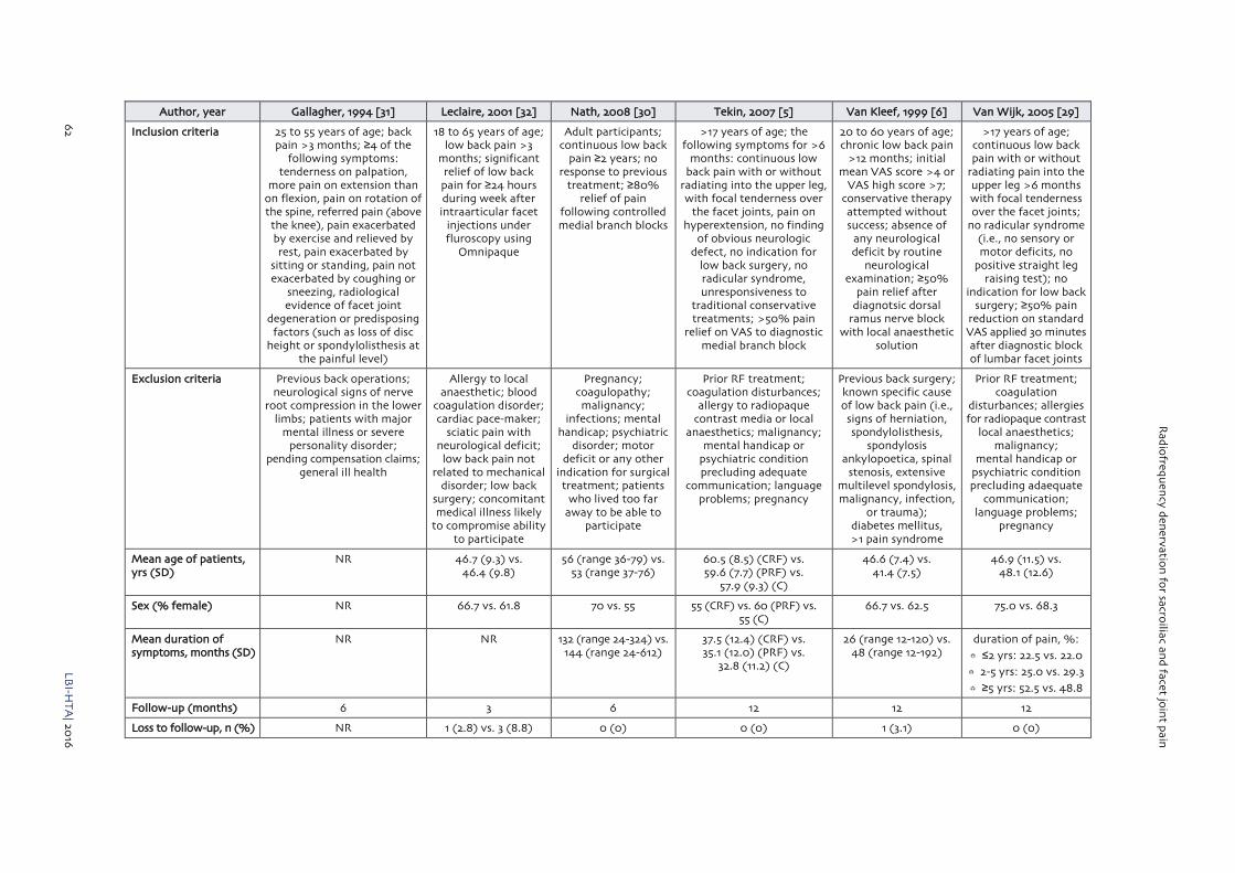
Rad
iofreq
uen
cy den
ervation
for sacro
iliac and
facet join
t pain
62
LBI-H
TA
| 2016
Author, year Gallagher, 1994 [31] Leclaire, 2001 [32] Nath, 2008 [30] Tekin, 2007 [5] Van Kleef, 1999 [6] Van Wijk, 2005 [29]
Inclusion criteria 25 to 55 years of age; back pain >3 months; ≥4 of the
following symptoms: tenderness on palpation,
more pain on extension than on flexion, pain on rotation of the spine, referred pain (above the knee), pain exacerbated by exercise and relieved by rest, pain exacerbated by
sitting or standing, pain not exacerbated by coughing or
sneezing, radiological evidence of facet joint
degeneration or predisposing factors (such as loss of disc
height or spondylolisthesis at the painful level)
18 to 65 years of age; low back pain >3
months; significant relief of low back pain for ≥24 hours during week after intraarticular facet
injections under fluroscopy using
Omnipaque
Adult participants; continuous low back
pain ≥2 years; no response to previous
treatment; ≥80% relief of pain
following controlled medial branch blocks
>17 years of age; the following symptoms for >6
months: continuous low back pain with or without
radiating into the upper leg, with focal tenderness over
the facet joints, pain on hyperextension, no finding
of obvious neurologic defect, no indication for
low back surgery, no radicular syndrome, unresponsiveness to
traditional conservative treatments; >50% pain
relief on VAS to diagnostic medial branch block
20 to 60 years of age; chronic low back pain
>12 months; initial mean VAS score >4 or
VAS high score >7; conservative therapy attempted without success; absence of
any neurological deficit by routine
neurological examination; ≥50%
pain relief after diagnotsic dorsal
ramus nerve block with local anaesthetic
solution
>17 years of age; continuous low back pain with or without
radiating pain into the upper leg >6 months with focal tenderness over the facet joints;
no radicular syndrome (i.e., no sensory or motor deficits, no
positive straight leg raising test); no
indication for low back surgery; ≥50% pain
reduction on standard VAS applied 30 minutes after diagnostic block of lumbar facet joints
Exclusion criteria Previous back operations; neurological signs of nerve
root compression in the lower limbs; patients with major
mental illness or severe personality disorder;
pending compensation claims; general ill health
Allergy to local anaesthetic; blood
coagulation disorder; cardiac pace-maker;
sciatic pain with neurological deficit; low back pain not
related to mechanical disorder; low back
surgery; concomitant medical illness likely
to compromise ability to participate
Pregnancy; coagulopathy; malignancy;
infections; mental handicap; psychiatric
disorder; motor deficit or any other
indication for surgical treatment; patients
who lived too far away to be able to
participate
Prior RF treatment; coagulation disturbances;
allergy to radiopaque contrast media or local
anaesthetics; malignancy; mental handicap or
psychiatric condition precluding adequate
communication; language problems; pregnancy
Previous back surgery; known specific cause of low back pain (i.e., signs of herniation, spondylolisthesis,
spondylosis ankylopoetica, spinal
stenosis, extensive multilevel spondylosis, malignancy, infection,
or trauma); diabetes mellitus, >1 pain syndrome
Prior RF treatment; coagulation
disturbances; allergies for radiopaque contrast
local anaesthetics; malignancy;
mental handicap or psychiatric condition precluding adaequate
communication; language problems;
pregnancy
Mean age of patients, yrs (SD)
NR 46.7 (9.3) vs. 46.4 (9.8)
56 (range 36-79) vs. 53 (range 37-76)
60.5 (8.5) (CRF) vs. 59.6 (7.7) (PRF) vs.
57.9 (9.3) (C)
46.6 (7.4) vs. 41.4 (7.5)
46.9 (11.5) vs. 48.1 (12.6)
Sex (% female) NR 66.7 vs. 61.8 70 vs. 55 55 (CRF) vs. 60 (PRF) vs. 55 (C)
66.7 vs. 62.5 75.0 vs. 68.3
Mean duration of symptoms, months (SD)
NR NR 132 (range 24-324) vs. 144 (range 24-612)
37.5 (12.4) (CRF) vs. 35.1 (12.0) (PRF) vs.
32.8 (11.2) (C)
26 (range 12-120) vs. 48 (range 12-192)
duration of pain, %:
≤2 yrs: 22.5 vs. 22.0
2-5 yrs: 25.0 vs. 29.3
≥5 yrs: 52.5 vs. 48.8
Follow-up (months) 6 3 6 12 12 12
Loss to follow-up, n (%) NR 1 (2.8) vs. 3 (8.8) 0 (0) 0 (0) 1 (3.1) 0 (0)

Ap
pen
dix
LBI-H
TA
| 2016
6
3
Author, year Gallagher, 1994 [31] Leclaire, 2001 [32] Nath, 2008 [30] Tekin, 2007 [5] Van Kleef, 1999 [6] Van Wijk, 2005 [29]
Outcomes
Efficacy
Pain intensity Change in VAS score:
at 1 month: -17 (A) vs. +13 (B) vs. -13 (C) vs. -12 (D),
s. diff. between groups A and C
at 6 months: -7 (A) vs. +5 (B) vs. -3 (C) vs. -17 (D),
s. diff. between groups A and C
Change in McGill Pain Questionnaire scores:
at 1 month: -3 (A) vs. +4 (B) vs. -2 (C) vs. -5 (D),
s. diff. between groups A and C
at 6 months: 0 (A) vs. 0 (B) vs. -1 (C) vs. -3 (D)
Change in VAS score:
at 4 weeks: -3.6 vs. +0.6, n.s.
at 12 weeks: +0.5 vs. -7.2, n.s.
Change in VAS score:
Back pain:
at 6 months: -2.1 vs. -0.7; n.s.
Generalised pain:
at 6 months: -1.9 vs. -0.4, s.s.
Change in VAS score:
post procedure: -4.2 (CRF) vs. -3.8 (PRF)
vs. -2.5 (C), s. diff. between CRF/PRF groups and control group
at 6 months: -4.2 (CRF) vs. -3.7 (PRF) vs. -3.7 (C),
s. diff. between CRF group and PRF/control groups
at 12 months: -4.1 (CRF) vs. -3.1 (PRF) vs. -2.9 (C),
s. diff. between CRF group and PRF/control groups
Change in VAS score:
at 8 weeks: -2.37 vs. -0.43,
s.s.
Change in VAS score:
Back pain:
at 3 months: -2.1 vs. -1.6, n.s.
Functional status NR Change in ODI score:
at 4 weeks: -2.7 vs. -2.1, n.s.
at 12 weeks: -4.7 vs. -2.7, n.s.
Change in Roland-Morris Questionnaire
(RMQ) score:
at 4 weeks: -8.4 vs. -2.2, n.s. (p=0.05)
at 12 weeks: -9.8 vs. -7.2, n.s.
NR6 Change in ODI score:
post procedure: -13.6 (CRF) vs. -15 (PRF) vs.
-9.6 (C), s. diff. between CRF/PRF groups and
control group
at 6 months: -14.1 (CRF) vs. –14.1 (PRF) vs. -11.2 (C), s. diff. between CRF/PRF groups and control group
at 12 months: -11.2 (CRF) vs. -10.9 (PRF)
vs. -6.5 (C), s. diff. between CRF/PRF groups and
control group
Change in ODI score:
at 8 weeks: -11.07 vs. +1.69, s.s. 7:
Change in COOP/WONCA chart
at 8 weeks: -3.13 vs. -1.62, n.s.
Mean change in physical activities:
at 3 months: +1.5 vs. +0.9, n.s.
Global improvement NR NR Change in subjective global assessment
(6-point scale):
at 6 months: -1.1 vs. -0.3, s.s.
NR Change in global perceived effect8:
at 8 weeks: +1.33 vs. +0.37, s.s.
Change in global perceived effect on
back pain, %:
at 3 months: ≥50% pain relief: 61.5 vs. 39.0, s.s.
6 only specific variables for back movement and hip movement provided; no overall score 7 Dartmouth COOP Functional Health Assessment Charts/World Organisation of Primary Care Physicians (WONCA) 8 Global perceived effect was scored by the patient on a 7-point scale (ranging from much worse, -3; to 0, no change; to total pain relief, +3)

Rad
iofreq
uen
cy den
ervation
for sacro
iliac and
facet join
t pain
64
LB
I-HT
A| 20
16
Author, year Gallagher, 1994 [31] Leclaire, 2001 [32] Nath, 2008 [30] Tekin, 2007 [5] Van Kleef, 1999 [6] Van Wijk, 2005 [29]
Health-related quality of life
NR NR NR9 NR NR s. diff. only in parameter “vitality”
(no total score of SF-36 provided)
Ability to work NR NR NR NR NR NR
Satisfaction with treatment
NR NR NR Patient satisfaction was lower in control group than other groups, and highest
in CRF group (s.s.)
NR NR
Safety
Complications no adverse effects
no evidence of any nerve root anaesthesia or damage
no complications no complications no complications no complications no significant differences in occurrence of
treatment-related pain and subjective sensory
or motor changes
Abbreviations: NR not reported; n.s. not significant; ODI Oswestry Disability Index; RF radiofrequency; s.s. statistically significant; VAS visual analogue scale; yrs years; CRF conventional RFD, PRF pulsed RFD
9 various quality of life variables provided, but no overall score

Ap
pen
dix
LBI-H
TA
| 2016
6
5
Table A-2: Facet joint: Radiofrequency denervation vs. steroid injection: Results from randomised controlled trials
Author, year Civelek, 2012 [23] Duger, 2012 [33] Hashemi, 2014 [2] Lakemeier, 2013 [24]
Country Turkey Turkey Iran Germany
Sponsor NR NR NR no funding
Study design RCT RCT RCT RCT
Number of pts 100 (50 vs. 50) 120 (40 vs. 40 vs. 40) 80 (40 vs. 40) 56 (27 vs. 29)
Intervention Facet joint radiofrequency (FJRF) denervation at 80°C for 120 seconds
(n=50)
RF denervation group (group R): localisation of RF electrode in the facet joint causing pain was determined by sensorial stimulus and C-armed scope device. Pulsed RF thermocoagulation for 6 minutes at 40°C with RF lesion
generator (n=40)
Pulsed radiofrequency (n=40) Radiofrequency denervation: curved RF needles with 100-mm active tips were placed at the site of the dorsal ramus
medial branch of the relevant L3/L4-L5/ S1 LFJs, 1mL of 0.5% bupivacaine was
injected, the RF probe was then inserted into the cannula and lesion at a
temperature of 80° C for 90 seconds using an RF generator (n=27)
Comparator Facet joint injection (FJI) with medial branch block of posterior
primary ramus with 1 cc of methylprednisolone acetate (40
mg) (diluted with 1 cc SF) combined with 2 cc bupivacaine
hydrochloride (diluted with 2 cc SF) (n=50)
Injection group (group B): after C-arm scope guided determination of injection
point, injection of 1.5mL of 20mg methyl-prednisolone acetate mixed with 5mg
bupivacaine into the facet joint (n=40)
RF denervation and injection group (group RB): localisation of RF electrode
in the facet joint causing pain was determined by sensorial stimulus and
C-armed scope device. Pulsed RF thermo-coagulation for 6 minutes at 40°C and
injection of 1.5 ml mixture of 20 mg methylprednisolone acetate and 5 mg bupivacaine at the same localization
(n=40)
Injection by steroids (1 mL (40mg) triamcinolone) and
0.5mL bupivacaine 0.5% (n=40)
Intraarticular injection of steroids: same setting was used for LJF
infiltrations and RF denervation; the RF probe was inserted into the cannula and the denervation process (80° C for
90 seconds) was begun, but the electrodes were not connected to the
pain generator device (n=29)
Inclusion criteria Chronic and debilitating low back pain leading to diagnosis of lumbar facet syndrome; not responding to conservative treatment for up to 6 weeks, including various analgesics and physical therapy and additional pain relief after FJI for participants with FJRF; symptoms of facet syn-
drome include local tenderness over 1 or more FJs, back pain aggravated
by hyperextension and rotation, morning stiffness or pain increasing in the morning and hip and buttock pain of a non-radicular distribution
18 to 60 years of age; single-sided low back pain arising from facet joint;
complaints longer than 6 months, limited functions and daily life; presented with
at least 2 of the 4 symptoms of facet syndrome (back pain aggravation by
hyperextension and rotation, morning stiffness or pain increasing in the
morning, local tenderness over one or more facet joints and hip and buttock
pain of a nonradicular distribution)
>18 years of age; spondylolisthesis grade I in MRI
at 1 single level (2 adjacent vertebrae and 1 disc); chronic low
back pain >6 months; supra-vertebral facet tenderness; pain in
hyperextension; minimum NRS score of 4 (moderate to severe pain) and positive diagnostic
medial branch block test
≥18 years of age; lumbar facet joint (LFJ)-related low back pain ≥24 months;
ability to understand study protocol and to provide voluntary written
informed consent and participate in outcome measurements; a benefit in
pain reduction ≥50% after test injection of local anaesthetics into the L3/L4-L5/S1 LFJ; MRI-proven LFJ osteoarthritis and
hypertrophy in the L3/L4-L5/S1 segments

Rad
iofreq
uen
cy den
ervation
for sacro
iliac and
facet join
t pain
66
LB
I-HT
A| 20
16
Author, year Civelek, 2012 [23] Duger, 2012 [33] Hashemi, 2014 [2] Lakemeier, 2013 [24]
Exclusion criteria Radicular pain, neurogenic claudication and neurological deficits; acute or uncontrolled
medical illness; known history of adverse reactions to local
anaesthetics, pregnancy or lactation
Coagulation defect, major depression and uncontrolled psychiatric disorder, pregnancy or lactation, respiratory or
cardiac problems in prone position, opioid treatment during the previous month, undergoing surgical procedure at the
same site, infection at the procedure site, disc-related radicular symptoms; patients
not accepting the procedure and not giving informed consent
Radicular pain; neurologic deficit; indication for surgery (neurologic deficit and urinary inconsistent);
stenosis of the spinal canal; spondylolysis; positive straight
leg raising test; suppressed reflex; known psychiatric disease; spinal
deformity; neoplastic or infectious disease
Lack of positive response to L3/L4-L5/S1 test infiltration; history of osteoporosis
or malignancies; allergies to local anaesthetics; pregnancy or lactation;
lumbar spinal stenosis or spinal instability; vertebral fracture; symptomatic radiculopathy;
uncontrolled psychiatric disorder; uncontrolled medical illness; condition that could interfere with interpretation
of outcome assessments; history of adverse reactions to corticosteroids
Mean age of patients, yrs (SD)
51.8 (17.0) vs. 56.5 (17.7) 50.2 (12.1) (R) vs. 50.1 (12.3) (B) vs. 51.2 (11.9) (RB)
64.3 (13.3) vs. 63.9 (11.5) 57.6 (12.8) vs. 56.3 (10.8)
Sex (% female) 70.0 vs. 70.8 53.3 (R) vs. 56.7 (B) vs. 56.7 (RB)a 52.5 vs. 55.0 34.6 vs. 38.5
Mean duration of symptoms, months (SD)
18.9 (12.9) vs. 18.7 (12.3) 10.7 (5.2) (R) vs. 11.3 (5.1) (B) vs. 11.0 (5.0) (RB)
3.4 (2.3) vs. 3.8 (2.5) NR
Follow-up (months) 12 12 6 6
Loss to follow-up, n (%) 0 (0) NR 1 (2.5) vs. 1 (2.5) 1 (4) vs. 3 (10)
Outcomes
Efficacy
Pain intensity Change in VNS score:
at 1 month: -6.0 vs. -5.1, s.s
at 6 months: -5.7 vs. -4.1, s.s.
at 12 months: -5.6 vs. -3.6, s.s.
Change in VAS score:
at 1 month: -4.3 (R) vs. -1.7 (B) vs. -4.2 (RB), s. diff. btw. R and B,
and btw. RB and B
at 6 months: -4.2 (R) vs. -0.6 (B) vs. -4.3 (RB), s. diff. btw. R and B,
and btw. RB and B
at 12 months: -3.3 (R) vs. -0.1 (B) vs. -3.4 (RB), s. diff. btw. R and B,
and btw. RB and B
Change in NRS score:
at 6 months: -5 (PRF group), s. diff. between PRF and steroid
group at 12 weeks and 6 months, n.s. at 6 weeks
Change in VAS score:
at 6 months: -1.9 vs. -1.6, n.s.
Functional status NR NR Change in ODI score:
at 6 months: -56.3% (PRF group), s. diff. between PRF and
steroid group at 12 weeks and 6 months, n.s. at 6 weeks
Change in ODI score:
at 6 months: -12.8 vs. -5.7, n.s.
Change in RMQ score:
at 6 months: -3.7 vs. -4.2, n.s.
Global improvement NR NR NR NR
Health-related quality of life
Change in EQ-5D score:
at 1 month: -8.2 vs. -8.7, n.s.
at 6 months: -7.3 vs. -7.5, n.s.
at 12 months: -7.1 vs. -6.7, n.s.
NR NR NR

Ap
pen
dix
LBI-H
TA
| 2016
6
7
Author, year Civelek, 2012 [23] Duger, 2012 [33] Hashemi, 2014 [2] Lakemeier, 2013 [24]
Ability to work NR NR NR NR
Satisfaction with treatment
There was no significant difference in the 1st and 6th month follow-ups,
however there was a significant difference respect to 12 month
NASS patients’ satisfaction scores10
All the values except those obtained at day 2 and week 1 were higher in group R when compared to group B; in group RB,
early satisfaction values (day 1, day 2, week 1) were higher compared to group
R; in group RB, all satisfaction values except at day 2 and week 1 were higher
compared to group B
Patient satisfaction was significantly higher in PRF group
at 12 weeks and 6 months compared to steroid group
NR
Safety
Complications no infections or new motor or sensory deficits
rare complaints of small superficial burns after RFD
increase in severity of low back pain in 2 RFD pts (4%) in the early
follow-up period
no complications NR no major adverse events
Abbreviations: FJI Facet joint injection, FJRF Facet joint radiofrequency, NASS North American Spine Society patient satisfaction questionnaire; NRS numeric rating scale; NR not reported; n.s. not significant; ODI Oswestry Disability Index; RF radiofrequency; RMQ Roland-Morris Questionnaire; s.s. statistically significant; VAS visual analogue scale; VNS visual numeric pain scale; yrs years a instead of 40 pts per group only 30 each are described in the demographic data table. An email request to the author (8.2.2016) remained without reply.
Table A-3: Facet joint: Radiofrequency denervation vs. other treatment (other RFD method): Results from randomised controlled trials
Author, year Kroll, 2008 [34] Moon, 2013 [35] Sanders, 1999 [36]
Country USA Korea Netherlands
Sponsor NR no industry funding NR
Study design RCT RCT RCT
Number of pts 26 (13 vs. 13) 82 (41 vs. 41) 34 (17 vs. 17)
Intervention Continuous RF group (CRF): continuous RF thermocoagulation lesioning performed at
80° C for 75 seconds (n=13)
Distal approach group (D): fluoroscopic distal approach was used for L1 to L4 medial branches
(n=41)
Percutaneous intra-articular facet denervation (PIFD): after injection of 1ml lidocaine 2%, 3 RF lesions (60 seconds) were made in the articular cavity, central, rostral, and caudal of the facet
joint (n=17)
10 NASS Score: 1=fully meeting of patient’s expectations, 2=less improvement than the hoped for result but the patient would undergo the same procedure again,
3=the procedure helped but the patient would not undergo again, 4=the same or worse status with respect to pre-operative status

Rad
iofreq
uen
cy den
ervation
for sacro
iliac and
facet join
t pain
68
LB
I-HT
A| 20
16
Author, year Kroll, 2008 [34] Moon, 2013 [35] Sanders, 1999 [36]
Comparator Pulsed RF group (PRF): pulsed RF lesioning at 42° with pulse duration of 20 ms and pulse rate of
2 Hz for 120 seconds (n=13)
Tunnel vision approach group (TV): lumbar medial branch radiofrequency denervation was performed under fluoroscopic guidance in an oblique “tunneled”
view, as described by Bogduk 2005 (n=41)
Extra-articular facet denervation (PEFD) following the method described by Mehta and
Sluijter (n=17)
Inclusion criteria ≥18 years of age; ASA11 physical status I, II and III; unilateral or bilateral lumbar back pain for longer than 1 month; no radiating symptoms below the knee; >50% pain reduction based on mean VAS after 2 separate diagnostic medial branch blocks
with 1.0mL of 0.5% bupivacaine
>18 years of age; predominantly axial lower back pain for ≥3 months; paraspinal tenderness overlying the L2 to L4 lumbar facet joints; failure to respond to conservative therapy such as physical therapy or pharmacotherapy; concordant pain relief of
>50% after a comparative local anaesthetic block with 0.5mL lidocaine 1% (≥1 hour) and
levobupivacaine hydrochloride 0.5% (≥3 hours) at the L1 to L4 medial branches
Low back pain >6 months; pain exacerbated by extension of the lumbar spine, prolonged standing or sitting; deep pressure pain over the lumbar facet joints and absence of neurological abnormalities;
no improvement from physical therapy; pain intensity on VAS >4;
≥50% pain relief after diagnostic block
Exclusion criteria History of previous back surgery; presence of neurological deficits; claudication; active
psychiatric disorder; bleeding disorder; active infection; pregnancy; involved in current litigation;
ongoing workers’ compensation claims; disc herniation and spinal stenosis
(ruled out radiographically)
Any focal neurological signs or symptoms; radiologic evidence of a symptomatic herniated disc; severe spinal stenosis or structural lumbar spinal deformity; a positive response to previous spinal
interventions such as sacroiliac joint block or epidural steroid injection; discogenic pain verified
by discography; lumbar spine fusion; untreated coagulopathy; concomitant medical or psychiatric condition likely to undermine diagnostic workup
or assessment of treatment response
Radicular pain (neurological signs of nerve root compression); previous back operation(s);
age <18 years; bleeding disorders; presence of prominent functional or non-physiological signs
Mean age of patients, yrs (SD)
59.5 (11.6) (CRF) vs. 57.0 (8.4) (PRF) 68.6 (13.8) (D) vs. 62.6 (14.5) (TV) 60.9 (18.5) vs. 62.6 (14.6)
Sex (% female) 46.1 (CRF) vs. 61.5 (PRF) 61.8 (D) vs. 70,6 (TV) 23.5 vs. 29.4
Mean duration of symptoms, months (SD)
NR 40.8 (40.6) (D) vs. 29.3 (23.9) (TV) NR
Follow-up (months) 3 6 3
Loss to follow-up, n (%) 12 (48) vs. 12 (48) 5 (12.2) (D) vs. 6 (14.6) (TV)12 NR
Outcomes
Safety
Complications No adverse events were encountered during CRF or PRF lesioning, and no complications were
documented 3 months post-procedure
RF-associated complication during the 6-month follow-up: 1 (D) vs. 5 (TV)
(mild, localized pain at the RF lesion site lasting <1 month (1 vs 2); new neuropathy-like pain
lasting >3 months (0 vs 3))
No complications or morbidity were observed
Abbreviations: FJI Facet joint injection, FJRF Facet joint radiofrequency, NASS North American Spine Society patient satisfaction questionnaire; NRS numeric rating scale; NR not reported; n.s. not significant; ODI Oswestry Disability Index; RF radiofrequency; RMQ Roland-Morris Questionnaire; s.s. statistically significant; VAS visual analogue scale; VNS visual numeric pain scale; yrs years
11 is a five-category physical status classification system adopted by the American Society of Anesthesiologists (ASA) for assessing the fitness of patients before surgery:
I = healthy person; II = mild systemic disease; III = severe systemic disease 12 Additionally, 3 patients (1 in the tunnel vision approach group and 2 in the distal approach group) were excluded because of failure in both motor and sensory stimulation at 2 or
more nerves.

Ap
pen
dix
LBI-H
TA
| 2016
6
9
Table A-4: Sacroiliac joint Radiofrequency denervation vs. placebo: Results from randomised controlled trials
Author, year Cohen, 2008 [25] Patel, 2012 [4]
Country USA USA
Sponsor academic/Baylis Medical Baylis Medical
Study design RCT RCT
Number of pts 28 (14 vs. 14) 51 (34 vs. 17)
Intervention Cooled RF denervation group received L4-L5 primary dorsal rami and S1-S3 lateral branch RF denervation (80°C, 90 seconds) using cooling
probe technology after local anaesthetic block (n=14)
RF energy was delivered for 150 seconds at set temperature of 60°C; first L5 dorsal ramus was lesioned; after coagulation of L5 dorsal ramus,
sacral lateral branches of S1, S2 and S3 were targeted (n=34)
Comparator Control group received local anaesthetic block followed by placebo denervation, in which 0.5 ml lidocaine 2% was administered with no
current (n=14)
Participants who did not respond to placebo injections crossed over and were treated with RF denervation using conventional technology (n=11)
Same procedure as in experimental group, except that RF energy was not delivered. Probe placements, procedure duration, equipment sounds and
visual indications for participants were identical in both groups (n=17)
Inclusion criteria >18 years of age; axial low back or buttock pain ≥ 6 months; tenderness overlying SI joint(s); failure to respond to conservative therapy
(e.g., physical therapy and pharmacotherapy); long-term (>2 months) pain relief with SI joint corticosteroid injections; pain relief ≥ 75% as
calculated from a 6-hour post-block pain diary after a single diagnostic SI joint injection
>18 years of age; predominantly axial pain below L5 vertebrae; axial pain lasting longer than 6 months; three-day average NRS between 4 and 8;
failure to achieve adequate improvement with comprehensive non-operative treatments, including but not limited to activity alteration, non-steroidal
antiinflammatory drugs, physical and/or manual therapy and fluoro-scopically guided injections of steroids into SIJ or sacroiliac ligaments;
other possible sources of low back pain reasonably excluded (by means of physical exam, medical history and magnetic resonance imaging/computed
tomography/X-ray as required), including but not limited to bone fracture, hip joint, symptomatic spondylolisthesis, tumour and other regional soft
tissue structures
Exclusion criteria Focal neurological signs or symptoms; radiological evidence of symptomatic herniated disc; spondyloarthropathy; untreated
coagulopathy; unstable medical or psychiatric illness that might preclude an optimal treatment response
Beck Depression Inventory score >20; irreversible psychological barriers to recovery; spinal pathology that may impede recovery, such as
spondylolisthesis at L5/S1, or scoliosis; symptomatic moderate or severe foraminal or central canal stenosis; systemic infection or localised
infection at anticipated introducer entry site; concomitant cervical or thoracic pain >2/10 on a NRS scale; uncontrolled or acute illness; chronic severe conditions such as rheumatoid/inflammatory arthritis; pregnancy;
active radicular pain; immunosuppression (e.g. AIDS, cancer, diabetes, surgery < 3 months before); workers’ compensation, injury litigation or disability remuneration; allergy to injectate or medications used during
procedure; high narcotics usage (> 30 mg morphine daily or equivalent); active smokers (termination ≥ 6 months with no smoking during follow-up
period acceptable with caution);); history of potentially confounding intervertebral disc disease or zygapophyseal joint pain
Mean age of patients, yrs (SD) 51.9 (13.6) vs. 51.8 (13.1) 56 (15) vs. 64 (14)
Sex (% female) 64 vs. 57 68 vs. 82

Rad
iofreq
uen
cy den
ervation
for sacro
iliac and
facet join
t pain
70
LBI-H
TA
| 2016
Author, year Cohen, 2008 [25] Patel, 2012 [4]
Mean duration of symptoms NR 6-12 months: 18% vs. 6% 12-24 months: 15% vs. 18% >24 months: 65% vs. 77%
Follow-up (months) 6 (crossover of control group patients after 1 or 3 months) 9 resp. 12 [3] (but crossover of control group patients after 3 months)
Loss to follow-up, n (%) 0 (0) 0 (0) at 3 months
Outcomes
Efficacy
Pain intensity Change in NRS scores: at 1 month: -3.7 vs. -0.2, s.s. at 3 months: -3.7 vs. -0.513
Change in NRS scores: at 1 month: -2.7 vs. -1.7, n.s. at 3 months: -2.4 vs. -0.8, s.s.
Functional status Change in ODI scores: at 1 month: -16.2 vs. -4.3, s.s. at 3 months: -18.6 vs. -23.913
Change in ODI scores: at 1 month: -12 vs. -4, s.s. at 3 months: -11 vs. +2, s.s.
Global improvement Positive GPE14, % (95% CI): at 1 month: 93 (78-100) vs. 21 (2-45), s.s.
at 3 months: 83 (59-100) vs. 013
Positive GPE15, % (95% CI): at 3 months: 47 (29-65) vs. 8 (0-36), s.s.
Health-related quality of life NR Change in AQoL scores: at 1 month: +0.07 vs. +0.07, n.s. at 3 months:+0.09 vs. +0.02, s.s.
Ability to work NR NR
Satisfaction with treatment NR NR
Safety
Complications no serious complications (both groups)
majority of patients: temporary worsening pain (lasting 5-10 days after the procedure)
one RFD patient (1/25 (4%) due to crossover) reported transient nonpainful buttock paresthesia that resolved without therapy
no serious complications (both groups)
small proportion of subjects: soreness or numbness at the introducer sites in the 2 weeks following treatment
one subject: shingles at the introducer site (deemed unrelated to treatment)
Abbreviations: AQoL Assessment of Quality of Life; GPE Global Perceived Effect; NR not reported; NRS numerical rating pain scale; n.s. not significant; ODI Oswestry Disability Index; RF radiofrequency; s.s. statistically significant; VAS visual analogue scale; yrs years
13 Due to crossover of control group, 3-months results are based on only 2 patients in the original placebo group 14 A positive GPE was defined as an affirmative response to the following 3 questions: 1. My pain has improved/worsened/stayed the same since my last visit; 2. The treatment I received
improved/did not improve my ability to perform daily activities; 3. I am satisfied/not satisfied with the treatment I received and would recommend it to others. 15 GPE was measured by having subjects rate their index pain on a 7-item scale; a GPE score was considered positive if the subject rated GPE as „pain has decreased a lot“ or „pain
is completely gone“

Ap
pen
dix
LBI-H
TA
| 2016
71
Risk of bias table
The risk of bias of included studies was assessed by the Cochrane Risk of Bias tool and judged by two researchers. A more detailed description of the criteria used to assess the internal validity of the individual study designs can be found in the Cochrane Handbook for Systematic Reviews of Interventions [39].
Table A-5: Risk of bias – study level (randomised studies)
Trial Adequate generation
of randomisation sequence
Adequate allocation
concealment
Blinding Selective outcome reporting unlikely*
No other aspects which increase the
risk of bias
Risk of bias – study level Patient
Treating Physician/ follow up examination
RFA in fact joints vs. placebo
Gallagher, 1994 [31] unclear unclear yes unclear/yes unclear noa high
Leclaire, 2001 [32] yes yes yes unclear/yes unclear yes low
Nath, 2008 [30] yes yes yes yes/yes unclear no b high
Tekin, 2007 [5] yes yes yes unclear/yes unclear yes low
Van Kleef, 1999 [6] yes yes yes yes/yes unclear yes low
Van Wijk, 2005 [29] yes yes yes yes/yes unclear yes low
RFA in fact joints vs injection therapy
Civelek, 2012 [23] yes unclear unclear no/yes unclear yes high
Duger, 2012 [33] unclear unclear unclear no/unclear unclear no a high
Hashemi, 2014 [2] yes unclear no no/yes unclear yes high
Lakemeier, 2013 [24] yes yes yes no/yes unclear yes low
RFA in SI joints
Cohen, 2008 [25] yes yes yes unclear/yes yes no b high
Patel, 2012 [4] yes yes yes no/yes yes yes low
RFA in fact joints vs. other method of RFA (for safety only)
Kroll, 2008 [34] yes unclear unclear unclear unclear no c high
Moon, 2013 [35] unclear unclear unclear unclear/unclear yes yes high
Sanders, 1999 [36] unclear unclear unclear unclear/unclear unclear noa high
*This risk was defined as unclear if author’s didn’t explicitly state that a protocol was published before/the study was not registered; a number of drop-outs unclear; b baseline characteristics not similar; c high number of drop-outs

Radiofrequency denervation for sacroiliac and facet joint pain
72 LBI-HTA | 2016
Applicability tables
Table A-6a: Summary table characterising the applicability of a body of studies (RFD for facet joint pain)
Domain Description of applicability of evidence
Population The patient population (n=679) differed considerably between studies. Patients had suffered from back pain for more than 3 months up to at least 2 years. The mean age of included patients ranged from 41 to 64 years. The percentage of female participants was more than half of the patients in all, but one trial.
Intervention The RCTs on RFD for facet joint pain applied continuous or pulsed RFD methods (40-85° C, 60-120s). The optimal “dose” of RFD seems to be still under examination.
Comparators 6 trials compared radiofrequency denervation with a placebo/sham treatment, the remaining 4 studies used steroid injections as comparator.
Outcomes All outcomes defined as crucial for the decision were reported in the included trials: All trials reported pain outcomes (generally measured by the VAS), half of the trials changes in functional status mesured by the ODI. 3 trials reported ‘global impovements’ and only 2 assed (generic) health-related quality of life. 9 of 10 studies mentioned (at least the non-occurance of) complications, but there seemed to be no standardised assessment/documentation. Of the important (not crucial) outcomes, 4 studies reported on patient satisfaction, non on the ability to work. There are no data on outcomes at >12 months after intervention.
Setting 3 trials were conducted in Turkey, 2 in the Netherlands, the remaining 5 studies in Canada, Germany, Iran, Sweden, and the UK. The steroid injection compared trials were conducted more recently (published between 2012 and 2014) than the placebo-controlled trials (1994-2008).
Table A-6b: Summary table characterising the applicability of a body of studies (RFD for sacroiliac joint pain)
Domain Description of applicability of evidence
Population The total number of patients was 79. Patients had suffered from low back pain for more than 6 months and had not achieved adequate improvement after conservative therapies. The mean age of patients was 52-64 years. 57-82% of the patients were female.
Intervention Both RCTs used a “cooled radiofrequency denervation” method (with temperatures from 60 to 80°C and durations from 90 to 150 seconds). The optimal “dose” of RFD seems to be still under examination.
Comparators Both RCTs compared radiofrequency denervation with placebo/sham treatment.
Outcomes All outcomes defined as crucial for the decision were reported in the included trials: Both trials reported pain outcomes (measured by the NRS scores) and changes in functional status mesured by the ODI. Both trials also reported the ‘global impovement’ (determined by the same instrument (GPE). Only 1 trial asked (generic) health-related quality of life. Both studies described complications (in more detail than the facet joint pain studies). None of the studies provided data on the outcomes defined as important (not crucial: patient satisfaction, the ability to work). There are no data on outcomes at >3 month after intervention.
Setting On the contrary to the above mentioned trials on RFD for facet joint pain (no north American study), both trials on sacroiliac joint pain were conducted in the US (and published 2008-2012).

Ap
pen
dix
LBI-H
TA
| 2016
73
List of ongoing randomised controlled trials
Table A-7: List of ongoing randomised controlled trials of RFD for facet joint pain
Identifier/ Trial name
Patient population Intervention Comparison
Primary Outcome
Primary completion
date Sponsor
NCT02478437
A Trial of Cooled Radiofrequency Ablation of Medial Branch Nerves for the Treatment of Lumbar Facet Syndrome
18-79 years of age, lumbar facet syndrome pain, low back pain >6 months, pain resistant to
conventional therapy, pain diagram suggesting possibility of facet-mediated pain, positive response to at least 1 set of diagnostic intra-articular facet
injections or medial branch blocks
Conventional radio-frequency ablation
(RFA)
Cooled radio-frequency ablation
(CRFA)
Pain improvement, global pain score, McGill Pain Questionnaire
(MPQ), Pain Anxiety Symptom Scale short form (PASS-20), Center for Epidemiologic Studies Depression
short form index (CESD-10)
June 2017 North-western University,
American Pain Society
NCT02148003
Effect of the Temperature Used in Thermal Radiofrequency Ablation
>18 years of age, low back pain, chronic back pain attributed to lumbar facet joints arthropathy,
adequate response to the diagnostic blocks without the use of steroids
Radio-frequency ablation (performed at
90 degrees Celsius)
Radio-frequency ablation (performed
at 80 degrees Celsius)
Assessing a change of pain relief, number of repeats of procedure
February 2016
The Cleveland Clinic
Table A-8: List of ongoing randomised controlled trials of RFD for sacroiliac pain
Identifier/ Trial name
Patient population Intervention Comparison
Primary Outcome
Primary completion
date Sponsor
NCT02382289
Radiofrequency in Sacroiliac Arthropathy; Bipolar RF 6 Points Versus Monopolar RF at 6 and 3 Points
>18 years, chronic sacroiliac joint arthropathy; moderate to severe low back pain for >6 months with positive Patrick’s and Yeoman’s tests with
tenderness over the SI joint. Pain is not responding to the usual medical treatment. >50% pain relief after diagnostic injection with local anesthetic.
Procedure: radio-frequency
ablation for sacroiliac joint arthropathy
Comparing bipolar RF at 6 points with monopolar RF at 3 and 6 points for the treatment of
SI arthropathy
Patient satisfaction March 2015 King Hamad University Hospital, Bahrain
NCT01726608
RFN for SIJ Disease Study16
18-80 years of age, low back pain >6 months duration, >80% reduction in pain following
each diagnostic, intra-articular block.
Active radio-frequency neurotomy
Sham radio-frequency neurotomy
Pain intensity, quality of pain, health related quality of life, anxiety and depression, functional disability,
health related quality of life and quality-adjusted life years, portion
of patients randomised to sham requiring rescue therapy with RFN
December 2013
Barts & The London NHS
Trust
16 Due to the provided study completion date (2013), we contacted the principal investigator (V. Metha) on Feb 28th, but received no answer so far.

Rad
iofreq
uen
cy den
ervation
for sacro
iliac and
facet join
t pain
74
LBI-H
TA
| 2016
Additional information: List of (registered) completed but unpublished or terminated randomised controlled trials
Table A-9a: List of completed but unpublished randomised controlled trials (RFD for facet joint pain)
Identifier/ Trial name Patient population Intervention Comparison Primary Outcome
Primary completion date Sponsor
NCT00476684
The Effect of Radiofrequency-treatment on Patients With Facet-joint Pain in Cervical- and Lumbar-columna17
20-75 years of age, one-sided neck and low back chronic pain, pain durability of
at least 1 year
Radio-frequency treatment
Sham neurotomy Reduction in self-reported pain intensity
May 2010 Norwegian University of Science and Technology
NCT0048415918
Efficacy and Cost: Benefit Ratio of 0, 1, and 2 Medial Branch Blocks for Lumbar Facet Joint Radiofrequency Denervation
>18 years of age, axial low back pain unresponsive to conservative treatment,
duration of pain >6 months
Radio-frequency denervation of medial
branches that innervate the lumbar facet joints
Comparison of 3 different RFD groups
Cost per successful procedure, successful treatment
January 2009 Johns Hopkins University
Table A-9b: List of terminated randomised controlled trials (RFD for sacroiliac pain)
Identifier/ Trial name Patient population Intervention Comparison Primary Outcome
Primary completion date Sponsor
NCT0115809219
Trial Comparing Treatment With SInergy™ System to Conservative Treatment for Chronic Sacroiliac Joint Pain
>18 years of age, predominantly axial pain below the L5 vertebrae >75% pain relief
from 2 separate lateral branch blocks done on different days, chronic axial pain lasting
>six months, no improvement with comprehensive non-operative treatments
Lateral branch denervation using the
SInergy™ System (cooled RFD)
Conservative Treatment: physical therapy, chiropractic care, and medication
Pain status change for sacroiliac region, change in bodily pain and
physical functioning evaluated using SF-36, change in disability evaluated
using Oswestry Disability Index, change in quality of life
March 2012 Baylis Medical Company
17 A web search revealed the information that study data has not been published yet. Principal investigator (P. Borchgrevink) was contacted on Feb 28th, but no answer was received 18 Basic Results are already provided at clinicaltrials.gov. Principal investigator (S.Cohen) contacted on Feb 28th; his answer: “This study has enrolled 160 out of 225 patients.
It is still ongoing and nothing has been published yet.” 19 This study has been terminated. E. Ross (principal investigator ) contacted 28.2.2016, his answer: “I believe funding was stopped by the sponsoring company”

Appendix
LBI-HTA | 2016 75
Literature search strategies
Search strategy for MEDLINE
Search Name: Update search (of Cochrane Review) explicitly including the sacroiliac joint
Database: Ovid MEDLINE(R) <1946 to November Week 3 2015>, Ovid MEDLINE(R) In-Process & Other Non-Indexed Citations <December 28, 2015>, Ovid MEDLINE(R) Daily Update <November 18, 2015>, Ovid OLDMEDLINE(R) <1946 to 1965>
Search Date: 09.12.2015
ID Search
#1 1 ((randomized controlled trial or controlled clinical trial).pt. or randomi#ed.ab. or placebo.ab,ti. or drug therapy.fs. or randomly.ab,ti. or trial.ab,ti. or groups.ab,ti.) not (animals not (humans and animals)).sh. (3275407)
#2 dorsalgia.ti,ab. or exp Back Pain/or backache.ti,ab. or (lumbar adj pain).ti,ab. or coccyx.ti,ab. or coccydynia.ti,ab. or sciatica.ti,ab. or sciatic neuropathy/or spondylosis.ti,ab. or lumbago.ti,ab. (41368)
#3 exp Spine/or discitis.ti,ab. or exp Spinal Diseases/or (disc adj degeneration).ti,ab. or (disc adj prolapse).ti,ab. or (disc adj herniation).ti,ab. or spinal fusion.sh. or (facet adj joints).ti,ab. or intervertebral disc.sh. or postlaminectomy.ti,ab. or arachnoiditis.ti,ab. or (failed adj back).ti,ab. (179612)
4 2 or 3 (205558)
5 exp Radio Waves/or exp Pulsed Radiofrequency Treatment/or radiofrequency.mp. or radio frequency.mp. or exp Electrocoagulation/or electrocoag$.mp. or thermocoag$.mp. or (neurotom$ or neuroly$).mp. (73259)
6 1 and 4 and 5 (235)
7 exp Sacroiliac Joint/(3419)
8 sacroiliac joint*.mp. (4726)
9 sacro-iliac joint*.mp. (283)
10 7 or 8 or 9 (4842)
11 4 or 10 (207914)
12 1 and 5 and 11 (243)
13 12 not 6 (8)
Total: 8 Hits
Additional information: Search strategy for PubMed
Search Name: Radiofrequenc Denervation for sacroiliac joint pain due to “other indications”
Search Date: 04.02.2016
((Sacroiliac Joint[MeSH] OR sacroiliac joint* OR sacro-iliac joint*) AND (Pseudarthrosis[MeSH] OR pseudarthros* OR pseudoarthros* OR pseudo-arthros* OR Arthrodesis[MeSH] OR arthrodes* OR tumor* OR tumour*OR cancer* OR neoplasm* OR carcinoma* OR Carcinoma[MeSH] OR Neoplasms[MeSH] OR Arthritis, Rheumatoid[MeSH] OR rheumatoid OR Spondylarthropathies[MeSH] OR Spondylarthropath*) AND (Denervation[MeSH] OR denervation* OR de-nervation* OR Radio Waves[MeSH] OR Pulsed Radiofrequency Treatment[MeSH] OR radiofrequency OR radio-frequency OR Electrocoagulation[MeSH] OR electrocoag* OR thermocoag* OR neurotom* OR neuroly*))
Total: 4 Hits