RADIATION THERAPY ONCOLOGY GROUP RTOG 0234 A PHASE …
67
RADIATION THERAPY ONCOLOGY GROUP RTOG 0234 A PHASE II RANDOMIZED TRIAL OF SURGERY FOLLOWED BY CHEMORADIOTHERAPY PLUS C225 (CETUXIMAB) FOR ADVANCED SQUAMOUS CELL CARCINOMA OF THE HEAD AND NECK Study Chairs (2/15/11) Pathology/Biomarker Studies Co-Chair Kian Ang, MD MD Anderson Cancer Center 1515 Holcombe Boulevard, Suite 97 Houston TX 77030 713-563-2323 FAX 713-563-2331 [email protected] Principal Investigator/Radiation Oncology Paul M. Harari, MD University of Wisconsin-Madison 600 Highland Avenue Madison, WI 53792 608-263-8500 FAX 608-263-9167 [email protected] Pathology/Biomarker Studies Co-Chair M. Elizabeth Hammond, MD LDS Hospital Eighth Ave. & C Street Salt Lake City, UT 84143 801-408-1314 FAX 801-408-5020 [email protected] Medical Oncology Co-Chair Merrill S. Kies, MD MD Anderson Cancer Center 1515 Holcombe Boulevard, Unit 432 Houston, TX 77030 713-792-6363 FAX 713-796-8655 [email protected] ACOSOG Co-Chair David I. Rosenthal, M.D. MD Anderson Cancer Center 1515 Holcombe Boulevard Houston, TX 77030 713-792-3400 FAX 713-794-5573 [email protected] Surgical Oncology Co-Chair Jeffrey N. Myers, MD, PhD MD Anderson Cancer Center 1515 Holcombe Boulevard, Box 441 Houston, TX 77030 713-792-6920 FAX 713-794-4662 [email protected] Senior Statistician Qiang Zhang, PhD Radiation Therapy Oncology Group/ACR 1818 Market Street, Suite 1600 Philadelphia, PA 19103 215-574-3197/FAX 215-928-0153 [email protected] Activation Date: April 20, 2004 Closure Date: December 1, 2006 Update Date: March 2, 2011 Version Date: February 15, 2011 Includes Amendments 1-6 (Broadcast 2/24/11) RTOG HQ/Statistical Center 800-227-5463, X4189 (215) 574-3189 This protocol was designed and developed by the Radiation Therapy Oncology Group (RTOG) of the American College of Radiology (ACR). It is intended to be used only in conjunction with institution-specific IRB approval for study entry. No other use or reproduction is authorized by RTOG nor does RTOG assume any responsibility for unauthorized use of this protocol.
Transcript of RADIATION THERAPY ONCOLOGY GROUP RTOG 0234 A PHASE …

RADIATION THERAPY ONCOLOGY GROUP
RTOG 0234
A PHASE II RANDOMIZED TRIAL OF SURGERY FOLLOWED BY CHEMORADIOTHERAPY PLUS C225 (CETUXIMAB) FOR ADVANCED SQUAMOUS CELL
CARCINOMA OF THE HEAD AND NECK
Study Chairs (2/15/11) Pathology/Biomarker Studies Co-Chair Kian Ang, MD MD Anderson Cancer Center 1515 Holcombe Boulevard, Suite 97 Houston TX 77030 713-563-2323 FAX 713-563-2331 [email protected]
Principal Investigator/Radiation Oncology Paul M. Harari, MD University of Wisconsin-Madison 600 Highland Avenue Madison, WI 53792 608-263-8500 FAX 608-263-9167 [email protected]
Pathology/Biomarker Studies Co-Chair M. Elizabeth Hammond, MD LDS Hospital Eighth Ave. & C Street Salt Lake City, UT 84143 801-408-1314 FAX 801-408-5020 [email protected]
Medical Oncology Co-Chair Merrill S. Kies, MD MD Anderson Cancer Center 1515 Holcombe Boulevard, Unit 432 Houston, TX 77030 713-792-6363 FAX 713-796-8655 [email protected]
ACOSOG Co-Chair David I. Rosenthal, M.D. MD Anderson Cancer Center 1515 Holcombe Boulevard Houston, TX 77030 713-792-3400 FAX 713-794-5573 [email protected]
Surgical Oncology Co-Chair Jeffrey N. Myers, MD, PhD MD Anderson Cancer Center 1515 Holcombe Boulevard, Box 441 Houston, TX 77030 713-792-6920 FAX 713-794-4662 [email protected]
Senior Statistician Qiang Zhang, PhD Radiation Therapy Oncology Group/ACR 1818 Market Street, Suite 1600 Philadelphia, PA 19103 215-574-3197/FAX 215-928-0153 [email protected]
Activation Date: April 20, 2004 Closure Date: December 1, 2006 Update Date: March 2, 2011 Version Date: February 15, 2011 Includes Amendments 1-6
(Broadcast 2/24/11)
RTOG HQ/Statistical Center
800-227-5463, X4189 (215) 574-3189
This protocol was designed and developed by the Radiation Therapy Oncology Group (RTOG) of the American College of Radiology (ACR). It is intended to be used only in conjunction with institution-specific IRB approval for study entry. No other use or reproduction is authorized by RTOG nor does RTOG assume any responsibility for unauthorized use of this protocol.

INDEX (3/27/06)
Schema
Eligibility Check
1.0 Introduction
2.0 Objectives
3.0 Patient Selection
4.0 Pretreatment Evaluations
5.0 Registration Procedures
6.0 Radiation Therapy
7.0 Drug Therapy
8.0 Surgery
9.0 Other Therapy
10.0 Tissue/Specimen Submission
11.0 Patient Assessments
12.0 Data Collection
13.0 Statistical Considerations
References Appendix I - Sample Consent Forms Appendix II - Performance Status Scoring Appendix III - Staging System Appendix IV - Management of Dental Problems Appendix V - Study Agent Shipment Form Appendix VI - Specimen Plug Kit and Instructions

RADIATION THERAPY ONCOLOGY GROUP
RTOG 0234
A PHASE II RANDOMIZED TRIAL OF SURGERY FOLLOWED BY CHEMORADIOTHERAPY PLUS C225 (CETUXIMAB) FOR ADVANCED SQUAMOUS CELL CARCINOMA OF THE HEAD AND NECK
SCHEMA (11/17/05, 2/2/06)
Pa
O S T O P P A T I E N T S
S T R A T I F Y
Zubrod Score 1. 0 2. 1 Risk Categoryb 1. Positive marginsc
2. High risk (≥ 2 positive nodes or extranodal capsular spread)
Use of IMRT 1. No 2. Yes
Rd A N D O M I Z E
Arm 1e
Week 1: Cetuximab (C225) loading dose Weeks 2-7: 60 Gy (2 Gy/day) plus weekly cisplatin plus weekly C225 Arm 2e
Week 1: Cetuximab (C225) loading dose Weeks 2-7: 60 Gy (2 Gy/day) plus weekly docetaxel plus weekly C225
a. Gross total resection must be completed within 7 weeks of randomization. b. If both risk factors are present, patient will be stratified as “positive margins”. c. See Section 3.1.1.1 for details. d. See Sections 5.1-5.2 for pre-registration requirements. e. It is strongly recommended that radiation therapy begin within 8 weeks after surgery; see Sections 6.0
and 7.0 for details. NOTE: It is mandatory that the treating physician determine if IMRT will be used prior to the site registering the patient.
Patient Population (See Section 3 for eligibility) Pathologic stage III or IV (note that the preoperative clinical stage may be I-IV if nodes are not appreciated) squamous carcinoma of the head and neck (site of tumor origin oral cavity, oropharynx, larynx, or hypopharynx) following gross total resection and requiring postoperative XRT for high-risk features Required Sample Size: 230 patients

Institution # RTOG 0234 ELIGIBILITY CHECKLIST (11/17/05)
(Page 1 of 3) RTOG Case# ____________ (III/IV) 1. What is the pathologic tumor stage? ____________ (Y) 2. Has histologically proven squamous cell cancer of the oral cavity, oropharynx,
larynx, or hypopharynx (excluding lip, nasopharynx, or sinuses) been confirmed? ____________ (Y) 3. Has a complete gross total resection been done within 7 weeks of
randomization? ____________ (Y) 4. Are one of the risk factors listed in Section 3.1.1.1 present? ____________ (N) 5. Any evidence of distant metastasis? ____________ (Y) 6. Were pre-treatment labs done within 4 weeks prior to study entry? _____________(Y) 7. Are the pre-treatment lab values within ranges specified in Section 3.1.4? ____________ (Y) 8. Were pre-treatment radiographic studies done within 90 days prior to study
entry? _____________(N) 9. Any active coronary artery disease (angina) or myocardial infarction within the
last 6 months or ≥3 heart-related hospitalizations in the past year? ____________ (N) 10. Any history of prior chemotherapy in the last three years? ____________ (N) 11. Any prior anti-epidermal growth-factor receptor antibody therapy or therapy with
a tyrosine-kinase inhibitor? ____________ (N) 12. Any prior radiation to the head or neck area? ____________ (N) 13. Was the patient hospitalized three or more times for COPD complications over
the past year? ____________ (N/NA) 14. If female, is patient pregnant or lactating? ____________ (Y) 15. Is the patient willing to use effective contraception while on treatment and for at
least 3 months after end of treatment? _____________(N) 16. Does the patient have ≥ Grade 2 peripheral neuropathy? ____________ (N) 17. Any uncontrolled seizure disorder, or active neurological disease? ____________ (Y) 18. At least 18 years of age? ____________ (N) 19. Any history of severe hypersensitivity reaction to docetaxel or other drugs
formulated with polysorbate 80? _____________(N) 20. Did the patient require staged surgery? ____________ (Y/N) 21. Is there any history of prior invasive malignancy? ____________ (Y) 22. If yes, is it within parameters of Section 3.2.13?
(Continued on next page)

Institution # RTOG 0234 ELIGIBILITY CHECKLIST (11/17/05)
(Page 2 of 3) RTOG Case# _____________(N) 23. Any synchronous or concurrent head and neck primary tumors? _____________(N) 24. Any gross (visible or palpable) disease left after surgery? _____________(Y/N) 25. Is the primary tumor tonsillar? _____________(Y) If yes, was a neck dissection performed confirming histologic extracapsular nodal
extension or histologic involvement of ≥ 2 regional lymph nodes? _____________(N) 26. Prior severe infusion reaction to a monoclonal antibody? _____________(Y) 27. Has the patient signed a study-specific consent form? The following questions will be asked at study registration: ___________________ 1. Name of institutional person registering this case? ___________________(Y) 2. Has the Eligibility Checklist (above) been completed? ___________________(Y) 3. Is the patient eligible for this study? ___________________ 4. Date the study-specific Consent Form was signed? (must be prior to study
entry) ___________________ 5. Patient’s Initials (First Middle Last) [May 2003. If no middle initial, use
hyphen] ___________________ 6. Verifying Physician ___________________ 7. Patient’s ID number ___________________ 8. Date of Birth ___________________ 9. Race 10. Ethnic Category (Hispanic or Latino, Not Hispanic or Latino) 11. Gender 12. Patient’s Country of Residence 13. Zip Code 14. Patient’s Insurance Status 15. Will any component of the patient’s care be given at a military or VA facility? 16. Tissue/blood used for research in current study?
(Continued on next page)

Institution # RTOG 0234 ELIGIBILITY CHECKLIST (11/17/05)
(Page 3 of 3) RTOG Case# 17. Tissue/blood kept for cancer research? 18. Tissue/blood kept for medical research? 19. Allow contact for future research 20. Medical Oncologist 21. Specify Zubrod Performance Status (0 vs. 1) 22. Specify Risk Category (Positive margins vs. High risk [≥ 2 positive nodes or
extranodal capsular spread]) 23. Specify use of IMRT (no vs. yes) 24. Treatment Assignment 25. Treatment Start Date ____________________ 26. For ACOSOG Investigators only: Name of the radiation treatment facility
(RTF) at which the patient will receive treatment and the RTF number of that facility.
__________________________ RTF number The Eligibility Checklist must be completed in its entirety prior to web registration. The completed, signed, and dated checklist used at study entry must be retained in the patient’s study file and will be evaluated during an institutional NCI/RTOG audit. Completed by Date

1
1.0 INTRODUCTION 1.1 Background (3/16/05)
There are approximately 43,000 cases of head and neck squamous cell carcinoma diagnosed annually in the United States. Approximately two thirds of these patients present with advanced disease (Stage III or IV).1,2 Successful nonoperative treatment strategies have advanced considerably over recent years through the refinement of intensified radiation fractionation schedules and/or the use of combination chemoradiation approaches.3-6 Nonetheless, for many patients with advanced but resectable squamous cell carcinoma of the head and neck, surgical resection followed by postoperative radiation therapy remains a common treatment approach.7-11 Despite aggressive surgery and adjuvant radiation, many patients still succumb to locoregional disease recurrence. Distant metastases (most commonly to the lungs) can also occur in advanced head and neck cancer patients, and the frequency of this event is increased in patients who suffer locoregional disease recurrence following definitive therapy.
Published reports suggest that approximately one quarter to one third of advanced head and neck cancer patients treated with surgery and postoperative radiation therapy experience locoregional disease recurrence. Efforts to diminish these recurrence rates have focused primarily on combined chemoradiation in the postoperative setting. Two recent Phase III cooperative group studies have addressed this precise question. Both studies (RTOG 95-0112 and EORTC 22931) randomized postoperative head and neck cancer patients to radiation alone or radiation in combination with cisplatin chemotherapy.12,13,14 These two randomized trials yielded positive results for their respective endpoints but contrasting results for overall survival. The EORTC trial enrolled 334 patients and identified a clear benefit in locoregional disease control, disease free survival (primary endpoint) and overall survival for those patients receiving cisplatin chemotherapy concurrent with radiation. The RTOG trial enrolled 459 patients and identified a clear benefit in locoregional disease control (primary endpoint) but not for overall survival with the addition of cisplatin. Both trials confirmed greater acute and overall toxicity with the addition of cisplatin chemotherapy. Therefore, it remains somewhat unclear whether the addition of cisplatin chemotherapy to postoperative radiotherapy for advanced head and neck cancer patients is routinely warranted. Further data maturation for both trials is ongoing.
This protocol is therefore designed to examine the addition of a promising new class of molecular growth inhibitor (C225: cetuximab) delivered in conjunction with adjuvant chemoradiation therapy for advanced head and neck cancer patients. In this Phase II setting, C225 will be combined with radiation in conjunction with either cisplatin or docetaxel at relatively low weekly doses in an effort to enhance locoregional disease control.
1.2 EGFR Signal Inhibition (7/17/07) The epidermal growth factor receptor (EGFR) represents a particularly promising molecular target for modulation regarding the growth and spread of squamous cell carcinomas of the head and neck.15-17 In fact, among human solid tumors, the highest frequency of EGFR overexpression is found in squamous cell carcinomas of the head and neck.18 The knowledge that some 85%-100% of head and neck squamous cell carcinomas robustly express EGFR has justified the rationale for not performing a priori testing of EGFR expression for patient selection in head and neck cancer trials which incorporate the use of EGFR inhibitors. A series of high precision molecular agents have been designed to target the EGFR for growth inhibition in recent years.19,20 C225 is a monoclonal antibody directed against the extracellular domain of the EGFR and represents a lead investigational agent in this arena.21 Preclinical studies show that EGFR inhibition with C225 has the capacity to augment the effectiveness of radiation as well as that of a variety of cytotoxic chemotherapy agents.22-28 These combinations may therefore facilitate therapeutic gains for the advanced head and neck cancer patient.29, 30 Cetuximab, administered as monotherapy or in combination with concomitant chemotherapy or radiotherapy, exhibits nonlinear pharmacokinetics. The pharmacokinetics of cetuximab were similar in patients with squamous cell carcinoma of the head and neck (SCCHN) and those with colorectal cancer (Cetuximab [Erbitux™] package insert, 2006).
There is an established experience for the use of C225 in advanced head and neck cancer patients both in the definitive and in the metastatic/recurrent treatment setting. In a Phase III trial, 424 locoregionally advanced head and neck cancer patients were treated with high-dose radiation with or without weekly infusions of C225.31 Results from this recently completed trial will likely become available in 2004. In another Phase III trial, metastatic or recurrent head and neck

2
cancer patients were treated with cisplatin plus C225 or cisplatin plus placebo.32 Results from this trial identified a higher response rate for patients receiving C225, although no overall survival advantage was identified. These randomized Phase III studies have provided considerable information regarding the toxicity profile of C225 in head and neck cancer patients, delivered either concurrently with radiation or concurrently with cisplatin chemotherapy. In general, the toxicity profile for C225 has been considerably less than that observed with conventional cytotoxic chemotherapeutic agents. The most frequent adverse event is the development of skin rash (folliculitis, acne-like reaction), which occurs in approximately two thirds of patients treated with C225 (similar to other EGFR inhibitory agents). The spectrum of adverse events for C225 is more fully delineated in Section 7.2. To date, there is no clear evidence that C225 significantly augments the toxicity profile for radiation or cisplatin chemotherapy in head and neck cancer patients. Nevertheless, caution with regard to multimodality therapy remains warranted as suggested by a recently published abstract.33 This study combined high dose radiation and high dose cisplatin chemotherapy with weekly C225 in 22 advanced head and neck cancer patients. Despite very impressive two-year survival rates, there were 2 deaths on treatment (one pneumonia and one unknown cause) prompting early study closure. Although this study employed a 17% higher radiation doses given with a more aggressive fractionation schedule, and several-fold higher doses of cisplatin chemotherapy than in the current postoperative trial, careful monitoring of toxicity profiles with combination studies remains important.
1.3 Cisplatin More clinical experience exists for the use of cisplatin chemotherapy in head and neck cancer patients than for any other single cytotoxic agent.34-37 Cisplatin has been studied in head and neck patients in the neoadjuvant setting, the concurrent setting with radiation, and in the adjuvant setting following completion of surgery or radiation. Several dose/delivery schedules of administration have been used for cisplatin in head and neck cancer patients. The most common of these include 100 mg/m2 delivered every three weeks, 30-40 mg/m2 delivered weekly and 5-8 mg/m2 delivered daily, all as IV doses. Cisplatin is a known radiosensitizer and for the current protocol, cisplatin will be delivered weekly at a dose of 30 mg/m2 to optimize radiosensitization potential during a six-week course of adjuvant radiation. This low dose weekly cisplatin schedule is considerably less toxic than the 100mg/m2 schedule. This delivery schedule will also best match the weekly administration of C225 and/or docetaxel.
1.4 Docetaxel Docetaxel as a single agent has established activity in patients with squamous cell carcinoma of the head and neck.38-40 Studies have also been carried out combining docetaxel with cisplatin and with radiation in both head and neck as well as lung cancer populations. Preclinical data suggests that docetaxel serves as a potent radiosensitizer, which has prompted Phase I and Phase II studies combining docetaxel with radiation and cisplatin in head and neck cancer patients. A Phase I study recently completed evaluated docetaxel, cisplatin, and high dose radiation in locally advanced head and neck cancer patients and confirmed activity and feasibility of this regimen.41 Doses between 15 and 40 mg/m2 of docetaxel were used in conjunction with doses of cisplatin ranging from 20 to 60 mg/m2 weekly. For the current study, the docetaxel dose will be maintained at 15 mg/m2 in light of the triple combination of radiation, C225 and docetaxel following major head and neck surgery.
1.5 Clinical Studies of Cetuximab in Head and Neck Cancer (7/17/07) The efficacy and safety of cetuximab in combination with radiation therapy was studied in a
randomized controlled trial of 424 patients with locally or regionally advanced squamous cell carcinoma of the head and neck (SCCHN) versus radiation therapy alone. In addition, cetuximab alone was studied in a single-arm, multi-center clinical trial in 103 patients with recurrent or metastatic SCCHN with documented progression within 30 days after 2-6 cycles of platinum-based chemotherapy. Since expression of EGFR has been detected in nearly all patients with head and neck cancer, patients enrolled in the head and neck cancer clinical studies were not required to have immunohistochemical evidence of EGFR expression prior to study entry.
1.5.1 Randomized, Controlled Trial in SCCHN The efficacy and safety of cetuximab were studied in combination with radiation therapy in a
randomized, controlled trial of 424 patients with locally or regionally advanced squamous cell carcinoma of the head and neck versus radiation alone. In a multi-center controlled clinical trial, 424 patients with Stage III/IV SCC of the oropharynx, hypopharynx, or larynx with no prior therapy were randomized 1:1 to receive cetuximab plus radiation therapy (211 patients) or radiation therapy alone (213 patients). Stratification factors were Karnofsky Performance Status (60-80 versus 90-100); nodal stage (N0 versus N+); tumor stage (T1-3 versus T4 using

3
AJCC 1998 staging criteria); and radiation therapy fractionation (concomitant boost versus once-daily versus twice daily). Radiation therapy was administered from 6-7 weeks as once daily, twice daily, or concomitant boost. The planned radiation therapy regimen was chosen by the investigator prior to enrollment. For patients with ≥ N1 neck disease, a post-radiation therapy neck dissection was recommended. Starting 1 week before radiation, cetuximab was administered as a 400-mg/m2 initial dose, followed by 250 mg/m2 weekly for the duration of radiation therapy (6-7 weeks). All cetuximab-treated patients received a 20-mg test dose on Day 1. Cetuximab was administered 1 hour prior to radiation therapy, beginning week 2.
Of the 424 randomized patients, 80% were male and 83% were Caucasian. The median age
was 57 years (range 34-83). There were 258 patients enrolled in U.S. sites (61%) and 166 patients (39%) in non-U.S. sites. Ninety percent of patients had baseline Karnofsky Performance Status ≥ 80; 60% had oropharyngeal, 25% laryngeal, and 15% hypopharyngeal primary tumors; 28% had AJCC T4 tumor stage. The patient characteristics were similar across the study arms. Fifty-six percent of the patients received radiation therapy with concomitant boost, 26% received once-daily regimen, and 18% twice-daily regimen.
The main outcome measure of this trial was duration of locoregional control. Overall survival
was also assessed. Results are presented below:
Cetuximab + Radiation
(n = 211)
Radiation Alone
(n = 213)
Hazard Ratio
(95% CL3)
Stratified
Log-rank
p-value
Locoregional control
Median Duration
24.4 mo
14.0 mo
0.68 0.52-0.89)
0.005
Overall Survival
Median duration
49.0 mo
29.3 mo
0.74 (0.57-0.97)
0.03
a Cl = confidence interval 1.5.2 Single-Arm Trials of SCCHN Cetuximab alone was studied in a single-arm, multi-center clinical trial in 103 patients with
recurrent or metastatic SCCHN with documented progression within 30 days after 2-6 cycles of a platinum-based chemotherapy. Patients received a 20-mg test dose of cetuximab on Day 1, followed by a 400-mg/m2 initial dose, and 250 mg/m2 weekly until disease progression or unacceptable toxicity. Upon progression, patients were given the option of receiving cetuximab plus the platinum regimen that they failed prior to enrollment. Tumor response and progression were assessed by an Independent Radiographic Review Committee (IRC). The median age was 57 years (range 23-77), 82% were male, 100% Caucasian, and 62% had a Karnofsky performance status of ≥ 80. The objective response rate on the monotherapy phase was 13% (95% confidence interval (7%-21%). Median duration of response was 5.8 months (range 1.2-5.8 months).
1.6 Safety of Cetuximab in SCCHN Clinical Studies (7/17/07) Except where indicated, the data described below reflect exposure to cetuximab in 208 patients
with locally or regionally advanced SCCHN who received cetuximab in combination with radiation and as monotherapy in 103 patients with recurrent or metastatic SCCHN. Of the 103 patients receiving cetuximab monotherapy, 53 continued to a second phase with the combination of cetuximab plus chemotherapy. Patients receiving cetuximab plus radiation therapy received a median of 8 doses (range 1-11 infusions). The population had a median age of 56; 81% were male and 84% Caucasian. Patients receiving cetuximab monotherapy, received a median of 11 doses (range 1-45 infusions). The population had a median age of 57; 82% were male and 100%
4
Caucasian. The most serious adverse reactions associated with cetuximab in combination with radiation therapy in patients with head and neck cancer were:
Infusion reaction (3%); Cardiopulmonary arrest (2%); Dermatologic toxicity (2.5%); Mucositis (6%); Radiation dermatitis (3%); Confusion (2%); Diarrhea (2%).
Fourteen (7%) patients receiving cetuximab plus radiation therapy and 5 (5%) patients receiving
cetuximab monotherapy, discontinued treatment primarily because of adverse events. The most common adverse events seen in 208 patients receiving cetuximab in combination with
radiation therapy were acneform rash (87%), mucositis (86%), radiation dermatitis (86%), weight loss (84%), xerostomia (72%), dysphagia (65%), asthenia (56%), nausea (49%), constipation (35%), and vomiting (29%).
The most common adverse events seen in 103 patients receiving cetuximab monotherapy were
acneform rash (76%), asthenia (45%), pain (28%), fever (27%), and weight loss (27%). The data in the table below are based on the experience of 208 patients with locoregionally
advanced SCCHN treated with cetuximab plus radiation therapy compared to 212 patients treated with radiation therapy alone (Cetuximab [Erbitux™] package insert, 2006).
Incidence of Selected Adverse Events (≥10%) in Patients with Locoregionally Advanced
SCCHN Cetuximab plus Radiation
(n=208) Radiation Therapy Alone
(n=212) Grades 1 – 4 Grades 3 and 4 Grades 1 – 4 Grades 3 and 4 Body System
Preferred Term % of Patients Body as a Whole Asthenia/Malaise 56 4 48 5 Fever1 29 1 13 <1 Headache 19 <1 8 <1 Infusion Reaction2 15 3 2 0 Infection 13 1 9 1 Chills1 16 0 5 0 Digestive Mucositis/Stomatitis 93 56 94 52 Xerostomia 72 5 71 3 Dysphagia 65 26 63 30 Nausea 49 2 37 2 Constipation 35 5 30 5 Vomiting 29 2 23 4 Anorexia 27 2 23 2 Diarrhea 19 2 13 1 Dyspepsia 14 0 9 1 Metabolic/Nutritional Weight Loss 84 11 72 7 Dehydration 25 6 19 8 Respiratory Pharyngitis 26 3 19 4 Cough Increased 20 <1 19 0 Skin/Appendages Acneform Rash3 87 17 10 1 Radiation Dermatitis 86 23 90 18

5
Incidence of Selected Adverse Events (≥10%) in Patients with Locoregionally Advanced SCCHN
Cetuximab plus Radiation (n=208)
Radiation Therapy Alone (n=212)
Grades 1 – 4 Grades 3 and 4 Grades 1 – 4 Grades 3 and 4 Body System Preferred Term % of Patients Application Site Reaction 18 0 12 1 Pruritus 16 0 4 0 1 Includes cases also reported as infusion reactions 2 Infusion reaction is defined as any event described at any time during the clinical study as “allergic reaction” or “anaphylactoid reaction” or any event on the first day of dosing described as “allergic reaction”, “anaphylactoid reaction”, “fever”, “chills”, “chills and fever” or “dyspnia”. 3 Acneform rash as defined as any event described as “acne”, “rash”, “maculopapular rash”, “pustular rash”, “dry skin” or “exfoliative dermatitis”.
1.6.1 Late Radiation Toxicity The overall incidence of late radiation toxicities (any grade) was higher in cetuximab in
combination with radiation therapy compared with radiation therapy alone. The following sites were affected: salivary glands (65% versus 56%), larynx (52% versus 36%), subcutaneous tissue (49% versus 45%), mucous membrane (48% versus 39%), esophagus (44% versus 35%), skin (42% versus 33%), brain (11% versus 9%), lung (11% versus 8%), spinal cord (4% versus 3%), and bone (4% versus 5%). The incidence of Grade 3 or 4 late radiation toxicities were generally similar between the radiation therapy alone and the cetuximab plus radiation treatment groups.
1.7 Summary of Results of Investigational Program (3/16/05) 1.7.1 Clinically Relevant Adverse Events Related to Cetuximab (C225)
Safety data is available for 1473 patients enrolled in 26 trials who have received C225 alone or in combination with chemotherapy or radiotherapy. The most common composite groupings of adverse events deemed related to C225 as reported by investigators in all C225 trials (N = 1473) include skin reaction (73%), acne-like rash (69%), fatigue/malaise (30%), nausea/vomiting (24%), fever/chills (23%), mucositis/stomatitis (15%), diarrhea (14%), and hypersensitivity reaction (5%).
The development of acute interstitial pneumonitis in patients treated with EGFR-targeted agents has recently been described (Investigator’s Brochure; see Section 7.2.1 to obtain a copy). A detailed list of Serious Adverse Events (SAE) is presented in the Investigator Brochure. Noteworthy are SAEs leading to death; one from infusion reaction, and one from interstitial pneumonitis.
Except where indicated, the data described below reflect exposure to cetuximab in 774 patients with advanced metastatic colorectal cancer. Cetuximab was studied in combination with irinotecan (n=354) or as monotherapy (n=420). Patients receiving cetuximab plus irinotecan received a median of 12 doses (with 88/354 [25%] treated for over 6 months), and patients receiving cetuximab monotherapy received a median of 7 doses (with 36/420 [9%] treated for over 6 months). The population had a median age of 59 and was 59% male and 91% Caucasian. The range of dosing for patients receiving cetuximab plus irinotecan was 1-84 infusions, and the range of dosing for patients receiving cetuximab monotherapy was 1-63 infusions.
The most serious adverse reactions associated with cetuximab were:
Infusion reaction (3%); Dermatologic toxicity (1%); Interstitial lung disease (0.4%); Fever (5%); Sepsis (3%); Kidney failure (2%); Pulmonary embolus (1%);

6
Dehydration (5%) in patients receiving cetuximab plus irinotecan, 2% in patients receiving cetuximab monotherapy;
Diarrhea (6%) in patients receiving cetuximab plus irinotecan, 0% in patients receiving cetuximab monotherapy.
Thirty-seven (10%) patients receiving cetuximab plus irinotecan and 17 (4%) patients receiving cetuximab monotherapy discontinued treatment primarily because of adverse events. The most common adverse events seen in 354 patients receiving cetuximab plus irinotecan were acneform rash (88%), asthenia/malaise (73%), diarrhea (72%), nausea (55%), abdominal pain (45%), and vomiting (41%). The most common adverse events seen in 420 patients receiving cetuximab monotherapy were acneform rash (90%), asthenia/malaise (48%), nausea (29%), fever (27%), constipation (26%), abdominal pain (26%), headache (26%), and diarrhea (25%). Because clinical trials are conducted under widely varying conditions, adverse reaction rates observed in the clinical trials of a drug cannot be directly compared to rates in the clinical trials of another drug and may not reflect the rates observed in practice. The adverse reaction information from clinical trials does, however, provide a basis for identifying the adverse events that appear to be related to drug use and for approximating rates. Data in patients with advanced colorectal carcinoma in the tables below are based on the experience of 354 patients treated with cetuximab plus irinotecan and 420 patients treated with cetuximab monotherapy.
Incidence of Adverse Events (≥10%) in Patients with Advanced Colorectal Carcinoma
Cetuximab plus Irinotecan (n=354)
Cetuximab Monotherapy (n=420)
Grades 1 - 4 Grades 3 and 4 Grades 1 - 4 Grades 3 and 4 Body System Preferred Term1 % of Patients Body as a Whole Asthenia/Malaise2 73 16 48 10 Abdominal Pain 45 8 26 9 Fever3 34 4 27 <1 Pain 23 6 17 5 Infusion Reaction4 19 3 21 2 Infection 16 1 14 1 Back Pain 16 3 10 2 Headache 14 2 26 2 Digestive Diarrhea 72 22 25 2 Nausea 55 6 29 2 Vomiting 41 7 25 3 Anorexia 36 4 23 2 Constipation 30 2 26 2 Stomatitis 26 2 10 <1 Dyspepsia 14 0 6 0 Hematic/Lymphatic Leukopenia 25 17 <1 0 Anemia 16 5 9 3 Metabolic/Nutritional Weight Loss 21 0 7 1 Peripheral Edema 16 1 10 1 Dehydration 15 6 10 3 Nervous Insomnia 12 0 10 <1 Depression 10 0 7 0 Respiratory Dyspnea3 23 2 17 7 Cough Increased 20 0 11 1

7
Incidence of Adverse Events (≥10%) in Patients with Advanced Colorectal Carcinoma Cetuximab plus Irinotecan
(n=354) Cetuximab Monotherapy
(n=420) Grades 1 - 4 Grades 3 and 4 Grades 1 - 4 Grades 3 and 4 Body System
Preferred Term1 % of Patients Skin/Appendages Acneform Rash5 88 14 90 8 Alopecia 21 0 4 0 Skin Disorder 15 1 4 0 Nail Disorder 12 <1 16 <1 Pruritus 10 1 11 <1 Conjunctivitis 14 1 7 <1 1 Adverse events that occurred (toxicity Grades 1 through 4) in ≥10% of patients with refractory colorectal
carcinoma treated with cetuximab plus irinotecan or in ≥10% of patients with refractory colorectal carcinoma treated with cetuximab monotherapy.
2 Asthenia/malaise is defined as any event described as “asthenia”, “malaise”, or “somnolence”. 3 Includes cases reported as infusion reaction. 4 Infusion reaction is defined as any event described at any time during the clinical study as “allergic reaction” or
“anaphylactoid reaction”, or any event occurring on the first day of dosing described as “allergic reaction”, “anaphylactoid reaction”, “fever”, “chills”, “chills and fever” or “dyspnea”.
5 Acneform rash is defined as any event described as “acne”, “rash”, “maculopapular rash”, “pustular rash”, “dry skin”, or “exfoliative dermatitis”. 1.7.2 Acne-Like Rash
In clinical studies of cetuximab, dermatologic toxicities, including acneform rash, skin drying and fissuring, and inflammatory and infectious sequelae (e.g., blepharitis, cheilitis, cellulitis, cyst) were reported. In patients with advanced colorectal cancer, acneform rash was reported in 89% (686/774) of all treated patients, and was severe (grade 3 or 4) in 11% (84/774) of these patients. Subsequent to the development of severe dermatologic toxicities, complications including S. aureus sepsis and abscesses requiring incision and drainage were reported. Non-suppurative acneform rash described as “acne”, “rash”, “maculopapular rash”, “pustular rash”, “dry skin”, or “exfoliative dermatitis” was observed in patients receiving cetuximab plus irinotecan or cetuximab monotherapy. One or more of the dermatological adverse events were reported in 88% (14% grade 3) of patients receiving cetuximab plus irinotecan and in 90% (8% grade 3) of patients receiving cetuximab monotherapy. Acneform rash most commonly occurred on the face, upper chest, and back but could extend to the extremities and was characterized by multiple follicular- or pustular-appearing lesions. Skin drying and fissuring were common in some instances, and were associated with inflammatory and infectious sequelae (e.g., blepharitis, cellulitis, cyst). Two cases of S. aureus sepsis were reported. The onset of acneform rash was generally within the first two weeks of therapy. Although in a majority of the patients the event resolved following cessation of treatment, in nearly half of the cases, the event continued beyond 28 days.
1.7.3 Nail Disorder An uncommon adverse event reported is a nail disorder characterized as paronychial inflammation with associated swelling of the lateral nail folds of the toes and fingers. The most commonly affected digits are the great toes and thumbs. According to Investigators, the nail disorder may persist for up to 3 months after discontinuation of C225. Preliminary analysis in subjects treated at the doses to be administered in this trial (400 mg/m2 initial dose, followed by 250 mg/m2 weekly) revealed that incidence of nail disorder is greater in subjects who received > 6 C225 infusions (~10%) compared with subjects treated with ≤ 6 infusions (~3%).
1.7.4 Infusion Reactions In clinical trials, severe, potentially fatal infusion reactions were reported, one leading to death (see Section 1.5.1). These events include the rapid onset of airway obstruction (bronchospasm, stridor, hoarseness), urticaria, and/or hypotension. In studies in advanced colorectal cancer, severe infusion reactions were observed in 3% of patients receiving cetuximab plus irinotecan and 2% of patients receiving cetuximab monotherapy. Grade 1 and 2 infusion reactions, including chills, fever, and dyspnea usually occurring on the first day of initial dosing, were observed in 16% of patients receiving cetuximab plus irinotecan and 19% of patients receiving cetuximab monotherapy.

8
A 20-mg test dose was administered intravenously over 10 minutes prior to the initial dose to all patients in earlier studies. The test dose did not reliably identify patients at risk for severe allergic reactions.
Severe infusion reactions occurred with the administration of cetuximab in approximately 3% of patients, rarely with fatal outcome (<1 in 1000). Approximately 90% of severe infusion reactions were associated with the first infusion of cetuximab despite the use of prophylactic antihistamines. These reactions were characterized by the rapid onset of airway obstruction (bronchospasm, stridor, hoarseness), urticaria, and/or hypotension.
1.7.5 Pulmonary Toxicity Interstitial lung disease (ILD) was reported in 3 of 774 (< 0.5%) patients with advanced
colorectal cancer receiving cetuximab. Interstitial pneumonitis with non-cardiogenic pulmonary edema resulting in death was reported in one case. Two patients had pre-existing fibrotic lung disease and experienced an acute exacerbation of their disease while receiving cetuximab in combination with irinotecan. In the clinical investigational program, an additional case of interstitial pneumonitis was reported in a patient with head and neck cancer treated with cetuximab and cisplatin. The onset of symptoms occurred between the fourth and eleventh doses of treatment in all reported cases.
1.8 Rationale for this Proposed Phase II Trial Locoregional disease recurrence following surgical resection and adjuvant radiation represents a dominant failure pattern for advanced head and neck cancer patients. Large scale randomized cooperative group studies have recently been completed examining the potential benefit of adding cisplatin chemotherapy during adjuvant radiation in the postoperative setting. These two randomized trials suggest that the addition of cisplatin to radiation in the postoperative setting for high risk may improve overall outcome. However, both studies confirm significant enhancement of acute and overall toxicity from the addition of cisplatin. The current study is designed to incorporate one of the new molecular EGFR signaling inhibitors (C225) into the postoperative head and neck cancer treatment paradigm in an effort to improve outcome. The known radiosensitizing effects of cisplatin and docetaxel will also be examined in conjunction with C225 in this 2-arm Phase II study. Gaining experience regarding the feasibility, toxicity profile, and outcome for patients treated in this Phase II trial will provide a logical platform to consider future Phase III comparisons of one of these approaches against standard postoperative therapy with radiation plus cisplatin.
1.9 Molecular Biomarker Studies There is accumulating evidence that increased expression of EGFR correlates with poor clinical outcome in advanced head and neck cancer patients. Building upon recent RTOG biomarker studies from head and neck trial 90-03, quantitative evaluation of EGFR by immunohistochemistry emerged as the most promising marker for clinical outcome correlation. With the support of multivariate analysis, it was concluded that EGFR expression was a strong independent prognostic determinant for overall and disease-free survival and a strong predictor for locoregional relapse but not for distant metastasis.42 The current study will allow further opportunity to investigate the relationship between EGFR expression and clinical outcome in a surgically treated cohort of advanced head and neck cancer patients. In light of the larger tumor specimens that will be available from this surgical trial, evaluation of not only total EGFR expression, but also that of phosphorylated EGFR, phosphorylated MAPK, phosphorylated AKT and Stat-3 will be examined with respect to ultimate treatment outcome. These phosphorylated or “activated” forms of EGFR downstream signaling molecules may provide a more accurate reflection of the “activity state” of EGFR signaling status than simple measurement of total EGFR. In addition, Ki-67 will be examined as a proliferative marker that correlates well with tumor growth status. Further, to advance preliminary data from recent head and neck RTOG trials, this study will explore any correlation between COX-2 and Cyclin B1 expression with ultimate treatment outcome in advance head and neck cancer patients treated with up front surgery. A preliminary study in patients with high-risk surgical-pathologic features receiving postoperative radiation revealed that cyclin B1 expression represents a strong prognostic factor. A subsequent study with specimens from patients enrolled into RTOG 90-03 demonstrated that COX-2 expression predicts for locoregional disease control, albeit to a lesser magnitude than EGFR. These preliminary data results will be further investigated with analysis of tissue specimens from the current study.

9
Finally, the fact that all patients will receive C225 (cetuximab) in the postoperative setting in the current trial will afford additional opportunities for correlative biomarker study. Specifically, it is hypothesized that patients with high EGFR tumor expression may be most likely to respond to EGFR inhibitory therapies such as C225 when combined with radiation or chemotherapy. This hypothesis will be further explored in the current study as a prelude to potential further examination in a subsequent Phase III study setting.
1.10 Intensity-Modulated Radiotherapy (IMRT) [11/17/05] The use of intensity-modulated radiotherapy (IMRT) is permitted (as part of Amendment 6 of the
study) and will be recorded as a stratification variable. An increasing number of participating centers routinely implement this precision radiation technique to spare normal tissue. It is possible that IMRT may reduce toxicity, but the assumption is made that it will have no enhancement on the primary endpoint, disease-free survival.
2.0 OBJECTIVES
2.1 Primary Objective 2.1.1 To evaluate, using a random assignment phase II design, two treatment regimens that utilize
the EGFR inhibitor C225 in combination with chemoradiation in high-risk postoperative head and neck patients. This trial is designed to determine if either regimen is promising enough to be pursued in a subsequent phase III study. This decision will be primarily based on whether there is improvement in disease-free survival relative to the RTOG database of similar patients treated with chemoradiation in the completed intergroup trial RTOG 9501.
2.2 Secondary Objectives 2.2.1 To determine whether each of the treatment regimens can be delivered safely and successfully
following surgical resection for advanced head and neck cancer; 2.2.2 To estimate the locoregional control and overall survival rates for patients treated with the each
regimen; 2.2.3 To examine the correlation between EGFR (total and phosphorylated), pMAPK, pAKT, Stat-3,
Ki-67, COX-2, and cyclin B1 expression with the ultimate treatment outcome. 3.0 PATIENT SELECTION
3.1 Eligibility (11/17/05) 3.1.1 AJCC pathological stage III or IV (note that the preoperative clinical stage may be I-IV)
squamous cell carcinoma of the head and neck meeting the following criteria: 3.1.1.1 Gross total resection must be completed within 7 weeks of randomization, with pathology
demonstrating one or more of the following risk factors: Histologic extracapsular nodal extension; Histologic involvement of ≥ 2 regional lymph nodes; Invasive cancer seen on microscopic evaluation of the resection margin, with no
evidence of gross tumor residual. NOTE: Tonsillar cancer patients who undergo transoral excision of all gross tumor are eligible if the patient has formal neck dissection confirming histologic extracapsular nodal extension or histologic involvement of ≥ 2 regional lymph nodes.
3.1.2 Site of tumor origin in the oral cavity, oropharynx, larynx, or hypopharynx (excluding lip, nasopharynx, or sinuses);
3.1.3 Zubrod performance status of 0-1; 3.1.4 Pretreatment evaluations required for eligibility include:
History and physical examination within four weeks prior to study entry Dental evaluation with management according to the guidelines in Appendix IV prior to start
of radiation Medical oncology examination to evaluate medical contraindications prior to start of
chemotherapy Surgical evaluation and clearance prior to start of RT
Laboratory studies within four weeks prior to study entry: CBC with differential and platelet counts; serum chemistry tests to include sodium, potassium, glucose, calcium, magnesium, BUN, serum creatinine, total protein, albumin, alkaline phosphatase, total bilirubin, AST and ALT

10
Serum pregnancy test, if applicable, within one week prior to study entry; urine dipstick test on the first day of treatment
Radiographic Studies:
Pre-operative CT or MRI of the primary tumor and neck for clinical staging is required Chest x-ray or thoracic CT scan within 90 days prior to study entry
3.1.5 ANC ≥ 2,000/mm3; platelets ≥ 100,000/mm3; hemoglobin > 8.0 g/dl; bilirubin ≤ 1.5 X the ULIN; serum creatinine ≤ 1.5 mg/dl; AST or ALT and alkaline phosphatase must be within the range allowing for eligibility, as in the following table:
AST or ALT
≤ ULN >1x but ≤ 1.5x ULN
>1.5x but ≤5x ULN
>5x ULN
≤ ULN Eligible Eligible Eligible Ineligible >1x but ≤ 2.5x Eligible Eligible Ineligible Ineligible >2.5x but ≤ 5x Eligible Ineligible Ineligible Ineligible
ALK
PH
OS:
>5x ULN Ineligible Ineligible Ineligible Ineligible 3.1.6 Patients must be ≥ 18 years of age; 3.1.7 Women of childbearing potential (WOCBP) and male participants must be willing to consent to
using effective contraception while on treatment and for at least 3 months thereafter; 3.1.8 Pregnant or lactating women are ineligible as treatment involves unforeseeable risks to the
participant and to the embryo or fetus. WOCBP must have a negative serum or urine pregnancy test (minimum sensitivity 25 IU/L or equivalent units of human chorionic gonadotropin [hCG], or in accordance with local regulations, whichever is more sensitive);
3.1.9 Patients participating in 0234 also are eligible for and are strongly encouraged to participate in RTOG 0514, the Head and Neck tissue banking protocol.
3.1.10 Patients must sign a study-specific informed consent form prior to registration. 3.2 Conditions for Patient Ineligibility (3/16/05) 3.2.1 Histology positive for other than squamous cell carcinoma or lymphoepithelioma; 3.2.2 Evidence of distant metastases; 3.2.3 Gross (visible or palpable) disease left after surgery; 3.2.4 Complete resection with negative margins and absence of extracapsular nodal extension or <
2 histologically positive regional nodes; 3.2.5 Less than gross total resection or patients requiring staged surgery; 3.2.6 Prior head and neck radiotherapy; 3.2.7 Prior cytotoxic chemotherapy, unless disease free > 3 years; 3.2.8 Active cardiac disease defined as unstable angina, uncontrolled hypertension, myocardial
infarction in the last six months (unless successfully treated with CABG or PTCA), uncontrolled arrhythmia, or congestive heart failure; ≥ 3 heart-related hospitalizations in the past year;
3.2.9 Severe COPD requiring ≥ 3 hospitalizations over the past year; 3.2.10 Women of childbearing potential (WOCBP) and male participants who are unwilling or unable
to use an acceptable method to avoid pregnancy for the entire study period and for up to 3 months after the study;
3.2.11 Pre-existing ≥ Grade 2 peripheral neuropathy; 3.2.12 Uncontrolled seizure disorder or active neurological disease; 3.2.13 Prior invasive malignancy (excluding non-melanoma skin cancer) within the previous 3 years; 3.2.14 Prior anti-epidermal growth factor receptor antibody therapy or therapy with a tyrosine kinase
inhibitor; 3.2.15 Patients with a history of severe hypersensitivity reaction to docetaxel or other drugs
formulated with polysorbate 80 must be excluded; 3.2.16 Presence of synchronous or concurrent head and neck primary tumors; 3.2.17 Prior severe infusion reaction to a monoclonal antibody. 3.3 (8/27/04) ACOSOG Investigators: All questions regarding eligibility should be directed to the
RTOG Coordinating Center at (215) 574-3189.
4.0 RECOMMENDED PRETREATMENT EVALUATIONS (In addition to required evaluation in Section 3.0) 4.1 Prophylactic placement of a gastrostomy (PEG) tube is recommended only as per physician
discretion.
11
5.0 REGISTRATION PROCEDURES (11/17/05, 2/2/06)
5.1 Pre-Registration Requirements for IMRT Treatment Approach It is mandatory that the treating physician determine if IMRT will be used prior to the site registering the patient. In order to utilize IMRT, the institution must have met technology requirements and have provided the baseline physics information described on the Advanced Technology Consortium (ATC) web site, http://atc.wustl.edu. As it pertains to this study, the ATC includes the Image-Guided Therapy Center (ITC) at Washington University; the Radiological Physics Center (RPC) at MD Anderson Cancer Center; and, St. Louis and RTOG RT Quality Assurance. Institutions that have been certified by the ATC to participate in RTOG head and neck-specific studies (e.g., RTOG 0022 or RTOG 0225) may enroll patients on this study without further credentialing by the ITC. Institutions that have not been certified by the ATC to participate in head and neck-specific IMRT studies (e.g., RTOG 0022 or RTOG 0225) MUST apply for IMRT certification as described in Sections 5.1.1-5.1.3.
5.1.1 IMRT Certification Process (For institutions not previously certified for RTOG head and neck –specific IMRT studies)
5.1.1.1 First, the institution or investigator anticipating the use of IMRT on this study must complete a new IMRT Facility Questionnaire (see http://atc.wustl.edu). The IMRT Facility Questionnaire requests information regarding the training and experience of the IMRT team; IMRT treatment planning and treatment equipment; and in-house QA procedures.
5.1.1.2 Second, the institution must successfully complete an IMRT “dry-run” or benchmark case with the ITC. This will require that the institution set up an FTP account for digital data submission by contacting the ITC ([email protected]).
5.1.1.3 Third, if the institution has not previously met this credentialing requirement on another RTOG IMRT study, it is necessary to complete a paper “benchmark” planning exercise to demonstrate the ability to generate acceptable IMRT plans. The benchmark plan is available through the Radiological Physics Center (RPC) at MD Anderson Cancer Center. Instructions for downloading the benchmark plan are available at the RPC web site, http://rpc.mdanderson.org/rpc/ by selecting “Credentialing” and “RTOG”.
5.2 Preregistration Requirements for Cetuximab (11/17/05) 5.2.1 U.S. sites must mail or send overnight the completed, signed, original study-specific FDA
1572 form to the CTSU Regulatory Office, Coalition of National Cancer Cooperative Groups, 1818 Market Street, Suite 1100, Philadelphia, PA 19103.
U.S. sites must fax copies of the documentation below to the CTSU Regulatory Office (215-569-0206) prior to registration of the institution’s first case:
IRB approval letter; IRB approved consent form; IRB assurance number; CV for the PI and all sub PIs; Lab accreditation certificate and institutional normals.
Financial disclosure forms are not required. 5.2.2 (8/27/04) Canadian sites must mail or send overnight the completed, signed, original study-
specific FDA 1572 form to RTOG Headquarters, 1818 Market Street, Suite 1600, Philadelphia, PA , 19103.
Canadian sites must fax copies of the documentation below to RTOG Headquarters (215-574-0300) prior to registration of the institution’s first case:
IRB approval letter; IRB approved consent form; IRB assurance number; CV for the PI and all sub PIs; Health Canada’s TPD Forms Lab accreditation certificate and normals.
Financial disclosure forms are not required.

12
5.2.3 For the initial shipment of Cetuximab: (11/17/05, 2/2/06) The Study Agent Shipment Form for this study is available on the RTOG web site, www.rtog.org, next to the protocol. U.S. and Canadian institutions must email the shipment form for this study to [email protected] as soon as the individual responsible for the study agent has been identified and prior to registration of the institution’s first case. (Fax 215-574-0300 only if unable to email; please write legibly). Allow adequate processing time (7-10 days) before calling to randomize your first patient. Required regulatory documents (see Sections 5.1.1) must be received and approved by BMS and RTOG notified of this approval before drug can be shipped. See Appendix V for the procedure for resupply requests.
5.3 Registration 5.3.1 Online Registration (11/17/05)
Patients can be registered only after eligibility criteria are met (and BMS approval and the SASF have been received and entered into the RTOG database). Institutions must have an RTOG user name and password to register patients on the RTOG web site. To get a user name and password:
The Investigator must have completed Human Subjects Training and been issued a certificate (Training is available via http://69.5.4.33/c01).
The institution must complete the Password Authorization Form at www.rtog.org/members/webreg.html (bottom right corner of the screen), and fax it to 215-923-1737. RTOG Headquarters requires 3-4 days to process requests and issue user names/passwords to institutions.
An institution can register the patient by logging onto the RTOG Web site (http://www.rtog.org), going to 'Data Center Login" and selecting the link for new patient registrations. The system triggers a program to verify that all regulatory requirements (OHRP assurance, IRB approval) have been met by the institution. The registration screens begin by asking for the date on which the eligibility checklist was completed, the identification of the person who completed the checklist, whether the patient was found to be eligible on the basis of the checklist, and the date the study-specific informed consent form was signed.
Once the system has verified that the patient is eligible and that the institution has met regulatory requirements, it assigns a patient-specific case number. The system then moves to a screen that confirms that the patient has been successfully enrolled. This screen can be printed so that the registering site will have a copy of the registration for the patient’s record. Two e-mails are generated and sent to the registering site: the Confirmation of Eligibility and the patient-specific calendar. The system creates a case file in the study’s database at the DMC (Data Management Center) and generates a data submission calendar listing all data forms, images, and reports and the dates on which they are due.
If the patient is ineligible or the institution has not met regulatory requirements, the system
switches to a screen that includes a brief explanation for the failure to register the patient. This screen can be printed.
Institutions can contact RTOG web support at [email protected] for assistance with
web registration.
In the event that the RTOG Web registration site is not accessible, participating sites can register a patient by calling RTOG Headquarters, as discussed in Section 5.2.2. The registrar will ask for the site’s user name and password. This information is required to assure that mechanisms usually triggered by web registration (e.g., drug shipment, confirmation of registration, and patient-specific calendar) will occur.
5.3.2 Dial-in Registration Patients can be registered only after eligibility criteria are met. Patients are registered prior to any protocol therapy by calling RTOG Headquarters at (215) 574-3191, Monday through Friday, 8:30 a.m. to 5:00 p.m. ET. The patient will be registered to a treatment arm and a case number will be assigned and confirmed by mail. The Eligibility Checklist must be completed in its entirety prior to calling RTOG. The completed, signed, and dated Checklist used at study entry must be retained in the patient’s study file and will be evaluated during an institutional NCI/RTOG audit.

13
5.4 Pre-Registration Requirements for ACOSOG Investigators (8/27/04) 5.4.1 U.S. Investigators must mail or send overnight the completed, signed, original, study-
specific FDA 1572 form to Coalition of National Cancer Cooperative Groups, 1818 Market Street, Suite 1100, Philadelphia, PA 19103.
U.S. Investigators must fax copies of the documentation below to the CTSU Regulatory Office (215-569-0206) prior to registration of the institution’s first case:
IRB approval letter; IRB approved consent form; IRB assurance number; CV for the PI and all sub PIs; Lab accreditation certificate and institutional normals.
Financial disclosure forms are not required. CTSU requires 24-72 hours to process and enter the regulatory documentation into the CTSU database. Then the regulatory documentation is forwarded to BMS for final approval; this process can require an additional 24 hours. BMS will notify RTOG via email of final approval of the regulatory documents received from CTSU. When notified, RTOG will note receipt of BMS approval in the RTOG Database.
5.4.2 Simultaneously with the submission of regulatory documentation to CTSU, ACOSOG (U.S.) Investigators must email the study agent shipment form (SASF) for the initial shipment of Cetuximab (available at
http://www.rtog.org/members/protocols/0234/0234shipmentform.doc) to [email protected]. The SASF should be emailed as soon as the individual responsible for the study agent has been identified and prior to registration of the institution’s first case. (Fax 215-547-0300 if unable to email). The SASF will be reviewed for completeness, processed, and entered as received in the RTOG database. Registration will not be possible unless both BMS approval and the SASF have been received and entered into the RTOG database. See Appendix V for the procedure for resupply requests.
5.4.3 ACOSOG Investigators must provide the name of the radiation treatment facility (RTF) at which the patient will receive treatment and the RTF number of that facility at the time the patient is registered (Question 26, page 3 of the Eligibility Checklist). The radiation treatment facility must be monitored by the Radiological Physics Center (RPC) http://rpc.mdanderson.org/rpc/ (See Section 6.1.6).
5.5 Registration 5.5.1 Online Registration
Patients can be registered only after eligibility criteria are met (and BMS approval and the SASF have been received and entered into the RTOG database). ACOSOG physician groups must have an RTOG user name and password to register patients on the RTOG web site. To get a user name and password:
The Investigator must have completed Human Subjects Training and been issued a certificate (Training is available via http://69.5.4.33/c01).
The institution must complete the Password Authorization Form at www.rtog.org/members/webreg.html (bottom right corner of the screen), and fax it to 215-923-1737. RTOG Headquarters requires 3-4 days to process requests and issue user names/passwords to sites.
The ACOSOG physician group will register the patient by logging onto the RTOG web site (http://www.rtog.org/), going to “Data Center Login" and selecting the link for new patient registrations. The system triggers a program to verify that all regulatory requirements (OHRP assurance, IRB approval) have been met by the institution. The registration screens begin by asking for the date on which the eligibility checklist was completed, the identification of the person who completed the checklist, whether the patient was found to be eligible on the basis of the checklist, and the date the study-specific informed consent form was signed.
Once the system has verified that the patient is eligible and that the institution has met regulatory requirements, it assigns a patient-specific case number. The system then moves to a screen that confirms that the patient has been successfully enrolled. This screen can be printed so that the registering site will have a copy of the registration for the patient’s record.

14
Two e-mails are generated and sent to the registering site and to ACOSOG: the Confirmation of Eligibility and the patient-specific calendar. The system creates a case file in the study’s database at the DMC (Data Management Center) and generates a data submission calendar listing all data forms, images, and reports and the dates on which they are due.
If the patient is ineligible or the institution has not met regulatory requirements, the system
switches to a screen that includes a brief explanation for the failure to register the patient. This screen can be printed.
In the event that the RTOG web registration site is not accessible, investigators can register a patient by calling RTOG Headquarters, (215) 574-3191, Monday through Friday, 8:30 a.m. to 5:00 p.m. ET.
6.0 RADIATION THERAPY (11/17/05)
NOTE: IMRT is permitted for this study. Additional information is required if an IMRT treatment planning approach is used (see Section 5.1). 6.1 Radiation Dose 6.1.1 Patients will be randomized post-operatively, and it is strongly recommended that radiation
therapy begin within 8 weeks after surgery. If there are wound complications after surgery, e.g., a major active fistula or wound dehiscence, and radiation therapy will be delayed, contact the Principal Investigator/Radiation Oncology Study Chair, Dr. Harari.
Once daily (2 Gy/d) radiation therapy is given to a total minimum dose of 58 Gy and maximum dose of 66Gy to involved areas, over 5.5-6.5 weeks. If the first scheduled radiation day falls on a Thursday, Friday, weekend, or holiday, then RT should be deferred to the next business day (unless the patient is treated over the weekend/holiday) so that the patient receives at least three consecutive early RT fractions before a two-day non-work day interruption.
For simple field arrangements and multi-section CT-based 2D planning, the fields should provide prescribed dose coverage to 95 to 100% of the PTV. For 3D conformal planning, prescribed dose should also cover at least 95% of this volume. For IMRT planning, plan normalization should guarantee that exactly 95% of the PTV is covered by the 58 Gy prescribed dose. For all treatment techniques, the maximum dose should not exceed 110% of the prescribed dose. However, if a boost to involved areas is used, the maximum dose can be increased to a value that is 10% higher than the value of the boost dose. This maximum dose is allowed to spill outside of the involved region into the 58 Gy region. For IMRT planning, 98% of the PTV that receives the prescribed dose of 58 Gy should be covered by a dose that is 92% of this prescribed dose (i.e., by 53 Gy). This limits the amount of underdose of this target See Section 6.7.1 for a complete statement of the review criteria used to determine protocol compliance.
6.1.2 Spinal Cord The dose to any point within the spinal cord should not exceed 48 Gy to any volume larger than
0.03 cc (approximately equivalent to a 3x3x3 mm cube). Spinal cord dose must be clearly documented. For non-IMRT plans, spinal cord blocks should be inserted into all fields at a dose of 40 -44 Gy to achieve this goal.
6.1.3 Primary Tumor Bed Final dose (using shrinking field technique): Minimum 58 Gy to resected regions. Boost to 62-
66 Gy for high-risk factors (see Section 3.1.1.1). A simultaneous boost technique should be used for IMRT.
6.1.4 Neck Lymph Nodal Bed Final dose (using shrinking field technique): Minimum 58 Gy to resected regions. Boost to 62-
66 Gy for high-risk factors (Section 3.1.1.1). A simultaneous boost technique should be used for IMRT.
6.1.5 Contralateral and other non-dissected lymph node regions (Levels 2-5 [plus level 1 for oral cavity cancers], and for pharyngeal cancers, the retropharyngeal lymph node region): 50 Gy minimum dose.

15
6.1.6 (8/27/04) ACOSOG Investigators: ACOSOG Investigators must provide the name of the radiation treatment facility (RTF) at which the patient will receive treatment and the RTF number of that facility at the time the patient is registered (Question 26, page 3 of the Eligibility Checklist). The radiation treatment facility must be monitored by the Radiological Physics Center (RPC) http://rpc.mdanderson.org/rpc/. All questions regarding radiation treatment should be directed to the RTOG Principal Investigator/Radiation Oncology Study Chair, Dr. Harari.
6.2 Treatment Planning All fields must be designed on a conventional simulator or by using CT-scan based virtual simulation. Immobilization with a mask is strongly recommended. Bite blocks to displace the tongue, palate, or mandible may also be helpful. Three-dimensional planning is not required, although the use of CT-planning (CT scan with the patient in the treatment position) for dosimetry is required. Computerized 2-dimensional plans with isodose distributions at a minimum of two levels (at isocenter and at least one other level) are required. Irregular field dose calculations alone without CT-based treatment planning are not permitted.
6.2.1 Volume Definitions For IMRT, the treatment plan used for each patient will be based on an analysis of the volumetric dose, including dose-volume histogram (DVH) analyses of the PTV (CTV with a 5 mm margin) and critical normal structures. An “inverse” planning technique that employs computerized optimization should be used. Gross Tumor Volume (GTV): Strictly speaking, there should be no formal GTV as a region of interest as eligible patients for this post-operative trial have undergone complete surgical resection of all gross disease.
6.2.2 Clinical Target Volume (CTV): This is the region of interest that the treating physician deems at risk for occult or microscopic residual disease involvement following complete surgical resection. A high-risk CTV1 and a lower risk CTV2 may be designated per physician discretion.
6.2.3 PTV: This includes the CTV plus a margin to compensate for various uncertainties, such as systematic treatment setup variables, organ motion, and organ displacement (e.g., laryngeal motion). A minimum of 5 mm around the CTV is recommended in all directions, except where the CTV is immediately adjacent to the spinal cord or brainstem (in which case, the margin from CTV to PTV may be as small as 3 mm). The recommended margin from CTV to PTV where the spinal cord or brainstem is not a concern is 10 mm (1.0 cm).
6.3 Field Arrangements for 3D Radiotherapy 6.3.1 It is expected that most patients will be treated with conventional comprehensive radiotherapy
technique, including opposed lateral fields to encompass the primary tumor bed and upper cervical lymph nodes, matched on to an anterior low neck/supraclavicular field. The decision on the “site” of the match is left to the individual investigator, with the recommendation that the match point not be within 2 cm of gross tumor. Electron boosting to the posterior neck will commonly be used to supplement nodal dose following off-cord reduction of the primary photon beams.
6.3.2 For relatively superiorly located tumors, it is acceptable to utilize a “high” match at a level 1-2 cm below the hyoid bone, in order to minimize irradiation of the central larynx. With this technique, the glottic larynx may be shielded in the low neck/supraclavicular field. When using the “high-match” technique in the setting of adenopathy, it should be remembered that there may be underdosing of relatively posteriorly located lymph nodes. Treatment of the low neck/supraclavicular field AP-PA or conedowns of the low-neck field may be necessary to comply with Section 6.1.4.
6.4 Dosimetry for 3D Radiotherapy and Simple CT-Based Plans Using At Least Two CT Cross-Sections
6.4.1 Opposed Lateral Fields: For opposed lateral fields the prescription dose should reflect the isodose line selected from CT planning to appropriately encompass the treatment volume.
6.4.2 Low/Neck Field: For the low/neck supraclavicular field, the prescription dose can be prescribed to a depth of 3 cm or to an isodose line selected to cover the lower neck nodes. With a “high match,” this may result in a relative underdose of the posterior cervical nodal chain and field and/or prescription adjustments may be necessary (See Sections 6.1.3 and 6.1.4).
6.4.3 Conedowns: Conedowns to areas of prior gross disease may be performed using opposed laterals with “shrinking field” technique, or may be performed with other techniques for lateralized lesions (tonsil), such as a wedge pair or ipsilateral mixed photon-electron beam technique. More complex “conformal” plans are also acceptable. Guidelines for conedowns are as follows:

16
6.4.3.1 For any plan other than shrinking field opposed laterals, CT-planned dosimetry is required. 6.4.3.2 The conedown plan must encompass the preoperative gross tumor volume within the
prescription isodose curve. 6.4.3.3 The maximum acceptable “hot spot” on the plan is 10%, with a strong recommendation to
keep the maximum “hot spot” below 5%. 6.4.3.4 The maximum spinal cord dose (Section 6.1.2) should be < 48 Gy to any volume larger than
0.03 cc (approximately equivalent to a 3x3x3 mm cube). 6.5 Localization, Simulation, and Immobilization for IMRT 6.5.1 Immobilization Head and neck immobilization device(s) must be utilized. A thermoplastic head mask is
recommended. If the treatment volume includes the lower neck, immobilization should include the shoulders as well (e.g., combination head and shoulder mask). If the target volume includes oral tongue, a form of tongue immobilization also is recommended.
6.5.2 Treatment Planning CT Scan A treatment planning CT scan is mandatory. CT scan thickness should be 0.5 cm or smaller (preferably 0.3 cm) through the treatment volume. Intravenous contrast is recommended in patients who do not have a contraindication to it. MRI and/or PET scans with image fusion also may be helpful in treatment planning, particularly if these scans can be performed with the same immobilization device as was used for the planning CT scan.
6.6 Radiation Therapy Interruptions 6.6.1 Radiotherapy interruptions or delays only will be permitted for Grade IV in-field mucous and/or
skin toxicity. Radiation can be interrupted for 3-5 days (systemic chemotherapy also should be held) until the reaction subsides to Grade III and radiation (and chemotherapy) is resumed; however, every effort should be made to keep this treatment break as short as possible. The maximum radiation treatment break should be 7 days. Total dose, number of fractions, and elapsed days should be carefully reported. For resumption of cetuximab (C225), see Section 7.5.3.
6.7 Protocol Compliance Criteria 6.7.1 RT Quality Assurance Reviews (2/2/06) The Radiation Oncology Chair, Paul M. Harari, MD, will perform an RT Quality Assurance
Review after complete data for the first 60 cases enrolled has been received at RTOG Headquarters. Dr. Harari will perform the next review after complete data for the next 60 cases enrolled has been received at RTOG Headquarters. The final cases will be reviewed within 3 months after this study has reached the target accrual or as soon as complete data for all cases enrolled has been received at RTOG Headquarters, whichever occurs first. These reviews will be on going and performed at the RTOG semi-annual meetings as well as at RTOG Headquarters. IMRT RT Quality Assurance reviews will be remotely performed by the ITC (see section 12.2).
OPPOSED FIELDS, SIMPLE WEDGE
PAIRS, OR 3DCRT
IMRT ALL FIELD ARRANGEMENTS
SCORE
TARGET VOLUME
COVERED BY
PRESCRIB-ED DOSE
TARGET VOLUME
COVERED BY
PRESCRIB-ED DOSE
MIN DOSE IN
TARGET VOLUME (for 98% volume
coverage)
MAX DOSE IN TARGET
VOLUME
SPINAL CORD DOSE
(volume 0.03 cc)
XRT ELAPS-ED
TIME
Per Protocol
≥ 95% Normalized to give 95%
≥ 92% of prescribed dose
≤ 110% of prescribed dose
≤ 48 Gy 47 to 56 days
Variation Acceptable
≥ 90% but < 95%
Normalized to give 95%
≥ 90% of prescribed dose but < 92%
≤ 110% of prescribed dose
> 48 Gy but ≤ 50 Gy
57 to 63 days
Deviation < 90% Normalized < 90% of ≤ 110% of > 50 Gy > 64 days

17
Unaccept-able
to give 95% prescribed dose
prescribed dose
6.8 Radiation Toxicity 6.8.1 Reversible radiation mucositis is expected to develop in the majority of patients. This will
commonly manifest as Grades I to III in severity. In those rare cases of Grade IV mucositis, radiation can be interrupted (see Section 6.5.1). Other common radiation toxicities include fatigue, weight loss, regional alopecia, xerostomia, hoarseness, transient ear discomfort, hypogeusia, dysgeusia, dysphagia, and skin erythema and desquamation within the treatment fields. If a feeding tube is placed for nutritional supplementation, this should be recorded. Less common long-term radiation toxicities include hypothyroidism, loss of hearing, chronic swallowing dysfunction requiring permanent feeding tube, and cervical fibrosis. Much less common radiation toxicities include mandibular osteoradionecrosis (< 5% incidence with attention to the dental recommendations provided in Appendix IV), and cervical myelopathy (< 1% with restriction of spinal cord dose to ≤ 45 Gy).
6.9 Radiation Toxicity Reporting (11/17/05) 6.9.1 All acute and late adverse events from protocol radiation therapy will be reported and scored
for severity using the NCI Common Terminology Criteria for Adverse Events (CTCAE) v3.0. A copy of the CTCAE can be downloaded from the CTEP homepage (http://ctep.cancer.gov/reporting/ctc.html). See Section 7.8 for Adverse Event Reporting Requirements.
7.0 DRUG THERAPY (11/17/05)
During week 1 (within seven weeks after surgery), C225 will be administered alone, without radiation therapy. If there are wound complications after surgery, e.g., a major active fistula or wound dehiscence, and C225 will be delayed, contact the Medical Oncology Study Chair, Dr. Kies. After the initial dose of C225, systemic therapy with C225 and chemotherapy is to commence within 24 hours from the start of radiotherapy and be administered on Monday, Tuesday, or Wednesday (and on the same day each week). For patients starting radiotherapy on Wednesday, the systemic treatment should also start on Wednesday. To accommodate for holidays, the drug treatment may be advanced or delayed by one day and then return to the original schedule for subsequent weeks. Systemic therapy is administered for radiosensitization, and the intent is to deliver systemic therapy during radiotherapy. When radiotherapy concludes, no drug treatment will be given, whether or not drug treatment was delayed. See Section 9.0 for other permitted therapies. 7.1 Treatment Plan (3/16/05) 7.1.1 Both Arms: Cetuximab Loading Dose (Week 1, Day 1)
Patients will receive an initial dose of cetuximab (C225), 400 mg/m², intravenously (IV) over 120 minutes on Day 1. No chemotherapy or radiation therapy will be given this day or week. The initial dose of C225 should precede start of radiation by >4 and <10 days. Note that C225 should commence no later than postoperative day 52 in an effort to commence radiation by postoperative day 56. All patients will be premedicated with diphenhydramine hydrochloride 50 mg (or similar agent) by IV 30-60 minutes prior to the first dose of cetuximab in an effort to prevent an infusion reaction. At the discretion of the treating physician, Decadron® 20 mg and an H2 blocker also may be administered IV. Premedications are recommended prior to subsequent doses, but at the Investigator’s discretion, the dose of diphenhydramine or Decadron® may be reduced. The medical staff must closely observe patients for signs of anaphylaxis or any other potential adverse events. Vital signs (blood pressure, heart rate, respiratory rate, and temperature) should be checked and recorded prior to the administration of cetuximab, midway through the infusion, at the completion of the infusion, and 60 minutes post the infusion in an area with resuscitation equipment and other agents (epinephrine, prednisone equivalents, etc.) available. A nurse must be present in the immediate treatment area throughout the infusion and observation period. A physician must be in close proximity to the patient treatment area. In the

18
event that a patient experiences an allergic/hypersensitivity or cytokine release reaction, see Section 7.5.5.1 for proper management. Patients should be instructed to report any delayed reactions to the investigator immediately.
7.1.2 Arm 1, Radiation Plus Weekly C225 Plus Cisplatin: Weeks 2-7: C225 at 250 mg/m2 IV plus cisplatin 30 mg/m2 IV in combination with radiation
therapy • C225 is to be administered prior to cisplatin and radiation therapy over 60 minutes. • Dolasetron 100 mg IV (or equivalent antiemetic) is to be administered 30 minutes prior to
delivery of cisplatin. • Patients must be adequately hydrated prior to receiving cisplatin. It is highly recommended
that all patients receive 1 liter of sodium chloride 0.9% over 2 hours prior to treatment. Attention should be given to K+ and Mg++ levels with replacement as needed, and chemotherapy should be administered as long as the patient is stable.
• Cisplatin should be infused over 1 hour. Additional hydration may be given at physician discretion. Patients should be sent home with adequate anti-emetic medication.
7.1.3 Arm 2, Radiation Plus Weekly C225 Plus Docetaxel: (11/17/05) Weeks 2-7: C225 at 250 mg/m2 IV plus docetaxel 15mg/m2 IV in combination with radiation therapy • C225 is to be administered prior to docetaxel and radiation therapy over 60 minutes. • Docetaxel will be administered in a 30-minute IV infusion at least 30 minutes following the
C225. Premedication with Decadron® 20 mg IV should be administered, unless there is a medical contraindication.
CAUTION: Infusion reactions may occur during or following cetuximab administration. Most infusion reactions occur with the first infusion of cetuximab, but some patients’ first infusion reactions have been reported following subsequent doses (a severe reaction occurred in one patient following the 8th dose). The infusion reaction may occur during the infusion or be delayed until any time after the infusion.
7.1.4 (8/27/04) ACOSOG Investigators: All questions regarding drug therapy should be directed to the RTOG Medical Oncology Study Chair, Dr. Kies.
7.2 Cetuximab (C225) [IND #5804] (3/16/05) 7.2.1 Formulation
Cetuximab is an anti-EGFR receptor humanized chimeric monoclonal antibody. Cetuximab is expressed in SP2/0 myeloma cell line, grown in large scale cell culture bioreactors, and purified to a high level purity using several purification steps including protein A chromatography, ion exchange chromatography, low pH treatment, and nanofiltration. Cetuximab is not known to be a vesicant. For more information on this agent, refer to the FDA approved Package Insert. To obtain a copy of the C225 Investigator Brochure or Package Insert, please contact Bristol-Myers Squibb (BMS) via Allison Hunt at (609) 897-3637 or [email protected] or Randy Gardner-McQuade at (609) 897-3922 or [email protected].
7.2.2 Supply Bristol-Myers Squibb (BMS) will supply cetuximab free of charge to patients on study. The product is a sterile, clear, colorless liquid of pH 7.0 to 7.4, which may contain a small amount of easily visible, white, amorphous cetuximab particulates. Each single-use 50-mL vial contains 100 mg of cetuximab at a concentration of 2 mg/mL and is formulated in a preservative-free solution containing 8.48 mg/mL sodium chloride, 1.88 mg/mL sodium phosphate dibasic heptahydrate, 0.42mg/mL sodium phosphate monobasic monohydrate, and Water for injection, USP.
7.2.3 Safety Precautions Appropriate mask, protective clothing, eye protection, gloves and Class II vertical-laminar-airflow safety cabinets are recommended during preparation and handling.
7.2.4 Preparation and Administration Cetuximab must not be administered as an IV push or bolus. Cetuximab must be administered with the use of a low protein binding 0.22-micrometer in-line filter. Cetuximab is supplied as a 50-mL, single-use vial containing 100 mg of cetuximab at a concentration of 2 mg/mL in phosphate buffered saline. The solution should be clear and colorless and may contain a small amount of easily visible white amorphous cetuximab particulates. DO NOT SHAKE OR DILUTE.

19
Cetuximab can be administered via infusion pump or syringe pump. Infusion Pump:
1. Draw up the volume of a vial using a sterile syringe attached to an appropriate needle (a vented spike or other appropriate transfer device may be used).
2. Fill cetuximab into a sterile evacuated container or bag such as glass containers, polyolefin bags (e.g., Baxter Intravia), ethylene vinyl acetate bags (e.g., Baxter Clintec), DEHP plasticized PVC bags (e.g., Abbott Lifecare), or PVC bags.
3. Repeat procedure until the calculated volume has been put in to the container. Use a new needle for each vial.
4. Administration must be through a low protein binding 0.22-micrometer in-line filter (placed as proximal to the patient as practical).
5. Affix the infusion line and prime it with cetuximab before starting the infusion. 6. Maximum infusion rate should not exceed 5 mL/min. 7. Use 0.9% saline solution to flush line at the end of infusion.
Syringe Pump:
1. Draw up the volume of a vial using a sterile syringe attached to an appropriate needle (a vented spike may be used).
2. Place the syringe into the syringe driver of a syringe pump and set the rate. 3. Administration must be through a low protein binding 0.22-micrometer in-line filter rated
for syringe pump use (placed as proximal to the patient as practical). 4. Connect up the infusion line and start the infusion after priming the line with cetuximab. 5. Repeat procedure until the calculated volume has been infused. 6. Use a new needle and filter for each vial. 7. Maximum infusion rate should not exceed 5 mL/min. 8. Use 0.9% saline solution to flush line at the end of infusion.
Cetuximab should be piggybacked to the patient’s infusion line. Following the cetuximab infusion, a one-hour observation period is recommended.
7.2.5 Storage Requirements/Stability Store vials under refrigeration at 2° C to 8° C (36° F to 46° F). DO NOT FREEZE. Increased particulate formation may occur at temperatures at or below 0°C. This product contains no preservatives. Preparations of cetuximab in infusion containers are chemically and physically stable for up to 12 hours at 2° C to 8° C (36° F to 46° F) and up to 8 hours at controlled room temperature (20° C to 25° C; 68° F to 77° F). Discard any remaining solution in the infusion container after 8 hours at controlled room temperature or after 12 hours at 2° to 8° C. Discard any unused portion of the vial.
7.2.6 Adverse Events • Hematologic: Leukopenia • Gastrointestinal: Nausea, vomiting, diarrhea, anorexia, mucous membrane disorder,
stomatitis, reduced kidney or liver function • Dermatologic: Rash, acne, dry skin, pruritus • Circulatory: Deep vein thrombosis • Neurological: Confusion, disorientation, seizure, coma; rarely, encephalitis • Allergy: Allergic reaction, anaphylactoid reaction • Other: Asthenia, fatigue/malaise, fever, dyspnea, headache, chills, nail disorder, myalgia,
arthralgia 7.2.7 Drug Ordering and Accountability (11/17/05, 2/2/06)
For the initial shipment of Cetuximab, The Study Agent Shipment Form for this study is available on the RTOG web site, www.rtog.org, next to the protocol. U.S. and Canadian institutions must email the shipment form for this study to [email protected] as soon as the individual responsible for the study agent has been identified and prior to registration of the institution’s first case. (Fax to 215-574-0300 only if unable to email; please write legibly). Allow adequate processing time (7-10 days) before calling to randomize your first patient. Required regulatory documents (see Section 5.3) must be received and approved by BMS and RTOG notified of this approval before drug can be shipped. See Appendix V for the procedure for resupply requests.
20
Quantities must be ordered in multiples of 24 (keeping in mind that each single-use 50-mL vial contains 100 mg of cetuximab at a concentration of 2 mg/mL and that you will need 7-9 vials for an initial dose, and 4-6 vials for weekly maintenance doses, dependent on patient's BSA). A suggested initial shipment is 24 vials. Allow 5 business days for shipment of drug from receipt of the C225 (Cetuximab) Clinical Supply Shipment Request form. The Drug Supply Shipment Form can be downloaded from the RTOG web site in WORD format. All product will be shipped via Federal Express in a temperature-controlled container. Shipments will be made from BMS on Monday through Thursday for delivery to the site Tuesday through Friday. There will be no weekend or holiday delivery of drugs. Each drug box (24 vials) will contain a large label on the side of the box with the RTOG 0234 protocol number. It is possible that sites may have more than one cetuximab clinical study ongoing at the same time. It is imperative that only product designated for RTOG 0234 be utilized for this study. Inside each shipping container will be a disposable electronic unit (TagAlert™) to ensure the product has remained at the appropriate temperature during shipping. This unit will be attached to an information card. The LCD display will show OK (indicating no alarm has been triggered) or a black bar and the number(s) 1-4 (indicating an alarm/alarms have been triggered). Should an alarm be triggered, follow the instructions on the attached information card. Display results should be recorded on the packing list. For questions regarding drug requisitioning, contact Bristol-Myers Squibb at 800-743-9224. Important Reorder Instructions Reorders should be emailed directly to BMS (See Appendix IV) for shipment within 5 days. When assessing need for resupply, institutions should keep in mind the number of vials used per treatment dose (~7-9 for initial dose, ~4-6 for weekly maintenance doses, dependent on patient's BSA) and that shipments may take 5 business days from BMS receipt of request. Quantities must be in multiples of 4 . Receipt Of Drug Shipment
Study drug shipments will include a TagAlert™ unit and attached information card (see above for description) and a clinical supply packing list (CSPL). The pharmacist/study personnel responsible for the clinical study product will need to indicate the condition of the shipment, record the TagAlert™ results, and sign the CSPL in the designated areas. The pharmacist/study personnel will keep a photocopy for the site's records, and return the original to BMS, using the enclosed, pre-addressed envelope. The TagAlert™ unit can be discarded after the reading is recorded on the CSPL.
7.2.8 Handling and Dispensing of Investigational Product Investigational product should be stored in a secure area according to local regulations. It is the responsibility of the Investigator to ensure that investigational product is only dispensed to study patients. The investigational product must be dispensed only from official study sites by authorized personnel according to local regulations.
7.2.9 Drug Destruction and Return (3/16/05) Opened vials must be disposed of at the site as chemotherapy or biohazardous waste, provided documented procedures for destruction are in place. Otherwise, opened vials must be returned to the BMS for disposal. At the completion of the study, all unused drugs will be destroyed at the site according to the institution's policy for drug destruction. It is the responsibility of the Investigator to ensure that a current record of investigational product disposition is maintained at each study site where investigational product is inventoried and disposed, including dates and quantities. If approved procedures for destruction are not in place and/or for questions regarding cetuximab destruction, please contact BMS at 800-743-9224 or [email protected].
7.3 Cisplatin (Cis-Diamminedichloroplatinum, DDP) 7.3.1 Formulation: Each vial contains 10 mg of cisplatin, 19 mg of sodium chloride, 100 mg of
mannitol, and hydrochloric acid for pH adjustment. One vial is reconstituted with 10 mL of sterile water. The pH range will be 3.5 to 4.5.
7.3.2 Storage and Preparation: The dry, unopened vials should be stored at refrigeration temperature (+4°C to +8°C). Reconstitution results in a solution stable for not more than one hour at room temperature when exposed to normal room illumination, and not more than 8 hours at room temperature when protected from light.

21
7.3.3 Administration: Intravenous. 7.3.4 Pharmacology: The mechanism of action of cisplatin has not been clearly elucidated. However,
the most likely mechanism of antitumor action of this drug resides in its ability to inhibit DNA synthesis and to a lesser degree, RNA and protein synthesis. It has also been shown that cisplatin binds to DNA and produces inter-strand cross-links. Also cisplatin is not phase-sensitive and its cytotoxicity is similar in all phases of the cell cycle.
7.3.5 Side Effects and Toxicities: The major effects in humans have been renal toxicity manifested by BUN and serum creatinine elevation, tinnitus and audiologic impairment in the high frequency range (4000 to 8000 Hz), nausea and vomiting, hyperuricemia, mild to moderate anemia, peripheral neuropathy, and electrolyte abnormalities.
7.3.6 Supplier: Commercially available. For further information, please see the package insert. 7.4 Docetaxel (Taxotere®) Therapy 7.4.1 Formulation: Taxotere® for Injection concentrate is supplied in a single-dose vial as a sterile,
pyrogen-free, non-aqueous, viscous solution with an accompanying sterile, non-pyrogenic, diluent (13% ethanol in water for injection) vial. The following strengths are available: • Taxotere® (docetaxel) (NDC 0075-8001-80) 80 mg Concentrate for Infusion: 80 mg
docetaxel in 2 mL polysorbate 80 and diluent for TAXOTERE® 80 mg. 13% (w/w) ethanol in Water for Injection. Both items are in a blister pack in one carton.
• Taxotere® (docetaxel) (NDC 0075-8001-20) 20 mg Concentrate for Infusion: 20 mg docetaxel in 0.5 mL polysorbate 80 and diluent for Taxotere® 20 mg. 13% (w/w) ethanol in Water for Injection. Both items are in a blister pack in one carton available as 80 mg/m² mL vials (15% overfilled) with a 7 mL vial of solvent (ethanol 95%in water, 15% overfilled). (The vials contain 94.4 mg/2.36 mL docetaxel and 7.33 mL ethyl alcohol 95% to compensate for liquid lost during preparation.)
7.4.2 Storage and Preparation: Store between 2 and 25°C (36 and 77ºF). Retain in the original package to protect from bright light. Freezing does not adversely affect the product. Docetaxel is stored at 4°C and should be protected from light. The solvent vials may be stored at room temperature or at 4°C. Taxotere® infusion solution, if stored between 2 and 25ºC (36 and 77ºF) is stable for 4 hours. Fully prepared Taxotere® infusion solution (in either 0.9% sodium chloride solution or 5% dextrose solution) should be used within 4 hours (including the administration time). Contact of the docetaxel concentrate with plasticized PVC equipment or devices used to prepare solutions for infusion is not recommended. In order to minimize patient exposure to the plasticizer DEHP (di-2-ethylhexyl phthalate), which may be leached from PVC infusion bags or sets, the final docetaxel dilution for infusion should be stored in bottles (glass, polypropylene) or plastic bags (polypropylene, polyolefin) and administered through polyethylene-lined administration sets.
7.4.2.1 Preparation and Administration Precautions 1) Docetaxel is a cytotoxic anticancer drug and, as with other potentially toxic compounds,
caution should be exercised when handling and preparing docetaxel solutions. The use of gloves is recommended. Please refer to Handling and Disposal in Section 7.4.2.3.
2) If docetaxel concentrate, initial diluted solution, or final dilution for infusion should come into contact with the skin, immediately and thoroughly wash with soap and water. If docetaxel concentrate, initial diluted solution, or final dilution for infusion should come into contact with mucosa, immediately and thoroughly wash with water.
3) Taxotere® for Injection Concentrate requires two dilutions prior to administration. Please follow the preparation instructions provided below. Note: Both the Taxotere® for Injection Concentrate and the diluent vials contain an overfill.
7.4.2.2 Preparation of the Initial Diluted Solution 1) Gather the appropriate number of vials of Taxotere® for Injection Concentrate and
diluent (13% ethanol in water for Injection). If the vials were refrigerated, allow them to stand at room temperature for approximately 5 minutes.
2) Aseptically withdraw the contents of the appropriate diluent vial into a syringe and transfer it to the appropriate vial of Taxotere® for Injection Concentrate. If the procedure is followed as described, an initial diluted solution of 10mg docetaxel/mL will result.
3) Mix the initial diluted solution by repeated inversions for at least 45 seconds to assure full mixture of the concentrate and diluent. Do not shake.
4) The initial diluted docetaxel solution (10 mg docetaxel/mL) should be clear; however, there may be some foam on top of the solution due to the polysorbate 80. Allow the

22
solution to stand for a few minutes to allow any foam to dissipate. It is not required that all foam dissipate prior to continuing the preparation process.
The initial diluted solution may be used immediately or stored either in the refrigerator or at room temperature for a maximum of 8 hours.
7.4.2.3 Preparation of the Final Dilution for Infusion 1) Aseptically withdraw the required amount of initial diluted docetaxel solution (10 mg
docetaxel/mL) with a calibrated syringe and inject into an infusion bag or bottle of either 0.9% sodium chloride solution or 5% dextrose solution to produce a final concentration of 0.3 to 0.74 mg/mL.
2) Thoroughly mix the infusion by manual rotation. 3) As with all parenteral products, docetaxel should be inspected visually for particulate
matter or discoloration prior to administration whenever the solution and container permit. If the docetaxel for Injection initial diluted solution or final dilution for infusion is not clear or appears to have precipitation, these should be discarded. The final docetaxel dilution for infusion should be administered intravenously as per protocol under ambient room temperature and lighting conditions. Handling and Disposal: Procedures for proper handling and disposal of anticancer drugs should be considered. Several guidelines on this subject have been published. There is no general agreement that all of the procedures recommended in the guidelines are necessary or appropriate.
7.4.3 Administration: Intravenous. 7.4.4 Pharmacology: Docetaxel is an anti-microtubule agent. Docetaxel, a semi-synthetic analog of
taxol, promotes the assembly of tubulin and inhibits microtubule depolymerization. Bundles of microtubules accumulate and interfere with cell division.
7.4.5 Side Effects and Toxicities: Cardiac: arrhythmias, pericardial effusions. Hematologic: dose-related neutropenia, leukopenia, thrombocytopenia, and anemia. Gastrointestinal: nausea and vomiting, diarrhea, oral mucositis. Neurologic: reversible dysthesias or paresthesias, peripheral neuropathy, mild or moderate lethargy or somnolence, headache, seizures. Hypersensitivity: hypersensitivity (local or general skin rash, flushing, pruritus, drug-fever, chills and rigors, low back pain), severe anaphylactoid reactions (flushing with hypo- or hypertension, with or without dyspnea). Dermatologic: alopecia, desquamation following localized pruriginous maculopapular eruption, skin erythema with edema, extravasation reaction (erythema, swelling, tenderness, pustules), reversible peripheral phlebitis, nail changes. Hepatic: increased transaminase, alkaline phosphatase, bilirubin; hepatic failure; hepatic drug reaction. Pulmonary: dyspnea with restrictive pulmonary syndrome, pleural effusions. Other: asthenia, dysgeusia, anorexia, conjunctivitis, arthralgia, muscle aches, myopathy, peripheral edema, fluid retention syndrome, ascites.
7.4.6 Supplier: Commercially available. For further information, please see the package insert. 7.5 Dose Modifications (11/17/05)
NOTE: Serum chemistries are to be monitored weekly during chemotherapy (see Section 11.1). It is expected that appropriate adjustments in electrolyte therapy will be addressed by the patient’s attending physician. 7.5.1 Cetuximab/Docetaxel/Cisplatin Dose Levels
Starting Dose Dose Level –1 Dose Level –2
Cetuximab (C225)
400 mg/m2 (week 1) 250 mg/m2 (weekly)
200 mg/m2 (weekly)
150 mg/m2 (weekly)
Docetaxel 15 mg/m2 (weekly) 12 mg/m2 (weekly)
–
Cisplatin 30 mg/m2 (weekly) 20 mg/m2 (weekly)
–
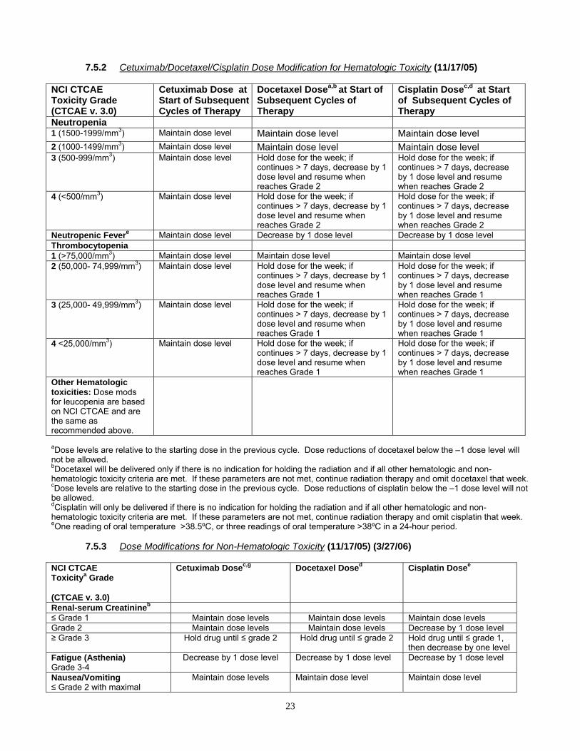
23
7.5.2 Cetuximab/Docetaxel/Cisplatin Dose Modification for Hematologic Toxicity (11/17/05)
NCI CTCAE Toxicity Grade (CTCAE v. 3.0)
Cetuximab Dose at Start of SubsequentCycles of Therapy
Docetaxel Dosea,b at Start of Subsequent Cycles of Therapy
Cisplatin Dosec,d at Start of Subsequent Cycles of Therapy
Neutropenia 1 (1500-1999/mm3) Maintain dose level Maintain dose level Maintain dose level 2 (1000-1499/mm3) Maintain dose level Maintain dose level Maintain dose level 3 (500-999/mm3) Maintain dose level
Hold dose for the week; if continues > 7 days, decrease by 1 dose level and resume when reaches Grade 2
Hold dose for the week; if continues > 7 days, decrease by 1 dose level and resume when reaches Grade 2
4 (<500/mm3) Maintain dose level
Hold dose for the week; if continues > 7 days, decrease by 1 dose level and resume when reaches Grade 2
Hold dose for the week; if continues > 7 days, decrease by 1 dose level and resume when reaches Grade 2
Neutropenic Fevere Maintain dose level Decrease by 1 dose level Decrease by 1 dose level Thrombocytopenia 1 (>75,000/mm3) Maintain dose level Maintain dose level Maintain dose level 2 (50,000- 74,999/mm3) Maintain dose level Hold dose for the week; if
continues > 7 days, decrease by 1 dose level and resume when reaches Grade 1
Hold dose for the week; if continues > 7 days, decrease by 1 dose level and resume when reaches Grade 1
3 (25,000- 49,999/mm3) Maintain dose level
Hold dose for the week; if continues > 7 days, decrease by 1 dose level and resume when reaches Grade 1
Hold dose for the week; if continues > 7 days, decrease by 1 dose level and resume when reaches Grade 1
4 <25,000/mm3) Maintain dose level
Hold dose for the week; if continues > 7 days, decrease by 1 dose level and resume when reaches Grade 1
Hold dose for the week; if continues > 7 days, decrease by 1 dose level and resume when reaches Grade 1
Other Hematologic toxicities: Dose mods for leucopenia are based on NCI CTCAE and are the same as recommended above.
aDose levels are relative to the starting dose in the previous cycle. Dose reductions of docetaxel below the –1 dose level will not be allowed. bDocetaxel will be delivered only if there is no indication for holding the radiation and if all other hematologic and non-hematologic toxicity criteria are met. If these parameters are not met, continue radiation therapy and omit docetaxel that week. cDose levels are relative to the starting dose in the previous cycle. Dose reductions of cisplatin below the –1 dose level will not be allowed. dCisplatin will only be delivered if there is no indication for holding the radiation and if all other hematologic and non-hematologic toxicity criteria are met. If these parameters are not met, continue radiation therapy and omit cisplatin that week. eOne reading of oral temperature >38.5ºC, or three readings of oral temperature >38ºC in a 24-hour period.
7.5.3 Dose Modifications for Non-Hematologic Toxicity (11/17/05) (3/27/06) NCI CTCAE Toxicitya Grade (CTCAE v. 3.0)
Cetuximab Dosec,g Docetaxel Dosed Cisplatin Dosee
Renal-serum Creatinineb ≤ Grade 1 Maintain dose levels Maintain dose levels Maintain dose levels Grade 2 Maintain dose levels Maintain dose levels Decrease by 1 dose level ≥ Grade 3 Hold drug until ≤ grade 2 Hold drug until ≤ grade 2 Hold drug until ≤ grade 1,
then decrease by one level Fatigue (Asthenia) Grade 3-4
Decrease by 1 dose level Decrease by 1 dose level Decrease by 1 dose level
Nausea/Vomiting ≤ Grade 2 with maximal
Maintain dose levels
Maintain dose level
Maintain dose level

24
medical management ≥ Grade 3 with maximal medical management
Hold drug until ≤ grade 2
Hold drug until ≤ grade 2
Hold drug until ≤ grade 2
Other non-hematologic Toxicitiesf,h
Neuropathy ≤ Grade 2 Grade 3-4
See footnote i
See footnote i
Decrease by 1 dose level
Discontinue cisplatin Other: Mucositis in RT field Grade 0-3 Grade 4
Maintain dose levels Hold drug until ≤ grade 3
Maintain dose levels Hold drug until ≤ grade 3
Maintain dose levels Hold drug until ≤ grade 3
Rash, in RT field ≤ Grade 2 Grade 3 Grade 4
Maintain dose levels
Hold drug until ≤ grade 2 Hold drug until ≤ grade 2
Maintain dose levels Maintain dose levels
Hold drug until ≤ grade 3
Maintain dose levels Maintain dose levels
Hold drug until ≤ grade 3 Rash, out of RT field ≤ Grade 2 Grade 3 Grade 4
Maintain dose levels
Hold drug until ≤ grade 2 Hold drug until ≤ grade 2
Maintain dose levels Maintain dose levels
Hold drug until ≤ grade 3
Maintain dose levels Maintain dose levels
Hold drug until ≤ grade 3 Grade 4/Other Hold drug until ≤ grade 1 Hold drug until ≤ grade 1 Hold drug until ≤ grade 1 Hypersensitivity Reaction See Section 7.5.4 aFor CTCAE Grade < 2 non-hematologic toxicity not described above, maintain dose level of drug. b Choose one or the other study to assess renal function and base treatment decision. cDose levels are relative to the previous dose. Dose reductions of C225 below the –2 dose level will not be allowed. dDose levels are relative to the previous dose. Dose reductions of docetaxel below the –1 dose level will not be allowed. eDose levels are relative to the previous dose. Dose reductions of cisplatin below the –1 dose level will not be allowed. fCetuximab: With the exception of allergic/hypersensitivity (see Section 1.5.4) gIn any case of C225 treatment delay, there will be no reloading infusion, and all subsequent treatments will be at the assigned dose level. hFor depressed K or Mg, administer replacement therapy. Chemotherapy should continue at the discretion of the treating physician. iNeuropathy is not expected with cetuximab or docetaxel. If toxicity seems related to cetuximab or docetaxel, contact Dr. Kies, Medical Oncology Co-Chair.
7.5.4 CTCAE v. 3.0 Infusion Reaction Management (11/17/05) (3/27/06) CTCAE Grade Treatment Guidelines
Cetuximaba Docetaxel Grade 1: Transient flushing or rash; drug fever < 38º C (< 100º.4 F)
For mild infusion reactions manifesting only as delayed drug fever, consider administering prophylactic antihistamine medications for subsequent doses. Maintain the cetuximab dose but slow the infusion rate by 50%. Acetaminophen or a non-steroidal anti-inflammatory drug (NSAID) may be administered prior to subsequent cetuximab infusions, if not otherwise contraindicated in subjects.
Consider decreasing the rate of infusion until recovery from symptoms. Stay at bedside and monitor patient, then complete docetaxel infusion at the initial planned rate.

25
Grade 2 : Rash; flushing; urticaria; dyspnea; drug fever ≥ 38º C (≥ 100º.4 F)
For moderate infusion reactions manifesting only as delayed drug fever, slow the infusion rate for cetuximab by 50%, and consider administration of antihistamine medications and/or steroidal medications. Maintain the cetuximab dose. Acetaminophen or a non-steroidal anti-inflammatory drug (NSAID) may be administered prior to subsequent cetuximab infusions, if not otherwise contraindicated in subjects.
-Interrupt docetaxel infusion and give diphenylhydramine 50 mg IV with or without dexamethasone 10mg IV. -Monitor patient until resolution of symptoms. -Resume docetaxel infusion after recovery of symptoms. -Depending on the physician’s assessment of the patient, docetaxel infusion should be resumed at a slower rate, then increased incrementally to the initial planned rate. (e.g., infuse at a 4-hr rate for 3 minutes, then at a 2-hr rate for 3 minutes, then at a 1-hr rate for 3 minutes, then finally, resume at the initial planned rate.) -Depending on the intensity of the reaction observed, additional oral or IV premedication with an antihistamine should also be given for the next cycle of treatment, and the rate of infusion should be decreased initially and then increased back to initial planned rate, (e.g., infuse at a 4-hr rate for 3 minutes, then at a 2-hr rate for 3 minutes, then at a 1-hr rate for 3 minutes, and finally, administer at the initial planned rate.)
Grade 3: Symptomatic bronchospasm with or without urticaria; parenteral medication(s) indicated; allergy-related edema/angioedema; hypotension
Severe infusion reactions require immediate interruption of cetuximab infusion and permanent discontinuation from further treatment with cetuximab. Appropriate medical therapy including epinephrine, corticosteroids, diphenhydramine, bronchodilators, and oxygen should be available for use in the treatment of such reactions. Subjects should be carefully observed until the complete resolution of all signs and symptoms.
Immediately discontinue docetaxel infusion. Give diphenylhydramine 50mg IV with or without dexamethasone 10mg IV and/or epinephrine as needed. Monitor patient until resolution of symptoms. Follow the same treatment guidelines outlined for Grade 2 symptoms.
7.5.4 CTCAE v. 3.0 Infusion Reaction Management (Continued) [11/17/05] [3/27/06]
CTCAE GRADE Treatment Guidelines
Cetuximaba Docetaxel
Grade 4: Anaphylaxis
NO FURTHER STUDY DRUG THERAPY Life-threatening infusion reactions require immediate interruption of cetuximab infusion and permanent discontinuation from further treatment with cetuximab. Appropriate medical therapy including epinephrine, corticosteroids, diphenhydramine, bronchodilators, and oxygen should be available for use in the treatment of such reactions. Subjects should be carefully observed until the complete resolution of all signs and symptoms.
NO FURTHER STUDY DRUG THERAPY Appropriate medical therapy including epinephrine, corticosteroids, diphenhydramine, bronchodilators, and oxygen should be available for use in the treatment of such reactions. Subjects should be carefully observed until the complete resolution of all signs and symptoms.
a Study Therapy Retreatment Following Infusion Reactions: Once a C225 infusion rate has been decreased due to an infusion reaction, it will remain decreased for all subsequent infusions. If the subject has a second infusion reaction with the slower infusion rate, the infusion should be stopped, and the subject should receive no further C225 treatment. If a subject experiences a Grade 3 or 4 infusion reaction at any time, the subject should receive no further C225 treatment. If there is any question as to whether an observed reaction is an infusion reaction of Grades 1-4, the Study Chair or designee should be contacted immediately to discuss and grade the reaction.

26
7.5.5 Cetuximab Special Instructions (11/17/05) If C225 is omitted for more than four consecutive infusions for toxicity due to C225, or for an intercurrent illness (e.g., infection) requiring interruption of therapy, the subject should be discontinued from further C225 therapy. If toxicities prevent the administration of C225, the subject may continue to receive radiation therapy.
7.5.5.1 Management of Acne-Like Rash (rash/desquamation) [3/16/05] Patients developing dermatologic toxicities while receiving cetuximab should be monitored for the development of inflammatory or infectious sequelae, and appropriate treatment of these symptoms initiated. Dose modifications (see Section 7.5.5.2) of any future cetuximab infusions should be instituted in case of severe acneiform rash. Treatment with topical and/or oral tetracyclines should be considered; topical corticosteroids are not recommended.
Dry skin will be noted in most patients receiving cetuximab. This should be treated with an emollient twice daily. With prolonged use of cetuximab, some patients may develop paronychial inflammation of the fingers and toes or fissuring of the fingertips. In general, good hygiene with appropriate local measures such as soaks in aluminum acetate (Burow’s) solution BID-QID will prevent secondary infection. Symptom relief may be achieved with standard bandages or with the application of liquid bandages (cyanoacrylate preparations such as Band-Aid Liquid Bandage®).
If a patient experiences severe acneiform rash, cetuximab treatment adjustments should be made according to Section 7.5.5.2. In patients with mild and moderate skin toxicity, treatment should continue without dose modification.
7.5.5.2 Drug Related Rash Management (11/17/05) Patients developing dermatologic toxicities while receiving cetuximab should be monitored for the development of inflammatory or infectious sequelae, and appropriate treatment of these symptoms initiated. Below are suggestions for managing cetuximab-induced rash*:
• Antibiotics: The benefit of routine antibiotics in uncomplicated (uninfected) rash is unclear. Some clinicians have used oral minocycline (Minocin), mupirocin (Bactroban), or topical clindamycin (Cleocin). Rash complicated by cellulitis should be treated with appropriate antibiotics based on clinical judgment or microbial sensitivity analysis.
• Antihistamines: Benadryl or Atarax may be helpful to control itching. • Topical Steroids: The benefit of topical steroids is unclear. • Retinoids: No data to support use. Use is not advised. • Benzoyl peroxide: Should NOT be used--may aggravate rash. • Makeup: Rash can be covered with makeup; this should not make it worse (use a
dermatologist-approved cover-up, e.g., Dermablend, or any other type of foundation). Remove makeup with a skin-friendly liquid cleanser, e.g., Neutrogena, Dove, or Ivory Skin Cleansing Liqui-Gel.
• Moisturizers: Use emollients to prevent and alleviate the skin dryness, e.g., Neutrogena Norwegian Formula Hand Cream or Vaseline Intensive Care Advanced Healing Lotion.
• Sunlight: It is recommended that patients wear sunscreen and hats and limit sun exposure while receiving cetuximab as sunlight can exacerbate any skin reactions that may occur.
• Over-the-counter medications: Over-the-counter acne vulgaris medications (e.g., benzoyl peroxide) are not advised. This rash is not like acne vulgaris and these treatments could make it worse.
*Adapted from Perez-Soler R, Delord J, Halpern A, et al. HER1/EGFR inhibitor-associated rash: Future directions for management and investigation outcomes from the HER1/EGFR Inhibitor Rash Management Forum. The Oncologist. 10:345–356, 2005.

27
7.5.6 Docetaxel Special Instructions (3/16/05) 7.5.6.1 Liver Function
Liver function tests should be evaluated at a minimum of every 4 weeks. Patients who develop abnormal liver function tests for any reason while on the study will have the following dose reductions:
Dose Modifications for Abnormal Liver Function (Docetaxel [Taxotere®])
AST or ALT: ALK PHOS:
≤ ULN >1X but ≤1.5X ULN >1.5X but ≤5X ULN >5X ULN
≤ ULN Full Dose Full Dose Full Dose Hold*
>1X but ≤ 2.5X
Full Dose Full Dose Reduce Dose Hold*
>2.5X but ≤ 5X
Full Dose Reduce Dose Hold* Hold*
>5X ULN Hold* Hold* Hold* Hold*
* Hold until recovered, maximum 21 days, then re-treat at a reduced dose. “Recovered” is defined as meeting the study baseline eligibility criteria.
7.5.6.2 Bilirubin
Docetaxel should not be administered to patients with serum total bilirubin > 1.5 X ULIN. If serum total bilirubin is >1.5 X ULIN on treatment day, hold docetaxel until serum total bilirubin is ≤ 1.5 X ULIN (maximum 21 days), then re-treat at a reduced dose.
7.6 Duration of Treatment 7.6.1 Discontinuation from Protocol Treatment
Study therapy MUST be immediately discontinued for the following reasons: - Withdrawal of informed consent (subject’s decision to withdraw for any reason). - Termination of the study by the sponsor. - Any clinical adverse event, laboratory abnormality or intercurrent illness, which, in the
opinion of the investigator, indicates that continued treatment with all study therapy is not in the best interest of the subject.
- Pregnancy. - Subject non-compliance with the protocol. - Imprisonment or the compulsory detention for treatment of either a psychiatric or physical
(e.g., infectious disease) - Progressive disease. - Any clinical event requiring discontinuation from therapy as detailed in Section 7.5.
The reason(s) for discontinuation from protocol treatment should be documented in the patient’s medical record and Case Report Form (CRF). All patients should be followed as specified in Sections 11.1 and 12.1.
7.6.2 Treatment Compliance Trained medical personnel will administer study therapy. Treatment compliance will be monitored by drug accountability, as well as recording treatment administration in the patient’s medical record and Case Report Forms.
7.6.3 Modality Review Institutional participation in chemotherapy studies must be in accordance with the medical oncology quality control guidelines stated in the RTOG Procedures Manual. All cases will undergo modality review by the modality study chair.
The Medical Oncology Co-Chair, Merrill S. Kies, MD, will perform a Chemotherapy Assurance Review of all patients who receive or are to receive chemotherapy in this trial. The goal of the review is to evaluate protocol compliance. The scoring mechanism is: per protocol; variation, acceptable; deviation unacceptable; not evaluable for chemotherapy review, or, incomplete chemotherapy. The review process is contingent on timely submission of chemotherapy treatment data as specified in Section 12.1. When complete data has been received at RTOG Headquarters for ten cases, these cases will be prepared and sent to Dr.

28
Kies for review. Subsequent cases will be prepared and sent to Dr. Kies for review, in increments of 20-25 cases, after complete data for those cases is received at RTOG Headquarters. A report is sent to each institution once per year to notify the institution about compliance for each case reviewed in that year.
7.7 Adverse Events (11/17/05) 7.7.1 This study will utilize the Common Terminology Criteria for Adverse Events (CTCAE) version
3.0 for grading of all treatment related adverse events. A copy of the CTCAE v3.0 can be downloaded from the CTEP home page (). The CTEP home page also can be accessed from the RTOG web page at http://www.rtog.org/regulatory/regs.html. All appropriate treatment areas should have access to a copy of the CTCAE v3.0. See the RTOG procedure manual for general Adverse Event Reporting Guidelines.
All adverse events (AEs) as defined in the tables below will be reported via the AdEERS (Adverse Event Expedited Reporting System) application accessed via the CTEP web site (https://webapps.ctep.nci.nih.gov/openapps/plsql/gadeers_main$.startup).
7.7.2 Adverse Events (AEs) — RTOG AE PHONE: 215-717-2762; 800-227-5463 ext. 4189 (available 24 hours/day) Definition of an AE: Any unfavorable and unintended sign (including an abnormal laboratory finding), symptom, or disease temporally associated with the use of a medical treatment or procedure regardless of whether it is considered related to the medical treatment or procedure (attribution of unrelated, unlikely, possible, probable, or definite). [CTEP, NCI Guidelines: Expedited Adverse Event Reporting Requirements. December 2004.] The following guidelines for reporting adverse events (AEs) apply to all NCI/RTOG research protocols. AEs, as defined above, experienced by patients accrued to this protocol should be reported via AdEERS. Use the patient’s case number as the patient ID when reporting via AdEERS. AEs reported using AdEERS also must be reported on the AE case report form (see Section 12.1). NOTE: If the event is a Serious Adverse Event (SAE) [see next section], further reporting may be required. Reporting AEs only fulfills Data Management reporting requirements.
7.7.3 Serious Adverse Events (SAEs) — All SAEs that fit any one of the criteria in the SAE definition below must be reported to RTOG (SAE PHONE: 215-717-2762, 800-227-5463 ext. 4189; available 24 hours/day) within 24 hours of discovery of the event. Definition of an SAE: Any adverse drug experience occurring at any dose that results in any of the following outcomes:
Death; A life-threatening adverse drug experience; Inpatient hospitalization or prolongation of existing hospitalization; A persistent or significant disability/incapacity; A congenital anomaly/birth defect.
Important medical events that may not result in death, be life threatening, or require hospitalization may be considered an SAE drug experience, when, based upon medical judgment, they may jeopardize the patient and may require medical or surgical intervention to prevent one of the outcomes listed in the definition. [CTEP, NCI Guidelines: Expedited Adverse Event Reporting Requirements. December 2004.] Outside of regular business hours (8:30-5:00 EST), leave a message that includes the study/case numbers and the caller’s contact information. A Data Manager will return the call the next business day requesting details of the event and also will inform the caller which type of report is required for that study (5 or 10 day AdEERS). The required report must be completed in AdEERS within 5 or 10 calendar days of the initial phone report, as directed by the Data Manager taking the call. SAEs reported using AdEERS also must be reported on the AE case report form (see Section 12.1).
Any late death (more than 30 days after last treatment) attributed to the protocol treatment (possible, probable or definite) should be reported to RTOG via the AE/SAE telephone line within 24 hours of discovery. An expedited report, if applicable, will be required within 5 or 10 calendar days.

29
All supporting source documentation, if applicable or if being faxed to NCI, must be properly labeled with the study/case numbers and the date of the adverse event and must be faxed to the RTOG dedicated SAE FAX, 215-717-0990, before the five or ten-calendar-day deadline to allow RTOG to comply with the reporting requirements of the pharmaceutical company/companies supporting the RTOG trial. All forms (and supporting source documentation) submitted to RTOG Headquarters must include the RTOG study/ case numbers; non-RTOG intergroup study and case numbers must be included, when applicable. Submitted AdEERS Reports are forwarded to RTOG electronically via the AdEERS system. Use the patient’s case number as the patient ID when reporting via AdEERS.
SAE reporting is safety related and separate and in addition to the Data Management reporting requirements as outlined in the previous AE reporting section. Any event that meets the above outlined criteria for an SAE but is assessed by the AdEERS System as “expedited reporting NOT required” must still be reported for safety reasons and to fulfill the obligations of RTOG to the pharmaceutical company/companies supporting the RTOG trial. Sites must bypass the “NOT Required” assessment and complete and submit the report. The AdEERS System allows submission of all reports regardless of the results of the assessment. Note: Sites must print the AdEERS report and fax it to the FDA, FAX 1-800-332-0178.
7.7.4 Acute myeloid leukemia (AML) or myelodysplastic syndrome (MDS) AML or MDS that is diagnosed during or subsequent to treatment in patients on NCI/CTEP-sponsored clinical trials must be reported using the NCI/CTEP Secondary AML/MDS Report Form available at http://ctep.cancer.gov/forms/index.html. The report must include the time from original diagnosis to development of AML/MDS, characterization such as FAB subtype, cytogenetics, etc., and protocol identification (RTOG study/case numbers). This form will take the place of a report via the AdEERS system and must be faxed to the Investigational Drug Branch, FAX 301-230-0159, and mailed to RTOG Headquarters (address below) within 30 days of AML/MDS diagnosis.
RTOG Headquarters AML/MDS Report 1818 Market Street, Suite 1600 Philadelphia, PA 19103
7.8 AdEERS Expedited Reporting Requirements (11/17/05)
Phase 2 and 3 Trials Utilizing an Agent under a Non-CTEP IND: AdEERS Expedited Reporting Requirements for Adverse Events that Occur within 30 Days1 of the Last Dose of the Investigational Agent, Cetuximab, in this Study
Grade 1
Grade 2
Grade 2
Grade 3
Grade 3
Grades 4 & 52
Grades 4 & 52
Unexpected Expected
Unexpected and
Expected
Unexpected
Expected with Hospitali-
zation
without Hospitali-
zation
with Hospitali-
zation
without Hospitali-
zation
Unex- pected Expected
Unrelated Unlikely
Not Required
Not Required
Not Required
10 CalendarDays
Not Required
10 Calendar
Days
Not Required
10 Calendar
Days
10 Calendar
Days
Possible Probable Definite
Not Required
10 Calendar Days
Not Required
10 CalendarDays
10 Calendar
Days
10 Calendar
Days
Not Required
24-Hour; 5 Calendar
Days
10 Calendar
Days 1 Adverse events with attribution of possible, probable, or definite that occur greater than 30 days after the last dose of
treatment with an agent under a non-CTEP IND require reporting as follows: AdEERS 24-hour notification followed by complete report within 5 calendar days for:
• Grade 4 and Grade 5 unexpected events AdEERS 10 calendar day report:
• Grade 3 unexpected events with hospitalization or prolongation of hospitalization • Grade 5 expected events
2 Although an AdEERS 24-hour notification is not required for death clearly related to progressive disease, a full report is required as outlined in the table.

30
Please see exceptions below under section entitled “Additional Instructions or Exceptions.”
March 2005
Note: All deaths on study require both routine and expedited reporting regardless of causality. Attribution to treatment or other cause must be provided. “On study” is defined as during or within 30 days of completing protocol treatment.
• Expedited AE reporting timelines defined: “24 hours; 5 calendar days” – The investigator must initially report the AE via AdEERS within 24
hours of learning of the event followed by a complete AdEERS report within 5 calendar days of the initial 24-hour report.
“10 calendar days” - A complete AdEERS report on the AE must be submitted within 10 calendar days of the investigator learning of the event.
• Any medical event equivalent to CTCAE grade 3, 4, or 5 that precipitates hospitalization (or prolongation of existing hospitalization) must be reported regardless of attribution and designation as expected or unexpected with the exception of any events identified as protocol-specific expedited adverse event reporting exclusions.
• Any event that results in persistent or significant disabilities/incapacities, congenital anomalies, or birth defects must be reported via AdEERS if the event occurs following treatment with an agent under a CTEP IND.
• Use the NCI protocol number and the protocol-specific patient ID assigned during trial registration on all reports.
Additional Instructions or Exceptions to AdEERS Expedited Reporting Requirements for Phase 2 and 3 Trials Utilizing an Agent under a Non-CTEP-IND: In this study, an investigational agent supplied under a non-CTEP-IND is being used in combination with commercial agents. The combination should be considered investigational, and reporting should follow the guidelines described above.
8.0 SURGERY
8.1 (8/27/04) Patients must have undergone gross total surgical resection of high-risk, pathological Stage III/IV squamous cell carcinoma of the head and neck within 7 weeks of randomization (see Section 3.1.1.1).
8.2 Surgical Quality Assurance Reviews The Surgical Oncology Co-Chair, Jeffrey N. Myers, M.D., will perform a Quality Assurance Review after complete data for the first 50 cases enrolled has been received at RTOG Headquarters. Dr. Myers will perform the next review after complete data for the next 50 cases enrolled has been received at RTOG Headquarters. The final cases will be reviewed within 3 months after this study has reached the target accrual or as soon as complete data for all cases enrolled has been received at RTOG Headquarters, whichever occurs first.
9.0 OTHER THERAPY (3/16/05)
All supportive therapy for optimal medical care will be given during the study period at the discretion of the attending physician(s) within the parameters of the protocol and documented on each site’s source documents as concomitant medication. The use of amifostine and pilocarpine is discouraged in light of the overlapping toxicity profile with the experimental agent, cetuximab, with resultant impact on the primary study endpoint. If amifostine or pilocarpine is administered, it should be documented on the Treatment Form (TF) and the Follow-up Form (F1).
10.0 PATHOLOGY (2/15/11) (For patients who have consented to participate in the tissue/blood component of the study; see
Appendix IB) 10.1 (6/11/09) Translational Research to be conducted with banked samples: 10.1.1 Rationale
The RTOG has been collecting pretreatment diagnostic tissue from head and neck cancer protocols over the last eight to ten years. A number of histologic, cell kinetic/proliferation, and molecular markers are under active investigation, with several showing promise for the stratification of patients in future trials. The EGFR represents one of the most promising head

31
and neck biomarkers studied to date with regard to clinical outcome in advanced head and neck cancer. The results of the current studies will expand and refine investigation of EGFR relationship to clinical outcome in head and neck cancer and may lead to identification of promising similar or new biomarkers with the goals of 1) identifying factors predictive of outcome such that patients may be better stratified in future trials, and 2) developing novel treatment strategies which target the molecular abnormalities identified. In this particular trial, we will gain specific information regarding any correlation between various forms of the EGFR (along with several downstream markers such as phosphorylated MAPK, AKT, and Stat-3) and clinical outcome in head and neck cancer patients who receive an EGFR inhibitory agent.
10.1.2 Specimen Collection (6/11/09)The following materials will be provided to the RTOG Biospecimen Resource for
translational research: 10.1.2.1 One H & E stained slide 10.1.2.2 (3/27/06) (6/11/09) A paraffin-embedded tissue block of the tumor or a 2 mm diameter core
of tissue punched from the tissue block containing the tumor with a skin punch and submitted in a plastic tube labeled with the surgical pathology number. NOTE: A kit with the punch, tube, and instructions can be obtained from the Biospecimen Resource (see contact information in Section 10.1.3). An example of the kit and instructions may be found in Appendix VI. Tissue blocks or cores must be clearly labeled with the pathology identification number that corresponds to the Pathology Report.
10.1.2.3 (3/27/06) A Pathology Report documenting that the submitted blocks or cores contain tumor; the report must include the RTOG protocol number and the patient’s case number. The patient’s name and/or other identifying information should be removed from the report. The surgical pathology numbers and information must NOT be removed from the report.
10.1.2.4 (3/27/06) A Specimen Transmittal Form clearly stating that the tissue is being submitted for translational research; the form must include the RTOG protocol number and the patient’s case number. Note: Institutions must clearly indicate on the form the “Type” of specimen (e.g., pre-treatment or post-treatment) and collection time frame (“Procedure Date”).
10.1.2.5 Specimens for translational research will be retained until the study is terminated. 10.1.2.6 (3/27/06) (6/11/09) Recurrent Tumors: Because a cohort of patients treated on RTOG 0234
will manifest tumor recurrence at some point following protocol treatment, specimens from the recurrent tumors are desired for study as these specimens represent valuable biologic materials following treatment with radiation and study drugs. Specimens from recurrent tumors should be sent to the RTOG Biospecimen Resource according to the specifications listed in Sections 10.1.2.1 to 10.1.2.4. Such tumor recurrences may derive from the locoregional head and neck area, or from a distant metastatic site if the clinical impression and pathology review are most consistent with recurrence of the squamous cell carcinoma of the head and neck that originally caused the patient to be enrolled on RTOG 0234.
10.1.3 (8/27/04, 3/27/06, 7/17/07, 6/11/09) Submit materials as follows:
U. S. Postal Service Mailing Address: For Non-frozen Specimens Only RTOG Biospecimen Resource University of California San Francisco Campus Box 1800 1657 Scott Street, Room 223 San Francisco, CA 94143-1800 Courier Address (FedEx, UPS, etc.): For Frozen Specimens RTOG Biospecimen Resource University of California San Francisco 1657 Scott Street, Room 223 San Francisco, CA 94115 Questions: 415-476-RTOG (7864)/FAX 415-476-5271; [email protected]
10.2 Reimbursement (6/11/09)
RTOG will reimburse submitting institutions $300 per case for fresh or flash frozen tissue or $200 per case for a block or core of material. After confirmation from the RTOG Biospecimen Resource that appropriate materials have been received, RTOG Administration will prepare the proper paperwork and send a check to the institution. Pathology payment cycles are twice a year

32
in January and July and will appear on the institution’s summary report with the institution’s regular case reimbursement.
10.3 Confidentiality/Storage (See Appendix IB and the RTOG Patient Tissue Consent Frequently Asked Questions, http://www.rtog.org/tissuebank/tissuefaq.html for further details.)
10.3.1 (6/11/09) Upon receipt, the specimen is labeled with the RTOG protocol number and the patient’s case number only. The Biospecimen Resource database only includes the following information: the number of specimens received, the date the specimens were received, documentation of material sent to a qualified investigator, type of material sent, and the date the specimens were sent to the investigator. No clinical information is kept in the database.
10.3.2 Specimens for translational research will be retained until the study is terminated, unless the patient consents to storage for future studies. If at any time the patient withdraws consent to store and use specimens, the material will be returned to the institution that submitted it.
11.0 PATIENT ASSESSMENTS 11.1 Study Parameters (2/15/11)
Assessment Pre-
Treatment Weekly During RT
3 months from start of RT
6 months from start of RT
Follow Upi
History/Physical Xa X X X Performance Status/Weight
Xa X X X X
CBC/Diff/PLT X a Xe X Xg Xg
Na+, K+, glucose, Ca+, Mag+, BUN, serum creatinine, TPro, Alb, AlkPhos, TBili , AST and ALT
X a Xe X Xg Xg
Pregnancy Test Xb CT/MRI Tumor Xc Xg X Xg
CXR or Thoracic CT
Xd Xh
Toxicity Evaluation X X X X Dental Evaluation X Nutritional Evaluation (Feeding tube recommended)
X
Surgical clearance X Medical Oncology Exam
Xa Xf Xg Xg Xg
a. Within 4 weeks prior to study entry b. Within 1 week prior to study entry c. Pre-operatively for clinical staging d. Within 90 days prior to study entry e. CBC, glucose, electrolytes, AST/ALT, creatinine, and Mag+ only f. Every three weeks during chemotherapy g. Only if clinically indicated h. Annually for 5 years i. Every 3 months from start of treatment for years 1 & 2; q 6 months for years 3 through 6, then annually.
11.2 Response Criteria/Outcome Definitions (11/17/05) 11.2.1 Response Criteria 11.2.1.1 No evidence of disease (NED): All patients must have no measurable tumor following
surgery. 11.2.1.2 Local-Regional Relapse: Recurrent cancer in the tumor bed and/or neck not clearly
attributable to a second primary neoplasm; biopsy confirmation is necessary. 11.2.1.3 Distant Relapse: Clear evidence of distant metastases (lung, bone, brain, etc.); Biopsy is
recommended where possible. A solitary lung mass/nodule is considered a second primary neoplasm unless proven otherwise.

33
11.2.1.4 Second Primary Neoplasm: The emergence of new tumor as a second primary should be documented. Modified rigorous criteria for a second primary have been adapted from Warren and Gates,43 as follows:
A distinct lesion separated from the primary tumor site by > 2 cm of normal epithelium;
A new cancer with different histology; Any cancer, regardless of site, occurring ≥ 3 years after initial treatment; Multiple lung nodules/masses are considered distant metastases from the index
cancer unless proven otherwise. 11.2.1.5 Disease-free survival (DFS): Duration for which the patient is without evidence for local-
regional or distant relapse, second primary, or death. 12.0 DATA COLLECTION (11/17/05)
Data should be submitted to: RTOG Headquarters
1818 Market Street, Suite 1600, Philadelphia, PA , 19103 Patients will be identified by initials only (first middle last); if there is no middle initial, a hyphen will be used (first-last). Last names with apostrophes will be identified by the first letter of the last name.
12.1 Summary of Data Submission (2/15/11)
Item Due Demographic Form (A5) Within 2 weeks of study entry Initial Evaluation Form (I1) Pathology Report (P1) Slides/Blocks (P2) Operative Note (S2) Surgical Path Report (S5)
Staging Diagrams (I6) Staging Diagram (nodes) [I7] Preliminary Dosimetry Information for non-IMRT Approaches:(For IMRT, see Section 12.2)
Within 1 week of start of RT
RT Prescription (Protocol Treatment Form) (T2) Films (simulation and portal) (T3) Calculations (T4) Preoperative CT Scan Report (C3) Final Dosimetry Information for non-IMRT Approaches: (For IMRT, see Section 12.2)
Within 1 week of RT end
Daily Treatment Record (T5) Isodose Distribution (T6) Boost Films (simulation and portal) (T8) Radiotherapy Form (T1) Within 1 week of RT end Treatment Form (TF) At end of treatment Follow-up Form (F1)
Thirteen weeks (90 days) from start of RT, then every 3 months thereafter for years 1 & 2; q 6 months for years 3 through 6, then annually. Also at death
Autopsy Report (D3) As applicable

34
12.2 Summary of Dosimetry Data Submission for IMRT (Submit to ITC; see Section 12.2.1) (2/2/06)
Item Due Preliminary Dosimetry Information †Digital Data Submission Form (DDSI) Within 1 week of start of RT CT data, critical normal structures, all GTV, CTV, and PTV contours
Simulation films and/or digital film images for all initial treatment fields and orthogonal set up pair
First day port films (or digital images) of all initial treatment fields and orthogonal set up pair
Digital beam geometry for initial and boost beam sets Doses for initial and boost sets of concurrent treated beams
Digital DVH data for all required critical normal structures, GTV, CTV, and PTVs for total dose plan
Hard copy isodose distributions for total dose plan as described in QA guidelines
Final Dosimetry Information Within 1 week of RT end Radiotherapy Form (T1) [copy to HQ and ITC] Daily Treatment Record Simulation films and/or digital film images (or digital images) of all boost treatment fields and orthogonal set up pair
First day port films of all boost treatment fields and orthogonal set up pair
Modified digital patient data as required through consultation with Image Guided Therapy QA Center
†Available on the ATC web site, http://atc.wustl.edu/
12.2.1 Digital Data Submission to ITC
Digital data submission may be accomplished using magnetic tape or the Internet. For network submission: The FTP account assigned to the submitting institution by the ITC shall be used, and e-mail identifying the data set(s) being submitted shall be sent to:
For tape submission: Please contact the ITC about acceptable tape types and formats. Hardcopies accompanying digital data should be sent by mail or Federal Express and should be addressed to:
Image-Guided Therapy Center (ITC) ATTN: Roxana Haynes 4511 Forest Park, Suite 200 St. Louis, MO 63110 314-747-5415 FAX 314-747-5423
12.3 (8/27/04) ACOSOG Investigators must submit data (Case Report Forms) directly to RTOG, as
specified in Section 12.0; data should not be submitted to ACOSOG. Include the RTOG protocol number and patient case number as well as the ACOSOG study number and patient number. The required forms can be accessed on the RTOG web site at http://www.rtog.org/members/forms/0234/main.html (no password required).
RTOG will send queries regarding data and forms due reports to ACOSOG Investigators. Investigators’ responses to queries should be submitted directly to RTOG.

35
NOTE: ACOSOG Investigators must submit Serious Adverse Event (SAE) regulatory requirements electronically via AdEERS, with an email copy to ACOSOG Headquarters. (RTOG will receive the SAE report directly from AdEERS).
13.0 STATISTICAL CONSIDERATIONS
13.1 Study Endpoints 13.1.1 Disease-free survival (failure: local, regional, or distant disease progression, second primary, or
death) 13.1.2 Patient tolerance of the treatment regimens 13.1.3 Frequency of Grade 5 and acute non-hematologic Grade 4 toxicity 13.1.4 Frequency of other acute and late toxicity 13.1.5 Overall Survival (Failure: death to any cause) 13.1.6 Local-regional control (Failure: local or regional disease progression) 13.1.7 Correlation of EGFR (total and phosphorylated), pMAPK, pAKT, Stat-3, Ki-67, COX-2, and
cyclin B1 expression with local-regional control, and overall and disease-free survival 13.2 Overview and Sample Size (3/16/05)
This trial is designed to determine if either regimen is promising enough to be pursued in a subsequent phase III study. This selection will be primarily based on whether there is improvement in disease-free survival relative to similar patients treated on the chemoradiation arm of the completed intergroup trial RTOG 9501.
Using the method of Dixon and Simon,44 104 analyzable patients are required for each arm to detect a ≥ 33% reduction in the failure rate (for DFS) as compared to RTOG 9501 with 80% statistical power (one-sided 0.05). This equates to an improvement in the two-year DFS rate from 53.9% (observed in RTOG 9501) to 66.1%.
Secondary considerations are patient tolerance and acute toxicity. Tolerability will be defined as having received ≥ 90% of the protocol radiation dose to the primary, and full protocol loading dose of C225 with ≥ 4 weeks of full protocol dose of C225 and chemotherapy (cisplatin or docetaxel). Dose within 5% of the protocol prescribed dose will be considered “full”. In RTOG 95-01, a chemoradiation program with cisplatin was evaluated in similar patients with high-risk, resectable tumors. Seventy-nine percent received within 10% of the protocol radiation dose and at least 2 (of 3) doses of cisplatin. With 104 patients for each arm, we have a ≥ 95% (two-sided) confidence interval around the estimated tolerance rate for each of the two arms with margin of error ≤ 9.6%.
Each regimen will be monitored for excessive acute toxicity (defined as non-hematologic Grade 4 toxicity within 90 days of the start of radiation therapy or any Grade 5 toxicity) in the initial 25, 50, and 104 evaluable patients (targeted sample size). In RTOG 9501, 13% of the evaluable patients experienced the above toxicity, and 2 (2%) treatment-related deaths were reported. For this study, we will assume a baseline rate of 15%, and a rate of acute toxicity > 30% will be considered unacceptable.
Adjusting the sample size by 10% to account for patient ineligibility or loss, a total sample size of 230 will be required for the study.
(11/17/05) The protocol has been amended to permit use of intensity-modulated radiotherapy
(IMRT). It is assumed that IMRT will have no enhancement on efficacy or on the primary endpoint, disease-free survival. However, it is possible that IMRT may reduce toxicity. Therefore, toxicity for each arm will be reported as follows: all eligible, assigned patients; those patients treated without IMRT; those patients treated with IMRT. Planned IMRT (no vs. yes) has been added as a stratifying variable to better ensure balance in the treatment assignments.
13.3 Patient Accrual Patient accrual will not only come from RTOG institutions but also from institutions in other
cooperative groups; ACOSOG has already agreed to participate in this trial. The patient accrual is projected to be 10 patients per month. At this rate, it will take approximately 29 months to reach the target accrual assuming that there will be little accrual during first six months while institutions are obtaining their IRB approvals. If the average monthly accrual is less than two cases after the initial six months, the study will be re-evaluated with respect to feasibility.

36
13.4 Randomization Scheme (11/17/05) Patients will be randomized to two treatment regimens in order to avoid any patient selection
bias. The treatment allocation scheme described by Zelen45 will be used because it balances patient factors other than institution. Patients will be stratified by Zubrod, risk category (positive margins vs. high risk [i.e. ≥ 2 positive nodes or extracapsular extension]), and use of IMRT (no vs. yes).
13.5 Toxicity Monitoring (11/17/05) As mentioned above, the acute toxicity rate in this patient population is approximately 13%. For
planning purposes, we assume a 15% acute toxicity rate. We wish to ensure that this treatment is tolerable and does not significantly increase acute toxicity. A toxicity rate of 30% is set as the highest acceptable rate. Fleming’s One-Stage Multiple Testing Procedure46 is utilized. Each regimen would be monitored as follows: If at least 8 patients (32%) demonstrate unacceptable toxicity among the initial 25 evaluable patients, or at least 14 (28%) among the initial 50 evaluable patients, or at least 23 (22%) among 104 evaluable patients (targeted sample size), the regimen would be considered to have an unacceptable toxicity profile. Analysis of the acute toxicity will be performed when the data are available for the first 25, first 50, and all patients entered. If the boundary for unacceptable toxicity is crossed in the initial 25 or 50 patients for a regimen, the study chairs will review all data pertaining to the events, and a recommendation will be made to the RTOG Research Strategy Committee for their consideration. The results of this review will determine the future course of action; accrual may be suspended for that regimen. If the boundary is crossed for the final analysis of the 104 evaluable patients, the associated regimen will be considered to have an unacceptable toxicity profile and would not be further tested in a phase III trial unless the RTOG Head and Neck Committee deems the efficacy results so positive as to warrant such toxicity. This monitoring plan has 0.97 probability of identifying a regimen with at least 30% unacceptable toxicity rate and 0.97 probability of accepting a regimen with no more than 15% unacceptable toxicity rate.
Total Number Entered
Rejected Ha (p = 0.30) # with toxicity <
Reject H0 (p= 0.15) # with toxicity >
25 3 8 50 11 14
13.6 Analysis and Reporting Plan (11/17/05) 13.6.1 This study will be monitored by the Clinical Data Update System (CDUS) version 3.0.
Abbreviated reporting of cumulative CDUS data will be submitted quarterly by electronic means. Reports are due January 31, April 30, July 31, and October 31.
13.6.2 Interim Reports: Interim reports will be prepared every six months until the final analysis. In general, the interim reports include information about: • patient accrual rate with a projected completion date for the accrual phase; • institutional accrual; • distribution of pretreatment characteristics of patients accrued; • compliance rate of treatment delivery with respect to the protocol prescription; • frequency and severity of toxicities.
13.6.3 Analysis and Reporting of Final Treatment Results: (11/17/05) The analysis to report the final results of treatment will be undertaken when each patient has been potentially followed for a minimum of 2 years. The usual components of this analysis are: • tabulation of all patients entered and any excluded from the analysis with reasons for
exclusion; • patient accrual rate; • institutional accrual; • distribution of the important baseline prognostic variables; • compliance rate of treatment delivery with respect to the protocol prescription; • frequency and severity of toxicities; • observed results with respect to the endpoints described in Section 13.1.
Overall and disease-free survival will be estimated using the Kaplan-Meier method.47 Local-regional control will be estimated using the method of cumulative incidence,48 as this accounts for

37
non-administrative censoring (i.e., death without local-regional failure). All failure time variables will be measured by the time interval from the date of registration to the date of the first failure. The one and two-year rates of local-regional control, disease-free and overall survival will be estimated along with 95% confidence intervals. The study was not designed to compare the efficacy of the two treatment programs against one another but rather each program will be tested against the RTOG 9501 chemoradiation arm. However, should both experimental arms demonstrate an improvement in disease free survival as compared to the RTOG 9501 chemoradiation arm, and the other factors (i.e. toxicity and tolerability) are not dissimilar, the RTOG will use statistical selection theory to choose which arm should be considered for further testing in a follow-up trial.49 Briefly, its criterion is to select the treatment arm with the highest response regardless of how small or “non-significant” the advantage is over the other treatment arm. Toxicity for each arm will be reported as follows: all eligible, assigned patients; those patients treated without IMRT; those patients treated with IMRT.
13.7 Tumor Marker Evaluation In addition to the clinical endpoints, this study will also evaluate several tumor markers (EGFR [total and phosphorylated], pMAPK, pAKT, Stat-3, Ki-67, COX-2, cyclin B1 expression). Each marker will be considered dichotomous (i.e., present/absent or overexpressed/not overexpressed) and correlated with local-regional control and disease-free and overall survival using Cox proportional hazards models,50 which will be stratified by the RTOG Recursive Partitioning Analysis (RPA) prognostic class.51
Because of the moderate number of failures expected, only very large differences in outcome can be detected with adequate statistical power. For this reason, all results will be considered hypothesis generating to be confirmed in a future study.
13.8 Inclusion of Women and Minorities In conformance with the National Institutes of Health (NIH) Revitalization Act of 1993 with regard
to inclusion of women and minorities in clinical research, we have considered the two possible interactions treatment by race and treatment by gender. The study was designed to evaluate disease-free survival, the treatment tolerance rate, and acute toxicity under the assumption of the same rates across the genders and across the races. A statistical analysis will be performed to examine the possible difference between the genders and among the races. In RTOG 95-01, 84% of the patients eligible for this trial were male; 16% were female; 74% were white; 26% were non-white. For planning purposes, we assume 85% of patients entered into this protocol will be male, 15% female, 75% white, and 25% non-white. We will use a binomial distribution for the two-year DFS rate (success – alive without disease). For males we have a 95% confidence interval for the two-year DFS rate with margin of error ≤ 10.4%; for females ≤ 24.5%; for whites ≤ 11.1%; for non-whites ≤ 19.2%. The following table gives the expected number of patients in each race and gender group:
Gender and Minority Accrual Estimates
Ethnic Category Sex/Gender Females Males Total
Hispanic or Latino 0 8 8 Not Hispanic or Latino 35 187 222 Ethnic Category: Total of all subjects 35 195 230
Racial Category American Indian or Alaskan Native 0 0 0 Asian 0 7 7 Black or African American 9 42 51 Native Hawaiian or other Pacific Islander 0 0 0 White 26 146 172 Racial Category: Total of all subjects 35 195 230

38
REFERENCES (11/17/05)
1. Forastiere A, Koch W, Trotti A, et al. Medical progress-Head and neck cancer. N Engl J Med. 345:1890-1900, 2001.
2. Vokes EE, Weichselbaum RR, Lippman SM, et al. Head and neck cancer. N Engl J Med. 328:184-194,
1993. 3. Ang KK, Thames HD, and Peters LJ. Altered fractionation schedules. In: Perez CA and Brady LW, eds.
Principles and Practice of Radiation Oncology. Philadelphia, PA: Lippincott-Raven; 1997:119-142. 4. Ang KK. Altered fractionation trials in head and neck cancer. Semin Radiat Oncol. 8:230-236, 1998. 5. Withers HR, Taylor JM, and Maciejewski B. The hazard of accelerated tumor clonogen repopulation
during radiotherapy. Acta Oncol. 27:131-146, 1988. 6. Vokes EE, Kies MS, Haraf DJ, et al. Concomitant chemoradiotherapy as primary therapy for
locoregionally advanced head and neck cancer. J Clin Oncol. 18:1652-61,2000. 7. Bachaud JM, David JM, Boussin, Daly N. Combined postoperative radiotherapy and weekly cisplatin
infusion for locally advanced head and neck carcinoma: Final report of a randomized trial. Int J Rad Onc Biol Phys. 36:(5) 99-1004, 1996.
8. Cooper JS, Pajak TF, Forastiere A, et al. Precisely defining high-risk operable head and neck tumors
based on RTOG #85-03 and #88-24: Targets for postoperative radiochemotherapy?” Head & Neck. 20: 588-594, 1998.
9. Head and Neck Contracts Program: Adjuvant chemotherapy for advanced head and neck squamous
carcinoma: Final report of the Head and Neck Contracts Program. Cancer. 60: 301-311, 1987. 10. Laramore GE, Scott CB, Al-Sarraf M, et al. Adjuvant chemotherapy for resectable squamous cell
carcinomas of the head and neck: report on Intergroup Study 0034. Int J Rad Onc Biol Phys. 23: 705-713, 1992.
11. Peters LJ, Goepfert H, Ang KK, et al. Evaluation of the dose of postoperative radiation therapy of head
and neck cancer: First report of a prospective randomized trial. Int J Rad Onc Biol Phys. 19:1449-1453, 1990.
12. Cooper JS, Pajak TF, Forastiere AA, et al. Postoperative concurrent radiation therapy and chemotherapy
in high-risk SCCA of the head and neck: the RTOG 9501/Intergroup phase III trial. In press, New Engl J Med, 2004.
13. Cooper JS, Pajak TF, Forastiere AA, et al. Postoperative concurrent radiochemotherapy in high-risk
SCCA of the head and neck: initial report of RTOG 9501/intergroup phase III trial. Proc Am Soc Clin Onc: 21: 226a, 2002.
14. Bernier J, van Glabbeke M, Domenge C, et al. Results of EORTC phase III trial 22931 comparing,
postoperatively, radiotherapy (RT) to concurrent chemo-radiotherapy (RT-CT) with high dose cisplatin in locally advanced head and neck (H&N) carcinomas (SCC). Euro J Can. 37(6): S267, 2001.\
15. Carpenter G, Cohen S: Epidermal growth factor. J Biol Chem. 265:7709-7712, 1990. 16. Mendelsohn, J. Epidermal growth factor receptor inhibition by a monoclonal antibody as anticancer
therapy. Clin Cancer Res. 3:2703-2707, 1997. 17. Salomon DS, Brandt R, Ciardiello F, and Normanno N. Epidermal growth factor-related peptides and their
receptors in human malignancies. Crit Rev Oncol Hemat. 19:183-232, 1995. 18. Grandis JR, Melhem MF, Gooding WE, et al. Levels of TGF-alpha and EGFR protein in head and neck
squamous cell carcinoma and patient survival. J Natl Cancer Inst. 90:824-32, 1998.

39
19. Baselga J. New therapeutic agents targeting the epidermal growth factor receptor. J Clin Oncol. 18:54-
59, 2000. 20. Fan Z, and Mendelsohn J. Therapeutic application of anti-growth factor receptor antibodies. Curr Opin
Oncol. 10: 67-73, 1998. 21. Kies MS, Harari PM. Cetuximab. Current Opinion Invest Drugs. 3(7):1092-1100, 2002. 22. Akimoto T, Hunter NR, Buchmiller L, Mason K, Ang KK, and Milas L. Inverse relationship between
epidermal growth factor receptor expression and radiocurability of murine carcinomas. Clin Cancer Res. 5: 2884-2890, 1999.
23. Bianco C, Bianco R, Tortora G, et al. Antitumor activity of combined treatment of human cancer cells with
ionizing radiation and anti-epidermal growth factor receptor monoclonal antibody C225 plus type I protein kinase A antisense oligonucleotide. Clin Cancer Res. 6: 4343-50, 2000.
24. Huang S-M, Bock JM, and Harari PM. Epidermal growth factor receptor blockade with C225 modulates
proliferation, apoptosis, and radiosensitivity in squamous cell carcinomas of the head and neck. Cancer Res. 59:1935-1940, 1999.
25. Huang S-M, Harari PM. Epidermal growth factor receptor inhibition in cancer therapy biology, rationale
and preliminary clinical results. Invest New Drugs. 17:259-269, 1999. 26. Huang S-M, and Harari PM. Modulation of radiation response following epidermal growth factor receptor
blockade in squamous cell carcinomas: inhibition of damage repair, cell cycle kinetics and tumor angiogenesis. Clin Cancer Res. 6:2166-2174, 2000.
27. Milas L, Mason K, Hunter N, et al. In vivo enhancement of tumor radioresponse by C225 antiepidermal
growth factor receptor antibody. Clin Cancer Res. 6: 701-708, 2000. 28. Perrotte P, Matsumoto T, Inoue K, et al. Anti-epidermal growth factor (VPF/VEGF) gene requires AP-2-
dependent DNA binding and transactivation. EMBO J. 16:750-759, 1997. 29. Harari P, and Huang S-M. Modulation of molecular targets to enhance radiation. Clin Cancer Res. 6:323-
325, 2000. 30. Schmidt-Ullrich RK, Mikkelsen RB, Dent P, et al. Radiation-induced proliferation of the human A431
squamous carcinoma cells is dependent on EGFR tyrosine phosphorylation. Oncogene. 15:1191-1197, 1997.
31. Harari PM, Huang S-M, Herbst R, Quon H. Molecular targeting of the epidermal growth factor receptor in
head and neck cancer. In: Harrison, Sessions, Hong, eds. Head & Neck Cancer: a Multidisciplinary Approach. Lippincott, Williams and Wilkins (in press), 2002.
32. Burtness BA, Li Y, Flood W, Mattar BI, Forastiere AA. Phase III trial comparing cisplatin (C) + placebo
(P) to C + anti-epidermal growth factor antibody (EGF-R) C225 in patients with metastatic/recurrent head & neck cancer. Proc Am Soc Clin Onc. 21:226a, 2002.
33. Pfister DG, Aliff TB, Kraus DH, et al. Concurrent cetuximab, cisplatin, and concomitant boost radiation
therapy (RT) for locoregionally advanced, squamous cell head and neck cancer (SCCHN): Preliminary evaluation of a new combined-modality paradigm. Proc Am Soc Clin Oncol 22: Abst #1993, 2003.
34. Douple EB. Platinum-radiation interactions. Natl Cancer Inst. Monogram 6:315-319, 1988. 35. Forastiere AA. Cisplatin and radiotherapy in the management of locally advanced head and neck cancer.
Int J Rad Onc Biol Phys. 27:465-470, 1993. 36. Jeremic B, Shibamoto Y, Milicic B, et al. Hyperfractionated radiation therapy with or without concurrent
low-dose daily cisplatin in locally advanced squamous cell carcinoma of the head and neck: a prospective randomized trial. J Clin Oncol. 18(7):1458-1464, 2000.

40
37. Harari PM, Mehta MP, Ritter MA, Petereit DG. Clinical promise tempered by reality in the delivery of
combined chemoradiation for common solid tumors. Semin Radiat Oncol (in press), 2002. 38. Glisson BS. The role of docetaxel in the management of squamous cell cancer of the head and neck.
Oncology. 16(6)supp: 83-87, 2002. 39. Glisson B, Murphy B, Frenette G, et al. Phase II trial of docetaxel and cisplatin combination
chemotherapy in patients with squamous cell carcinoma of the head and neck. J Clin Oncol 20:1593-1599, 2002.
40. Mason K, Hunter N, Milas L, et al. Docetaxel enhances tumor radioresponse in vivo. Clin Cancer Res.
3:2431-2438, 1997. 41. Glisson BS, Garden AS, Kies MS, et al. Phase I study of docetaxel (D), cisplatin (P), and concomitant
boost radiation for patients with locally advanced squamous cancer of the head and neck. Proc Am Soc Clin Onc. 21:231a, 2002.
42. Ang KK, Berkey BA, Tu X, Zhang HZ, Katz R, Hammond EH, Fu KK, Milas L. Impact of epidermal growth
factor receptor expression on survival and pattern of relapse in patients with advanced head and neck carcinoma. Cancer Res15; 62(24):7350-7356, 2002.
43. Warren S, Gates O: Multiple primary malignant tumors: A survey of the literature and statistical study.
Am J Cancer. 51:1358-1403, 1932. 44. Dixon DO, Simon R. Sample size considerations for studies comparing curves using historical controls. J
Clin Epidemiol 41: 1209-1213, 1988. 45. Zelen M. The randomization and stratification of patients to clinical trials. J Chronic Dis. 37:365-375,
1974. 46. Fleming T. One-sample multiple testing procedure for phase II clinical trials. Biometrics. 38:143-151,
1982. 47. Kaplan EL and Meier P. Nonparametric estimation from incomplete observations. J Amer Statist
Assoc53:457-481,1958. 48. Kalbfleisch JD and Prentice RL. The Statistical Analysis of Failure Data. John Wiley and Sons, New York,
1980. 49. Simon R, Wittes RE, Ellenber SS. Randomized Phase II Clinical Trials. Canc Treat Rep 69:1375-1381,
1985. 50. Cox DR. Regression models and life tables. J Royal Stat Soc; Series B, 34:187-229, 1972. 51. Cooper J, Berkey B, et al. Validation of the RTOG Recursive Partitioning Analysis for Head and Neck
Cancers. Proceedings of the 5th Int Conf on Head and Neck Cancer. Abstract 168, p.108, 2000.

41
APPENDIX IA
RTOG 0234
SAMPLE CONSENT FOR RESEARCH STUDY
A PHASE II RANDOMIZED TRIAL OF SURGERY FOLLOWED BY CHEMORADIOTHERAPY PLUS C225 (CETUXIMAB) FOR ADVANCED SQUAMOUS CELL CARCINOMA OF THE HEAD AND NECK
This is a clinical trial (a type of research study). Clinical trials include only patients who choose to take part. Please take your time to make your decision. Discuss it with your friends and family. The National Cancer Institute (NCI) booklet, “Taking Part in Clinical Trials: What Cancer Patients Need To Know,” is available from your doctor. You are being asked to take part in this study because you have cancer of the head and neck. WHY IS THIS STUDY BEING DONE? (3/16/05) (3/27/06) A standard form of treatment for advanced head and neck cancer patients is surgery under general anesthesia followed by radiation therapy. However, some patients still experience recurrence of their cancer despite the surgery and radiation. In this study, all participants will receive surgery and chemoradiation. All patients on this study will also receive weekly infusions of an agent called C225 (cetuximab). Patients will receive chemotherapy — cisplatin or docetaxel — depending on their treatment group. C225 was approved in 2004 as a treatment for patients with colorectal cancer, and when this study began, C225 was an experimental treatment for patients with head and neck cancer. In 2006, the FDA approved C225 for the treatment of head and neck cancer. C225 is an agent from a new family of drugs that block certain chemical pathways that lead to tumor cell growth. C225 may delay tumor growth. This study is being done because we do not know which of the radiation, C225, and chemotherapy combinations being studied may better control your cancer, or have fewer side effects, or prevent recurrence. In addition, if you agree, laboratory tests will be performed on your tumor tissue following surgery to analyze growth patterns that may help to predict and treat head and neck cancer patients in the future.

42
HOW MANY PEOPLE WILL TAKE PART IN THE STUDY Approximately 230 people will take part in this study. WHAT IS INVOLVED IN THE STUDY? (11/17/05) If you agree to take part in this study, after your surgery, you will be randomized into one of the two treatment groups described below. Randomization means that you are put into a group by chance. A computer will determine into which treatment group you are placed. Both Groups Radiation therapy and drug therapy will begin within 7 weeks of your surgery, when your doctors feel that you have healed adequately from your surgery. In general, most of your treatment will be done as an outpatient at your institution. Before your first dose of C225, you will be given some medications through your vein to prevent an allergic reaction to C225. Then you will be given the first dose of C225 through your vein for approximately 2 hours. You will not receive chemotherapy or radiation therapy on the day you receive the first dose of C225. Your blood pressure and overall physical condition will be closely monitored while you receive C225 and for at least one hour afterwards. If you have a severe allergic reaction to the first dose of C225 or any later doses, your doctor will treat you for the reaction, and you will not receive further treatment on this study. You and your doctor can discuss other treatments that you can receive off study. If you tolerate the first dose of C225 well, the following week you will begin receiving radiation therapy, C225, and chemotherapy. You have an equal chance of being assigned to either of the two treatment groups of the study.
Group 1 Radiation therapy
Once a day, five days a week, for six weeks; treatments take about 20-30 minutes
C225 Once a week before radiation and
chemotherapy for 6 weeks; treatment takes about an hour
Cisplatin Once a week before radiation therapy and
after C225 for 6 weeks; treatment takes about an hour

43
Group 2 Radiation therapy
Once a day, five days a week, for six weeks; treatment takes about 20-30 minutes
C225 Once a week before radiation and
chemotherapy for 6 weeks; treatment takes about an hour
Docetaxel Once a week before radiation therapy and
after C225 for 6 weeks; treatment takes about 30 minutes
In addition, if you take part in this study, you will have the following tests and procedures:
Prior to Study Entry
History and Physical Examination by several doctors Blood tests Pregnancy test for women able to have children CT/MRI of tumor Chest x-ray Evaluation of your teeth Evaluation of your nutrition – Insertion of a feeding
tube is strongly encouraged to make sure you get adequate nutrition during treatment because sores inside your mouth and throat will make chewing and swallowing difficult
Weekly During Radiation Therapy
You will be seen by your doctor to monitor any side effects you may be having
Blood Tests Every Three Weeks
Exam by Medical Oncologist
Three Months from start of Radiation Therapy
Physical exam Blood Tests CT/MRI of tumor (If your doctor advises it) Exam by Medical Oncologist (If your doctor advises it)
Five Months from start of Radiation Therapy
Physical Exam Blood tests (If your doctor advises) CT/MRI of tumor Exam by Medical Oncologist (If your doctor advises it)
3/2/11 Follow-up Visits: Every three months for 2 years, every six months for years 3 through 6, then once a year for your lifetime
Physical exam Blood tests (If your doctor advises) CT/MRI of tumor (If your doctor advises it) Chest X-ray (once a year) Exam by Medical Oncologist (If your doctor advises it)

44
HOW LONG WILL I BE IN THE STUDY? (2/15/11) You will receive treatment for about 2 months. You will be seen in follow-up visits every 3 months for years 1 and 2 and every 6 months for years 3 through 6, then once a year for your lifetime. The doctor may decide to take you off this study if it is in your medical best interest, your condition worsens, or new information becomes available and this information suggests the treatment will be ineffective or unsafe for you. It is unlikely, but the study may be stopped due to lack of funding or participation. You can stop participating at any time. However, if you decide to stop participating in the study, we ask you to talk to the study doctor and your regular doctor first. WHAT ARE THE RISKS OF THE STUDY? While on the study, you are at risk for these side effects. You should discuss these with the researcher and/or your regular doctor. There also may be other side effects that we cannot predict. Other drugs may be given to make side effects less serious and uncomfortable. Many side effects go away shortly after the docetaxel, cisplatin, C225 and radiation therapy are stopped, but in some cases side effects can be serious or long lasting or permanent. You may experience some or all of the side effects listed below.
Risks Associated with Radiation Therapy to the Head and Neck
Very Likely ▪ Sores in the mouth and/or throat which can be painful and make it very difficult to chew and or swallow foods ▪ Mouth dryness or changes in taste and/or smell that may be permanent ▪ Thick saliva ▪ Hoarseness ▪ Tanning or redness of the skin in the head and neck area being treated with radiation ▪ Ear pain and/or pressure ▪ Fatigue ▪ Weight loss ▪ Permanent hair loss in the area treated with radiation ▪ Loss of teeth, or cavities in the teeth, if strict dental care is not followed; hypersensitivity of teeth
Less Likely, But Serious ▪ Decrease in function of the thyroid gland that may require you to take thyroid replacement medicine to prevent you from feeling tired or sleepy ▪ Serious damage to the spinal cord, nerves in the neck, jawbone, voice box, skin, or other parts of the head and neck that may require a major operation to correct and, rarely, can even be life threatening

45
▪ Temporary pain or scarring around nerves in the shoulder that could cause numbness and/or weakness ▪ Breathing problems ▪ Difficulty with swallowing and eating for which you might need a long term or permanent feeding tube; possibility of inhaling food and/or liquids into the lungs – which could also result in pneumonia. ▪ Serious ear infections and/or hearing loss ▪ Damage to the spinal cord leading to permanent weakness and/or symptoms like a “stroke” ▪ Permanent hair loss (of the face/chin/neck)
Risks Associated with Cetuximab (C225) Very Likely
Weakness Headache Fever Nausea and/or vomiting Diarrhea Dry skin Localized acne-like skin reactions
Less Likely
Inflammation under fingernails and/or toenails, which can last for several months after C225 is stopped
Mouth sores Chills Muscle aches Joint pain Reduced appetite, which could lead to weight loss Confusion, not being oriented Shortness of breath
Less Likely, But Serious
Reduced white blood cell count which could lead to an increased risk of infection, weakness, and/or in bleeding and bruising easily; this lowering of blood counts, if severe, can lead to need for treatment with antibiotics, transfusions, or hospitalization.
Blood clots within a blood vessel in the legs or pelvis Seizure Coma Reduced kidney and/or liver function, which could lead to being hospitalized, or rarely, to death
Rare
Inflammation of the lining of the brain
46
Cetuximab also may cause allergic reactions such as hives, itching, and/or skin rash. Some patients have had allergic reactions with the first dose of cetuximab, but some patients have had reactions with later doses. The allergic reactions also can be severe, involving shortness of breath, wheezing, difficulty swallowing, lightheadedness, very low blood pressure, and rarely, heart attack and/or death.
Your condition will be closely monitored during doses of cetuximab and for at least one hour afterwards. If you have a severe reaction, your doctor will treat you for the reaction, and you will not receive further treatment on this study. If you have a delayed severe reaction after receiving cetuximab, you must immediately tell your doctor.
In addition, the combination of cetuximab with chemotherapy and radiation therapy could increase the likelihood and/or severity of the side effects of chemotherapy and radiation therapy.
Risks Associated with Cisplatin Very Likely ▪ Decrease in blood counts, which can lead to a risk of infection, decreased healing after surgery, and/or bleeding ▪ Anemia ▪ Loss of appetite and/or taste; metallic taste in your mouth ▪ Nausea and/or vomiting ▪ Fatigue ▪ Generalized loss of strength ▪ Hearing loss, ringing in the ears ▪ Loss of muscle or nerve function that may cause weakness or numbness in your hands and feet ▪ Loss of appetite and weight loss
Less Likely ▪ Allergic reactions (sweating, difficulty breathing, rapid heartbeat) ▪ Muscle cramps or spasm ▪ Facial swelling ▪ Loss of taste ▪ Loss of coordination ▪ Involuntary movement ▪ Restlessness Less Likely, But Serious ▪ Decrease in the kidneys’ ability to handle the body’s waste, which may be permanent ▪ Decrease in liver function ▪ Another cancer called acute leukemia
Risks Associated with Docetaxel
Very Likely ▪ Decrease in blood counts, which can lead to a risk of infection, decreased healing

47
after surgery, and/or bleeding ▪ Hair loss ▪ Weakness
▪ Loss of appetite ▪ Change in taste ▪ Inflammation of eye ▪ Fatigue, unusual sleepiness ▪ Mouth sores ▪ Muscle aches and/or joint pains ▪ Nausea and/or vomiting ▪ Headache ▪ Seizures ▪ Fever ▪ Allergic reaction that may cause rash, fever, swelling, chills, low back pain ▪ Bloating ▪ Diarrhea ▪ Numbness or tingling in the hands or feet
Less Likely, But Serious ▪ Allergic reaction, which can cause difficulty breathing, irregular heartbeat, low blood pressure, and can even be life threatening ▪ Changes in your nails ▪ Liver damage ▪ Lung damage
Reproductive Risks This study may be harmful to a nursing infant or an unborn child. Enough medical information is not available to determine whether the study treatment administered to a pregnant woman causes significant risks to the fetus. If you are a woman able to have children and have not been surgically sterilized (tubal ligation or hysterectomy), you should have a pregnancy test before enrolling in this study. If you are unwilling to use adequate birth control measures while on treatment and for at least three months thereafter to prevent pregnancy, you should not participate in this study. If you should become pregnant while on study, you must tell your doctor immediately.
If you are a man able to father children, the treatment you receive may risk harm to an unborn child unless you use a form of birth control approved by your doctor. If you are unwilling to use adequate birth control measures while on treatment and for at least three months thereafter to prevent pregnancy, you should not participate in this study. If you suspect you have caused anyone to become pregnant, you must tell your doctor immediately. ARE THERE BENEFITS TO TAKING PART IN THE STUDY? If you agree to take part in this study, there may or may not be direct medical benefit to you. We hope the information learned from this study will benefit other patients with head and neck cancer in the future.

48
The benefit of C225, chemotherapy, and radiation therapy to patients with head and neck cancer is unknown. This treatment may keep your cancer from growing, and this may provide relief from symptoms and improve your quality of life. This treatment may improve control of your head and neck cancer. However, none of these benefits is guaranteed, and the effects of a combination of C225, chemotherapy, and radiation therapy may be no different or worse than chemotherapy or radiation therapy alone. WHAT OTHER OPTIONS ARE THERE? You may choose to not participate in this study. Other treatments that could be considered for your condition may include the following: (1) once or twice a day radiation therapy; (2) chemotherapy that does not include the study drug (C225 is not available off study); (3) surgery; or (4) no treatment except medications to make you feel better. With the latter choice, your tumor may continue to grow and your disease may spread. These treatments could be given either alone or in combination with each other. There may also be other treatment trials in which you could participate. Your doctor can tell you more about your condition and the possible benefits of the different available treatments. Please talk to your regular doctor about these and other options. WHAT ABOUT CONFIDENTIALITY? (8/27/04) Efforts will be made to keep your personal information confidential. We cannot guarantee absolute confidentiality. Records of your progress while on the study will be kept in a confidential form at this institution and in a computer file at the headquarters of the Radiation Therapy Oncology Group (RTOG). Your personal information may be disclosed if required by law. Organizations that may inspect and/or copy your research records for quality assurance and data analysis include groups such as the Food and Drug Administration (FDA), the National Cancer Institute (NCI), Bristol-Myers Squibb, and ImClone Systems Incorporated (suppliers of C225), Aventis Pharmaceuticals (manufacturers of Taxotere® [docetaxel]), the American College of Surgeons Oncology Group (ACOSOG), and the Radiation Therapy Oncology Group (RTOG). WHAT ARE THE COSTS? Taking part in this study may lead to added costs to you or your insurance company. Please ask about any expected added costs or insurance problems. You may find a National Cancer Institute Guide: “Clinical Trials

49
and Insurance Coverage – a Resource Guide” helpful in this regard. You may ask your doctor for a copy, or it is available on the world wide web at http://www.nci.nih.gov/ClinicalTrials/insurance (and click on printable version). The study drug, C225, will be provided free of charge by Bristol-Myers Squibb in collaboration with ImClone Systems Incorporated for the participants in this study. Every effort has been made to ensure that adequate supplies of C225, free of charge, will be available for all participants. If, however, this study drug becomes commercially available for head and neck cancer like yours while you are being treated, there is a possibility that you or your insurance company will be charged for future supplies. In the case of injury or illness resulting from this study, emergency medical treatment is available but will be provided at the usual charge. No funds have been set aside to compensate you in the event of injury. You or your insurance company will be charged for all other costs of treatment, continuing medical care, and/or hospitalization. You will receive no payment for taking part in this study. WHAT ARE MY RIGHTS AS A PARTICIPANT? Taking part in this study is voluntary. You may choose not to take part or may leave the study at any time. Leaving the study will not result in any penalty or loss of benefits to which you are entitled. We will tell you about new information that may affect your health, welfare, or willingness to stay in this study. A group of experts in cancer of the head and neck from the RTOG Head and Neck Committee, the study chairs, and the RTOG study statistician will be reviewing the data from this research periodically throughout the study. WHOM DO I CALL IF I HAVE QUESTIONS OR PROBLEMS? (This section must be completed) For information about your disease and research-related injury, you may contact: ______________________________ _______________________ Name Telephone Number

50
For information about this study, you may contact: ______________________________ _______________________ Name Telephone Number For information about your rights as a research subject, you may contact: (OPRR suggests that this person not be the investigator or anyone else directly involved with the research) ______________________________ _______________________ Name Telephone Number
WHERE CAN I GET MORE INFORMATION? You may call the NCI’s Cancer Information Service at 1–800–4–CANCER (1–800–422–6237) or TTY: 1–800–332–8615. Visit the NCI’s Web sites for comprehensive clinical trials information at http://cancertrials.nci.nih.gov or for accurate cancer information including PDQ (Physician Data Query) visit http://cancernet.nci.nih.gov.
SIGNATURE
I have read all the above, asked questions, and received answers concerning areas I did not understand. I have had the opportunity to take this consent form home for review or discussion. I willingly give my consent to participate in this program. Upon signing this form I will receive a copy. I may also request a copy of the protocol (full study plan). Patient’s Name Signature Date Name of Person Obtaining Signature Date Consent

51
APPENDIX IB
RTOG 0234
SAMPLE CONSENT FORM FOR USE OF TISSUE FOR RESEARCH
A PHASE II RANDOMIZED TRIAL OF SURGERY FOLLOWED BY CHEMORADIOTHERAPY PLUS C225 FOR ADVANCED SQUAMOUS CELL CARCINOMA OF THE HEAD AND NECK ABOUT USING TISSUE FOR RESEARCH (3/16/05) (3/27/06)
In this study, you will have surgery to remove your cancer before receiving radiation and drug therapy. We would like to keep some of the tumor tissue from the surgery. If your tumor comes back after you complete study treatment, we would like to keep some of that tumor tissue as well. If you agree to allow us to keep your tissue, it may be used in research to learn more about cancer and other diseases.
The research that may be done with your tissue is not designed specifically to help you. It might help people who have cancer and other diseases in the future. All possible methods will be used to ensure your privacy and confidentiality. Identifying information will be taken off anything associated with your tissue before it is given to a researcher. Reports about research done with your tissue will not be given to you or your doctor. These reports will not be put in your health record. The research will not have an effect on your care.
THINGS TO THINK ABOUT (3/16/05)
The choice to let us keep the tumor tissue for future research is up to you. No matter what you decide to do, it will not affect your care or your participation in the main study. If you decide now that your tissue can be kept for research, you can change your mind at any time. Just contact us and let us know that you do not want us to use your tissue and then any tissue that remains will no longer be used for research; or, you may request that your tissue, if any remains, be returned to you or your designee.

52
In the future, people who do research may need to know more about your health. While __________ (doctor/institution) may give researchers reports about your health, your doctor/institution will not give researchers your name, address, phone number, or any other information that will let the researchers know who you are. Sometimes tissue is used for genetic research (about diseases that are passed on in families). Even if your tissue is used for this kind of research, the results will not be put in your health records. Your tissue will be used only for research. However, the research done with your tissue may help to develop new products in the future, or your tissue may be used to establish a cell line that could be patented and licensed. If this occurs, you will not be financially compensated.
BENEFITS
The benefits of research using tissue include learning more about what causes cancer and other diseases, how to prevent them, and how to treat them.
RISKS (3/16/05) Physical Risks Most patients will heal satisfactorily after having surgery or a needle biopsy to remove tissue. Your doctor will discuss the risks of surgery with you. Social-Economic Risks There is a very small chance that information from your health records could be incorrectly released. All possible methods will be used to protect your privacy and ensure confidentiality. Unless you have given your specific permission, your _______ (doctor/institution) will not release your personal results or information to third parties such as employers or insurers. In the case of injury or illness resulting from participating in this research, emergency medical treatment is available but will be provided at the usual charge. No funds have been set aside to compensate you in the event of injury.
MAKING YOUR CHOICE
If you have any questions about the research involving your tissue/blood or about this form, please talk to your doctor or nurse, or call the institution’s research review board at _________ (IRB’s phone number). Please read each sentence below and think about your choice. After reading each sentence, circle “Yes” or “No”. No matter what you decide to do, it will not affect your care or your participation in the main study.

53
1. My tissue may be used for the research in the current study. Yes No 2. My tissue may be kept for use in research to learn about, prevent, or treat cancer. Yes No 3. My tissue may be kept for use in research to learn about, prevent, or treat other health
problems (for example: diabetes, Alzheimer’s disease, or heart disease). Yes No
4. Someone from ______ (doctor’s office/institution) may contact me in the future to ask me to take part in more research.
Yes No Participant statement: I have read and received a copy of this consent form. I have been given an opportunity to discuss the information with my doctor/nurse, and all of my questions/concerns have been answered to my satisfaction. My answers above and my signature below indicate my voluntary participation in this research. Patient’s Name Signature Date
Witness statement: I have explained the information in this consent form to the patient and have answered any questions raised. I have witnessed the patient’s signature. _ _________ Name of Person Obtaining Consent Signature Date

54
APPENDIX II
KARNOFSKY PERFORMANCE SCALE
100 Normal; no complaints; no evidence of disease
90 Able to carry on normal activity; minor signs or symptoms of disease
80 Normal activity with effort; some sign or symptoms of disease
70 Cares for self; unable to carry on normal activity or do active work
60 Requires occasional assistance, but is able to care for most personal needs
50 Requires considerable assistance and frequent medical care
40 Disabled; requires special care and assistance
30 Severely disabled; hospitalization is indicated, although death not imminent
20 Very sick; hospitalization necessary; active support treatment is necessary
10 Moribund; fatal processes progressing rapidly
0 Dead
ZUBROD PERFORMANCE SCALE 0 Fully active, able to carry on all predisease activities without restriction (Karnofsky 90-100). 1 Restricted in physically strenuous activity but ambulatory and able to carry out work of a light or sedentary nature. For example, light housework, office work (Karnofsky 70-80). 2 Ambulatory and capable of all self-care but unable to carry out any work
activities. Up and about more than 50% of waking hours (Karnofsky 50-60). 3 Capable of only limited self-care, confined to bed or chair 50% or more of waking
hours (Karnofsky 30-40). 4 Completely disabled. Cannot carry on any self-care. Totally confined to bed or
chair (Karnofsky 10-20). 5 Death (Karnofsky 0).

55
APPENDIX III
AJCC STAGING SYSTEM HEAD & NECK, 6th Edition
STAGING-PRIMARY TUMOR (T) TX Primary tumor cannot be assessed T0 No evidence of primary tumor Tis Carcinoma in situ PHARYNX Nasopharynx T1 Tumor confined to the nasopharynx T2 Tumor extends to soft tissues of oropharynx and or nasal fossa T2a without parapharyngeal extension T2b with parapharyngeal extension T3 Tumor invades bony structures and/or paranasal sinuses
T4 Tumor with intracranial extension and/or involvement of cranial nerves, infratemporal fossa, hypopharynx, orbit, or masticator space.
Oropharynx T1 Tumor 2 cm or less in greatest dimension T2 Tumor more than 2 cm but not more than 4 cm in greatest dimension T3 Tumor more than 4 cm in greatest dimension T4a Tumor invades the larynx, deep/extrinsic muscle of tongue, medial pterygoid, hard palate, or
mandible. T4b Tumor invades lateral pterygoid muscle, pterygoid plates, lateral nasopharynx, or skull base or
encases carotid artery.
Hypopharynx T1 Tumor limited to one subsite of hypopharynx and 2 cm or less in greatest dimension. T2 Tumor invades more than one subsite of hypopharynx or an adjacent site, or measures more than
2 cm but not more than 4 cm in greatest diameter without fixation of hemilarynx. T3 Tumor measures more than 4 cm in greatest dimension or with fixation of hemilarynx. T4a Tumor invades thyroid/cricoid cartilage, hyoid bone, thyroid gland, esophagus or central
compartment soft tissue. T4b Tumor invades prevertebral fascia, encases carotid artery, or involves mediastinal structures.
LARYNX Supraglottis T1 Tumor limited to one subsite of supraglottis with normal vocal cord mobility T2 Tumor invades mucosa of more than one adjacent subsite of supraglottis or glottis or region
outside the supraglottis (e.g., mucosa of base of tongue, vallecula, medial wall of pyriform sinus) without fixation of the larynx.
T3 Tumor limited to larynx with vocal cord fixation and/or invades any of the following: postcricoid area, pre-epiglottic tissues, paraglottic space, and/or minor thyroid cartilage erosion (e.g., inner cortex).
T4a Tumor invades through the thyroid cartilage and/or invades tissues beyond the larynx (e.g., trachea, soft tissues of the neck including deep extrinsic muscle of the tongue, strap muscles, thyroid, or esophagus).
T4b Tumor invades prevertebral space, encases carotid artery, or invades mediastinal structures.

56
APPENDIX III (Continued) Glottis T1 Tumor limited to the vocal cord(s) (may involve anterior or posterior commissure) with normal
mobility T1a Tumor limited to one vocal cord T1b Tumor involves both vocal cords T2 Tumor extends to supraglottis and/or subglottis, or with impaired vocal cord mobility T3 Tumor limited to the larynx with vocal cord fixation, and/or invades paraglottic space, and/or minor
thyroid cartilage erosion (e.g., inner cortex). T4a Tumor invades through the thyroid cartilage and/or invades tissues beyond the larynx (e.g.,
trachea, soft tissues of neck including deep extrinsic muscle of the tongue, strap muscles, thyroid, or esophagus).
T4b Tumor invades prevertebral space, encases carotid artery, or invades mediastinal structures. Subglottis T1 Tumor limited to the subglottis T2 Tumor extends to vocal cord(s) with normal or impaired mobility T3 Tumor limited to larynx with vocal cord fixation T4a Tumor invades cricoid or thyroid cartilage and/or invades tissues beyond the larynx (e.g., trachea,
soft tissues of neck including deep extrinsic muscles of the tongue, strap muscles, thyroid, or esophagus).
T4b Tumor invades prevertebral space, encases carotid artery, or invades mediastinal structures. REGIONAL LYMPH NODES (N) Excluding Nasopharynx NX Regional lymph nodes cannot be assessed N0 No regional lymph node metastasis N1 Metastasis in a single ipsilateral node, 3 cm or less in greatest dimension. N2 Metastasis in a single ipsilateral node, more than 3 cm, but not more than 6 cm in greatest
dimension, or in multiple ipsilateral lymph nodes, none greater than 6 cm in greatest dimension, or bilateral or contralateral nodes, none more than 6 cm in greatest dimension.
N2a Metastasis in a single ipsilateral node more than 3 cm, but not more than 6 cm in greatest dimension.
N2b Metastasis in multiple ipsilateral nodes, none more than 6 cm in greatest dimension.
N2c Metastasis in bilateral or contralateral lymph nodes, none more than 6 cm in greatest dimension.
N3 Metastases in a lymph node, more than 6 cm in greatest dimension. DISTANT METASTASIS (M) MX Distant metastasis cannot be assessed M0 No distant metastasis M1 Distant metastasis

57
APPENDIX III (Continued) STAGE GROUPING Excluding Nasopharynx STAGE GROUPING Nasopharynx Stage 0 Tis, N0, M0 Stage 0 Tis, N0, M0 Stage I T1, N0, M0 Stage I T1, N0, M0 Stage II T2, N0, M0 Stage IIA T2a, N0, M0 Stage III T3, N0, M0 Stage IIB T1-T2a, N1, M0 T1-3, N1, M0 T2b, N0-1, M0 Stage III T1-T2b, N2, M0 Stage IVA T4a, N0-2, M0 T3, N0-2, M0 Any T, N2, M0 Stage IVA T4, N0-2, M0 Stage IVB T4b, Any N, MO Any T, N3, M0 Stage IVB Any T, N3, M0 Stage IVC Any T, Any N, M1 Stage IVC Any T, Any N, M1

58
APPENDIX IV
MANAGEMENT OF DENTAL PROBLEMS IN IRRADIATED PATIENTS
Dental Care for Irradiated Patients Goals for a dental care program include: 1. To reduce incidence of bone necrosis. 2. To reduce incidence of irradiation caries. 3. To allow proper fitting of dentures following treatment. Preirradiation Care and Procedures The patients may be grouped into four groups in accordance with the problems they present prior to irradiation. Group 1 Includes edentulous patients. They may require surgical removal of any symptomatic cysts, infected retained root tips, or alveolar hyperplasia. These patients require hygiene instruction and precautionary instruction about trauma with premature use of a prosthesis. Group 2 Includes those with poor dental hygiene, including those patients whose teeth are beyond repair by ordinary dental procedure, those with generalized oral sepsis, those with generalized periodontal disease, and those with chronic periapical abscesses or granulomas. Procedures performed on this group include removal of all remaining teeth prior to irradiation with primary closure and surgical preparation of the alveolar ridges to laterally support a prosthesis. There should be antibiotic coverage during the healing stage and adequate time prior to the start of radiation therapy. These patients need complete hygiene instruction and precautionary instruction about premature use of a prosthesis. Group 3 Includes those in whom dental condition is fair, including those patients whose teeth are restored, ordinary dental procedures, periodontal pockets are less than 3 mm deep, carious lesions are not in proximity to the pulp, and no more than 20 restorable carious lesions are present. X-ray examinations show at least 1/2 of the bone still present around root surfaces. These patients require removal of any teeth which are non-salvageable in accordance with the above and restorations of the remaining teeth as required. The patients are instructed for dental prophylaxis and the patients utilize custom-made fluoride carriers. Group 4 Includes those in whom dental hygiene is good. This includes patients who do not have severe malocclusion in whom few carious lesions are present. Carious lesions are not in close proximity to the pulp and are correctable by conventional methods. These patients require periodontal evaluation and dental prophylaxis training, restorations as needed, no extractions prior to radiation therapy, and fitting for custom carriers. Extraction of Teeth If extraction of teeth is necessary prior to radiation therapy, the bone must be contoured so that closure at the extraction site is possible. All loose spicules and sharp projections must be removed. The approximation of the gingival tissue must be such that the closure is neither too loose nor too tight. At least 10 days are required for adequate healing prior to initiation of therapy. Causative Factors The major causative factors appear to be the reduction of the amount of saliva and secondarily, reduced pH in the mouth. This occurs following high dose radiation to the major salivary glands using parallel opposed fields. The decay process usually occurs in the first year following radiation therapy. It tends to occur more quickly in teeth which have a large amount of root cementum exposed to those teeth with large amounts of plaque formation present. Doses of radiation in excess of 20 Gy to salivary tissue place the teeth at risk. Preventive Program The rationale behind the use of fluoride treatments is to make the tooth surfaces less susceptible to the decay process. This is accomplished by a combination of increasing fluoride concentration on the tooth surface and by the effect of fluoride on the plaque and flora that are present in the oral cavity. Adequate results are obtained by: 1) cleansing the teeth thoroughly, followed by a good home care dental prophylaxis program, 2) construction of fluoride carriers, custom-made mouth guards which provide local application of fluoride solution to the gingiva and tooth surfaces. Fluoride carriers are made individually with the use of casts. Material used for making a mouth guard is "Sta-Guard" plastic used in conjunction with vacutrole unit produced by Jelrus Technical Products, Corp.,

59
both of which are available through local dental supply. This material is molded to the cast impression and allowed to harden. A fluoride solution prepared at the M.D. Anderson Hospital is now available from the Emerson
APPENDIX IV (Continued)
Laboratories, Inc., Dallas, Texas 75221. It has been used to coat the plastic carrier for use in the mouth. The patients are instructed to cleanse their teeth prior to placement of the carrier. It is then worn in place for 5 minutes each day. The patients are instructed to rinse their mouths thoroughly following the use of the carrier. This will be continued for an indefinite period of time. Close follow-up is necessary. Results In the 5-1/2 year program at the M.D. Anderson Hospital beginning in 1966, a study of 304 patients shows that the incidence of necrosis of the jaw was reduced to approximately 21% compared to 37% prior to initiation of the study. Groups 3 and 4 patients randomized with and without fluoride treatment showed reduction in radiation carries from 67% to 34% among Group 3 patients, and from 65% to 22% among Group 4 patients. Failure to Control Decay Management of failure to control radiation decay includes silver fillings with continued use of fluoride treatments. If the decay process is sufficiently advanced that a filling will no longer stay in place, these teeth are merely smoothed so that there will be no sharp, irritating edges. The mere existence of such a decayed tooth is not necessarily reason for extraction, for it must be remembered that extraction could lead to complications such as bone necrosis. Pulp exposure resulting from the decay process can usually be handled by use of antibiotics and/or root-canal therapy. Hypersensitivity of Teeth Occasionally, a patient will exhibit extreme sensitivity of the teeth secondary to diminished amounts of saliva. This has been shown to be reduced in incidence with the fluoride treatments. Should this problem become manifest, increasing the fluoride treatment to 10 to 15 minutes 3 times a day is recommended. Infections Infections occurring in patients under or after radiation therapy are best managed conservatively with good oral hygiene, irrigation and flushing procedures, and systemic antibiotics. Bone Necrosis The patients receiving radiation therapy to a high dose to the head and neck region have increased susceptibility to bone necrosis for several reasons including: impairment of normal metabolism, increased susceptibility to infection and severely limited repair process. Bone necrosis occurs most often after post-irradiation surgery or other traumas. Conservative management should be tried first, though in more aggressive lesions a more radical approach may ultimately be necessary.

60
APPENDIX V (11/17/05, 2/2/06)
C225 (Cetuximab) CLINICAL SUPPLY SHIPMENT REQUEST TO INVESTIGATIONAL SITE
Cetuximab will be shipped only to institutions that have identified a single individual for receipt of shipment. For the initial shipment of Cetuximab, the Study Agent Shipment Form for this study is available on the RTOG web site, www.rtog.org, next to the protocol. U.S. and Canadian institutions must email the shipment form for this study to [email protected] as soon as the individual responsible for the study agent has been identified and prior to registration of the institution’s first case. (Fax to 215-574-0300 only if unable to email; please write legibly). Allow adequate processing time (7-10 days) before calling to randomize your first patient. Required regulatory documents (see Section 5.2) must be received and approved by BMS and RTOG notified of this approval before drug can be shipped. For Resupply Requests, email the shipment form to [email protected]. (Fax to 866-227-7229 only if unable to email; please write legibly). For questions call 800-743-9224.

61
APPENDIX VI (3/27/06) (7/17/07) (6/11/09)
Specimen Plug Kit and Instructions*
The Specimen Plug Kit contains a dermal needle and a shipping tube. Institutions should NOT dispose of the Plug Kit but should ship it back to the RTOG Biospecimen Resource with their specimen(s). Sites can call or email the RTOG Biospecimen Resource with questions (contact information below) or to request additional Specimen Plug Kits.
Step 1 Place the dermal needle on the paraffin block over the selected tumor area. (Ask a Pathologist to select the tumor area). Push the needle into the paraffin block. Twist the needle once around to separate the plug from the block, then pull the needle out of the block. The needle should be filled with tissue sample. Step 2 Label the dermal needle with the proper specimen identification. Do not try to remove the specimen from the needle. Use a separate dermal needle for every specimen. Do not mix specimens. Step 3 Once the specimen needle is labeled, place it in the shipping tube and mail the shipping tube to the RTOG Biospecimen Resource (address below). At the Biospecimen Resource, the specimen will be removed from the needle, embedded in a cassette, and labeled with the specimen identification.
*NOTE: If an institution is uncomfortable obtaining the plug but wants to retain the tissue block, the institution should send the entire tissue block to the RTOG Biospecimen Resource. The Biospecimen Resource will sample a plug from the tissue block and return the remaining block to the institution. Sites must document the request to perform the plug procedure and return the block on the Specimen Transmittal Form. Ship the Specimen Plug Kit, specimen in dermal needle inside shipping tube, and all paperwork as follows:
U. S. Postal Service Mailing Address: For Non-frozen Specimens Only RTOG Biospecimen Resource University of California San Francisco Campus Box 1800 1657 Scott Street, Room 223 San Francisco, CA 94143-1800 Courier Address (FedEx, UPS, etc.): For Frozen Specimens RTOG Biospecimen Resource University of California San Francisco 1657 Scott Street, Room 223 San Francisco, CA 94115 Questions: 415-476-RTOG (7864)/FAX 415-476-5271; [email protected]