RADIATION PROTECTION IN DIAGNOSTIC AND INTERVENTIONAL RADIOLOGY
21
IAEA International Atomic Energy Agency RADIATION PROTECTION IN DIAGNOSTIC AND INTERVENTIONAL RADIOLOGY Part 16.3: Optimization of protection in fluoroscopy Practical exercise IAEA Training Material on Radiation Protection in Diagnostic and Interventional Radiology
-
Upload
demetrius-duran -
Category
Documents
-
view
42 -
download
0
description
IAEA Training Material on Radiation Protection in Diagnostic and Interventional Radiology. RADIATION PROTECTION IN DIAGNOSTIC AND INTERVENTIONAL RADIOLOGY. Part 16.3: Optimization of protection in fluoroscopy Practical exercise. Overview / Objectives. - PowerPoint PPT Presentation
Transcript of RADIATION PROTECTION IN DIAGNOSTIC AND INTERVENTIONAL RADIOLOGY

IAEAInternational Atomic Energy Agency
RADIATION PROTECTION INDIAGNOSTIC AND
INTERVENTIONAL RADIOLOGY
Part 16.3: Optimization of protection in fluoroscopy
Practical exercise
IAEA Training Material on Radiation Protection in Diagnostic and Interventional Radiology

IAEA 16.3: Optimization of protection in fluoroscopy 2
Overview / Objectives
• To become familiar with quality control tests in fluoroscopy.
• To measure the high contrast resolution
• Interpretation of results

IAEAInternational Atomic Energy Agency
Part 16.3: Optimization of protection in fluoroscopy
High contrast spatial resolution
IAEA Training Material on Radiation Protection in Diagnostic and Interventional Radiology

IAEA 16.3: Optimization of protection in fluoroscopy 4
High contrast resolution
Purpose :• to measure the high contrast resolution of the
fluoroscopy system
Equipment :• image quality phantom (Leeds phantom)
• 2 mm Cu filter

IAEA 16.3: Optimization of protection in fluoroscopy 5
Leeds Test Phantom 18FG to be used for Leeds Test Phantom 18FG to be used for routine image quality controlroutine image quality control

IAEA 16.3: Optimization of protection in fluoroscopy 6
Details of the test object (Leeds Test Details of the test object (Leeds Test 18FG) to be used for routine image quality 18FG) to be used for routine image quality controlcontrol

IAEA 16.3: Optimization of protection in fluoroscopy 7
Leeds test object Leeds test object is placed at the is placed at the entrance of the entrance of the image intensifierimage intensifier

IAEA 16.3: Optimization of protection in fluoroscopy 8
Centering sometimes can be difficultCentering sometimes can be difficult

IAEA 16.3: Optimization of protection in fluoroscopy 9
An alternative solution to centring the An alternative solution to centring the Leeds test object for image quality Leeds test object for image quality evaluationevaluation
IAEA 16.3: Optimization of protection in fluoroscopy 10
Another possible solution— tape the Cu Another possible solution— tape the Cu filter together with the test object.filter together with the test object.

IAEA 16.3: Optimization of protection in fluoroscopy 11
Do not forget
• To angle test object at 45°
to TV scan lines
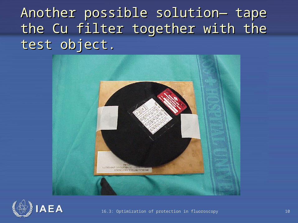
• The best results are in center of intensifier field

IAEA 16.3: Optimization of protection in fluoroscopy 12
Test object and filters placed at the Test object and filters placed at the image intensifierimage intensifier

IAEA 16.3: Optimization of protection in fluoroscopy 13
Test object image (23 cm field size)Test object image (23 cm field size)

IAEA 16.3: Optimization of protection in fluoroscopy 14
Test object image (17 cm field size)Test object image (17 cm field size)
IAEA 16.3: Optimization of protection in fluoroscopy 15
The best results are in the center of image field

IAEA 16.3: Optimization of protection in fluoroscopy 16
Do not forget to angle test object at 45° to TV raster lines to avoid Moire patterns

IAEA 16.3: Optimization of protection in fluoroscopy 17
High contrast resolution test object

IAEA 16.3: Optimization of protection in fluoroscopy 18
Leeds Fluoroscopy Phantom (spatial resolution)
Incorporates:• a test pattern with bar
patterns with spatial frequencies ranging from 0.5 to 5.0 cycles (line pairs) per mm.

IAEA 16.3: Optimization of protection in fluoroscopy 19
Leeds Fluoroscopy Phantom (spatial resolution)

IAEA 16.3: Optimization of protection in fluoroscopy 20
Spatial resolution
Interpretation of resultsInterpretation of results
Analysis depends on the phantom used (Leeds)
The resolution limit is normally measured at the centre of the image field. It should correspond to the following values:
Field size diameter
Resolution limit
cm Lp/mm 30 - 35 >1.0 22 - 25 >1.25 15 – 18 >1.6

IAEA
Where to Get More Information
15.3: Optimization of protection in radiography 21
Quality Control in Diagnostic Imaging, Gray JE, Winkler NT, Stears J, Frank ED. Available at no cost. http://www.diquad.com/QC%20Book.html