RADIATION ONCOLOGY RESIDENCY PROGRAM … Syllabi/Competency...RADIATION ONCOLOGY RESIDENCY PROGRAM...
16
RADIATION ONCOLOGY RESIDENCY PROGRAM Competency Evaluation of Resident Resident’s Name: Rotation: PHYS 703: Clinical Rotation 2 Inclusive dates of rotation: Feb. 26, 2016 – Aug. 25, 2016 Director or Associate Director: Evaluation criteria Not Competent Marginally Competent Fully Competent Explanatory Notes & Mentor Signature Treatment Techniques 1. Demonstrates understanding of 2D coplanar beam treatment planning 2. Demonstrates an understanding of the placement of non-coplanar beams (3D) in external beam treatment planning 3. Demonstrates an understanding of the following image-guided radiation therapy techniques: a. Planar MV imaging b. Planar kV imaging c. Cone beam computed tomography (CBCT) d. Ultrasound (US) e. Non-radiographic localization, e.g., US, surface camera, radiofrequency (RF) beacon tracking. 4. Demonstrates an understanding of image registration techniques, e.g., rigid and deformable registration 5. Demonstrates an understanding of site-specific techniques (photons and electrons): a. Performs 3D or IMRT treatment planning for breast and chest wall that includes axilla fields and the single isocenter technique
Transcript of RADIATION ONCOLOGY RESIDENCY PROGRAM … Syllabi/Competency...RADIATION ONCOLOGY RESIDENCY PROGRAM...
RADIATION ONCOLOGY RESIDENCY PROGRAM Competency Evaluation of Resident
Resident’s Name:
Rotation: PHYS 703: Clinical Rotation 2
Inclusive dates of rotation: Feb. 26, 2016 – Aug. 25, 2016
Director or Associate Director:
Evaluation criteria Not
Competent Marginally Competent
Fully Competent
Explanatory Notes & Mentor
Signature
Treatment Techniques
1. Demonstrates understanding of 2D coplanar beam treatment planning
2. Demonstrates an understanding of the placement of non-coplanar beams (3D) in external beam treatment planning
3. Demonstrates an understanding of the following image-guided radiation therapy techniques:
a. Planar MV imaging
b. Planar kV imaging
c. Cone beam computed tomography (CBCT)
d. Ultrasound (US)
e. Non-radiographic localization, e.g., US, surface camera, radiofrequency (RF) beacon tracking.
4. Demonstrates an understanding of image registration techniques, e.g., rigid and deformable registration
5. Demonstrates an understanding of site-specific techniques (photons and electrons):
a. Performs 3D or IMRT treatment planning for breast and chest wall that includes axilla fields and the single isocenter technique

b. Performs 3D or IMRT treatment planning for the brain, spine, and craniospinal irradiation
c. Performs 3D or IMRT treatment planning for the bladder, prostate, and testis
d. Performs 3D or IMRT treatment planning for gynecological tumors
e. Performs 3D or IMRT treatment planning for gastrointestinal tumors, e.g., colorectal tumors, tumors of the esophagus, stomach, and liver
f. Performs 3D or IMRT treatment planning for head and neck tumors
g. Performs 3D treatment planning for common lymphomas that includes the mantle field technique;
h. Performs 3D treatment planning for skin cancers
i. Demonstrates an understanding of common 3D or IMRT treatment planning techniques for pediatric cancers and performs 3D treatment planning for pediatric craniospinal irradiation
j. Demonstrates an understanding of common 3D or IMRT treatment planning techniques for sarcoma of the trunk and extremities
k. Performs 3D or IMRT treatment planning for the lungs, mediastinum, and thoracic region
Treatment Planning
1. Beam properties
a. Demonstrates an understanding of photon and electron percent depth dose in tissue and other media
b. Demonstrates an understanding of electron ranges (Rp, R80, R90, and dmax) for different energies
c. Demonstrates an understanding of proton percent depth dose in tissue and other media and proton ranges for different energies, e.g., stopping and scattering power and range

d. Demonstrates an understanding of the potential uncertainties in dose deposition in proton radiotherapy
e. Demonstrates an understanding of the flatness and symmetry of photon and electron beams
f. Demonstrates an understanding of the differences between source-to-axis distance (SAD) and source-to-skin distance (SSD) treatments;
g. Demonstrates an understanding of the applicability of electron and photon therapy with regard to disease, depth, and critical normal structures
h. Discusses the impact of dose and fractionation on normal and tumor tissues
i. Demonstrates an understanding of the impact of beam quality (e.g., linear energy transfer [LET]) on the relative biological effectiveness (RBE) of different forms of ionizing radiation (e.g., electrons, photons, and protons)
j. Discusses the uncertainties related to electron and photon therapy (e.g., in terms of physics, biology, machine and patient setup accuracy) and how they may be detected and mitigated during the planning and delivery process.
2. Beam modifiers
a. Demonstrates an understanding of the effect of beam modifiers (e.g., wedges, compensators) on the dosimetric characteristics of the incident beam
b. Demonstrates an understanding of wedges (wedge angle, hinge angle) and the different types of wedges used clinically (physical, universal, dynamic)
c. Demonstrates an understanding of the design of the different commercially available multileaf collimators (MLCs)
d. Demonstrates an understanding of blocking and shielding for therapy beams
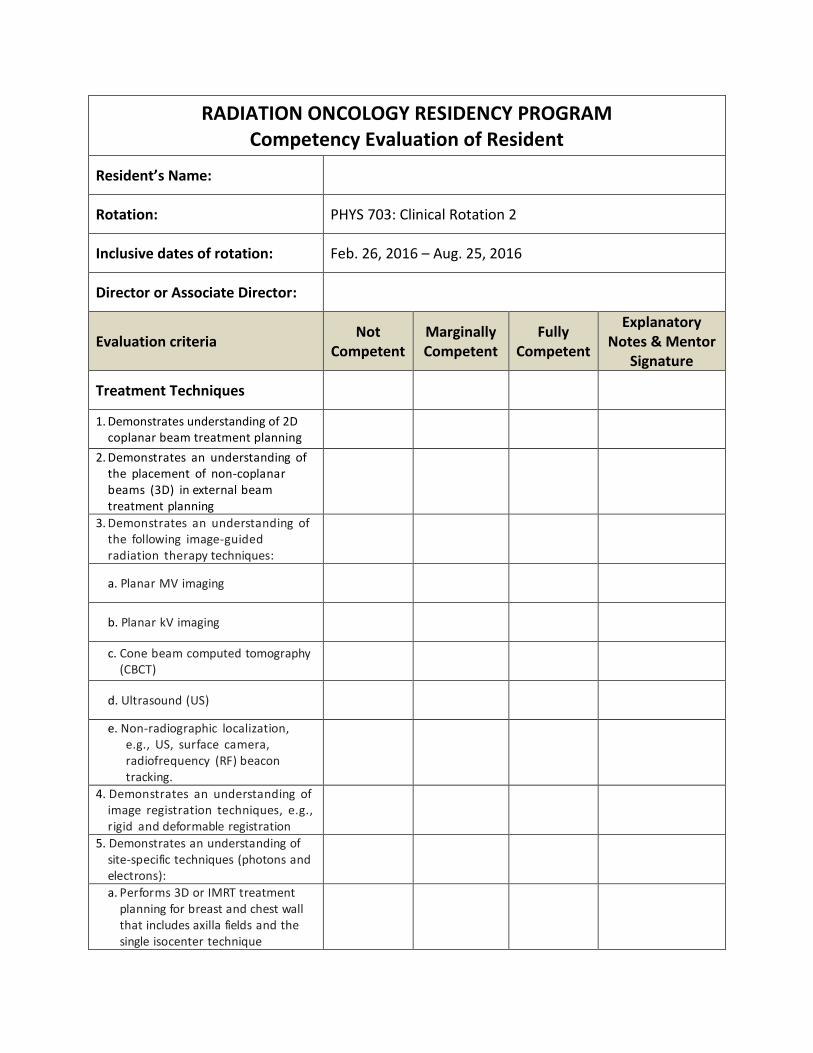
e. Demonstrates an understanding of the use of custom bolus
f. Demonstrates an understanding of the design and use of tissue compensators.
3. Treatment simulation techniques
a. Demonstrates an understanding of common patient-positioning and immobilization devices
b. Demonstrates an understanding of when and how to use specific treatment devices for specific treatments
c. Discusses how to account for beam attenuation from patient-positioning and immobilization devices in treatment planning.
4. Tumor localization and normal tissue anatomical contouring
a. Performs structure delineation on CT, MRI, PET, PET/CT, SPECT, or SPECT/ CT data sets
b. Demonstrates an understanding of target volume determination, including the design of ICRU target structures (involving concepts such as gross tumor volume [GTV], clinical target volume [CTV], internal target volume [ITV], planning target volume [PTV], and planning organ at risk volume [PRV]);
c. Demonstrates an understanding of how 4D data is used for target definition and relevant radiation treatment prescription parameters such as GTV, PTV, CTV, and ITV
d. Demonstrates an understanding of the role of maximum intensity projection (MIP) images in the treatment planning process
e. Demonstrates an understanding of the role of digitally reconstructed radiographs (DRRs) in the treatment planning process
f. Demonstrates an understanding of and performs image registration and fusion of data sets for modalities such as CT/CT, CT/MRI, and CT/PET; deformable

registration; and image/dose registration.
5. Plan evaluation. Defines and discusses each of the following treating planning evaluation tools, including their limitations:
a. Dose volume histograms (V(dose), D(volume), mean dose; cumulative and differential)
b. Conformity index
c. Homogeneity index
d. Biological evaluators (e.g., generalized equivalent uniform dose [gEUD], equivalent uniform dose [EUD], normal tissue complication probability [NTCP], and tumor control probability [TCP]).
Discusses dose tolerances for various normal tissue structures along with relevant volume effects.
Intensity-modulated Radiation Therapy (IMRT)
1. Inverse planning
a. Demonstrates an understanding of the use of objective functions for IMRT optimization
b. Demonstrates an understanding of the optimization processes involved in inverse planning
c. Performs inverse planning optimization for a variety of treatment sites in sufficient number to become proficient in the optimization process (see Section 4.5.2.1)
d. Demonstrates an understanding of commonly used planning procedures and guidelines as well as optimization and dose calculation algorithms.
2. IMRT/volumetric modulated arc therapy (VMAT) delivery
a. Demonstrates an understanding of various IMRT delivery techniques (e.g., compensators, static field IMRT, rotational delivery
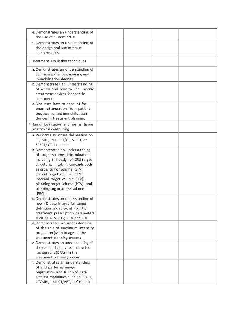
techniques) and their relative advantages and disadvantages
b. Explains the differences between dynamic multileaf collimator (DMLC) and segmental multileaf collimator (SMLC) leaf sequencing algorithms in terms of delivery parameters and dose distributions
c. Participates in IMRT or VMAT delivery for patients with a variety of treatment sites and demonstrates an understanding of the techniques and requirements for patient setup, immobilization, and localization.
Monitor Unit (MU) Calculations
1. Demonstrates an understanding and performs derivation of the following factors:
a. Percent depth dose (PDD)
b. Tissue-air ratio (TAR)
c. Tissue-maximum ratio (TMR)
d. Tissue-phantom ratio (TPR)
e. Scatter factors (i.e., Sc, Sp, Scp)
f. Off-axis factors
g. Inverse square factors
h. Calibration factor (monitor unit [MU] reference conditions)
i. Standard wedge factors
j. Virtual and dynamic wedge factors
k. Compensator factors
l. Tray and other insert factors
2. Performs manual MU calculations for photon or electron beams of the following configurations:
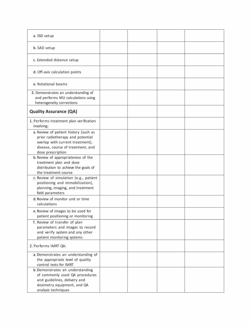
a. SSD setup
b. SAD setup
c. Extended distance setup
d. Off-axis calculation points
e. Rotational beams
3. Demonstrates an understanding of and performs MU calculations using heterogeneity corrections
Quality Assurance (QA)
1. Performs treatment plan verification involving:
a. Review of patient history (such as prior radiotherapy and potential overlap with current treatment), disease, course of treatment, and dose prescription
b. Review of appropriateness of the treatment plan and dose distribution to achieve the goals of the treatment course
c. Review of simulation (e.g., patient positioning and immobilization), planning, imaging, and treatment field parameters
d. Review of monitor unit or time calculations
e. Review of images to be used for patient positioning or monitoring
f. Review of transfer of plan parameters and images to record and verify system and any other patient monitoring systems
2. Performs IMRT QA:
a. Demonstrates an understanding of the appropriate level of quality control tests for IMRT
b. Demonstrates an understanding of commonly used QA procedures and guidelines, delivery and dosimetry equipment, and QA analysis techniques
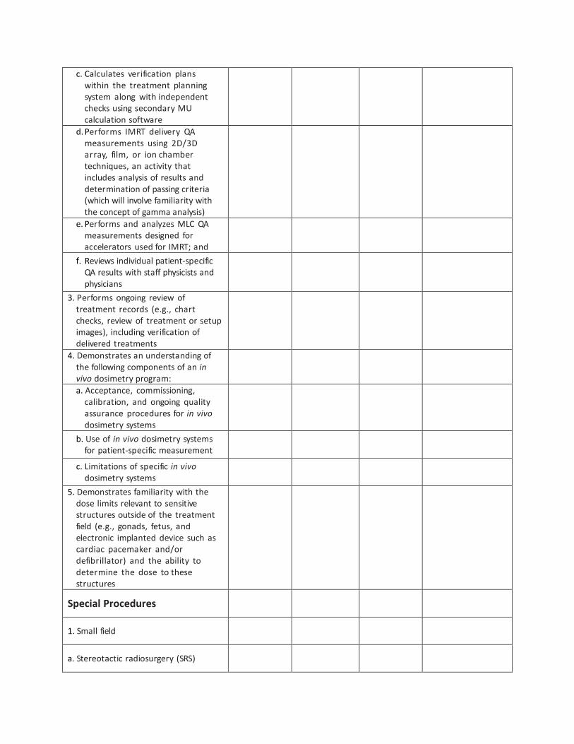
c. Calculates verification plans within the treatment planning system along with independent checks using secondary MU calculation software
d. Performs IMRT delivery QA measurements using 2D/3D array, film, or ion chamber techniques, an activity that includes analysis of results and determination of passing criteria (which will involve familiarity with the concept of gamma analysis)
e. Performs and analyzes MLC QA measurements designed for accelerators used for IMRT; and
f. Reviews individual patient-specific QA results with staff physicists and physicians
3. Performs ongoing review of treatment records (e.g., chart checks, review of treatment or setup images), including verification of delivered treatments
4. Demonstrates an understanding of the following components of an in vivo dosimetry program:
a. Acceptance, commissioning, calibration, and ongoing quality assurance procedures for in vivo dosimetry systems
b. Use of in vivo dosimetry systems for patient-specific measurement
c. Limitations of specific in vivo dosimetry systems
5. Demonstrates familiarity with the dose limits relevant to sensitive structures outside of the treatment field (e.g., gonads, fetus, and electronic implanted device such as cardiac pacemaker and/or defibrillator) and the ability to determine the dose to these structures
Special Procedures
1. Small field
a. Stereotactic radiosurgery (SRS)

i. Explains rationales for SRS treatments, examples of malignant and non- malignant lesions treated with SRS, and typical dose and fractionation schemes for linac-based and Co-60 SRS techniques
ii. Describes in general terms the components of commissioning an SRS sys- tem (e.g., accurate localization, mechanical precision, accurate and optimal dose distribution, and patient safety)
iii. E x p l a i n s the stereotactic localization of a target (e.g., on the basis of angiography as opposed to CT and MRI) and how the accuracy of this localization is measured
iv. Describes the alignment of coordinate systems (e.g., target frame of reference with linac frame of reference) and how the mechanical precision of this alignment is measured
v. Describes issues associated with dosimetry measurements for an SRS system (e.g., choice of dosimeter, phantom geometry, etc.)
vi. Describes the components of pre-treatment QA for an SRS system, including linac-based and Co-60 SRS techniques
b. Stereotactic body radiation therapy (SBRT)
i. Explains the rationale for SBRT treatments, common treatment sites, and typical dose and fractionation schemes
ii. Describes immobilization and localization systems for SBRT treatments
iii. Describes the use of simulation imaging for SBRT target definition, including multi-modality imaging and 4D imaging for cases requiring motion management
iv. Describes treatment planning objectives for SBRT treatments, including dose limits, dose heterogeneity, dose gradient and fall-off, and beam geometry
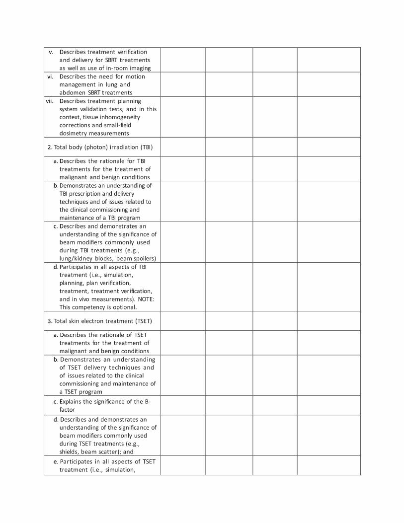
v. Describes treatment verification and delivery for SBRT treatments as well as use of in-room imaging
vi. Describes the need for motion management in lung and abdomen SBRT treatments
vii. Describes treatment planning system validation tests, and in this context, tissue inhomogeneity corrections and small-field dosimetry measurements
2. Total body (photon) irradiation (TBI)
a. Describes the rationale for TBI treatments for the treatment of malignant and benign conditions
b. Demonstrates an understanding of TBI prescription and delivery techniques and of issues related to the clinical commissioning and maintenance of a TBI program
c. Describes and demonstrates an understanding of the significance of beam modifiers commonly used during TBI treatments (e.g., lung/kidney blocks, beam spoilers)
d. Participates in all aspects of TBI treatment (i.e., simulation, planning, plan verification, treatment, treatment verification, and in vivo measurements). NOTE: This competency is optional.
3. Total skin electron treatment (TSET)
a. Describes the rationale of TSET treatments for the treatment of malignant and benign conditions
b. Demonstrates an understanding of TSET delivery techniques and of issues related to the clinical commissioning and maintenance of a TSET program
c. Explains the significance of the B-factor
d. Describes and demonstrates an understanding of the significance of beam modifiers commonly used during TSET treatments (e.g., shields, beam scatter); and
e. Participates in all aspects of TSET treatment (i.e., simulation,

planning, plan verification, treatment, treatment verification, and in vivo measurements). NOTE: This competency is optional.
4. Respiratory-correlated planning and delivery
a. Describes the rationale for using respiratory management systems in radiation therapy
b. Describes the common issues introduced by respiratory motion in imaging, planning, and treatment delivery
c. Describes common treatment sites affected by respiratory motion and the typical range of tumor excursion
d. Describes methods for evaluating and managing respiratory motion
e. Describes QA tests for common respiratory management systems and their recommended frequency
Treatment Planning Workstations
1. Data acquisition
a. Explains the connection between linac commissioning and the data required for operation of a treatment planning system
b. For a particular treatment planning system, describes the linac data needed for:
i. Photon beams
ii. Electron beams
iii. IMRT and VMAT
2. Acceptance testing
a. Describes what tests of the treatment planning system need to be performed before patient-specific planning can commence for:
i. Photon beams

ii. Electron beams, and
iii. Brachytherapy sources
3. Quality assurance
a. Describes the tests that need to be performed and their accuracy
b. Describes accuracy checks for the following input devices and types of images:
i. Digitizers
ii. Film scanners
iii. Imported images from instruments such as CT scanners, MRI scanners, and picture archiving and communication (PAC) systems
c. Describes accuracy checks for the following output devices:
i. Printers
ii. Record and verify systems
iii. DICOM output
4. Computer algorithms (models)
a. Describes how the computer algorithm calculates dose for at least one major treatment planning system with regard to:
i. Photon beams
ii. Electron beams
iii. Brachytherapy calculations, and
iv. Proton beams (Optional)
b. Describes the advantages and disadvantages of the various treatment planning calculation algorithms

c. Describes how the computer algorithm determines the number of monitor units per beam or segment (for step-and-shoot IMRT)
5. Plan normalization
a. Describes the numerous normalization capabilities available on a treatment planning system
b. Describes how different normalization schemes affect final isodose curve representation
c. Describes how the computer plan normalization relates to the calculation of monitor units for patient treatments
6. Inhomogeneity (heterogeneity) corrections
a. Describes the type of data that need to be taken on a CT scanner in preparation for treatment planning using inhomogeneous material
b. Describes how these CT data are converted into inhomogeneity data usable in a treatment planning system
c. Describes how computerized treatment planning systems take inhomogeneities into account
d. Identifies where the computer algorithm calculates dose with acceptable accuracy and in which regions calculational accuracy is suspect
e. Describes how the accuracy of the inhomogeneity corrections performed by a treatment planning system would be checked
7. Beam modeling
a. Completely models at least one photon beam energy for a treatment planning system
b. Completely models at least one electron beam energy for a treatment planning system
c. Completely models at least one proton beam energy for a treatment planning system (optional)

d. Tests the accuracy of his or her modeling for the beams and is able to describe the criteria for acceptability of the modeling
8. Imaging tests
a. Describes the tests that would be performed to ensure that the imported image data are correct
b. Demonstrates that images can be imported from CT, MR, and PET or PET/CT scanners
c. Demonstrates that the above imaging sets can be accurately fused with the primary treatment planning image set
d. Describes the different image fusion algorithms available on a treatment-planning system (e.g., CT-CT, CT-MR, CT-PET)
9. Secondary monitor unit check computer programs
a. Describes what input data need to be acquired
b. Describes the checks of that input data that need to be performed to ensure that the monitor unit check program is working correctly
c. Describes how imported data from treatment-planning systems are handled in a monitor unit check program
d. Describes how the monitor unit check program calculates the number of monitor units for off central-axis normalization points
e. Describes how the monitor unit check program calculates monitor units for treatments involving inhomogeneous material
Patient Safety
1. General
a. Understands the principles behind the development of a general patient and staff safety management program within the hospital

b. Describes the physicist’s role in developing and overseeing an overall quality assurance program for both equipment and procedures, including a discussion of allocation and management of resources necessary to carry out these tasks, incorporation of tools and techniques into these tasks, and inclusion of various groups within the structure of the radiation oncology department
c. Describes the principles and rationale of TJC Universal Protocol as well as the use of pre-procedure verification and time-outs for the prevention of treatment errors
d. Describes internal, voluntary, and mandatory incident reporting systems and the role of root cause analysis (RCA) as a tool for continuous quality improvement
e. Describes the concept of a failure mode and effect analysis (FMEA), design and implementation of an FMEA, and how to use the results of such an analysis to prevent errors and minimize risks to patients and staff
f. Describes charting systems for the prescription, delivery, and recording of treatment information, standardization of such systems, and the use of such systems within a record and verify electronic medical record system
g. Describes mechanisms for independent checking of treatment information
2. Equipment
a. Describes the implementation of an effective set of equipment operating procedures that would include preventative maintenance and repair, keeping of maintenance and repair records, emergency procedures, and systematic inspection of interlock systems

b. Describes the development of a program to prevent mechanical injury caused by the machine or accessory equipment, with consideration of the need for visual and audio contact with the patient while the patient is under treatment
c. Understands potential patient safety hazards related to the use of blocks, block trays, wedges, and other ancillary treatment devices and accessories as well as mechanisms to minimize these risks
d. Understands potential patient safety hazards posed by patient support and immobilization systems, as well as mechanisms to minimize these risks
e. Understands potential patient safety hazards of gantry–patient collision as well as mechanisms to minimize this risk
3. Other patient/staff safety issues
a. Understands potential electrical hazards affecting patients and staff
b. Understands the potential hazards to patients and staff posed by strong magnetic fields
c. Understands the mechanisms of ozone production and related potential hazards to patients and staff
d. Understands potential hazards to patients and staff arising from the use of cerrobend