Radiation Biology: A Handbook for Teachers and...
138
Radiation Biology: A Handbook for Teachers and Students Slide Series prepared in 2011 by J.H. Hendry. The IAEA officer responsible for this publication is J. Wondergem of the Division of Human Health, International Atomic Energy Agency
Transcript of Radiation Biology: A Handbook for Teachers and...

Radiation Biology: A Handbook for Teachers and
Students
Slide Series prepared in 2011 by
J.H. Hendry. The IAEA officer responsible
for this publication is J. Wondergem of the
Division of Human Health,
International Atomic Energy Agency

Section 2. Sources of Additional Illustrative Material and Slides • EJ Hall and A Giaccia. Radiobiology for the Radiologist: 6th edition.
(2006). JB Lippincott, Philadelphia, USA. Figures and Tables. • VAN DER KOGEL, A.J., JOINER, M.C., Editors, Basic Clinical Radiobiology:
4th edition. (2009). Hodder Arnold, London, UK. Figures and Tables. • TANNOCK, I.F., HILL, R.P., BRISTOW, R.G., HARRINGTON, L., Editors, The
Basic Science of Oncology, 4th edition. (2005). McGraw-Hill. Figures and Tables.
• http://www.iaea.org/Publications/Training/Aso/register.html IAEA slide series of Modules in Radiobiology.
• http://lowdose.energy.gov/radiobio_slideshow.aspx DOE slide sets on Low Dose effects.
• http://www.cancer.gov/search/results NCI slide sets on Understanding Cancer.
Radiation Biology Handbook 2

2.2 Physics and Chemistry of Radiation: Interactions with
Matter
3 Radiation Biology Handbook
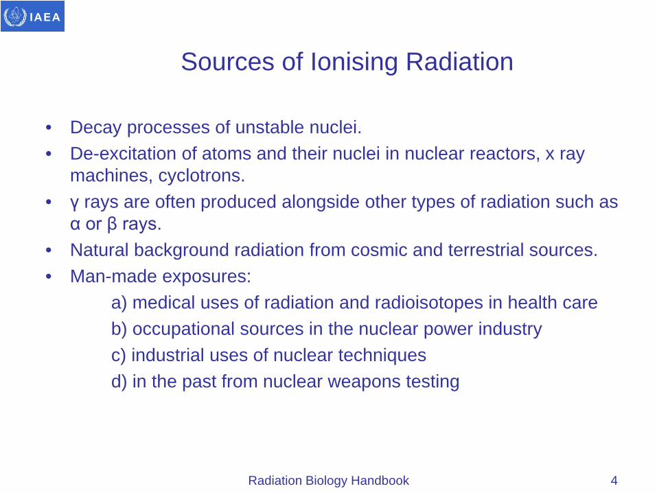
Sources of Ionising Radiation
4
• Decay processes of unstable nuclei. • De-excitation of atoms and their nuclei in nuclear reactors, x ray
machines, cyclotrons. • γ rays are often produced alongside other types of radiation such as
α or β rays. • Natural background radiation from cosmic and terrestrial sources. • Man-made exposures: a) medical uses of radiation and radioisotopes in health care b) occupational sources in the nuclear power industry c) industrial uses of nuclear techniques d) in the past from nuclear weapons testing
Radiation Biology Handbook
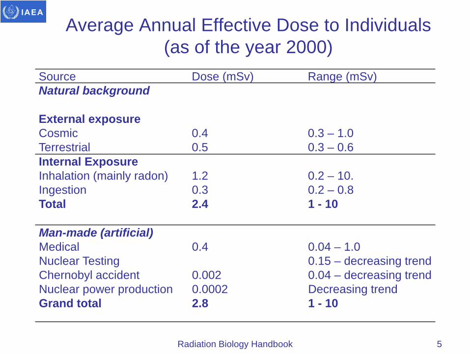
Average Annual Effective Dose to Individuals (as of the year 2000)
5
Source Dose (mSv) Range (mSv) Natural background
External exposure Cosmic Terrestrial
0.4 0.5
0.3 – 1.0 0.3 – 0.6
Internal Exposure Inhalation (mainly radon) Ingestion Total
1.2 0.3 2.4
0.2 – 10. 0.2 – 0.8 1 - 10
Man-made (artificial) Medical Nuclear Testing Chernobyl accident Nuclear power production Grand total
0.4 0.002 0.0002 2.8
0.04 – 1.0 0.15 – decreasing trend 0.04 – decreasing trend Decreasing trend 1 - 10
Radiation Biology Handbook

Types of Ionizing Radiation
• Directly ionising charged particles e.g. electrons, protons, ions, α particles.
• Indirectly ionising particles e.g. neutrons which are uncharged, collisions release charged particles.
• Indirectly ionising electromagnetic radiations e.g. X and γ ray photons, producing secondary electrons (charged particles) after energy absorption in the material.
6 Radiation Biology Handbook

Electromagnetic Radiation (1)
• Radiowaves, microwaves, visible light, ultra violet light, X rays, γ rays.
• Energy varies inversely with the wavelength.
• Photon energy progressively increases from radiowaves with least energy to X and γ rays with highest energy.
• X and γ ray photons can eject an electron from its orbit in an atom, and are ionizing radiations.
7 Radiation Biology Handbook
Electromagnetic Radiation (2)
• Ionization is the process of removing one or more electrons from atoms by the incident radiation leaving behind electrically charged particles (an electron and a positively charged ion).
• The ionized or excited atom or molecule may either fragment producing free radicals or return to the parent state.
• If the energy transferred by ionizing radiation to the atom is insufficient to eject orbital electrons, the electrons may be raised from lower to higher orbitals and the atom is said to be excited.
8 Radiation Biology Handbook
Electromagnetic Radiation (3)
Non-ionising radiations: • include cellular phones, radar, infrared, radiowaves, microwaves,
visible light, ultrasound. • fall short of the energy required to remove an electron from an
atom. • because of the longer wavelengths and, therefore, smaller
energy per quanta, they are not known to cause significant chemical changes in atoms or molecules of the medium.
• considered virtually harmless to biological tissues at levels below those that cause heating effects.
• the exact demarcation between ionizing and non-ionizing radiation parts of the spectrum is somewhat arbitrary.
9 Radiation Biology Handbook

The Electromagnetic Spectrum
10
Hall and Giaccia, 2006
Radiation Biology Handbook
Interactions of Electromagnetic Radiation (1)
• Quantum theory considers electromagnetic radiation as streams of packets/bundles of energy called photons.
• The energy of a photon of electromagnetic radiation is given by Planck’s equation, where
E = hν = hc/ λ
E is the energy of the photon, h is Planck’s constant, and ν is the frequency of the photon.
11 Radiation Biology Handbook

Interactions of Electromagnetic Radiation (2)
• The energy of a photon is directly related to its frequency and inversely to wavelength, λ.
• Wave velocity is obtained by the product of frequency and wavelength, c = λν, where c is the velocity of light.
• Photons passing through matter transfer their energy through the following three main processes: photoelectric absorption, Compton scattering, and pair production.
12 Radiation Biology Handbook

Dominant Types of Interactions vs the Atomic Number Z of the Absorber and the Photon Energy
13
Podgorsak, 2005
Radiation Biology Handbook

Photoelectric Absorption (1)
• The photon interacts with a bound inner shell electron in the atom of the absorbing medium and transfers its entire energy to the electron, ejecting it from the occupied atomic shell.
• The kinetic energy of the ejected photoelectron equals the energy of the incident photon minus the binding energy of the electron
Kinetic Energy (electron) = hν – E b
where hν is the energy of the incident photon, and Eb is the binding energy of the electron.
14 Radiation Biology Handbook

Photoelectric Absorption (2)
• An atom that participated in photoelectric interaction is left ionized. The vacancy created due to ejection of the electron is instantly filled by an electron from an outer orbital of the same atom, emitting the balance of energy as a photon between the respective orbits with characteristic low energy.
• The photoelectric effect is the dominant energy transfer mechanism for X and γ ray photons having energies below 50 keV in biological tissues, but it is much less important at higher energies.
15 Radiation Biology Handbook

Compton Scattering
• The Compton Effect occurs when the incident photon interacts with the outer orbital electron whose binding energy is very low compared with that of the incident photon.
• The incident photon transfers energy to an atomic electron causing its ejection from the atom.
• Compton scatter thus causes ionization of the absorbing atom due to loss of an electron.
• The probability of Compton scattering decreases with increasing photon energy. It is the principal absorption mechanism for X and γ rays in the intermediate energy range of 100 keV to 10 MeV.
• 100 keV = 1.602 x 10 –14 Joules; 10 MeV = 1.602 x 10 –12 Joules.
16 Radiation Biology Handbook
Pair Production
• When a photon of high energy ( >1.02 MeV) interacts with atoms of the medium, the incident photon can be spontaneously converted into the mass of an electron and positron pair by interaction of the Coulomb force in the vicinity of the nucleus.
• A positron is the anti-matter equivalent of an electron and it has the same mass as an electron, but it has a positive charge equal in strength to the negative charge of an electron.
• The energy of the interacting photon in excess of the equivalent rest mass of the two particles (1.02 MeV) appears as the kinetic energy of the pair and the recoil nucleus.
• The entire mass of these two particles is then converted into two γ photons each of 0.51 MeV energy emitted in opposite directions.
17 Radiation Biology Handbook

Dependence of Absorption on Atomic Number
• The radiation energy deposition depends on the energy of the radiation and the atomic number (Z) of the absorbing material.
• The mass absorption coefficient of photoelectric absorption varies directly with the third power of the atomic number of the absorber (Z3).
• The effective atomic number of bone is about twice that of soft tissues, and the probability that a photon will be absorbed in bone is about six times that in an equal thickness of soft tissues.
• The mass absorption coefficient for the Compton process is nearly independent of atomic number.
• In radiotherapy, high-energy photons in the range of 1-10 MeV are preferred because absorbed dose is nearly the same in bone and soft issues, whereas low energy photons are preferred in diagnosis because of the much desired large contrast in absorption of these tissues.
18 Radiation Biology Handbook

Half Value Layer
• The thickness of absorber that reduces the photon intensity (mainly due to photoelectric absorption and Compton scattering processes) to one half is called the half value layer (HVL).
• There is an exponential decrease in intensity with an increase in the thickness represented by:
I (x) = I0 . e- µx
• where I (x) = the intensity at thickness x, I0 = is the initial intensity on the surface of the absorber, μ = n×σ is the absorption coefficient measured in cm−1, n = the number of atoms per cm3 in the material, σ = the absorption cross section in cm2, and x = the thickness of material in cm.
• Low energy photons are much more likely to be absorbed than high energy photons, for example the first 1.5 cm of water absorbs 40 % of 50 kVp X rays.
19 Radiation Biology Handbook

Charged Elementary Particles
• Protons with one unit mass and one positive charge, cause less damage than α particles (helium nuclei) because the rate of deposition of energy varies inversely in proportion to the velocity of the particle and directly in proportion to the square of the charge.
• At the same energy, α particles have lower velocity because of their higher mass and carry twice the charge of a proton.
• High energy electrons ionize much less efficiently than α particles because of their lower mass (and resulting higher velocity) and lower charge. Therefore, they penetrate tissues to a greater depth than α particles.
• Generally, beta particles do not penetrate deeper than the skin of the human body.
20 Radiation Biology Handbook
Uncharged Particles (1)
• Neutrons (n) are uncharged particles with a mass very similar to that of a proton and are an indirectly ionizing radiation because without a charge they cannot participate in electrostatic interactions.
• Neutrons interact with the atomic nuclei of the medium and they lose energy by different interaction processes depending on their energy (velocity) and the mass of the encountered nucleus.
• In soft tissues, because of the abundance of protons with mass equal to that of neutrons, fast neutrons (>1 MeV) mostly lose energy by elastic scattering through collision processes producing high energy recoil protons, which in turn deposit energy by electrostatic interactions with electrons in the tissue.
• Neutrons begin to interact by inelastic scattering at energies above 6 MeV, and fast neutrons may interact with carbon and oxygen nuclei producing α particles, recoil protons and heavy nuclear particles.
21 Radiation Biology Handbook

Uncharged Particles (2)
• Fast neutrons can be made into thermal neutrons via a process called moderation. In reactors, typically heavy water, light water, or graphite are used to moderate neutrons.
• Thermal neutrons have a much larger effective cross-section than fast neutrons, and, therefore, can be absorbed more easily by any atomic nuclei with which they collide, creating a heavier and often unstable isotope of the irradiated element.
• Most fission reactors use a neutron moderator to slow down, or thermalize the neutrons that are emitted by nuclear fission so that they are more easily captured, causing further fission.
• This ability of neutrons to produce radioactive nuclei (neutron activation), which then produce ionizing radiation by their decay, can be used to analyse the atomic composition of certain materials.
22 Radiation Biology Handbook
Ions
• The nuclei of carbon, neon, silicon, argon atoms form charged ions when one or more orbital electrons have been stripped off.
• Ions can be accelerated to hundreds of MeV in special accelerator facilities.
• High energy charged ions offer special advantages in cancer radiotherapy because of the energy distribution along their track which has a high peak at its end (the Bragg peak).
• This peak allows the possibility of depositing high energy densities at depth in tissue, but these facilities are as yet very limited on account of high costs and sophisticated technical requirements.
23 Radiation Biology Handbook

Linear Energy Transfer
• The Linear Energy Transfer (LET) of a radiation type is defined as the average energy deposited per unit length of track of radiation, and the unit is keV/μm.
• For low LET radiations the energy deposition events along the track of the photon are sparse relative to the dimensions of biomolecules such as DNA with the result that photons may pass through such a molecule without depositing any energy.
• The rate of transferring energy (-dE/dX, loss of energy per unit distance) increases as a charged particle slows down, such that there is a peak of energy deposition at the end of the track (the Bragg peak).
• In general the RBE of a radiation increases with its LET up to a value of about 100 keV/μm, and above this value RBE starts to decline due to energy deposition in excess of that needed to cause the biological effect (overkill).
24 Radiation Biology Handbook
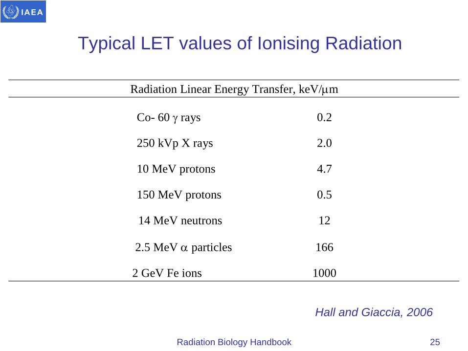
Typical LET values of Ionising Radiation
25
Hall and Giaccia, 2006
Radiation Biology Handbook
Radiation Linear Energy Transfer, keV/µm
Co- 60 γ rays 0.2
250 kVp X rays 2.0
10 MeV protons 4.7
150 MeV protons 0.5
14 MeV neutrons 12
2.5 MeV α particles 166
2 GeV Fe ions 1000

Radiation Dose and Units (1)
• The radiation exposure is a measure of radiation based on its ability to produce ionization in air under standard temperature and pressure, and is the quantity indicated by many radiation detectors such as ionization (e.g. Geiger-Muller) chambers. The (S.I.) unit for exposure is Coulombs/kg in air (or Roentgen R in old units: 1 R = 2.58 x 10⁻⁴ C/kg air).
• Radiation dose is the energy (Joules) absorbed per unit mass of tissue and has the (S.I.) units of gray (1 Gy = 1 J/kg). In the past the rad (radiation absorbed dose) was used, where 100 rad = 1 Gy (1 rad = 1 cGy).
26 Radiation Biology Handbook

Radiation Doses and Units (2)
27
Dose SI Unit Old unit Conversion factor Exposure C/kg air Roentgen 1 R = 2.58 x 10⁻⁴ C/kg air Absorbed dose gray (Gy) rad 100 rad = 1 Gy Equivalent dose sievert (Sv) rem 100 rem = 1 Sv
Radiation Biology Handbook
Radiation Doses and Units (3)
• For radiation protection purposes the term ‘equivalent dose’ is used to compare the effectiveness of different types of radiation in tissues. The (S.I.) dose equivalent (HT) in sievert (Sv) is the product of the absorbed dose (DT) in the tissue multiplied by a radiation weighting factor (WR), often called the quality factor.
• Effective Dose is used to estimate the risk of radiation in humans. It is sum of the products of equivalent doses to each organ/tissue (HT) and the tissue weighting factor (WT). The unit of effective dose is the Sievert (Sv).
E =∑ W T x HT
• Collective dose is defined as the dose received per person in Sv multiplied by the number of persons exposed per year i.e. man-sievert per year. This unit is generally used for protection purposes and in population response calculations.
28 Radiation Biology Handbook

Principles of Radiation Dosimetry
• Detectors can be divided broadly into three categories: those that measure directly the quantity of energy absorbed, detectors that measure ionization and those that quantify free radicals formed in the absorbing medium.
• Secondary chemical dosimeters are widely used commercially and have proved beneficial to clinical and scientific communities for both research and applications in photon radiation dosimetry.
• Among the most popular dosimeters are the Fricke chemical dosimeter, thermo-luminescence dosimeters (TLD) and ion chambers or diode dosimeters.
• The fundamental requirement for a suitable dosimeter is the linearity of response as a function of radiation dose within a wide dosage range.
29 Radiation Biology Handbook

Chemical Dosimeters
• The Fricke chemical dosimeter is based on chemical change by absorption of radiation and used to measure, X, γ and electron doses.
• The principle consists of the chemical change of ferrous ions (Fe +2) into ferric ions (Fe +3) by absorption of radiation energy.
• Measurement is accomplished by optical absorption of ferric ions, which has a high extinction coefficient allowing determination of concentration changes.
• The measurements are highly linear with increasing dose up to more than 150 Gy.
30 Radiation Biology Handbook

Thermoluminescence Dosimeters (TLD)
• Thermoluminescence is based on generation of trapped electrons by exposure of lithium fluoride to radiation.
• The method consists of measuring the luminescence induced by thermal treatment after radiation exposure. The light emitted is proportional to radiation dose.
• Lithium fluoride chips provide good spatial information but require careful calibration and rather laborious read-out. In addition, TLD are oxygen sensitive which imposes a limitation.
• The method is not as cost effective as the Fricke dosimeter, it lacks ease of preparation and the measurements become nonlinear at absorbed doses above 10 Gy.
31 Radiation Biology Handbook

Ionisation Chambers
• Ionization chambers consist of an air-filled chamber containing two electrodes to which a voltage is applied.
• They measure the current flow which occurs due to the ionization of the air molecules exposed to radiation.
• They are capable of giving instant readings with good accuracy. The chambers are easy to use but are poor in providing spatial information.
• Diode dosimeters are based on the principle of ion collection formed by radiation incident in the chamber.
• Measurement consists of collection of ions on the cathode, formed by exposure to radiation, but this technique requires intricate circuitry and is not cost effective.
32 Radiation Biology Handbook
Film Dosimetry
• Special radiographic films have been developed for verification of dose in radiotherapy practice.
• This has proved useful for measuring dose profiles but the method has limited accuracy and dose range for determination of absolute radiation doses.
33 Radiation Biology Handbook

Direct and Indirect Effects (1)
• The physical interactions of ionizing radiation leads to loss of energy of radiation and production of ionization and excitation of atoms and molecules which may convert into free radicals in pico to femto seconds after physical interaction with atoms (10-13 to -15 s).
• These radicals react with neighbouring molecules and produce secondary DNA or lipid radicals by reaction with another neighbouring molecule.
• Free radicals are fragments of molecules having unpaired electrons, which have high reactivity with cellular molecules and, therefore, have a short life.
• Free radicals can be detected by fast measuring techniques like pulse radiolysis and flow electron spin resonance (ESR).
34 Radiation Biology Handbook

Direct and Indirect Effects (2)
• Free radicals are highly reactive and are found in a number of biological processes: metabolism, oxidation, reduction, and pathological diseases and cancer induction.
• Both electromagnetic and particulate radiations act on cells to cause free radicals and subsequent molecular damage through direct as well as indirect actions.
• When ionizing radiation energy is deposited in a macromolecule that is important for the biological effect observed (often DNA for cell killing), it is called a direct effect of radiation.
• Alternatively, photons may be absorbed in the water of an organism causing excitation and ionization in the water molecules.
35 Radiation Biology Handbook

Direct and Indirect Effects (3)
• The radicals formed after passage of radiation and water radiolysis, namely the hydrated electron (eaq
-), the hydrogen atom (H.) and the hydroxyl radical (.OH) contribute in causing damage to biological systems.
• A compound with a high rate constant of reaction can scavenge primary free radicals of water radiolysis. Free radicals of biomolecules can be restituted by hydrogen donating compounds, such as thiols and cysteine.
• Alternatively, they can be fixed by reaction with oxygen or oxygen mimicking compounds, which makes them permanently damaged.
• These chemical reactions form the basis of searching for compounds which can sensitize cell/tissue damage or protect them against radiation, and which are of direct relevance to radioprotection and cancer radiotherapy.
36 Radiation Biology Handbook

Direct Effects
• Ionizing radiation (IR) can act on biological molecules (RH, representative of hydrocarbons) causing ionization and excitation. One or more chemical bonds may be broken giving atoms or molecules with unpaired electrons, which are very reactive and have a short life:
IR + RH → R•+H• • Both H· and R· radicals can react with another molecule e.g. DNA,
lipids, proteins:
R•+ R’H → R’·+ RH
• Radicals can produce cross linking reactions:
R•+ R· →R• - R·
• It is estimated that about one third of biological damage by γ radiation is caused by direct effects. This process becomes more dominant with high LET radiation, such as neutrons or α particles.
37 Radiation Biology Handbook

Indirect Effects - Water Radiolysis (1)
• Interaction of radiation with water causes ionization and excitation process producing short-lived H2O+ radical-cations, fast electrons, and electronically-excited water molecules (H2O+).
• H2O+ ions and excited water molecules are unstable and decompose within 10-13 s to form OH• and H• radicals:
IR + H2O → H2O+ + e-
H2O + H2O+→ H3O+ +OH•
IR + H2O → H2O*→ H2O + photon emitted
or
H2O*→ OH•+ H•
38 Radiation Biology Handbook
Indirect Effects - Water Radiolysis (2)
• The hydroxyl radical has an unpaired electron and is a highly reactive oxidizing agent. It can diffuse a short distance and react with critical target molecules producing another radical.
• This can react with water forming an anion which rapidly dissociates to give a hydrogen atom (H·).
• The ejected secondary electrons may interact with a water molecule to form hydroxyl ions and a hydrogen atom (a hydrogen radical), or they may lose energy by a sequence of interactions with the medium until they attain thermal energies after about 10-11 s.
• The thermalized electrons are then solvated by dielectric interactions with neighbouring water molecules to form e-
aq i.e. e-aq is
a free electron in a solvent cavity surrounded by a sheath of orientated water dipoles.
39 Radiation Biology Handbook

Indirect Effects - Water Radiolysis (3)
• The aqueous electron reacts with a proton to give a hydrogen atom (H·):
e- + H2O→ H2O-→ OH- + H•
e-aq + H+ → H•
• e-aq is the strongest known reducing species at pH 7.0. In oxygenated
solutions, e-aq is converted to O2
-, which is a strong oxidizing agent and the precursor of hydrogen peroxide:
e-aq+ O2 → O2
-
• These primary water radicals (eaq, OH, H•) have high reactivity towards molecules of cells, DNA, lipids and other subcellular constituents. In oxygenated solutions, hydrogen atoms can react with oxygen to give hydroperoxyl free radicals (HO2
•):
H• + O2 → HO2•
40 Radiation Biology Handbook

Indirect Effects - Water Radiolysis (4)
• The relative yields of the water radiolysis products depend on the pH and LET of the radiation.
• The concentration of these radicals are expressed in terms of a G value, which is defined as the number of radicals or molecules produced per 100 eV of energy absorbed in the medium.
• Typical G-values are Ge-aq = 2.6, GOH·= 2.6, GH·= 0.6.
41 Radiation Biology Handbook

Free Radical Scavengers (1)
• Certain compounds with a high rate constant of reaction may scavenge the primary radicals of water radiolysis (e.g. dimethylsulphoxide).
• Hydroxyl radicals can also be scavenged by a number of –SH containing compounds as a moiety in their chemical structure.
• The hydrated electron can be efficiently scavenged by oxygen producing a number of oxygen-centered radicals.
42 Radiation Biology Handbook

Free Radical Scavengers (2)
• Scavenging of hydroxyl radicals forms one basis for development of radioprotectors.
• Thiol compounds may also donate hydrogen atoms to radical sites on other biological molecules such as DNA but scavengers act primarily against the indirect effect induced by water radicals.
• Hence they have reduced efficacy for high LET radiation for which the direct effect plays a more prominent role in biological damage such as cell killing.
43 Radiation Biology Handbook

2.3 Molecular and Cellular Radiobiology
44 Radiation Biology Handbook

Sources of Ionising Radiation
45
• Radiation causes a wide range of lesions in DNA such as single strand breaks in the phosphodiester linkage, double strand breaks on opposing sites or displaced, base damage, protein-DNA crosslinks and protein-protein crosslinks involving nuclear proteins such as histones and non-histone proteins.
• The numbers of lesions induced in the DNA of a cell by a dose of 1-2 Gy are approximately: base damages > 1000; single strand breaks (ssb) ~1000; double strand breaks (dsb) ~40.
• Dsb play a critical role in cell killing, and there are experimental data showing initially-produced dsb correlate with radiosensitivity and survival at low dose, and unrepaired or mis-repaired dsb to correlate with survival after higher doses.
• Knowledge of radiation track structure has been used to explain the wide variation and wide distribution of lesions in DNA.
Radiation Biology Handbook

Major Types of DNA Repair (1)
• For double strand breaks there are two primary repair pathways, non-homologous end joining (NHEJ) and homologous recombination (HR).
• NHEJ repair operates on blunt ended DNA fragments resulting from broken phosphodiester linkages.
• There is a requirement for Ku70/Ku80 repair proteins to recognize the lesion termini, binding of the Ku-heterodimer to DNA-PK (protein kinase), and activation of the XRCC4 ligase enzyme by this complex for final religation of the fragments after enzymatic “cleaning up” of the broken ends of the DNA molecule, by a variety of other recruited proteins, so that ligation can occur.
• Repair by NHEJ operates throughout the cell cycle but dominates in G1/S-phases. The process is error prone because it does not rely on sequence homology.
46 Radiation Biology Handbook
Major Types of DNA Repair (2)
• Dsb repair by homologous recombination (HR) utilizes sequence homology with an undamaged copy of the broken region and hence can only operate in late S- or G2- phases of the cell cycle.
• HR starts by nucleolytic resection of blunt ends, binding of NBS/MRE11/rad50 protein complex to the DNA termini, followed by strand exchange facilitated by attachment of rad51/XRCC2 protein.
• Then there is DNA synthesis of the missing nucleotides on the undamaged templates and ligation. This creates a complex strand crossover between the damaged and undamaged strands known as a Holliday junction, which is finally resolved before the repair process is complete.
• Other DNA repair mechanisms such as base excision repair (BER), mismatch repair (MR) and nucleotide excision repair (NER) respond to damage such a base oxidation, alkylation, and strand intercalation.
47 Radiation Biology Handbook

Damage recognition and signalling (1)
• A first step in recognition of radiation damage (strand breaks) to DNA is ATM binding to DNA termini.
• This binding induces kinase activity in ATM which phosphorylates and activates the CHK kinases, which in turn phosphorylate p53.
• As a result p53 is released from MDM-2 and is stabilized to induce p21, which inhibits the cyclin-dependent kinase cyclinE-CDC-2 controlling the G1-S transition in the cell cycle.
• The resultant G1 arrest (G1 block) after irradiation ensures that the damaged DNA is not replicated before repair.
48 Radiation Biology Handbook

Damage recognition and signalling (2)
• Tumours showing mutant p53 or p53 null status, as the result of p53 destruction by viral protein E6, fail to initiate a G1 arrest and may not restitute damaged DNA before replication.
• But even p53 mutant cells display a G2 arrest and may exercise repair options (or induce apoptosis) and thus prevent mitotic propagation of defective DNA in M phase.
• Repair signalling starting at ATM proceeds via downstream activation of BRCA1, c-Abl, NBS1 and RAD 51 to initiate DNA repair. An alternative response to DNA damage is induction of apoptosis initiated by p53, although this occurs extensively after irradiation only in a few specific cell types.
• C-Abl, BID and the proapoptotic factor BAX (in the Bcl-2 family of proteins) respond to sequential phosphorylation cascades starting with ATM.
49 Radiation Biology Handbook

Consequences of unrepaired DNA damage: chromosome damage (1)
• Mutations from low dose exposure influence base pairing, coding, transcription and gene expression.
• Chromosome analysis in mitotic spreads (karyotyping), micronucleus formation and fluorescent in situ hybridisation (FISH) can detect unrepaired DNA damage in chromatids by a variety of DNA damaging agents including radiation.
• Aberrant chromosomes arise when broken ends rejoin with other broken ends to generate rings, dicentrics, translocations and other chromosome aberrations.
• Dicentric chromosome aberrations arise post replication from the joining of 2 broken chromatids in different chromosomes and can be use as a marker for radiation exposure.
50 Radiation Biology Handbook

Consequences of unrepaired DNA damage: chromosome damage (2)
• Acentric fragments and dicentrics are unstable aberrations and may not survive past the next mitosis.
• Micronuclei contain acentric fragments and may be detected by stimulating lymphocytes (or certain other cell types) into division followed by treatment with cytochasin B, which allows nuclear division but stops cellular division.
• The micronucleus assay, although somewhat less sensitive than chromosome analysis, is a simple and effective alternative.
• The use of the micronucleus assay has been studied for the purpose of radiosensitivity testing of patients using lymphocytes, but limitations exist due to assay variability.
51 Radiation Biology Handbook

Radiobiological Definition of Cell Death
• Cells are generally regarded as having been “killed” by radiation if they have lost reproductive integrity, not by whether they physically survive in the population.
• Loss of reproductive integrity can occur by apoptosis, necrosis, mitotic catastrophe, autophagy or by induced senescence. Although all (except the last) of these mechanisms ultimately results in physical loss of the cell this may take a significant time to occur.
• Reproductive cell death as a result of mitotic catastrophe can occur in the first few cell divisions after irradiation, and it occurs with increasing frequency after increasing doses.
• Cells that fail to divide successfully after irradiation can also undergo apoptosis at that stage. Cellular necrosis generally occurs after high radiation doses.
52 Radiation Biology Handbook

Apoptosis
• Apoptosis or “programmed cell death” is a strong feature in embryological development and in lymphocyte turnover.
• Previously, this early form of cell death was called interphase cell death.
• Apoptosis can be identified by microscopy and typical shrinkage of cellular morphology, condensation of chromatin, nucleosome laddering indicating chromatin degradation, cell membrane blebbing, activation of caspases and release of cytochrome c.
• Exposed phosphatidyl serine in the cell wall permits binding of annexin V and assessment of apoptosis by flow cytometry. The characteristics of apoptosis (which is non-inflammatory) are in contrast to those of necrosis, typified by cell edema, poor staining of nuclei, increase of membrane permeability, shut down of cell metabolism, and an accompanying inflammatory response.
53 Radiation Biology Handbook

Senescence and Necrosis
• Senescence or replicative senesence (RS) is observed when cells stop dividing, and this differs from the behaviour of stem cells and tumour cells which do not show these limitations.
• Senescent cells are somewhat edematous and show poor cell-cell contact, increased polyploidy, decreased ability to express heat shock proteins, and shortening of telomeres.
• Cellular necrosis generally occurs after high radiation doses.
54 Radiation Biology Handbook

Survival curves and Models
• The accepted “gold standard” for measuring the radiosensitivity of a cell population is the retention of reproductive integrity or mitotic intactness i.e. the ability of a cell to undergo more than 5-6 cell divisions (and produce a viable colony containing at least 50 cells).
• This is referred to as cell survival, and percent survival after irradiation is calculated by correcting for the ‘plating efficiency’ of unirradiated cells.
• Measurements of apoptosis, or MTT or SRB vital dye staining growth assays, are often used instead of a colony assay. Major disadvantages of these approaches are the narrow range of doses and survivals that can be used, greater assay variability, and particularly, that the assays do not test mitotic viability.
55 Radiation Biology Handbook

Linear-Quadratic Model
• Survival curves are best shown as a semilog plot of survival against irradiation dose, generally in the dose range of 1 – 10 Gy for single cells. The most common model used today is the linear-quadratic model, fitted using a second-order polynomial, with the constants α and β describing the decline of survival (S) with increasing dose (D).
S = e – ( αD + βD2 )
• Equal cell kill of linear and quadratic components is achieved when dose D = α/β.
• For high LET irradiation the quadratic component is small or non-existent.
56 Radiation Biology Handbook

Target Models
• An older model is the single hit/ single target model described by
S = e – D/Do
• Do is effectively the reciprocal of α and represents the dose which reduces survival to e-1 or 37%. The linear relationship is consistent with data from some bacteria but it does not apply in eukaryotic cells (except at high LET), which show shouldered survival curves that can be accommodated by a single-hit multitarget model described by:
S = 1- [1 - e- (D/Do)]n
• This is reliable at high dose but not at low dose, because it does not describe accurately the ‘shoulder’ region at low doses, even if another single-hit term is added.
57 Radiation Biology Handbook
Cell Cycle Effects (1)
• Replication of the genome occurs in S-phase and mitotic propagation to daughter generations occurs in G2/M phases.
• Typical cell generation times are 10 – 40 hours with the G1 phase taking about 30 %, S-phase 50 %, G2 phase 15 % and M-phase 5 % of the cell cycle time, although G1 phase time may vary and be much longer in slowing proliferating populations.
• In interphase the majority of cells are in G1 or Go.
• There are checkpoints at the G1/S and G2/M boundaries that monitor the fidelity of genomic processing. Binding of cyclins to cyclin-dependent kinases activates the kinase complex to negotiate the checkpoints: cyclin B1/ p34 CDC-2 for G2/M transition, cyclin D1/cdk-4 for M/G1 transition, cyclin E/cdk-2 for G1/S and cyclinA/cdc-2 for S/G2 transition.
58 Radiation Biology Handbook
Cell Cycle Effects (2)
• Drugs that abrogate cell cycle blocks e.g. caffeine and pentoxifylline, are radiosensitizing for cells, by rapidly re-establishing the B1/p34 CDC-2 pair, promoting early mitotic progression before complete recovery and directly inhibiting HR repair in G2.
• In p53 mutant cells (i.e. in most tumours) and in cells of p53 null status arising from p53 destruction after viral infection (by the HPV E6 protein), p21 induction is abolished and p21-controlled inhibition of G1/S transition cannot occur.
• In the absence of the G1 block, cells enter a block at G2/M. Most tumour cells are p53 mutant and hence would display altered checkpoint expression and limited repair routes, with opportunities for therapeutic intervention.
59 Radiation Biology Handbook

Cell Cycle Effects (3)
• Radiosensitivity differs throughout the cell cycle with, in general, late S-phase being most radioresistant, G2/M being most radiosensitive and G1 phase taking an intermediate position.
• The greater proportion of repair by HR than by NHEJ in late S phase may explain the resistance of late S phase cells.
• The open structure of DNA helps explain radioresistance in G1.
• Chromatin compaction and poor repair competence (reduced enzyme access) could explain the high radiosensitivity in G2/M.
• Attempts at cell synchronization in tumours by irradiation to increase overall sensitivity and to harness this scenario clinically have not been successful.
60 Radiation Biology Handbook

Relative Biological Effectiveness (RBE) • RBE is defined as the ratio of doses of γ rays (Dγ-ray) and the test
radiation (Dr) required to produce an equal amount of a particular biological effect i.e. Dγ / Dr.
• For determination of RBE in mammalian cells, a surviving fraction of, say 0.1 or 0.01, can be used. In animal experiments, the biological effect measured for some particular functional endpoint can be used.
• RBE is higher at lower doses because of the lesser efficacy of the reference radiation per unit dose at low versus high doses i.e. the wider shoulder for lower LET radiations.
• RBE increases with LET and peaks at about 100 keV/ μm. It declines at even higher LET in many cell types, usually explained by ‘overkill’ of ‘wasted’ ionizations at very high ionization densities.
• RBE is higher with low dose rates of the low-LET reference radiation, because in general there is a dose-rate effect with low LET radiations but not for high-LET radiations.
61 Radiation Biology Handbook

Cellular Repair exemplified in Survival Curves (1)
• Sublethal damage repair (SLDR) between dose fractions (Elkind repair) is an increase in cell survival (the recovery ratio) when the same dose is given as 2 fractions, 2 or more hours apart, compared to a single fraction.
• T ½ is the time when half the repair has taken place, and is usually about ½-1 hr for cells in culture but can be longer for tissues. Thus full repair may take 6-8 hours or more.
• Potentially lethal damage repair (PLDR) is another class of repair, assessed by delayed-plating experiments. Contact inhibited (plateau phase) cells are irradiated, followed by incubation for various periods and subsequent reseeding, with analysis of cell survival by colony assay to obtain a measure of this type of repair.
62 Radiation Biology Handbook

Cellular Repair exemplified in Survival Curves (2)
• Delayed stimulation of irradiated cells in vivo also demonstrates the same phenomenon.
• The ‘shoulder’ or the curvature of a survival curve is usually considered to be a reflection of the repair capacity of cells.
• The possibility that lesions can be repaired between split doses is associated with the shoulder in the low dose region of the curve.
63 Radiation Biology Handbook
Cellular Repair exemplified in Survival Curves (3)
• The increase in RBE with increasing LET is attributable to an increase in non-repairable lesions at high LET.
• Repair depends on dose and time, and the maximum repair velocity is observed when damage is saturating, analogous to enzyme kinetics.
• Repair during irradiation is negligible at the high dose rate of 1-5 Gy/min practiced in external beam therapy and high-dose-rate brachytherapy, but is very significant during the course of the 1.6 - 150 cGy/min practiced in lower-dose-rate brachytherapy.
64 Radiation Biology Handbook

Cellular hyper-radiosensitivity (HRS) and induced repair (IRR)
• Some but not all tumour cells cultivated in vitro show increased sensitivity per unit dose at doses up to 0.2-0.5 Gy compared to higher doses. This is known as hyper-radiosensitivity (HRS).
• The HRS effect suggests that repair needs to be induced by a certain dose above about 0.5 Gy, so that smaller doses inflict greater damage and hence result in a steep decline of survival.
• The differential in low-dose/high-dose slopes is greater in radioresistant cell types, and has been linked to G2 radiosensitivity and mutant p53. It is absent when using high LET irradiation.
• There have been attempts to exploit the HRS effect in clinical fractionation protocols but with little success to date.
65 Radiation Biology Handbook
Other molecular targets: bystander (epigenetic) effects
• Recent studies have suggested that cells close to irradiated cells, but not themselves exposed to radiation, may exhibit damage similar to that caused by radiation such as DNA damage and reduced survival (a bystander effect).
• These findings have been variously interpreted as suggesting a role of gap junctions between cells to communicate damage response signals, or that damaging molecules can be released into the medium surrounding the cells and/or that energy deposition in DNA is not required to trigger a bystander response.
• Currently the literature on bystander effects remains controversial. For example, there have been reported difficulties in repeating the irradiated-medium transfer experiments, and this is a topic area that requires further research and clarification.
66 Radiation Biology Handbook

Radiation Sensitisers (1)
• At pO2 levels decreasing below 10 mm Hg tissues are considered to be hypoxic and show increasing radioresistance. The oxygen enhancement ratio (OER, usually 2.5-3.0) is given by the dose in hypoxia divided by the dose in air to achieve the same survival level.
• At a dose level of 2 Gy used in the clinic the OER is somewhat less and approximates to 2.0.
• Sensitization by oxygen has generally been explained by the oxygen fixation hypothesis, in which oxygen is argued to be capable of binding to radicals on the DNA and prevention their immediate restitution by interaction with reducing equivalents (H +-donating molecules such as thiols).
• Recent work demonstrating that hypoxia can modify gene expression including DNA repair genes suggests that other mechanism may also play a role in oxygen sensitization.
67 Radiation Biology Handbook
Radiation Sensitisers (2)
• Many other molecules (than oxygen) have been found to increase the radiosensitivity of cells, including molecules that enhance DNA damage (e.g. halogenated pyrimidines), inhibitors of DNA repair, modifiers of cell cycle checkpoints (e.g. caffeine) and modifiers of mitogen-activated protein (MAP) kinase signalling pathways (e.g. inhibitors of RAS, epidermal growth factor receptor (EGFR), or protein kinase B (AKT)).
• To date these molecules have not shown great gains in clinical application, because of the difficulty of obtaining sufficient differential uptake between tumour and exposed normal tissue.
• Other experimental approaches include antisense oligonucleotides to inhibit the expression of anti-apoptotic factors such as Bcl-2; gene directed enzyme prodrug therapy (GDEPT) targeting DNA synthesis; inhibitors of checkpoint kinases (Chk1 and Chk2) required for expression of cell cycle blocks and repair.
68 Radiation Biology Handbook

Radiation Protectors
• Addition of cysteine, cysteamine, mercaptoethylamine, or Amifostine is protective to cells and animals, quantified by a dose reduction factor (DRF).
• Amifostine is currently used in the clinic as a normal tissue protector based on data which suggests that the drug permeates normal tissue but not much in the tumour because of hypoxia and chaotic vasculature. It is also claimed to protect against mutation and carcinogenesis, as well as against nephrotoxicity from cisplatin.
• Sodium selenite, pentoxifylline, and vitamin E all show clinical benefits in reducing morbidity e.g. less xerostomia, mucositis, proctitis, enteritis, and fibrosis.
69 Radiation Biology Handbook

2.4 Tumour Radiotherapy
70 Radiation Biology Handbook

Tumour growth
71
• Tumour growth occurs because of the proliferation of the tumour cells and the development of supporting stroma and vasculature (by angiogenesis).
• Cell kinetic analysis of tumours has established that even in small tumours not every tumour cell is actively proliferating (i.e the growth fraction is less than unity) and that there is substantial cell loss from tumours.
• The reduced growth fraction means that the underlying potential doubling time (Tpot) of the tumour is longer than the cell cycle time (TC) and the cell loss means that the measured volume doubling time (TD) is even longer.
• Human tumours have an average TD that is in the range of 2-3 months (with wide variation for different tumour types) but the average TC is 2-3 days and Tpot values are in the range of 4-20 days.
Radiation Biology Handbook

Tumour Response to Irradiation (1)
• Widely used in situ techniques to assess tumour response to irradiation include determining growth delay, i.e. measuring the difference in time for treated and untreated tumours to grow to a defined size, and tumour control.
• Intrinsic to tumour growth is the concept that tumours contain a fraction of cells that have unlimited proliferative capacity (cancer stem cells). To achieve tumour control, all the cancer stem cells must be killed.
• Treatments that only induce a growth delay use lower doses than are needed for tumour control, and do not kill all the tumour stem cells. Hence it is necessary to assume that radiation modifiers that are tested by this approach, would be able to affect all the remaining surviving stem cells equally.
72 Radiation Biology Handbook

Tumour Response to Irradiation (2)
• The terms radiosensitive and radioresistant are often used to describe tumours that regress rapidly or slowly after radiation treatment. However, the rate of regression may not correlate with the ability to cure a tumour with tolerable doses of radiation so it is better to describe a tumour that regresses rapidly after treatment as radioresponsive.
• The response rate of a tumour depends on the proliferative rate of its cells because tumour cells often express their radiation damage (and die) by mitotic catastrophe. Thus, a tumour that contains a large proportion of proliferating cells will tend to express radiation damage in its cells early and will regress rapidly. Although radioresponsive, the tumour may contain surviving tumour stem cells that will be responsible for its recurrence.
73 Radiation Biology Handbook

Tumour Response to Irradiation (3)
74
Illustration of two assays for tumour response: In (A), growth curves for groups of treated and untreated tumours are shown and the measurement of growth delay is indicated. Growth delay is plotted as a function of radiation dose in (B). After large doses some of the tumours may not regrow and the percentage of controlled tumours can be plotted as a function of dose as in (C).
(Tannock et al, 2005)
Radiation Biology Handbook

Tumour Response to Irradiation (4)
• The cancer stem cells within a tumour are unlikely to exhibit a uniform radiosensitivity.
• The microenvironment of the cells in the tumour can affect their sensitivity to radiation. This is well documented for hypoxia, but alsothere may be interactions of the cells with the extracellular matrix (ECM) that can affect cellular sensitivity to radiation.
• There is also increasing evidence that vascular damage and the induction of inflammatory cytokines play an important role in the responses of normal tissues to radiation treatment. Radiation-induced apoptosis of microvascular endothelial cells in a tumour has been suggested recently to play an important role in its response to radiation treatment.
75 Radiation Biology Handbook

Dependence of tumour control on dose and tumour size
• A simple model assumes that the dose of radiation required to control a tumour only depends on: (1) the radiation sensitivity of the stem cells and (2) their number.
• Because of the random nature of radiation damage there will be statistical fluctuation around the number that survives irradiation (theoretically predicted by a Poisson distribution).
• A theoretical tumour-control versus dose curve can be constructed, which shows a sigmoid relationship with dose.
• The position of the curve relative to dose will depend on the number of stem cells whereas the slope will depend on their radiosensitivity, and on the extent of heterogeneity in these parameters.
• In general it can be expected that larger tumours would need to be treated with larger doses for control to be achieved.
76 Radiation Biology Handbook

Dose Fractionation Effects
• The finding that late-reacting tissues appear to have greater repair capacity than most tumours is one factor favouring fractionated treatments.
• Exceptions are e.g. breast cancer and prostate cancer which show high fractionation effects.
• Prolonging fractionated treatments over too long a duration may be counterproductive, since proliferation and repopulation of the surviving tumour cells will occur during the treatment thus increasing the number of cells to be killed. This is the case in particular in squamous cell cancers which retain some of the repopulation characteristics of their normal tissues of origin.
77 Radiation Biology Handbook

Predicting the radiation response of tumours (1)
• Studies of a wide range of cell lines derived from human tumours have shown intrinsic variations in radiation sensitivity. It is the size of the shoulder of the curves that varies most widely.
• Assuming that each fraction of a multiple-dose treatment is equally effective with survival level (SF2) of 0.8 after 2 Gy, and that there is no cell repopulation between dose fractions, the survival following thirty fractions of 2 Gy would be (0.8)30 =10-3. In contrast, for a tumour in which the cell survival level following 2 Gy is 0.6, survival after 30 fractions would be (0.6)30 = 2 x10⁻⁷.
• Estimates of the surviving fraction following a dose of 2 Gy for different histopathological types of human tumour show a trend toward higher levels of survival at 2 Gy for the cells from tumour groups expected to be less radiocurable.
78 Radiation Biology Handbook
Predicting the radiation response of tumours (2)
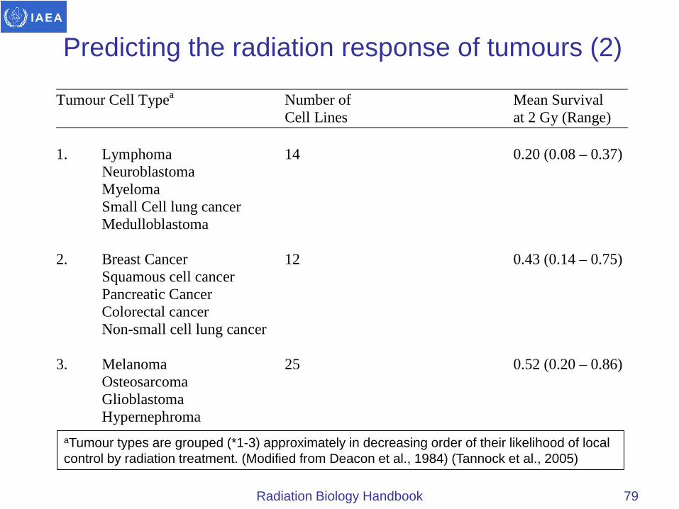
79 Radiation Biology Handbook
Tumour Cell Typea Number of Mean Survival Cell Lines at 2 Gy (Range) 1. Lymphoma 14 0.20 (0.08 – 0.37) Neuroblastoma Myeloma Small Cell lung cancer Medulloblastoma 2. Breast Cancer 12 0.43 (0.14 – 0.75) Squamous cell cancer Pancreatic Cancer Colorectal cancer Non-small cell lung cancer 3. Melanoma 25 0.52 (0.20 – 0.86) Osteosarcoma Glioblastoma Hypernephroma
aTumour types are grouped (*1-3) approximately in decreasing order of their likelihood of local control by radiation treatment. (Modified from Deacon et al., 1984) (Tannock et al., 2005)

Predicting the radiation response of tumours (3)
• There are some reports that patients with tumours in particular sites containing radioresistant cells (SF2>median) had significantly worse local control and survival than those with tumours containing radiosensitive cells (SF2 <median). However, this is not a universal finding, and the widespread application of clonogenic assays is limited by technical problems.
• Other proposed predictive assays evaluate radiation-induced apoptosis or senescence within solid tumours, or the expression of genes or proteins which relate to cell cycle control, cell death, and DNA repair. However, the predictive value of parameters such as the apoptotic index is uncertain given the limited correlation with cell death as assessed by a colony forming assay.
• Thus, a suitable, robust assay to detect such differences between individual patients has yet to be developed.
80 Radiation Biology Handbook

Predicting the radiation response of tumours (4)
81
Acturial survival in patients with cervical cancer treated by radical radiotherapy as a function of intrinsic radiosensitivity of tumours stratified as above or below the median SF2 of 0.41. Both survival and local control (not shown) were significantly worse for patients with SF2>0.41 (Tannock et al., 2005).
Radiation Biology Handbook
Tumour Hypoxia (1)
• The cells in a tumour are influenced both by their interactions with the extracellular matrix (ECM) and by their pathological microenvironment, which is characterized by regions of nutrient deprivation, low extracellular pH, high interstitial fluid pressure (IFP), and hypoxia.
• Tumours often contain regions where the oxygen concentration (pO2) is less than 5 mm Hg. A proportion of tumour cells may lie in hypoxic regions beyond the diffusion distance of oxygen, where they are exposed to chronically low oxygen tensions.
• Tumour cells may also be exposed to shorter (often fluctuating) periods of (acute) hypoxia due to intermittent flow in individual blood vessels.
82 Radiation Biology Handbook

Tumour Hypoxia (2)
• Acute and chronic hypoxia can coexist in the same tumour. Hypoxic regions are often diffusely distributed throughout the tumour and rarely concentrated only around a central core of necrosis.
• The cells in a tumour are influenced both by their interactions with the extracellular matrix (ECM) and by their pathological microenvironment, which is characterized by regions of nutrient deprivation, low extracellular pH, high interstitial fluid pressure (IFP), and hypoxia.
• The cells within hypoxic regions of tumours constitute an important target for cancer treatment since many such cells are viable and capable of regrowing the tumour if they survive treatment.
83 Radiation Biology Handbook
Tumour Hypoxia (3)
• Many tumours contain a proportion of hypoxic cells in the range 1 to 20%. These proportions represent the cells that are maximally resistant to radiation and there will also be a substantial proportion of cells in tumours that are at intermediate oxygen levels.
• Immediately after a dose of radiation, the proportion of the surviving cells that is hypoxic will be elevated. With time, some of the surviving hypoxic cells may gain access to oxygen, and this reoxygenation can result in a substantial increase in the sensitivity of tumours during fractionated treatment.
• Many techniques have provided evidence that hypoxic cells in human tumours can affect the outcome of radiation therapy.
84 Radiation Biology Handbook

2.5 Normal tissue response to radiotherapy
85 Radiation Biology Handbook
Cellular and Tissue Response (1)
86
• Acute responses occur primarily in tissues with rapid cell renewal where cell division is required to maintain the function of the organ. Because many cells express radiation damage during mitosis, there is early death and loss of cells killed by the radiation treatment.
• Late responses tend to occur in organs whose parenchymal cells divide infrequently (e.g. liver or kidney) or rarely (e.g. central nervous system or muscle) under normal conditions.
• Damage to the connective tissue and vasculature of the organ may lead to progressive impairment of its circulation. If the damage to the circulation is severe enough, secondary parenchymal cell death may occur due to nutrient deprivation.
Radiation Biology Handbook

Cellular and Tissue Response (2)
• The radiosensitivity of the cells of a number of normal tissues can be determined directly using in situ assays. Survival curves obtained for the cells of different normal tissues in mice and rats show considerable variability in sensitivity, and as with tumour cells, most of the difference appears to be in the shoulder region of the survival curve.
• For study of the response of individual organs, one widely used approach is to define a level of functional deficit and to determine the percentage of irradiated animals that express at least this level of damage following different radiation doses. This approach results in sigmoidal dose-response curves, generally quite steep and well defined.
• Increased cytokine and chemokine expression has been observed within hours after irradiation in both early and late responding tissues, when there are no apparent functional or histopathological changes, simulating a chronic inflammatory condition.
87 Radiation Biology Handbook
Acute Tissue Responses (1)
• Acute radiation responses occur mainly in renewal tissues and have been related to death of critical cell populations such as the stem cells in the crypts of the small intestine, in the bone marrow, or in the basal layer of the skin.
• Radiation-induced cell death in normal tissues generally occurs when the cells attempt mitosis, thus the tissue tends to respond on a time scale similar to the normal rate of loss of functional cells in that tissue and the demand for proliferation of the supporting stem cells.
• Radiation-induced apoptosis has also been detected in many cells and tissues, such as lymphoid, thymic, and hematopoietic cells, spermatogonia, and intestinal crypts.
• In the crypts of the small bowel there is a fraction of stem cells that die by apoptosis, and the others die a mitosis-linked death.
88 Radiation Biology Handbook

Acute Tissue Responses (2)
• Following irradiation of skin, there is early erythema within a few days of irradiation and this is related to the release of 5-hydroxytryptamine by mast cells, increasing vascular permeability.
• Expression of more severe acute skin reactions (moist desquamation and ulceration) depends on the relative rates of cell loss and cell proliferation of the basal cells, and they occur more rapidly in murine (7 to 10 days) than in human skin (2 to 3 weeks).
• The extent of these reactions and the length of time for recovery depend on the dose received and the volume (area) of skin irradiated.
• Erythema in human skin occurs at single doses greater than about 6 Gy, while moist desquamation and ulceration occur after single doses of 20 to 25 Gy.
89 Radiation Biology Handbook
Late Tissue Responses
• Late tissue responses ( > about 3 months after treatment) occur in organs whose parenchymal cells normally divide infrequently and hence do not express mitosis-linked death until later times when called upon to divide.
• The nature and timing of late reactions depends on the tissue involved and can be expressed as diminished organ function e.g. radiation-induced nephropathy (symptoms of hypertension, increased creatinine and blood urea nitrogen levels) or functional loss.
• Radiation-induced fibrosis development appears to be associated with the aberrant and chronic expression of inflammatory cytokines, particularly TGF-β.
• The volume of tissue or organ irradiated plays an important role in its response to irradiation, but its roles may be different in different tissues, depending on the functional structure and functionality of the tissue.
90 Radiation Biology Handbook

Apoptosis and Late Reactions
• Apoptosis has also been observed within hours after irradiation of a number of late responding normal tissues in rodents, such as the salivary glands, pulmonary and brain endothelial cells and spinal cord.
• For example, in rat spinal cord, endothelial cell apoptosis following irradiation appears to initiate the disruption of the blood/spinal cord barrier, which may be an early lesion leading on to the development of white matter necrosis and myelitis.
• Apoptotic endpoints, however, have often not correlated with clonogenic survival or functional or histopathological endpoints, and the relevance of apoptosis in radiation-induced late normal tissue damage remains to be established.
91 Radiation Biology Handbook

Predicting Normal Tissue Response
• Patients receiving identical radiation treatments may experience differing levels of normal tissue injury; thus predictive assays might be useful in identifying those patients at greater risk of experiencing the side effects of radiotherapy.
• Studies of breast cancer patients have also shown individual correlation of acute and late skin reactions in one treatment field with those in a different treatment field.
• Several studies have quantitated the in vitro variations in radiosensitivity of fibroblasts and peripheral lymphocytes as a potential predictive assay, but have been inconsistent in predicting late radiation fibrosis in individual patients.
• The differences in radiosensitivity between most patients may not be sufficient to override the effects of the other factors, such as cytokine induction, chronic inflammation and vascular damage that also influence the development of normal tissue damage.
92 Radiation Biology Handbook
Therapeutic Ratio (1)
• In the clinic the therapeutic ratio is often defined as the percentage of tumour cures that are obtained at a given level of normal tissue complications.
• In animal models it is more usual to define the therapeutic ratio in terms of the ratio of radiation doses Dn/Dt required to produce a given percentage of complications and tumour control (usually 50%).
• The therapeutic ratio is favourable when the tumour-control curve is displaced to the left of that for normal tissue damage. The greater this displacement, the more radiocurable is the tumour.
• If the tumour control curve is shallower than that for normal tissue damage, the therapeutic ratio is more favourable for low and intermediate tumour-control levels. If the two curves are close together, or the curve for tumour control is displaced to the right of that for complications, the therapeutic ratio is unfavourable because a high level of complications must be accepted to achieve even a minimal level of tumour control.
93 Radiation Biology Handbook

Therapeutic Ratio (2)
94
Illustration of the concept of a therapeutic ratio in terms of dose-response relationships for tumour control and normal tissue damage (Tannock et al., 2005)
Radiation Biology Handbook

Whole Body Irradiation (1)
• The response of animals to single doses of whole body irradiation can be divided into four separate syndromes (prodromal, haematological, gastrointestinal, and neurovascular) that manifest following different doses and at different times after irradiation.
• After doses greater than about 2 Gy, humans will develop early nausea and vomiting within hours of irradiation (prodromal syndrome), which may be controlled with 5-hydroxytryptamine antagonists.
• The hematopoietic syndrome occurs at doses in the range of 2 to 8 Gy in humans (3 to 10 Gy in rodents) and is caused by severe depletion of blood elements due to killing of precursor cells in the bone marrow.
• The gastrointestinal syndrome occurs after doses > about 5 -15 Gy.
• The neurovascular syndrome occurs following large doses of radiation (>20 Gy) and usually results in rapid death (hours to days) due to cardiovascular and neurological dysfunction.
95 Radiation Biology Handbook

Whole Body Irradiation (2)
• Death from the hematopoietic syndrome can sometimes be prevented by bone marrow transplantation (BMT) and cytokine therapy (e.g., GM-CSF, G-CSF, stem cell factor) provided that the radiation dose is not too high (<10 Gy) when damage to other organs may become lethal.
• Intensive nursing with antibiotics, fluid, and electrolyte replacement can prevent early death from the gastrointestinal syndrome in human victims of radiation accidents, but these patients may die later due to damage to other organs (e.g. kidney, lung).
• There are no known treatments for the neurovascular syndrome.
96 Radiation Biology Handbook
2.6. Radiobiological basis of radiation protection
97 Radiation Biology Handbook

Health Consequences after Total Body Irradiation from Radiation Accidents (1)
98
• Radiation exposure of the total body with doses >2 Gy will cause clinical symptoms which, after higher doses may be so severe that they become life threatening.
• The best known accident of the nuclear industry is the Chernobyl accident which lead to high total body doses in >200 rescue workers and firemen. Twenty eight of them died within 2 months from radiation sickness.
• More frequent than accidents in the nuclear industry are accidental exposures of non-involved people from lost or discarded radioactive sources.
• In contrast to nuclear industry accidents, where radiation exposure of the body often has been acute and fairly homogeneous, radiation exposure from accidents with lost radioactive sources is usually very inhomogeneous and protracted over days and weeks.
Radiation Biology Handbook
Health Consequences after Total Body Irradiation from Radiation Accidents (2)
• The signs and symptoms of radiation sickness after an acute total body exposure are predominantly the consequences of radiation injury to the haemopoietic tissues in the bone marrow.
• Proliferating cells of the bone marrow decrease their proliferative activity, and fewer cells are available for differentiation and maturation to white blood cells, red blood cells and platelets.
• Mature myeloid cells are not damaged by a few Gy. Since mature granulocytes have a lifespan of only 1 day, the decrease in supply of granulocytes will occur first, followed by a decrease in platelet number.
• Since the time of granulocyte maturation between the last mitosis of myelocytes and early-granulocyte transit to blood is ~4 days and radioresistant, the decrease of granulocytes in the blood starts after a delay of 4 days. Granulocyte hypoplasia (leukopenia or granulocytopenia) increases until day 12.
99 Radiation Biology Handbook
Health Consequences after Total Body Irradiation from Radiation Accidents (3)
• The further development of the haemopoietic radiation syndrome depends on the number of bone marrow stem cells which survived radiation exposure and are stimulated into very fast regeneration.
• If, after day 12, the concentration of granulocytes in the blood is maintained at a plateau of about 1,000/μl, this can be taken as a prognostically favourable sign.
• If the granulocyte count continues to decrease after day 12, there is a high risk that the number of surviving bone marrow stem cells is insufficient to lead to rapid regeneration of haemopoiesis before severe, potentially fatal consequences of leukopenia and of thrombopenia develop.
• In these cases, the only therapeutic option is allogeneic stem cell transfusion (“bone marrow transplantation”). In less severe cases G-CSF may be given to lead to maximal stimulation of proliferation and maturation of granulocyte progenitors.
100 Radiation Biology Handbook

Health Consequences after Total Body Irradiation from Radiation Accidents (4)
• The time course of decrease and recovery of platelets is similar to that of granulocytes but somewhat slower. Due to the long life span of erythrocytes, no significant anaemia is expected as a consequence of bone marrow hypoplasia.
• However, after the Chernobyl accident anemia was common, caused by intravascular coagulation due to widespread skin burns and severe radiation injury from skin contamination with radioactive fission products.
• The probability of survival depends not only on the dose of radiation but also on other concomitant risk factors such as chronic infections, and on the quality of medical interventions.
• The LD-50 value for humans is considered around 3-4 Gy with minimal medical attention, and 6-7 Gy with good medical treatment.
101 Radiation Biology Handbook
Health Consequences after Total Body Irradiation from Radiation Accidents (5)
• In animal experiments, the severity of the haemopoietic radiation damage increases with dose between 2 and 10 Gy.
• In mice, some animals are likely to die from septicaemia after a dose of around 5 Gy, caused by severe agranulocytosis and diffuse interstitial haemorrhage.
• Deaths occur at the time of the nadir of granulocyte depletion in the blood, i.e. in the third week after acute radiation exposure.
• Half of the animals are likely to die after a total body dose of approximately 7 Gy (LD-50). After 9 Gy, the chances of survival are small unless a specific therapy is initiated.
102 Radiation Biology Handbook

Health Consequences after Total Body Irradiation from Radiation Accidents (6)
• The medical treatment of acute sickness after total body irradiation, which may occur 2-5 weeks later, is purely symptomatic (except rare cases where stem cell infusion is indicated).
• Since in most cases, spontaneous regeneration of the bone marrow from surviving stem cells is likely, the primary aim of medical intervention is to bridge the period of critical granulocytopenia and thrombopenia.
• Bacterial decontamination of the gut and the oropharynx, replacement of platelets, treatment of infections with antibiotics, and prophylactic treatment to prevent fungal and herpes infections, are recommended. Since the Chernobyl accident, prophylactic treatment with haemopoietic growth factors (G-CSF), has been used. This leads to rapid restoration of the granulocyte count in the blood, but the overall impact on survival is uncertain.
103 Radiation Biology Handbook

Health Consequences after Total Body Irradiation from Radiation Accidents (7)
• Since the full signs and symptoms of the acute haemopoietic radiation syndrome after high radiation doses, which would require intensive treatment, does not occur until after a delay of 3-4 weeks, there is no time pressure for assessing the prognosis and for planning adequate treatment.
• Criteria for triage to assess the prognosis and the need for treatment are shown in the following Table. These criteria were used with great success after the Chernobyl accident.
• The clinical signs such as vomiting in the first few hours after exposure, as well as hair loss and lymphocyte counts in the first week after exposure, are more important for medical decision making than any results of physical or biological dosimetry.
104 Radiation Biology Handbook

TRIAGE CRITERIA USED AFTER THE CHERNOBYL ACCIDENT
Radiation Biology Handbook page xx Section xx 105
Severity Vomiting Lymphocytes Hair loss Cytogenetic Lethality time day 3 within 2 radiation dose including
per µl weeks skin burns
Mild no >600 no < 2Gy 0/105
Intermediate after 1-2 h 300-600 no 2 – 4 Gy 0/53
Severe after 30-60 min 100-300 yes 4 – 6 Gy 6/23
Very severe immediate <100 yes 6 – 16 Gy 19/22
Radiation Biology Handbook 105
Health Consequences after Total Body Irradiation from Radiation Accidents (8)
• The determination of radiation dose from accidental exposure in the first few weeks after the accident is commonly done by a combination of physical reconstruction of exposure scenarios and calculation of organ doses and total body doses as well as by biological dosimetry.
• The preferred method of biological dosimetry which has proven its value in many minor and major accidents is the determination of the frequency of unstable chromosome aberrations in stimulated lymphocytes.
• The method has been well standardised: phytohaemagglutinin is added to 5 – 10 ml heparinised blood to stimulate resting lymphocytes into proliferation. After incubation for 48 hours at 37C, cells entering mitosis are arrested in metaphase by adding colchicine.
106 Radiation Biology Handbook

Health Consequences after Total Body Irradiation from Radiation Accidents (9)
• It is important to arrest cells in their first mitosis since many of the severe chromosome aberrations which are used as “dosemeters” are eliminated in the first cell division.
• As a general rule, the number of dicentric chromosomes is counted in 500 arrested metaphases. If there are 25 dicentrics among 500 metaphases, a total body dose of 0.3 Gy can be assumed. After a dose of 3 Gy, there is, on average, one dicentric chromosome to be found in each metaphase.
• After homogeneous total body irradiation, the number of dicentric chromosomes per cell follows a Poisson distribution. Marked deviations from a Poisson distribution are an indicator of very inhomogeneous dose distribution which may have consequences for the prognosis.
107 Radiation Biology Handbook
Long Term Risks from Low Radiation Doses (1)
• The dramatic experience of the people of Hiroshima and Nagasaki in 1945 was the initiator for a proposal by the National Academy of Sciences of the USA to develop a programme for life-long follow-up of all A-bomb survivors.
• This programme, continued by US-Japanese co-operation in the Radiation Effects Research Foundation (RERF), is arguably the largest, most comprehensive and most detailed epidemiological study ever performed.
• The results of this study are the most importance source of information on which rules and regulations of radiation protection are based.
• Through a massive effort, the radiation doses to all critical organs of each member of the cohort has been individually assessed by various methods of retrospective dosimetry.
108 Radiation Biology Handbook

Long Term Risks from Low Radiation Doses (2)
The various studies are:
(1) The Life-Span-Study (LSS). This prospective cohort study has been studying approximately 120,000 people. Radiation doses have been reconstructed in nearly 90,000. Approximately 5,000 had received a total body dose > 1Gy.
(2) The Adult Health Study comprises 20,000 people, a subgroup of the LSS, predominantly those exposed to higher doses. Each member of this cohort is invited to a free health check every two years in the outpatient clinics of RERF.
(3) The F-1 Study comprises approximately 70,000 children of parents who were exposed by the A-bomb explosions.
(4) The In-Utero Study investigated the children born in Hiroshima and Nagasaki between September 1945 and May 1946, to detect potential radiation damage to the development of the embryo and fetus in utero.
109 Radiation Biology Handbook
Long Term Risks from Low Radiation Doses (3)
• The most important long-term health damage observed in the LSS of the A-bomb survivors is a dose dependent increased mortality from cancer. Among the 44,771 deceased members of the LSS with detailed dosimetric information, there were 9,335 deaths from solid cancer and 582 deaths from leukaemia.
• Approximately 440 cancer deaths (i.e. ~4%) and nearly 100 leukaemia deaths (i.e. ~15%) can be attributed to the radiation exposure from the bomb in 1945.
• Significant relationships to radiation exposure were found for the following types of malignant disease (in decreasing probability of mortality): stomach, colon, lung, leukaemia, breast, oesophagus, bladder, ovary, liver.
• Some of the most common types of cancer in the general population are not induced to any significant extent by radiation such as cancer of the prostate, cervix or rectum.
110 Radiation Biology Handbook
Long Term Risks from Low Radiation Doses (4)
• Members of the LSS who were exposed to radiation as children or adolescents developed radiation-induced cancer after a longer latency than those who were exposed later in life.
• This observation was the basis for the commonly used risk projection model, the “relative risk model”. It states that irradiation causes a dose-dependent increase of the relative risk of developing certain types of cancer.
• The overall risk of a person is estimated by his or her spontaneous cancer risk at the age of estimation (taken from national cancer statistics) multiplied by the dose-dependent risk factor.
• The life-time risk of dying from radiation-induced cancer after an acute exposure to 1 Gy (or 1 Sv) is 10% (ICRP 60). If the dose is given over a period of weeks or months the risk factor is 5%, and if spread over a working life it is 4%.
111 Radiation Biology Handbook
Long Term Risks from Low Radiation Doses (5)
• From the information collected by the LSS, the dependence of the risk of dying from radiation-induced cancer on factors other than dose has been determined, such as age, sex and irradiated organ.
• The relative contribution in the total radiation risk of radiation exposure of individual organs have been estimated. Three classes of organs have been identified and given organ weighting factors. The mean radiation dose to each organ is multiplied with the respective organ weighting factor.
• All weighted organ doses are added up to arrive at the radiation dose which would result in the same radiation risk if given homogeneously to the total body. This sum of weighted organ doses is called the effective dose.
112 Radiation Biology Handbook

Organ Weighting Factors from ICRP (2007)
113 Radiation Biology Handbook
Organs Organ weighting factor High sensitivity bone marrow, stomach, colon, lung 0.12 breast Intermediate sensitivity bladder, liver, oesophagus, thyroid 0.04 Low sensitivity skin, bone surface 0.01 remainder tissues (13) 0.12 (in total) Genetic and somatic gonads (ovary, testis) 0.08 effects from gonadal exposure

Epidemiological Studies in Other Radiation-exposed Populations
The most important studies can be classified into:
• Radiation workers
• Studies in people who were exposed to radiation in the treatment of various diseases, such as: Tuberculosis, Ankylosing spondylitis (M. Bechtherew), Mastitis, Cancer.
• Studies in people who were exposed to high levels of naturally radioactive materials, in particular radon: Miners, People in homes.
• Studies in people who were exposed to the fall-out of nuclear explosions and accidents in the nuclear industry: Chernobyl, Marshall Islands, Techa River.
114 Radiation Biology Handbook

Radiation Workers
• The first information on the risk of radiation-induced cancer was from studies comparing mortality of different medical professions.
• A significant increase in cancer mortality was found for radiologists who joined the profession before 1930, i.e. before strict rules and regulations were introduced to reduce occupational radiation exposure.
• Most occupationally exposed people today, also workers in the nuclear industry, receive very small radiation doses and no significant increase in cancer rates have been found with two exceptions which refer to the pioneering times of nuclear industry, i.e. workers in the reprocessing plant of Sellafield (UK) and workers in the plutonium factories of Mayak (former USSR).
• Mayak is of great interest for radiation protection since it is the only major source of information on the radiation risks from plutonium.
115 Radiation Biology Handbook

Patients (1)
• Pneumothorax, to improve blood perfusion in the tuberculosis-affected lung, required careful control of the collapsing lung which was done under permanent fluoroscopy. The chest wall received very high radiation doses of up to >10 Gy. In females, there was a risk of developing breast cancer which depended strongly on dose and age at exposure. From ages of 20 to 40 risk decreased dramatically, with little risk after menopause.
• Mastitis has been one of the most successful indications for radiotherapy of non-malignant diseases, using <2Gy. Radiation caused a dose dependent increase in the risk of breast cancer later in life. The relative risk model results (taking the country specific base-line cancer risks into account) and the age dependence of risk are similar for mastitis radiotherapy outcome in European and American women and for the A-bomb Japanese LSS women.
116 Radiation Biology Handbook

Patients (2)
• The study on the risk of leukaemia and cancer among 14,000 British patients who suffered from ankylosing spondylitis (M. Bechtherew) treated with radiotherapy to the spine with total doses of 6-12 Gy was the first study which provided convincing evidence that low radiation doses which do not cause any acute or late normal tissue damage still may cause leukaemia and cancer.
• 80%-90% of second cancers after radiotherapy are due to the longer life span of a cured patient.
• For prostate cancer patients receiving radiotherapy, the risk of developing a radiation-induced second cancer is ~ 0.3%.
• Half of the 0.3% risk is in the low-dose regions such as lung, and is probably induced by the same mechanism responsible for the increased cancer risk in the A-bomb survivors. The other half is in the high-dose regions inducing tissue atrophy associated with chronic inflammation, which is a well-known pre-cancerous lesion.
117 Radiation Biology Handbook

People Exposed to High Radon Concentrations
• Numerous studies on miners, particularly uranium miners, have defined a proportional relationship between the product of exposure time and radon decay-product concentration in the air and the risk of lung cancer.
• Radon levels in houses may also be associated with an increased risk of lung cancer. The results of these studies are unequivocal in demonstrating that a mean radon concentration of >200 Bq/m³ is associated with an increased risk.
• This value is therefore chosen as an intervention level which, if exceeded should initiate measures to reduce radon levels in houses. Of particular importance is the finding, both in the miner studies and in the indoor radon studies that radiation risk and smoking risk are supra-additive, or even multiplicative.
118 Radiation Biology Handbook

Victims of Nuclear Accidents (1)
• The Chernobyl accident led to exposure of large populations to radioactive fall-out, mainly in Ukraine, Belarus and Russia. Several-100,000 rescue workers (called liquidators) were exposed to 0.05-0.25 Gy, mainly through external irradiation from radioactivity deposited to the ground.
• The main health consequence of the Chernobyl accident is a massive increase in thyroid cancer among children, caused by uptake of large amounts of I-131 in milk giving a mean dose ~0.2 Gy in the thyroid cancer cases. The younger the child, the higher the risk.
• The dose dependence of risk of thyroid cancer among the Chernobyl children is similar to the dose dependence established before in various epidemiological studies on people who were irradiated with X rays for various diseases such as tinea capitis or thymic hyperplasia.
119 Radiation Biology Handbook

Victims of Nuclear Accidents (2)
• The United States of America tested their nuclear weapons between 1946 and 1956 on Bikini and Eniwetok in the Marshall Islands.
• 82 inhabitants of Rongelap received external total body doses of approximately 2 Gy, and 159 inhabitants of Utrik received 0.1 Gy γ-rays from ground contamination. In addition, high radiation doses to the thyroid of children resulted from I-131. Some children developed clinical signs of myxoedema, and there was a high incidence of benign and malignant thyroid nodules in the most affected children.
• However, an epidemiological study on the entire population of the Marshall Islands did not provide undisputed evidence of an increased incidence rate of thyroid cancer as a result of radioactive fall-out from the bomb tests.
120 Radiation Biology Handbook

Victims of Nuclear Accidents (3)
• The plutonium factory of Mayak near Chelyabinsk in Siberia discharged, in the early years of its operation from 1946, large amounts of radioactive waste into the River Techa, this way exposing the populations downstream to high doses of γ-rays from sediments of the river banks.
• The Techa river population accumulated the radiation dose over several years while the A-bomb LSS population received the same doses within seconds.
• The results of the Techa river population studies are hoped to answer one of the most import questions of radiation protection, namely whether radiation doses protracted over many months are equally, or more, or less effective than the same doses inflicted in a few seconds.
121 Radiation Biology Handbook
Mechanisms of Radiation-induced Cancer (1)
• The development of cancer occurs over a long period of time following a multi-step process, classified into initiation, promotion and progression.
• In some radiation-induced leukaemias, the specific molecular changes have been identified. However, no typical “fingerprint-mutation” has been found so far for radiation-induced cancer, which would betray the causation by radiation exposure.
• Both the inactivation of tumour suppressor genes by deleting the whole gene or important parts of them, as well as activation of oncogenes by translocations of promotors to proto-oncogenes as a result of breakage/re-union mechanisms, could occur after radiation exposure.
• The results of basic radiobiological research do not yet permit the identification of the crucial molecular steps in radiation carcinogenesis.
122 Radiation Biology Handbook
Mechanisms of Radiation-induced Cancer (2)
• There is strong evidence from animal studies and some human studies that the risk of radiation-induced cancer may be determined by various genes, such as mutations of the Rb gene.
• Other genes discussed in this context include BRCA 1 and 2.
• However, at the present state of knowledge, the role of genetic susceptibility on individual risks of radiation-induced cancer cannot be resolved definitively, although there is general agreement that it will be important.
• The implications of such findings on the selection of people for special occupations with a high risk of radiation exposure also need serious consideration.
123 Radiation Biology Handbook

Radiation effects in the developing embryo and fetus
• In Hiroshima and Nagasaki, >1,500 children born between September 1945 and March 1946 were investigated at regular intervals between 1948 and 1964 to study the effects of radiation doses between 0.01 and >1 Gy on intra-uterine development at different stages of pregnancy.
• No malformations were found increased in a dose dependent way in the children of Hiroshima and Nagasaki. However, 18 children presented with microcephaly and severe mental retardation. The mothers of 15 children had been exposed to bomb radiations near the hypocentre, in week 8 to 15 of pregnancy, and 3 in later stages.
• Findings of dystopic grey matter by MRI investigations of some of those severely-retarded people are in accordance with the results from experimental studies of the effects of radiation doses < 1 Gy given to pregnant mice in late pregnancy.
124 Radiation Biology Handbook

Radiation effects in the developing embryo and fetus (2)
• In the Hiroshima children there was evidence for a threshold of 0.1 Gy. At higher doses, the risk of severe mental retardation increased rapidly to a value of 40% after 1 Gy. In later stages of pregnancy, the threshold dose may be higher.
• At the age of 10, there was a dose dependent decrease of the mean IQs as well as the mean school performance scores of those groups exposed in weeks 8 to 15 and 16 to 25 after doses >0.1 Gy.
• No decrease of intellectual development was recorded if irradiation had occurred before week 8 or after week 25, even if doses were >0.5 Gy.
125 Radiation Biology Handbook
Radiation effects in the developing embryo and fetus (3)
• Embryos in the pre-implantation stage are very radiosensitive, but the damage inevitably will lead to death of the conceptus and early abortion. Those embryos that survive develop normally.
• In human early fetus, in the first few weeks after implantation during the period of major organogenesis, a comparable all-or-nothing effect is likely i.e. either an early, spontaneous abortion or normal development.
• The findings of a probable threshold of 0.1 Gy will influence the advice to be given to pregnant women after a diagnostic radiology procedure. In particular after abdominal CT investigations, careful analysis of radiation doses in the uterus as well as medical anamnestic exploration have to be performed.
126 Radiation Biology Handbook
Radiation-induced Heritable Diseases (1)
• A major concern of radiation protection in the past was the possible deleterious effects of ionising radiation on the health of future generations.
• The large epidemiological programme among the children of the A-bomb survivors (the F-1 Study) was not informative nor was any of the other studies which investigated the children of radiation workers or of radiotherapy patients.
• Most of the mouse studies used the 7 locus method, where irradiated wild-type males homozygous for 7 un-mutated genes coding for recessive traits, were mated with unirradiated “tester” females homozygous for the same 7 genes in the mutated state.
127 Radiation Biology Handbook
Radiation-induced Heritable Diseases (2)
• With increasing radiation (protracted) dose the spontaneous mutation rate of 1:100,000 increased following a linear dose response curve.
• The spontaneous mutation rate was doubled by a dose of 1 Gy (doubling dose DD).
• The mutation doubling dose does not give any useful information on the risk of heritable disease.
• Therefore, the mouse doubling dose is combined with information from human population genetics to estimate the risk of heritable disease in the progeny of irradiated people.
128 Radiation Biology Handbook
Radiation-induced Heritable Diseases (3)
• Heritable diseases may occur as direct result of a mutation in a single gene (single gene disorders). Inheritance of these diseases may be autosomal dominant, autosomal recessive or sex-linked recessive.
• Data from human population genetics give an overall frequency of Mendelian diseases in the population of 2.4% (1.5% autosomal dominant, e.g. Huntington´s disease, 0.75% autosomal recessive e.g. Phenylketonuria, and 0.15% sex-linked recessive, e.g. Haemophilia).
• In addition, approximately 6% of live births are affected by a congenital abnormality and 65% of the population will develop chronic disease later in life, with some genetic component.
129 Radiation Biology Handbook

Radiation-induced Heritable Diseases (4)
• Multifactorial diseases comprise common diseases such as diabetes, essential hypertension and coronary heart disease. This complexity of heritable diseases is incorporated in the present method of estimating the heritable risk among the progeny of irradiated people.
• The genetic risk calculation combines population genetic data in humans and radiation genetic data in mice as follows:
• Risk = Prevalence x 1/Doubling Dose x Mutation Component x PRCF
• Risk is the probability that an offspring of the exposed person will develop heritable disease (Mendelian or multifactorial).
• For protracted irradiation, the accumulated dose in the gonads before conception is divided by 1. The Mutation Component is a factor which describes the relationship between the increase in the mutation rate and the rate of additional disease.
130 Radiation Biology Handbook

Radiation-induced Heritable Diseases (5)
• Even for dominant diseases the mutation component is not 1, since the majority of existing mutations are inherited from parents and grandparents, often through many generations.
• A cautious estimate suggests that doubling of the rate of new dominant mutations will cause only a 30% increase of diseases with dominant inheritance in the first generation and 15% in the second generation.
• The same value of the Mutation Component is allocated to sex-linked recessive diseases. Since the development of single gene disease with recessive inheritance requires mutations in both alleles of the same gene, the relationship between a mutation and disease is very remote and the Mutation Component is therefore assumed to be close to zero.
• For multifactorial diseases, the relationship between mutation and disease is also not very close, and presently the Mutation Component is assumed to be 0.01.
131 Radiation Biology Handbook

Radiation-induced Heritable Diseases (6)
• The potential recoverability correction factor (PRCF) has been introduced to account for the fact that the molecular structure of radiation-induced mutations differs markedly from the molecular structure of “spontaneous” mutations.
• Most spontaneous mutations are point mutations with a single base pair altered or a minute deletion whereas radiation-induced mutations are mostly large deletions, often affecting whole genes.
• These mutations are usually not compatible with inter-uterine development and most will lead to premature termination of pregnancy.
• The value of PRCF suggested today is 0.15 to 0.3.
132 Radiation Biology Handbook

Radiation-induced Heritable Diseases (6)
• Using this equation for estimating the risk of heritable diseases of a young man who had been exposed to a radiation dose of 1 Gy from radiotherapy, e.g. of pelvic lymph nodes of Hodgkin´s disease, the risk of radiation-induced dominant and sex-linked heritable disease would be:
Risk = 0.0165 x 1 x 0.3 x 0.3 x 0.5
• The last factor of 0.5 has been introduced to account for the fact that only the father was irradiated.
• The result is a risk of less than 0.1%.
• The risk of multifactorial disease is similar at < 0.1% and the risk for radiation-induced recessive disease among the children is essentially zero.
133 Radiation Biology Handbook
Radiation-induced Heritable Diseases (7)
• Recent considerations of the potential molecular, genotypic manifestations of genetic diseases of genetic damage, induced in the germ cells of irradiated individuals and transmitted to the progeny, took account of the relationship between the observed molecular changes in the DNA of irradiated cells and gene function and gene position.
• It has been suggested that the major genetic effect of radiation exposure is related to microdeletions, i.e. deletions of multiple, functionally unrelated, yet physically contiguous genes that are compatible with survival of the individual receiving them.
• Such microdeletions are known to cause rare multisystem congenital abnormalities which share some common features: mental retardation, growth retardation, various malformations.
• The risk of these multi-organ congenital disorders after exposure to 1 Gy is approximately 0.1%.
134 Radiation Biology Handbook

REFERENCES TO SECTION 2 (1)
• BARENDSEN, G.W., Responses of cultured cells, tumors, and normal tissues to radiation of different linear energy transfer. Curr Top Radiat Res (1968), Q 4, 293-356
• HALL, E.J., GIACCIA, E., Editors, Radiobiology for the Radiologist, 6th edition (2006), J.B. Lippincott Company, Philadelphia, USA.
• INTERNATIONAL COMMISSION OF RADIATION PROTECTION Publication 103: 2007recommendations of the ICRP, including Annexes on Quantities used in radiation protection, Biological effects of radiation, and Bases for judging the significance of the effects of radiation. Ann ICRP 37: issues 2-4, 1-332 (2008).
• JOINER, M.C., JOHNS, H., Renal damage in the mouse: the effect of d(4)-Be neutrons. Radiat Res (1987), 109, 456-458
• PODGORSAK, E.B., Editor, Radiation Oncology Physics: a handbook for teachers and students, IAEA, Vienna, 2005.
135 Radiation Biology Handbook

REFERENCES TO SECTION 2 (2)
• TANNOCK, I.F., HILL, R.P., BRISTOW, R.G., HARRINGTON, L., Editors, The Basic Science of Oncology, Edition 4, Published by McGraw-Hill (2005).
• UNITED NATIONS SCIENTIFIC COMMITTEE on the Effects of Ionizing Radiations: Sources and Effects of Ionizing Radiations, Report 1994, Annex A: Epidemiological studies of radiation carcinogenesis, New York: United Nations (1994).
• VAN DER KOGEL, A.J., JOINER, M.C., Editors, Basic Clinical Radiobiology: 4th edition; (2009), Hodder Arnold, London, UK.
• WEST, C., DAVIDSON, S.E., ROBERTS, S.A., HUNTER, R.D., The independence of intrinsic radiosensitivity as a prognostic factor for patient response to radiotherapy of carcinoma of the cervix. Br J Cancer (1997), 76, 1184-1190.
136 Radiation Biology Handbook

BIBLIOGRAPHY TO SECTION 2 (1)
• BRENNER, D.J., CURTIS, R.E., HALL, E.J., RON, E., Second malignancies in prostate carcinoma patients after radiotherapy compared with surgery, Cancer (2000), 88, 398-406.
• FLIEDNER, T.M., MEINEKE, V., editors: Radiation-induced multi-organ involvement and failure: A challenge for pathogenetic, diagnostic and therapeutic approaches and research. Brit. J. Radiol. Supplement 27 (2005).
• INTERNATIONAL ATOMIC ENERGY AGENCY, Radiobiology modules in the “Applied Sciences of Oncology” distance learning course. http://www.iaea.org/NewsCenter/News/2010/aso.html
• INTERNATIONAL COMMISSION OF RADIATION PROTECTION Publication 60: 1990 recommendations of the ICRP, Ann. ICRP 21: issues 1-3, 1-201 (1991).
137 Radiation Biology Handbook

BIBLIOGRAPHY TO SECTION 2 (2)
• OTAKE, M., SCHULL, W.J., “Radiation-related brain damage and growth retardation among the prenatally exposed atomic bomb survivors”, Int. J. Radiat. Biol.(1998), 74, 159-171.
• PRESTON, D.L., SHIMIZU, Y., PIERCE, D.A., et al., “Studies of mortality of atomic bomb survivors”, Report 13: Solid cancer and non-cancer disease mortality: 1950-1997, Radiat. Res. (2003), 160, 381-407.
• SANKARAYARAYANAN, K., “Estimation of the hereditary risks of exposure to ionizing radiation: history, current status, and emerging perspectives”, Health Physics (2001), 80, 363-369.
• WORLD HEALTH ORGANIZATION: Health Effects of the Chernobyl Accident and Special Health Care Programmes (2006).
138 Radiation Biology Handbook