Radiation. Ionising Radiation Alpha Radiation Beta Radiation Gamma Rays X-Rays Neutrons.
Upload
sudhir-benCategory
view
261download
0
RADIATION
Radiation is an energy in the form
of electro-magnetic waves or
particulate matter, traveling in the
air.”
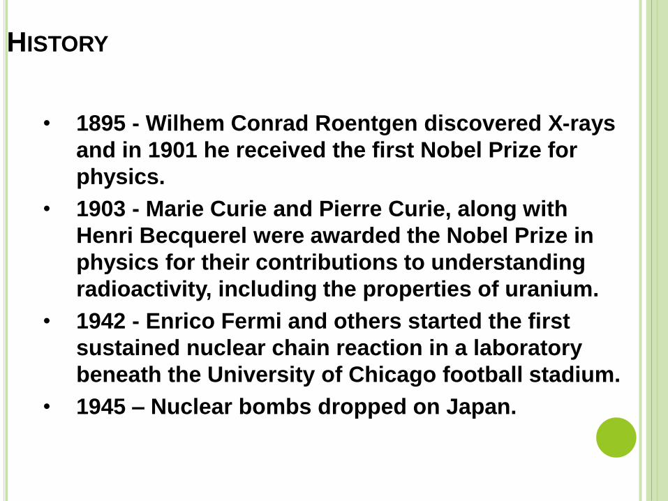
• 1895 - Wilhem Conrad Roentgen discovered X-rays
and in 1901 he received the first Nobel Prize for
physics.
• 1903 - Marie Curie and Pierre Curie, along with
Henri Becquerel were awarded the Nobel Prize in
physics for their contributions to understanding
radioactivity, including the properties of uranium.
• 1942 - Enrico Fermi and others started the first
sustained nuclear chain reaction in a laboratory
beneath the University of Chicago football stadium.
• 1945 – Nuclear bombs dropped on Japan.
HISTORY

RADIATION SOURCES
Sources of radiation can be divided
into two categories:
Natural Background Radiation
Man-Made Radiation
NATURAL BACKGROUND RADIATION
Cosmic Radiation
Terrestrial Radiation
Internal Radiation
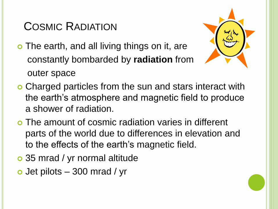
COSMIC RADIATION
The earth, and all living things on it, are
constantly bombarded by radiation from
outer space
Charged particles from the sun and stars interact with
the earth’s atmosphere and magnetic field to produce
a shower of radiation.
The amount of cosmic radiation varies in different
parts of the world due to differences in elevation and
to the effects of the earth’s magnetic field.
35 mrad / yr normal altitude
Jet pilots – 300 mrad / yr

ATMOSPHERIC RADIATION
Terrestrial
Radioactive material is also found throughout
nature in soil, water, and vegetation.
Important radioactive elements include uranium,
thorium, radium & isotopes of potassium(K40)
Some radioactive material is ingested with food and
water.
The amount of terrestrial radiation aprox. 50 mrad/yr
Atmospheric
Gases like Radon & thorum – 2 mrad/yr

INTERNAL RADIATION
People are exposed to radiation from radioactive
material inside their bodies. Besides radon, the most
important internal radioactive element is naturally
occurring potassium-40. Others - uranium, thorium,
strontium & carbon.
25 mrad / yr.
May go up to 70 or 80.
Total natural radiation - 0.1 rad / yr.
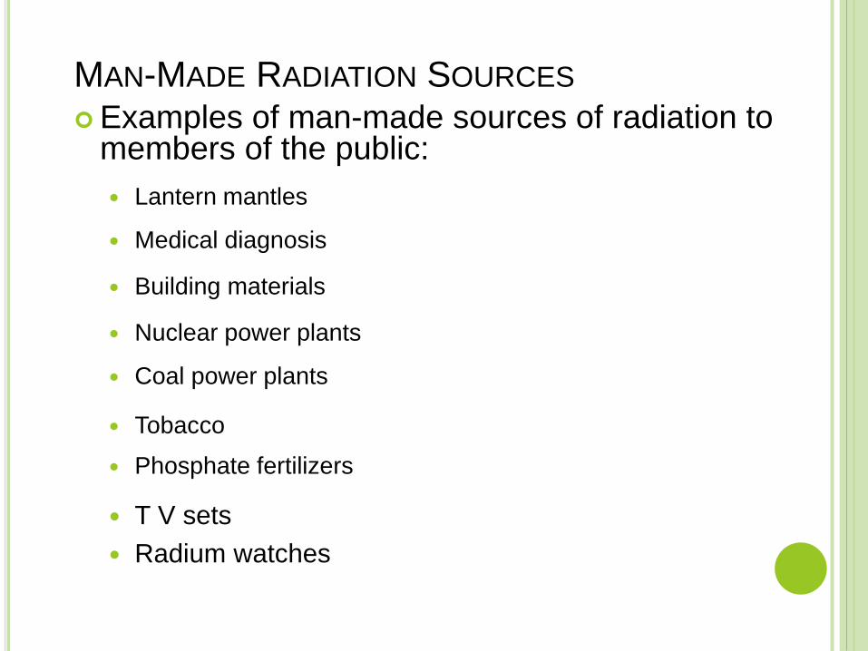
MAN-MADE RADIATION SOURCES
Examples of man-made sources of radiation to members of the public:
Lantern mantles
Medical diagnosis
Building materials
Nuclear power plants
Coal power plants
Tobacco
Phosphate fertilizers
T V sets
Radium watches
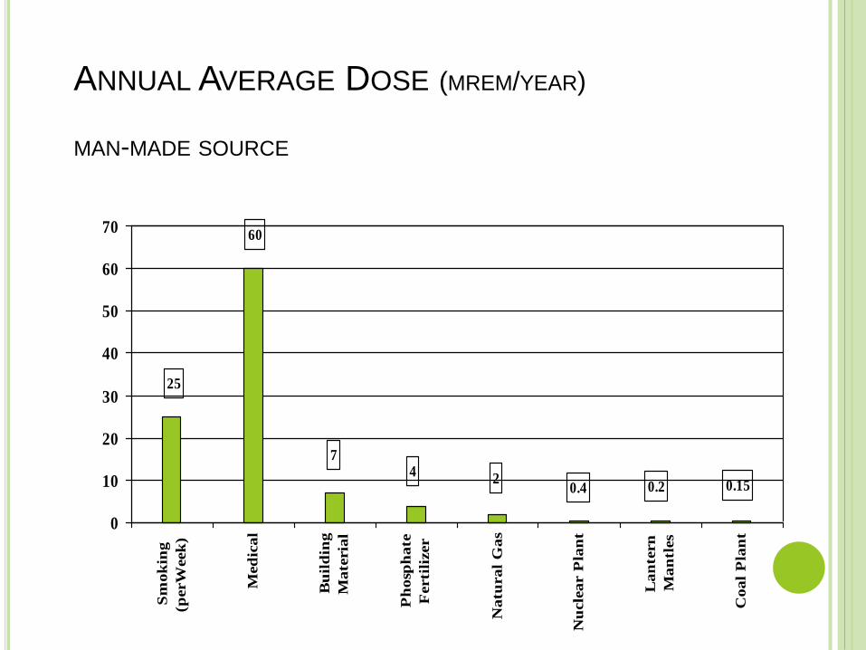
ANNUAL AVERAGE DOSE (MREM/YEAR)
MAN-MADE SOURCE
0.150.20.42
47
60
25
0
10
20
30
40
50
60
70
Sm
ok
ing
(perW
eek
)
Med
ical
Bu
ild
ing
Mate
ria
l
Ph
osp
hate
Ferti
lizer
Natu
ral
Gas
Nu
cle
ar P
lan
t
Lan
tern
Man
tles
Coal
Pla
nt
NUCLEAR FALL OUT
Fallout is the residual radioactive material
propelled into the upper atmosphere following
a nuclear blast
Carbon C14 , iodine I131, cescium Cs137 &
strontium Sc90
The Chernobyl disaster was a nuclear reactor
accident in the Chernobyl Nuclear Power Plant in
the Soviet Union on 26 April 1986.

RADIATION
Ionizing Radiation
Higher energy electromagnetic waves (gamma) or heavy
particles (beta and alpha).
High enough energy to pull electron from orbit.
Non-ionizing Radiation
Lower energy electromagnetic waves.
Not enough energy to pull electron from orbit, but can
excite the electron.
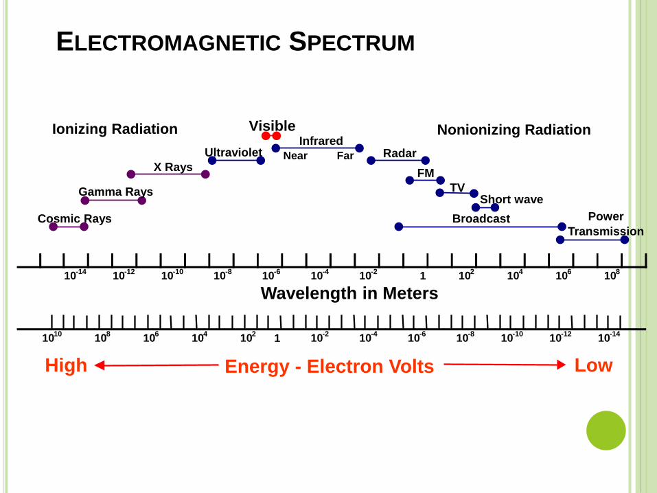
ELECTROMAGNETIC SPECTRUM
10-14
10-12
10-10
10-8
10-6
10-4
10-2
1 102
104
106
108
Wavelength in Meters
1010
108
106
104
102
1 10-2
10-4
10-6
10-8
10-10
10-12
10-14
Broadcast
Short waveTV
FM
RadarInfrared
Near Far
Visible
Ultraviolet
X Rays
Gamma Rays
Cosmic Rays Power
Transmission
Ionizing Radiation Nonionizing Radiation
Energy - Electron VoltsHigh Low

NON-IONIZING RADIATION
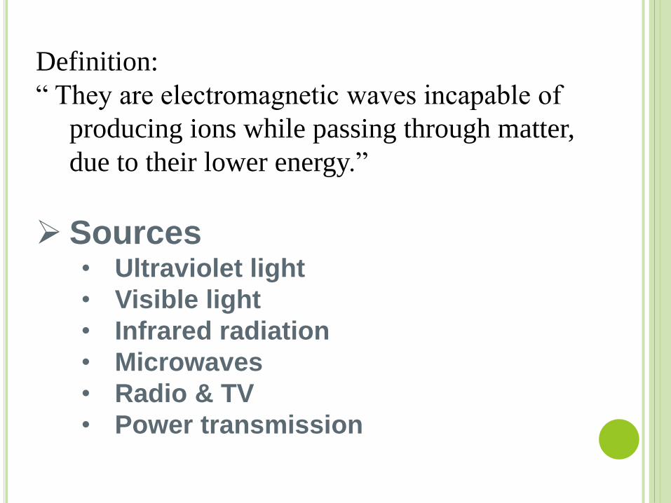
Definition:
“ They are electromagnetic waves incapable of
producing ions while passing through matter,
due to their lower energy.”
Sources• Ultraviolet light
• Visible light
• Infrared radiation
• Microwaves
• Radio & TV
• Power transmission
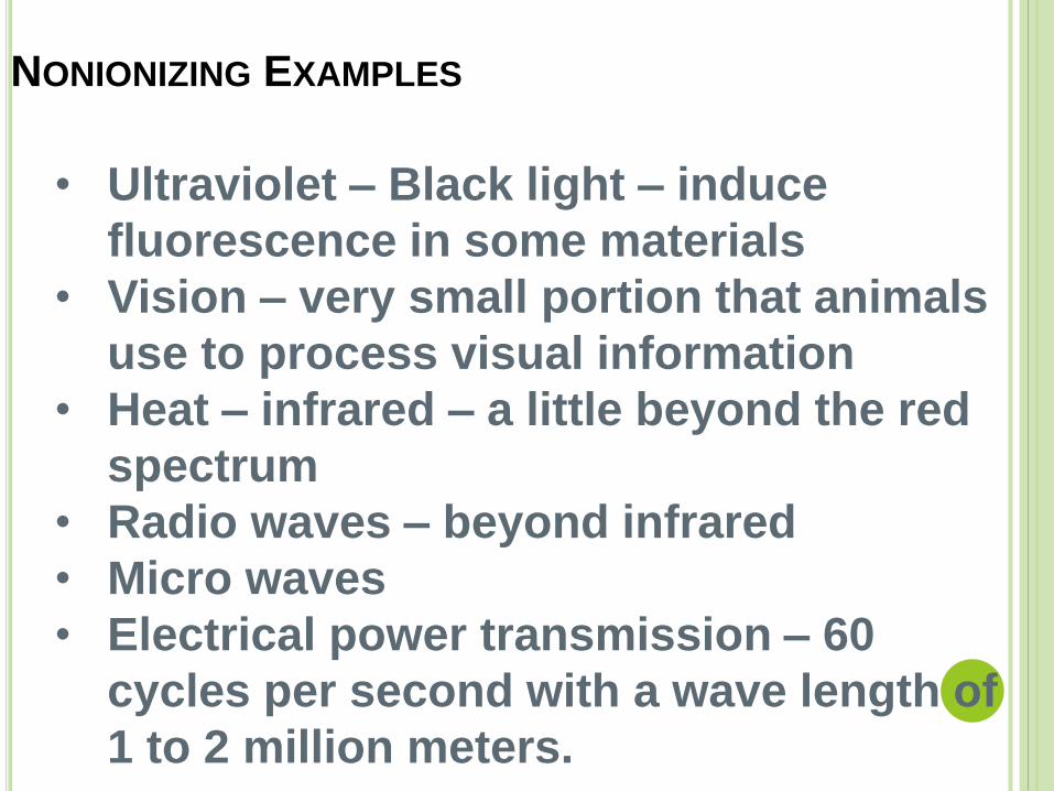
NONIONIZING EXAMPLES
• Ultraviolet – Black light – induce
fluorescence in some materials
• Vision – very small portion that animals
use to process visual information
• Heat – infrared – a little beyond the red
spectrum
• Radio waves – beyond infrared
• Micro waves
• Electrical power transmission – 60
cycles per second with a wave length of
1 to 2 million meters.
IONIZING RADIATION

IONIZING RADIATION
Definition
“ It is a type of radiation that is able to disrupt
atoms and molecules on which they pass
through, giving rise to ions and free radicals”.
Sources – x-rays, radioactive material
produce alpha, beta, and gamma
radiation, cosmic rays from the sun and
space.
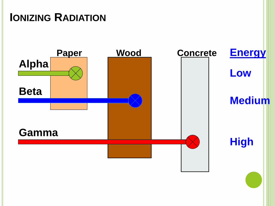
IONIZING RADIATION
Paper Wood Concrete
Alpha
Beta
Gamma
Energy
Low
Medium
High
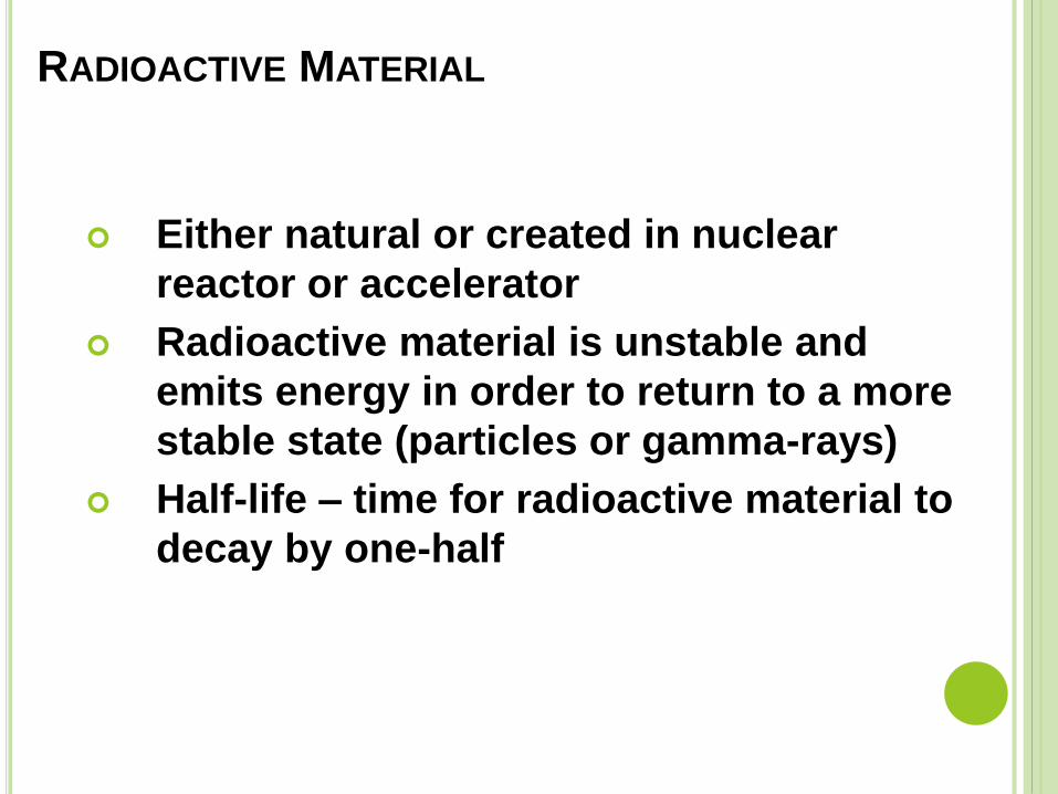
RADIOACTIVE MATERIAL
Either natural or created in nuclear
reactor or accelerator
Radioactive material is unstable and
emits energy in order to return to a more
stable state (particles or gamma-rays)
Half-life – time for radioactive material to
decay by one-half
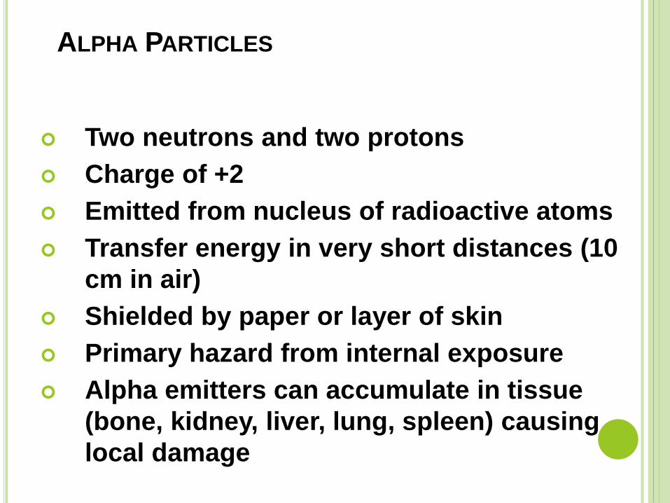
ALPHA PARTICLES
Two neutrons and two protons
Charge of +2
Emitted from nucleus of radioactive atoms
Transfer energy in very short distances (10
cm in air)
Shielded by paper or layer of skin
Primary hazard from internal exposure
Alpha emitters can accumulate in tissue
(bone, kidney, liver, lung, spleen) causing
local damage

BETA PARTICLES
Small electrically charged particles
similar to electrons
Charge of -1
Ejected from nuclei of radioactive atoms
Emitted with various kinetic energies
Shielded by wood, body penetration 0.2
to 1.3 cm depending on energy
Can cause skin burns or be an internal
hazard of ingested

GAMMA-RAYS
Electromagnetic photons or radiation
(identical to x-rays except for source)
Emitted from nucleus of radioactive
atoms – spontaneous emission
Emitted with kinetic energy related to
radioactive source
Highly penetrating – extensive shielding
required
Serious external radiation hazard

X-RAYS
Overlap with gamma-rays
Electromagnetic photons or radiation
Produced when electrons strike a target material inside and x-ray tube
Emitted with various energies & wavelengths
Highly penetrating – extensive shielding required
External radiation hazard

RADIATION UNITS
Exposure: Roentgen 1 Roentgen (R) = amount of X or gamma radiation that produces ionization resulting in 1 electrostatic unit of charge in 1 cm3 of dry air.
Absorbed Dose: rad (Roentgen absorbed dose) = absorption of 100 ergs of energy from any radiation in 1 gram of any material; 1 Gray (Gy) = 100 rads = 1 Joule/kg; Exposure to 1 Roentgen approximates 0.9 rad in air.
Biologically Equivalent Dose: Rem (Roentgen equivalent man) = dose in rads x QF, where QF = quality factor. 1 Sievert (Sv) = 100 rems.
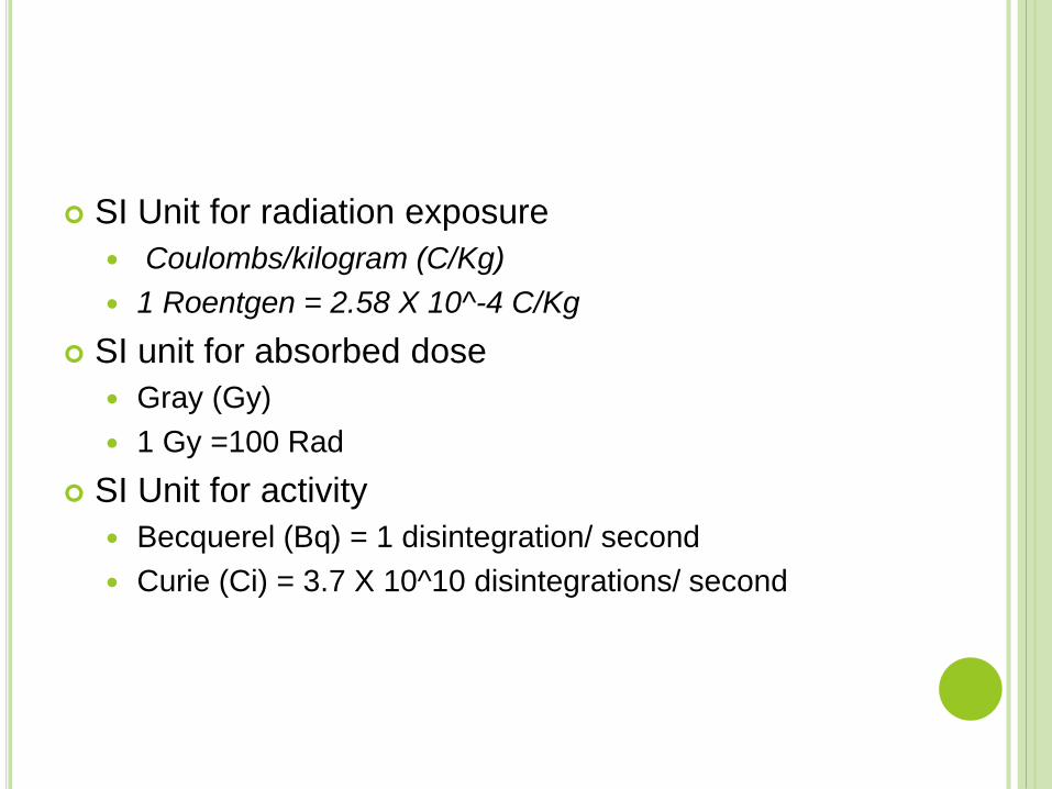
SI Unit for radiation exposure
Coulombs/kilogram (C/Kg)
1 Roentgen = 2.58 X 10^-4 C/Kg
SI unit for absorbed dose
Gray (Gy)
1 Gy =100 Rad
SI Unit for activity
Becquerel (Bq) = 1 disintegration/ second
Curie (Ci) = 3.7 X 10^10 disintegrations/ second

BIOLOGICAL EFFECTS
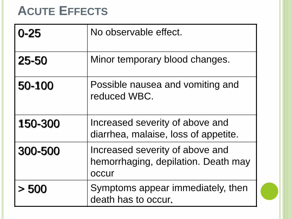
ACUTE EFFECTS
0-25 No observable effect.
25-50 Minor temporary blood changes.
50-100 Possible nausea and vomiting and
reduced WBC.
150-300 Increased severity of above and
diarrhea, malaise, loss of appetite.
300-500 Increased severity of above and
hemorrhaging, depilation. Death may
occur
> 500 Symptoms appear immediately, then
death has to occur.
Delayed Somatic Effects: Delayed effects to exposed person include: Cancer, leukemia, cataracts, life shortening from organ failure, and abortion. Probability of an effect is proportional to dose (no threshold). Severity is independent of dose. Doubling dose for cancer is approximately 10-100 rems.
Genetic Effects: Genetic effects to off-spring of exposed persons are irreversible and nearly always harmful. Doubling dose for mutation rate is approximately 50-80 rems. (Spontaneous mutation rate is approx. 10-100 mutations per million population per generation.)

DOSE RESPONSE TISSUE
Examples of tissue Sensitivity
Very High White blood cells (bone marrow)
Intestinal epithelium
Reproductive cells
High Optic lens epithelium
Esophageal epithelium
Mucous membranes
Medium Brain – Glial cells
Lung, kidney, liver, thyroid,
pancreatic epithelium
Low Mature red blood cells
Muscle cells
Mature bone and cartilage

ORGAN SPECIFIC
Skin
Erythema – desquamation (reversible)
Hair loss
Mucous Membranes
Fibrin Plaquing
Urinary and Bladder Changes
Visceral Changes (secretory)
Reproductive Organs
Irreversible damage to gametes
Sterility
Bone
Suppress osteoblast activity
Decrease number of osteocytes

• Permissible dose from man made < 5 rad/yr
• X- ray greatest hazard – 4 rad in one minute
STANDARDS
RADIATION PROTECTION
TimeReduce the spent near the source of radiation.
DistanceIncrease the distance from the source of radiation.
ShieldingPlace shielding material between you and the
source of radiation.
REDUCING EXPOSURE

MONITORING
Personal Dosimeters: Provide a record of
accumulated exposure for an individual worker
over extended periods of time and are small
enough for measuring localized exposures
Common types: Film badges; pocket dosimeters, &
Thermoluminescence detectors (TLD);

Direct Reading Survey Meters and Counters: Useful in identifying source of exposures and in evaluating potential sources, such as surface or sample contamination, source leakage, inadequate decontamination procedures, background radiation.
Common types:
Alpha Proportional or Scintillation counters Beta, gamma Geiger-Mueller or
Proportional countersX-ray, Gamma Ionization chambers Neutrons Proportional counters

Continuous Monitors: Continuous direct reading
ionization detectors (same detectors as above) can
provide read-out and/or alarm to monitor
hazardous locations and alert workers to leakage,
thereby preventing exposures.
Long-Term Samplers: Used to measure average
exposures over a longer time period. For example,
charcoal canisters or electrets are set out for days
to months to measure radon in basements (should
be <4 pCi/L).