Radial nerve - gmch.gov.in lectures/Anatomy/UL-radial nerve.pdf · Radial nerve It is the largest...
22
Radial nerve It is the largest branch of the posterior cord of brachial plexus with a root value of C5,6,7,8, T1
Transcript of Radial nerve - gmch.gov.in lectures/Anatomy/UL-radial nerve.pdf · Radial nerve It is the largest...

Radial nerve
It is the largest branch of the posterior cord of brachial plexus with a root value of C5,6,7,8, T1

Radial nerve in axilla• In axilla it passes behind
the third part of axillary artery
• Anterior to subscapularis, latissimus dorsi & and teres major
• Medial to it is axillary vein• Lateral to it are axillary
nerve and coracobrachialis
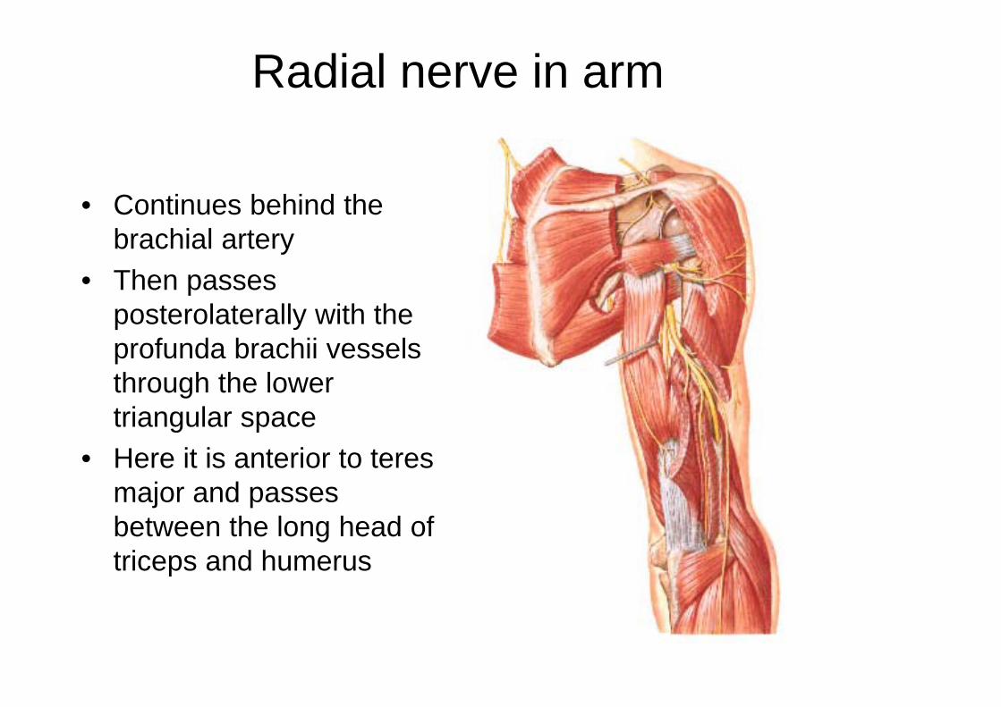
Radial nerve in arm
• Continues behind the brachial artery
• Then passes posterolaterally with the profunda brachii vessels through the lower triangular space
• Here it is anterior to teres major and passes between the long head of triceps and humerus

After this the nerve enters the radial groove with the profunda vessels
In the radial groove nerve lies between the lateral and medial heads of triceps in contact with the humerus
At the lower end of the groove, 5 cm below the deltoid tuberosity, the nerve pierces the lateral intermuscular septum and passes into anterior compartment of arm
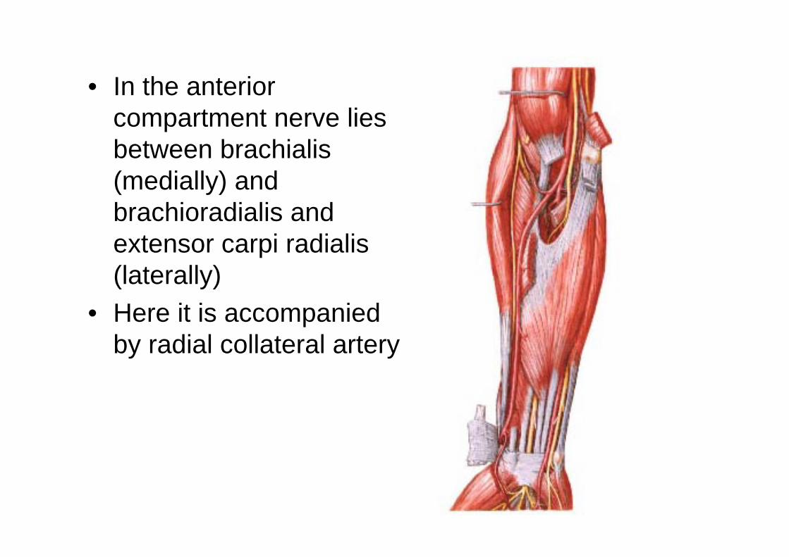
• In the anterior compartment nerve lies between brachialis (medially) and brachioradialis and extensor carpi radialis (laterally)
• Here it is accompanied by radial collateral artery

Branches of radial nerve • Muscular branches:• Before entering spiral
groove- to long andmedial heads of triceps
• In the spiral groove-lateral and medial headsof triceps and throughnerve to medial head toanconeus
• Below the radial groove,on the front of the arm, itsupplies, the brachialis,brachioradialis and theextensor carpi radialislongus

• Cutaneous branches:• Above the radial groove-posterior cutaneous nerve of the arm• In the radial groove-lower lateral cutaneous nerve of the arm and
posterior cutaneous nerve of the forearm• Articular branches: to elbow

Superficial terminal branch
• In the forearm the superficial branch descend between brachioradialis anteriorly and supinator posteriorly lying lateral to radial artery
• In the middle third it lies posterior to brachioradialis, lying successively on pronator teres, FDS (radial head) and FPL and lateral to radial artery

• About 7 cm proximal to the wrist nerve leaves the artery • passes deep to the tendon of brachioradialis• Curves around the lateral side of radius• Pierces the deep fascia• Divides into 4 or 5 digital nerves on the dorsum of hand. it communicates with posterior & lateral cutaneous nerves of forearm• Supplies radial half of the dorsum of the hand, proximal part of the dorsal surface of thumb, index finger & the lateral half of the middle finger

Deep branch of radial nerve
• Deep terminal branch reaches the back of forearm by passing between two heads of supinator
• before piercing supplies ECRB and supinator

Posterior interosseous nerve of forearm
• it is deep branch of radial nerve in forearm
• Reaches the back of forearm by passing between the two heads of supinator
• Descends between the superficial &and deep group of extensor muscles lying on the interosseous membrane

• The upper part of nerve is accompanied by posterior interosseous artery but the lower part is accompanied by anterior interosseous artery
• Terminates in to a pseudoganglion and ends by supplying the wrist and carpal joint
• Branches :• Muscular –supinator
ED,EDM &ECU-Divides into lateral & medial branch

• lateral branch supplies APL &EPB• Medial branch supplies EPL& EI• Articular branch: to wrist joint, distal
radioulnar joint, some intercarpal & intermetacarpal joint
• Sensory branches :to interosseous membrane, radius & ulna

Damage to the main trunk of the brachial plexus results in
• Paralysis of triceps –loss of extension of elbow• Paralysis of all the extensors of wrist –wrist drop
i.e.. loss of extension of wrist• Extension of metacarpophalangeal joints can not
be performed• Supination in extended elbow is not possible• Area of anaesthesia over dorsum of hand
between the first and second metacarpal

Lesions of the radial nerve in arm and axilla
• Injury can be due to-– pressure of the crutch (crutch palsy)– Prolonged hanging of the arm over the arm of
a chair (Saturday night palsy)– fracture of humeral shaft in spiral groove

Lesion of posterior interosseous nerve
• Lesion produces wrist drop• Produced due to paralysis of extensors &
unopposed action of flexors• Supination though not lost, is weak since it
can be produced by biceps
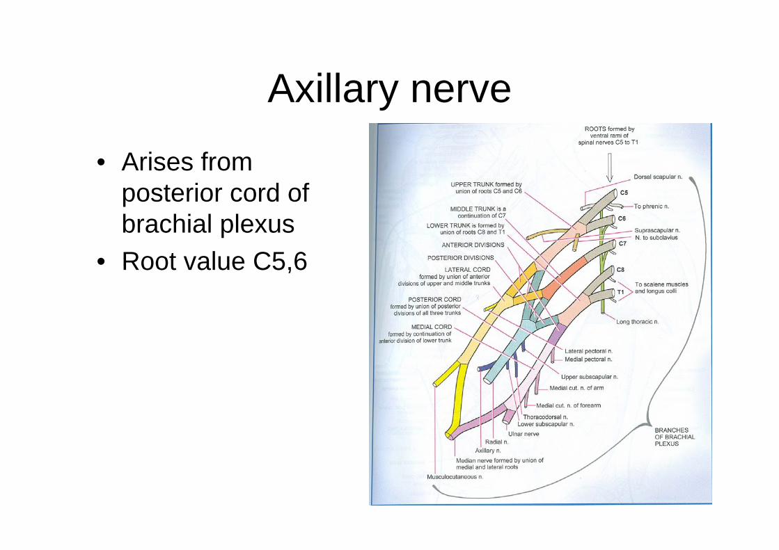
Axillary nerve
• Arises from posterior cord of brachial plexus
• Root value C5,6

Axillary nerve in the axilla
• At first lateral to radial nerve
• Posterior to axillary artery
• Anterior to subscapularis

•At the lower border of subscapulris nerve curves back inferior to shoulder joint capsule and with the posterior circumflex humeral vessels, traverses quadrangular space

Branches of axillary nerve• In quadrangular space the
nerve divides in to anterior and posterior branches
• Anterior branch, with posterior circumflex humeral vessels, curve round the humeral neck, deep to deltoid, to its anterior border' supplying it
• Further this nerve gives few cutaneous branches,,which pierce the muscle to ramify in the skin over its lower part

• Posterior branch: • Supplies teres minor and
posterior part of deltoid• On the branch to the
teres minor pseudo ganglion exists
• Continues as the upper lateral cutaneous nerve of arm which supplies skin over the lower part of deltoid and upper part of long head of triceps
• This branch also gives a articular branch to shoulder joint

Lesion of axillary nerve• Injured in the fractures of surgical neck of
humerus• Also in dislocation of shoulder joint• Results in loss of rounded contour of shoulder
and greater tuberosity of humerus becomes prominent due to wasting of deltoid
• Loss of abduction of shoulder due to paralysis of deltoid
• Sensory loss over the skin on the lower part of deltoid