RAB PROTEIN EXPRESSION REDUCES DIMER/OLIGOMER...
30
RAB PROTEIN EXPRESSION REDUCES DIMER/OLIGOMER ALPHA-SYNUCLEIN AND FACILITATES DEGRADATION By AMANDA HERRING A THESIS PRESENTED TO THE GRADUATE SCHOOL OF THE UNIVERSITY OF FLORIDA IN PARTIAL FULFILLMENT OF THE REQUIREMENTS FOR THE DEGREE OF MASTER OF SCIENCE UNIVERSITY OF FLORIDA 2013
Transcript of RAB PROTEIN EXPRESSION REDUCES DIMER/OLIGOMER...
RAB PROTEIN EXPRESSION REDUCES DIMER/OLIGOMER ALPHA-SYNUCLEIN AND FACILITATES DEGRADATION
By
AMANDA HERRING
A THESIS PRESENTED TO THE GRADUATE SCHOOL
OF THE UNIVERSITY OF FLORIDA IN PARTIAL FULFILLMENT OF THE REQUIREMENTS FOR THE DEGREE OF
MASTER OF SCIENCE
UNIVERSITY OF FLORIDA
2013

© 2013 Amanda Herring

To my future children, for all the times you complain about having to write a page-long book report

4
ACKNOWLEDGMENTS
I thank my family, friends, and most of all, my awesome boss who has had to
deal with me for the past three years.
5
TABLE OF CONTENTS page
ACKNOWLEDGMENTS ...............................................................................................................4
LIST OF FIGURES........................................................................................................................6
ABSTRACT ....................................................................................................................................7
CHAPTER
1 RABS AND α-SYNUCLEIN ..................................................................................................9
Introduction .............................................................................................................................9 Materials and Methods ........................................................................................................12
Plasmids and Antibodies .............................................................................................12
Cell Culture and Transfections ...................................................................................13 Gaussia Luciferase Protein-Complementation and MTS Assays .........................13
Size Exclusion Chromatography (SEC) to Separate Monomeric and Oligomeric α-Syn .......................................................................................................14
Western Blot Analysis ..................................................................................................15
Statistical Analysis ........................................................................................................16
2 RESULTS ..............................................................................................................................17
mRab1 and Rab8A Decrease α-Syn Oligomers .............................................................17
Rab8A Facilitates Degradation of α-Syn ..........................................................................19
3 DISCUSSION .......................................................................................................................21
LIST OF REFERENCES ............................................................................................................27
BIOGRAPHICAL SKETCH ........................................................................................................30
6
LIST OF FIGURES
Figure page Figure 1-1. mRab1 and Rab8A decrease α-Syn oligomers. ......................................... 25
Figure 1-2. Rab8A facilitates α-Syn degradation. ......................................................... 26
7
Abstract of Thesis Presented to the Graduate School of the University of Florida in Partial Fulfillment of the Requirements for the Degree of Master of Science
RAB PROTEIN EXPRESSION REDUCES DIMER/OLIGOMER ALPHA-SYNUCLEIN
AND FACILITATES DEGRADATION
By
Amanda Herring
December 2013
Chair: Nikolaus McFarland Major: Medical Science
The protein alpha-synuclein (α-Syn) is implicated in the etiology and
pathogenesis of Parkinson’s disease. Elevated levels of α-Syn can cause detrimental
effects to cellular homeostasis, including deficits in mitochondrial maintenance and
vesicle trafficking, such as ER-Golgi transport, depletion of reserve vesicle pool and
reduced synaptic vesicle response. Recent data suggests that the trafficking deficits
caused by α-Syn may be due to an interaction with Rab proteins. Rab proteins are small
GTPases critical for membrane transport, including intracellular trafficking and
neurotransmitter release. In several PD animal models, overexpression of murine Rab1
and human Rabs 3A and 8A can ameliorate the trafficking deficits caused by α-Syn
overexpression. However, it is unknown whether this rescue is due to Rab protein
interaction with the oligomeric forms of α-Syn thought to be the more toxic species in
disease pathogenesis. To assay for the effects of these Rabs on oligomeric α-Syn, we
utilized a previously-established bi-luminescent protein complementation in which fusion
of Gaussia luciferase hemi-constructs to α-Syn results in an active luciferase enzyme
upon α-Syn dimerization/oligomerization, allowing for direct quantification of these α-
8
Syn species. Supplementing this assay with size-exclusion chromatography and native
immunoblot, we tested the effects of mRab1, Rab3A, and Rab8A to reduce the level of
α-Syn oligomers. While Rab3A had no effect on reducing α-Syn oligomers,
overexpression of mRab1 and Rab8A reduced the level of both intracellular and
secreted α-Syn oligomers. Furthermore, Rab8A facilitated the degradation of α-Syn,
reducing the level of both oligomeric and total α-Syn. Together, these data provide
evidence that 1) toxic oligomeric α-Syn interferes with specific Rab pathways over
others and that, 2) Rab8A, which has not been identified as a Rab involved in protein
degradation, may have a previously unknown function upon increased α-Syn burden.
Whether this new role affects normal Rab8A functioning remains to be seen.
9
CHAPTER 1 RABS AND α-SYNUCLEIN
Introduction
Parkinson’s disease (PD) is the second most common neurodegenerative
disorder in the United States, affecting approximately one million individuals (1).
Pathological hallmarks of PD include the progressive loss of midbrain dopaminergic
neurons essential for the control of movement and the presence of Lewy bodies, or
intracellular accumulations enriched with the protein alpha-Synuclein (α-Syn). The
implication for α-Syn as a potent factor in the pathophysiology of PD is evident from
mutations in the SNCA gene, encoding for α-Syn, as well as duplications and
triplications in the SNCA locus, all of which are known to cause early-onset, familial
forms of PD (2). Elevated levels of α-Syn have also been found in the blood plasma and
cerebrospinal fluid of PD patients, and it is thought that trans-synaptic transmission of
small oligomeric forms of α-Syn cause the spreading pathology and disease
progression (3-7).
In the cell, cytoplasmic α-Syn exists primarily as a natively unfolded monomer
and adopts an α-helical structure upon binding to lipid membranes (8). Although the
exact physiological role of α-Syn remains unclear, it is thought that α-Syn may play a
function in neurotransmitter release (9-12). α-Syn was originally identified as a
presynaptic protein shown to associate with vesicles (11). Mice lacking α-Syn are
grossly normal, but display functional deficits in activity-dependent dopamine release in
the nigrostriatal system, significant impairments in synaptic response, and a depletion of
the reserve vesicle pool upon repetitive neuronal stimulation (9, 10, 12). α-Syn has also
been found to interact with the amino-terminus of the SNARE protein synaptobrevin-2
10
and to enhance SNARE-complex assembly in vivo, suggesting a role for α-Syn in
modulating the SNARE-complex at the presynaptic terminal (13). Furthermore, the
neurodegenerative phenotype in transgenic mice lacking CSPα, a molecular chaperone
involved in SNARE-complex assembly, can be rescued by α-Syn (14). These studies
suggest that the primary endogenous function of α-Syn is most likely involved in vesicle
trafficking at the neuronal synapse.
However, elevated α-Syn levels have been shown to affect earlier stages of
intracellular trafficking pathways, also (15, 16). Overexpression of α-Syn in yeast
causes ER stress and inhibits ER-Golgi trafficking (15). Increased levels of α-Syn also
promote the accumulation of small vesicle clusters at the peripheral ER and plasma
membrane in a dose-dependent manner (16). A genome wide screen of yeast open
reading frames revealed the largest class of suppressors for α-Syn toxicity to be those
associated with early stages of membrane trafficking, including ER-Golgi COPII
components, SNARE proteins, and— of particular importance here— Ypt1p, a Rab
GTPase (15).
Rab GTPases represent the largest class of RAS-related small GTPases, and
are vital to maintaining efficient vesicle transport. It was demonstrated that Ypt1p and
Rab1, the murine YPT1 ortholog and an early ER-Golgi Rab, rescued dopaminergic cell
loss associated with α-Syn overexpression in several animal models of PD, including
Drosophila and Caenorhabditis elegans (15). More importantly, Rab1 expression also
rescued α-Syn-induced cell loss in mammalian dopaminergic neurons (15). Following
the identification of Ypt1p/Rab1 as a potent inhibitor of α-Syn toxicity in yeast and PD
animal models, additional Rab proteins were shown to rescue α-Syn-induced transport
11
deficits and toxicity. Rab8A, the paralog to human Rab1A, localizes to post-Golgi
vesicles and has been shown to facilitate trafficking to the plasma membrane (17).
Rab3A, a neuronal-specific Rab, is enriched at the synaptic membrane and is involved
in neurotransmitter release (18, 19). In addition to Rab1, both Rab3A and Rab8A rescue
α-Syn-induced DA neuronal cell loss in C. elegans and rat primary neurons (16).
Furthermore, immunoprecipitation studies in mutant α-Syn transgenic mice suggest that
Rab3A and Rab8A may either directly or indirectly interact with α-Syn (20). In several
studies of the PD-related disorders diffuse Lewy body disease and multiple system
atrophy—both of which contain aggregated α-Syn inclusions— Rab3A was found to
immunoprecipitate with α-Syn in diseased brains but not in controls (21, 22). Together,
these studies suggest that toxic levels of α-Syn can affect multiple transport pathways
and that the trafficking deficits seen in disease may be a consequence of α-Syn
interaction with Rab proteins.
It is thought that the primary toxic species of α-Syn is not the monomeric form or
the fibrillar structures found in Lewy bodies, but small oligomers of α-Syn similar to
those found in the blood plasma and CSF of PD patients (3, 4, 23, 24). In several PD
model systems, Karpinar et al. (2009) expressed mutant forms of α-Syn with varying
abilities to form fibrils and demonstrated that impairment of oligomeric α-Syn to form
fibrils was correlated to a higher level of neurodegeneration (25). Although several Rab
proteins have been shown to rescue the trafficking deficits caused by overexpression of
α-Syn, the mechanism of reduced α-Syn toxicity remains unclear. We hypothesized that
the mechanism by which Rab proteins may ameliorate trafficking deficits is by reducing
“toxic” oligomeric species of α-Syn.
12
To examine the effects of Rab proteins on α-Syn oligomer formation, we utilized
a protein complementation assay developed by fusion of the N- or C- terminal fragment
of Gaussia luciferase (hemi-constructs) to full-length wild-type human α-Syn (26). Co-
expression of both hemi-constructs and subsequent dimer/oligomerization of α-Syn
results in reconstitution of an active luciferase enzyme that can be quantitatively
measured with the addition of substrate, coelenterazine. Here, we demonstrate that α-
Syn oligomers, as measured by changes in luciferase activity and high-molecular weight
(HMW) native protein, are reduced by expression of either mRab1 or Rab8A.
Furthermore, Rab8A also facilitates the degradation of α-Syn. Although we did not
assay for direct interaction of Rab8A with α-Syn, the interaction between Rab8A and α-
Syn shown in previous studies combined with the data presented here suggests that the
ability of Rab8A to facilitate degradation of oligomeric α-Syn may provide a useful
therapeutic target in future.
Materials and Methods
Plasmids and Antibodies
Fusion constructs α-Syn-hGLuc1 (S1) and α-Syn-hGLuc2 (S2) was generated by
subcloning α-Syn into NotI/ClaI sites of humanized Gaussia luciferase and were kindly
provided by Dr. Pamela McLean (Mayo Clinic, Jacksonville, FL; ref. 13). pcDNA-
mRab1, pcDNA-Rab3A, and pcDNA-Rab8A were kindly provided by Dr. Susan
Lindquist (Massachusetts Institute of Technology, Cambridge Massachusetts). FLAG-
mRab1, FLAG-Rab3A, and FLAG-Rab8A were generated by subcloning the sequence
GGATTACAAGGATGACGACGAT to the amino-terminus of pcDNA-Rab constructs.
Antibodies were as follows: Millipore rabbit anti-α-Syn (1:2000), BD Transduction
13
Laboratories mouse anti-α-Syn (1:5000), Sigma mouse anti-FLAG M2 (1:500), Sigma
mouse anti-β-actin (1:100,000).
Cell Culture and Transfections
Human H4 neuroglioma cells (HTB-148; American Type Culture Collection) were grown
in Opti-MEM + GlutaMAX media (Life Technologies) containing 5% Fetal Bovine Serum
and antibiotic (50U/mL penicillin, 50mg/mL streptomycin) in culture flasks under
standard tissue culture conditions (37°C under 5% CO2). Cells were plated 24 h prior to
transfection, and transiently transfected at 80-90% confluency with SuperFect
Transfection Reagent (Quiagen) according to the manufacturer’s instructions. For
conditioned medium (CM) experiments, media was collected 48 h post-transfection. To
eliminate floating cells, media was centrifuged prior to use for 2 min, 3000rpm, at RT
and analyzed immediately for luciferase assays, or 5 min, 3000rpm, at RT and stored at
-20°C for size exclusion assays. For native and total α-Syn studies, cells were lysed 48
h post-transfection in a non-detergent lysis buffer (50mM Tris pH 7.4, 175mM NaCl,
5mM EDTA pH 8.0, protease inhibitor cocktail (Roche Applied Science). For
cycloheximide studies, cells were transiently transfected in a 6-well plate. 24 h post-
transfection, cycloheximide was added to new media at a final concentration of 3ug/mL.
Cells were washed with PBS, lysed at indicated time points in RIPA buffer (50mM Tris
pH 7.4, 150mM NaCl, 0.1% SDS, 0.5% Na deoxycholate, 1% TritonX-100, 10mM N-
ethylmaleimide [Fisher], protease [Roche complete mini] and phosphatase [Fisher]
inhibitor), and stored at –20°C until all time points had been collected.
Gaussia Luciferase Protein-Complementation and MTS Assays
To assay for changes in oligomeric α-Syn, H4 cells were transiently transfected in 96-
well plates with fusion constructs S1, S2 and either pcDNA control vector or the
14
indicated FLAG-Rab plasmid. 48 hours post-transfection, culture medium was
transferred to a new 96-well plate and centrifuged as indicated above. Cells were
washed with PBS and replaced with serum- and phenol-red free Opti-MEM. Native
coelenterazine (Nanolight), a cell-permeable substrate of Gaussia luciferase, was
resuspended in methanol to 1 mg/mL and dispensed to a final concentration of 20µM
onto live cells or CM by an automated plate reader, (Synergy HT, Bio-Tek). The
bioluminescent signal generated by protein complementation of the luciferase enzyme
was integrated over 2 seconds before measurement at 480nm.
Following luciferase assays, cell viability was measured using the CellTiter 96®
AQueous One Solution Cell Proliferation Assay kit as per manufacture instructions.
Briefly, media was removed following luciferase assay to a final volume of 100µL/well.
20µL MTS reagent was added and cells were incubated at 37°C for 1-3 hours.
Absorbance was measured at 490nm with a microplate reader (SynergyHT, Bio-Tek).
Control plasmid (pcDNA) was used to measure background luciferase signal and
provide a baseline for cell viability assays. Each condition was transfected in 8
replicates per plate, and data from a minimum of 3 plates was collected for statistical
analysis.
Size Exclusion Chromatography (SEC) to Separate Monomeric and Oligomeric α-Syn
SEC was performed as described previously (23). To assay extracellular α-Syn
oligomerization, 500µL CM from H4 cells transfected with S1, S2, and pcDNA control or
the indicated FLAG-Rab was thawed and filtered through an Ultrafree®-MC Centrifugal
Filter (Millipore; PVDF 0.22µm) for 4 min, 10,000g at room temperature. 350µL filtered
15
CM was injected into a Superose 6 10/300 GL gel-filtration column (GE Healthcare) and
eluted with PBS at a flow rate of 0.4mL/min. 200µL fractions were collected into
microtubes (Fisher), and 100µL of each fraction was transferred to a 96-well plate and
analyzed for Gaussia luciferase protein-complementation, as described above.
To measure intracellular α-Syn oligomerization, cells from the same CM samples
were scraped down, washed once with PBS, and mechanically sheered by passage
through a 1mL 27-gauge needle in 170µL of a non-detergent lysis buffer (50mM Tris pH
7.4, 175mM NaCl, 5mM EDTA pH 8.0, protease inhibitor cocktail (Roche Applied
Science). Lysates were spun at 13,000 rpm for 10 min at 4°C and stored at -20°C. For
SEC, lysates were thawed and filtered through an Ultrafree®-MC Centrifugal Filter
(Millipore; PVDF 0.22µm) for 4 min, 10,000g at room temperature. 140uL of filtered cell
lysate was used for SEC under the same conditions as the CM and analyzed for
protein-complementation.
Western Blot Analysis
Denatured cell lysates from cycloheximide or total α-Syn experiments were subjected to
NuPAGE 4-12% Bis-tris SDS-PAGE electrophoresis (Life Sciences) followed by
western blot analysis with antibodies described above. Protein bands were visualized by
the ECL method (Amersham) or by the Odyssey system (Licor) using IR700/800
secondary antibodies (Rockland). Native western blot was performed under non-
denaturing conditions using NativePAGE 4-16% Tris-glycine gels (Life Sciences)
following manufacturer instructions.

16
Statistical Analysis
Statistical analyses were carried out using Prism 5.0 (GraphPad). One-way analysis of
variance is used to evaluate groups with Turkey’s multiple comparisons post-hoc.
Values in the figures are expressed as means ± SEM.
17
CHAPTER 2 RESULTS
mRab1 and Rab8A Decrease α-Syn Oligomers
Expression of mRab1, Rab3A, and Rab8A has been shown to ameliorate toxicity
and transport deficits caused by α-Syn overexpression in several PD animal models,
including worms, flies, and rat midbrain dopaminergic neurons (15, 16). It has also been
suggested that α-Syn may interact with Rab proteins in disease, providing a potential
mechanism by which toxic forms of α-Syn can impair vesicle transport (21, 22).
However, it is unknown whether Rab proteins can interact with different cytoplasmic
forms of α-Syn, including small oligomeric forms that are thought to play a primary role
in pathology. To determine the effects of Rab overexpression on α-Syn oligomerization,
we utilized a protein complementation assay employing the bioluminescent protein
Gaussia luciferase (26). In this model, two halves of the Gaussia luciferase enzyme are
fused in frame to full-length, wild-type α-Syn. Co-expression of Syn-hGLuc1 (S1) and
Syn-hGLuc2 (S2), but not S1 or S2 with either of the hGLuc hemi-constructs alone,
results in high levels of luciferase signal indicative of α-Syn dimer/oligomer formation
(26). This highly sensitive, quantitative assay can be used in both live cells and
conditioned media (CM) to measure α-Syn.
We used transient transfection in H4 neuroglioma cells to evaluate the level of α-
Syn dimer/oligomer formation in the presence mRab1 (a murine homolog of the human
Rab1A), Rab3A, and Rab8A. We expressed either S1 or S2 constructs with a control
plasmid (EGFP) to determine the background levels of the assay. To take into account
cell death caused by the transfection process, we performed an MTS assay immediately
following luciferase assay. The luciferase levels were then normalized to live cells and
18
are shown as the percent reduction in luciferase activity compared to the model
condition (S1/S2 with EGFP) (Fig. 1A). After 2 d, expression of S1 or S2 constructs
alone with control plasmid resulted in minimal luciferase signal (Fig. 1A). Co-expression
of either mRab1 or Rab8A caused a significant reduction in luciferase activity (mRab1,
49%; Rab8A, 74%; n=4, p<.001) compared to control EGFP plasmid. Previous studies
have indicated that Rab3A can rescue α-Syn-induced toxicity (16); however we did not
detect any change in luciferase activity in the presence of Rab3A.
We hypothesized that the reduction in α-Syn luciferase signal by Rab proteins
may be due to a shift of α-Syn into smaller molecular weight forms. Previous
characterization of the S1/S2 model has shown that co-expression of S1/S2 in H4
neuroglioma cells results in a high molecular weight (HMW) smear of α-Syn under
native conditions indicative of a wide range of α-Syn oligomers (26). Our data is
consistent with this, and expression of S1, S2, or S1/S2 together results in a HMW
smear of α-Syn ranging from 146kDa to over 700kDa (Fig. 1B, lane 1). Native-PAGE of
wild-type α-Syn demonstrates a similar HMW smear and confirms that wild-type α-Syn
behaves in a similar manner under our transfection conditions (Fig. 1B, lane 7).
Using Native and SDS-PAGE Western followed by α-Syn immunoblot, we saw no
change in HMW α-Syn with co-expression of mRab1 and Rab3A, but a significant
reduction in HMW α-Syn was observed with Rab8A (Figure 1-1B, lane 6). Contrary to
our hypothesis however, this does not appear to be due to a shift of α-Syn to smaller
MW forms, but due to a reduction in the total level of α-Syn as shown when run under
denaturing conditions.
19
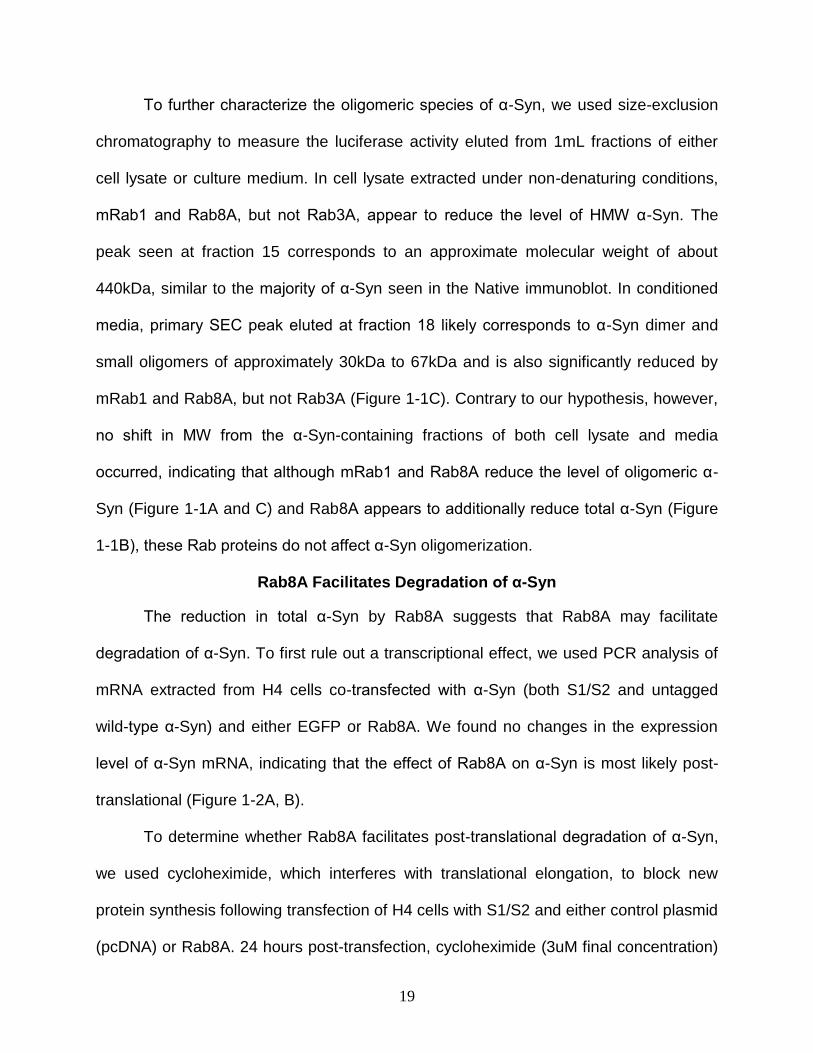
To further characterize the oligomeric species of α-Syn, we used size-exclusion
chromatography to measure the luciferase activity eluted from 1mL fractions of either
cell lysate or culture medium. In cell lysate extracted under non-denaturing conditions,
mRab1 and Rab8A, but not Rab3A, appear to reduce the level of HMW α-Syn. The
peak seen at fraction 15 corresponds to an approximate molecular weight of about
440kDa, similar to the majority of α-Syn seen in the Native immunoblot. In conditioned
media, primary SEC peak eluted at fraction 18 likely corresponds to α-Syn dimer and
small oligomers of approximately 30kDa to 67kDa and is also significantly reduced by
mRab1 and Rab8A, but not Rab3A (Figure 1-1C). Contrary to our hypothesis, however,
no shift in MW from the α-Syn-containing fractions of both cell lysate and media
occurred, indicating that although mRab1 and Rab8A reduce the level of oligomeric α-
Syn (Figure 1-1A and C) and Rab8A appears to additionally reduce total α-Syn (Figure
1-1B), these Rab proteins do not affect α-Syn oligomerization.
Rab8A Facilitates Degradation of α-Syn
The reduction in total α-Syn by Rab8A suggests that Rab8A may facilitate
degradation of α-Syn. To first rule out a transcriptional effect, we used PCR analysis of
mRNA extracted from H4 cells co-transfected with α-Syn (both S1/S2 and untagged
wild-type α-Syn) and either EGFP or Rab8A. We found no changes in the expression
level of α-Syn mRNA, indicating that the effect of Rab8A on α-Syn is most likely post-
translational (Figure 1-2A, B).
To determine whether Rab8A facilitates post-translational degradation of α-Syn,
we used cycloheximide, which interferes with translational elongation, to block new
protein synthesis following transfection of H4 cells with S1/S2 and either control plasmid
(pcDNA) or Rab8A. 24 hours post-transfection, cycloheximide (3uM final concentration)
20
was added to the cells and lysates were collected over a 48 hour period. As expected,
there was a marked reduction of α-Syn when co-transfected with Rab8A in cell lysate
collected at Time 0 (Figure 1-2D, lanes 1 and 5). However, analysis of the relative α-
Syn level reveals that degradation of α-Syn is significantly enhanced in the presence of
Rab8A but not control plasmid (Figure 1-2C). To rule out a possible effect of the
luciferase portion of the protein, we performed identical experiments with wild-type α-
Syn with similar results (data not shown).
21
CHAPTER 3 DISCUSSION
Rab proteins have been shown to ameliorate toxicity caused by α-Syn
overexpression in several PD animal models (15, 16). We hypothesized that one
potential mechanism of Rab effects on α-Syn could be that Rab proteins reduce α-Syn
oligomers. Using a previously established cell model for α-Syn dimer/oligomerization,
we tested our hypothesis using mRab1, Rab3A, and Rab8A. In a bioluminescent protein
complementation assay (PCA) utilizing co-expression of α-Syn-luciferase hemi-
constructs, we found that mRab1 and Rab8A (but not Rab3A) cause a significant
reduction in luciferase signal, with Rab8A decreasing luciferase activity by almost 75%.
We further characterized this using size-exclusion chromatography to identify whether
the reduction in luciferase activity corresponded to a decrease in high molecular weight
α-Syn. We found that, in agreement with our luciferase assay, both mRab1 and Rab8A
(but not Rab3A) reduce the luciferase signal of α-Syn eluted from SEC fractions in both
cell lysate and conditioned media, suggesting that mRab1 and Rab8A have an effect on
α-Syn dimer/oligomer formation. In contrast to our hypothesis however, we did not see a
shift in the molecular weight of α-Syn, suggesting that although mRab1 and Rab8A may
affect dimer/oligomer formation, they are not reduced into smaller molecular weight
forms.
Of novel importance, we found that in addition to a decrease in luciferase signal
by Rab8A in the PCA and SEC assays— presumably corresponding to a reduction in α-
Syn dimer/oligomers— Rab8A also reduces the total level of α-Syn. This was not due to
a transcriptional effect, as we did not see any change in α-Syn (both S1/S2 and wtSyn)
mRNA expression in the presence of Rab8A. To determine whether Rab8A might have
22
a post-translational effect on α-Syn, we used cycloheximide to block translation and
measure the degradation rate of α-Syn. At 24 h post-transfection there is already a
marked decrease in α-Syn expression in the presence of Rab8A (Fig 1, lanes 1 and 5).
However, by comparing the relative rate of α-Syn degradation, we found a significant
increase in the rate of α-Syn degradation in the presence of Rab8A. This is most likely
not due to an immediate turnover of α-Syn by Rab8A following transfection, as we have
seen approximately equal levels of α-Syn as well as luciferase signal with or without the
presence of Rab8A at 12 h post-transfection, suggesting that the effects of Rab8A on
decreasing α-Syn levels are only effective once α-Syn reaches a certain level
(unpublished observation). However, the mechanism by which this occurs and whether
this is due to a direct or indirect interaction of α-Syn with Rab8A still remains to be seen.
We have demonstrated that although all three of the Rab proteins used in this
study have previously been shown to reduce the toxicity and transport deficits caused
by α-Syn overexpression, the effects they have on oligomeric forms of α-Syn are
markedly different. One potential reason for this may be due to the different pathways in
which each of the Rabs are involved. Rab3A, a neuronal-specific Rab, localizes to
synaptic vesicles and most likely plays a role in vesicle release (27). One of the known
effector proteins of Rab3A is rabphilin-3A, a synaptic vesicle Ca2+/phospholipid-binding
protein (28). Rabphilin-3A, which assists in synaptic targeting, depends on Rab3A for
normal function. Immunoprecipitation studies of Rab-α-Syn interactions in diffuse Lewy
body disease, a PD-related disorder with similar α-Syn inclusions, demonstrated that in
control cases, rabphilin is pulled down with Rab3A (22). However, in diseased brains,
rabphilin is no longer precipitated by Rab3A, which instead pulls down α-Syn (22).
23
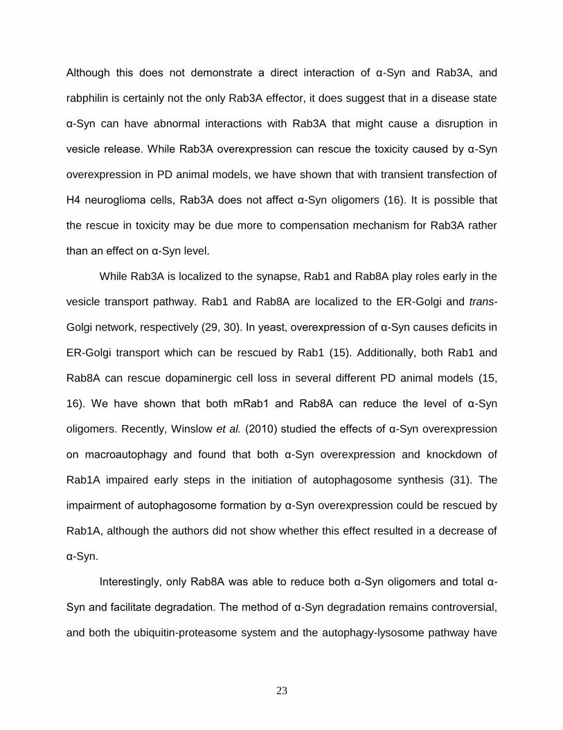
Although this does not demonstrate a direct interaction of α-Syn and Rab3A, and
rabphilin is certainly not the only Rab3A effector, it does suggest that in a disease state
α-Syn can have abnormal interactions with Rab3A that might cause a disruption in
vesicle release. While Rab3A overexpression can rescue the toxicity caused by α-Syn
overexpression in PD animal models, we have shown that with transient transfection of
H4 neuroglioma cells, Rab3A does not affect α-Syn oligomers (16). It is possible that
the rescue in toxicity may be due more to compensation mechanism for Rab3A rather
than an effect on α-Syn level.
While Rab3A is localized to the synapse, Rab1 and Rab8A play roles early in the
vesicle transport pathway. Rab1 and Rab8A are localized to the ER-Golgi and trans-
Golgi network, respectively (29, 30). In yeast, overexpression of α-Syn causes deficits in
ER-Golgi transport which can be rescued by Rab1 (15). Additionally, both Rab1 and
Rab8A can rescue dopaminergic cell loss in several different PD animal models (15,
16). We have shown that both mRab1 and Rab8A can reduce the level of α-Syn
oligomers. Recently, Winslow et al. (2010) studied the effects of α-Syn overexpression
on macroautophagy and found that both α-Syn overexpression and knockdown of
Rab1A impaired early steps in the initiation of autophagosome synthesis (31). The
impairment of autophagosome formation by α-Syn overexpression could be rescued by
Rab1A, although the authors did not show whether this effect resulted in a decrease of
α-Syn.
Interestingly, only Rab8A was able to reduce both α-Syn oligomers and total α-
Syn and facilitate degradation. The method of α-Syn degradation remains controversial,
and both the ubiquitin-proteasome system and the autophagy-lysosome pathway have

24
been implicated (32-36). However, as Rab8A was able to reduce high molecular weight
α-Syn oligomers, it is unlikely that the ubiquitin-proteasome system is involved. A recent
study by Ejlerskov et al. (2013) showed that overexpression of A30P α-Syn in the
presence of Rab8A in PC12 cells redistributed α-Syn to the plasma membrane and
resulted in increased levels of extracellular α-Syn (33). This was proposed to be due to
unconventional secretion by exophagy, a process in which autophagosomal machinery
is re-directed to the plasma membrane typically under conditions of cell starvation.
While this is consistent with our finding of lower levels of total α-Syn in the presence of
Rab8A, we did not find an elevated level of extracellular α-Syn. Mutant A30P α-Syn is
known to cause a higher level of toxicity in the cell; it is possible that under transient
conditions of increased α-Syn burden, such as that shown in our model, Rab8A directs
α-Syn oligomers to the autophagy-lysosomal pathway for degradation. Then, under
conditions of cell stress, Rab8A may be re-routed to assist in trafficking α-Syn to the
plasma membrane (33).
In conclusion, we have demonstrated that three Rab proteins, all of which have
been shown to rescue α-Syn toxicity and trafficking deficits in various PD models, have
differential effects on oligomeric forms of α-Syn. The most important result of this study
was a previously unidentified role for Rab8A in facilitating the degradation of α-Syn.
Future studies will aim at identifying which degradation pathway is involved and whether
this is correlated to a rescue in membrane trafficking and toxicity.
25
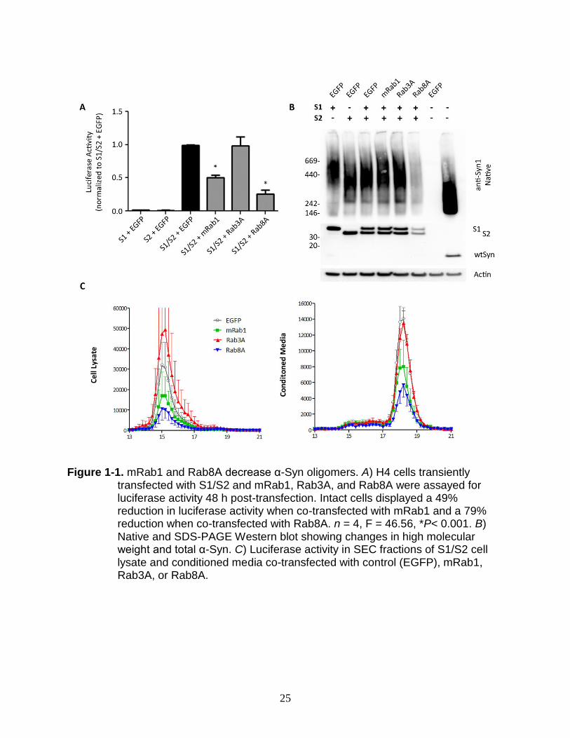
Figure 1-1. mRab1 and Rab8A decrease α-Syn oligomers. A) H4 cells transiently
transfected with S1/S2 and mRab1, Rab3A, and Rab8A were assayed for luciferase activity 48 h post-transfection. Intact cells displayed a 49% reduction in luciferase activity when co-transfected with mRab1 and a 79% reduction when co-transfected with Rab8A. n = 4, F = 46.56, *P< 0.001. B) Native and SDS-PAGE Western blot showing changes in high molecular weight and total α-Syn. C) Luciferase activity in SEC fractions of S1/S2 cell lysate and conditioned media co-transfected with control (EGFP), mRab1, Rab3A, or Rab8A.
26
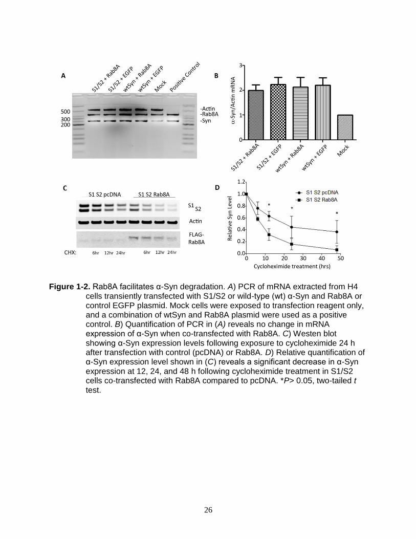
Figure 1-2. Rab8A facilitates α-Syn degradation. A) PCR of mRNA extracted from H4
cells transiently transfected with S1/S2 or wild-type (wt) α-Syn and Rab8A or control EGFP plasmid. Mock cells were exposed to transfection reagent only, and a combination of wtSyn and Rab8A plasmid were used as a positive control. B) Quantification of PCR in (A) reveals no change in mRNA expression of α-Syn when co-transfected with Rab8A. C) Westen blot showing α-Syn expression levels following exposure to cycloheximide 24 h after transfection with control (pcDNA) or Rab8A. D) Relative quantification of α-Syn expression level shown in (C) reveals a significant decrease in α-Syn expression at 12, 24, and 48 h following cycloheximide treatment in S1/S2 cells co-transfected with Rab8A compared to pcDNA. *P> 0.05, two-tailed t test.

27
LIST OF REFERENCES
1. Dorsey, E. R., Constantinescu, R., Thompson, J. P., Biglan, K. M., Holloway, R. G., Kieburtz, K., Marshall, F. J., Ravina, B. M., Schifitto, G., Siderowf, A., and Tanner, C. M. (2007) Projected number of people with Parkinson disease in the most populous nations, 2005 through 2030. Neurology 68, 384-386
2. Hardy, J., Lewis, P., Revesz, T., Lees, A., and Paisan-Ruiz, C. (2009) The genetics of Parkinson's syndromes: a critical review. Curr Opin Genet Dev 19, 254-265
3. Bruggink, K. A., Kuiperij, H. B., Ekholm-Pettersson, F., and Verbeek, M. M. (2011) Detection of elevated levels of alpha-synuclein oligomers in CSF from patients with Parkinson disease. Neurology 77, 510; author reply 510-511
4. El-Agnaf, O. M., Salem, S. A., Paleologou, K. E., Cooper, L. J., Fullwood, N. J., Gibson, M. J., Curran, M. D., Court, J. A., Mann, D. M., Ikeda, S., Cookson, M. R., Hardy, J., and Allsop, D. (2003) Alpha-synuclein implicated in Parkinson's disease is present in extracellular biological fluids, including human plasma. FASEB J 17, 1945-1947
5. Desplats, P., Lee, H. J., Bae, E. J., Patrick, C., Rockenstein, E., Crews, L., Spencer, B., Masliah, E., and Lee, S. J. (2009) Inclusion formation and neuronal cell death through neuron-to-neuron transmission of alpha-synuclein. Proc Natl Acad Sci U S A 106, 13010-13015
6. Luk, K. C., Kehm, V. M., Zhang, B., O'Brien, P., Trojanowski, J. Q., and Lee, V. M. (2012) Intracerebral inoculation of pathological alpha-synuclein initiates a rapidly progressive neurodegenerative alpha-synucleinopathy in mice. J Exp Med 209, 975-986
7. Lee, S. J., Desplats, P., Lee, H. J., Spencer, B., and Masliah, E. (2012) Cell-to-cell transmission of alpha-synuclein aggregates. Methods Mol Biol 849, 347-359
8. Davidson, W. S., Jonas, A., Clayton, D. F., and George, J. M. (1998) Stabilization of alpha-synuclein secondary structure upon binding to synthetic membranes. J Biol Chem 273, 9443-9449
9. Murphy, D. D., Rueter, S. M., Trojanowski, J. Q., and Lee, V. M. (2000) Synucleins are developmentally expressed, and alpha-synuclein regulates the size of the presynaptic vesicular pool in primary hippocampal neurons. J Neurosci 20, 3214-3220
10. Abeliovich, A., Schmitz, Y., Farinas, I., Choi-Lundberg, D., Ho, W. H., Castillo, P. E., Shinsky, N., Verdugo, J. M., Armanini, M., Ryan, A., Hynes, M., Phillips, H., Sulzer, D., and Rosenthal, A. (2000) Mice lacking alpha-synuclein display functional deficits in the nigrostriatal dopamine system. Neuron 25, 239-252
11. Iwai, A., Masliah, E., Yoshimoto, M., Ge, N., Flanagan, L., de Silva, H. A., Kittel, A., and Saitoh, T. (1995) The precursor protein of non-A beta component of Alzheimer's disease amyloid is a presynaptic protein of the central nervous system. Neuron 14, 467-475
12. Cabin, D. E., Shimazu, K., Murphy, D., Cole, N. B., Gottschalk, W., McIlwain, K. L., Orrison, B., Chen, A., Ellis, C. E., Paylor, R., Lu, B., and Nussbaum, R. L. (2002) Synaptic vesicle depletion correlates with attenuated synaptic responses to prolonged repetitive stimulation in mice lacking alpha-synuclein. J Neurosci 22, 8797-8807
28
13. Burre, J., Sharma, M., Tsetsenis, T., Buchman, V., Etherton, M. R., and Sudhof, T. C. (2010) Alpha-synuclein promotes SNARE-complex assembly in vivo and in vitro. Science 329, 1663-1667
14. Chandra, S., Gallardo, G., Fernandez-Chacon, R., Schluter, O. M., and Sudhof, T. C. (2005) Alpha-synuclein cooperates with CSPalpha in preventing neurodegeneration. Cell 123, 383-396
15. Cooper, A. A., Gitler, A. D., Cashikar, A., Haynes, C. M., Hill, K. J., Bhullar, B., Liu, K., Xu, K., Strathearn, K. E., Liu, F., Cao, S., Caldwell, K. A., Caldwell, G. A., Marsischky, G., Kolodner, R. D., Labaer, J., Rochet, J. C., Bonini, N. M., and Lindquist, S. (2006) Alpha-synuclein blocks ER-Golgi traffic and Rab1 rescues neuron loss in Parkinson's models. Science 313, 324-328
16. Gitler, A. D., Bevis, B. J., Shorter, J., Strathearn, K. E., Hamamichi, S., Su, L. J., Caldwell, K. A., Caldwell, G. A., Rochet, J. C., McCaffery, J. M., Barlowe, C., and Lindquist, S. (2008) The Parkinson's disease protein alpha-synuclein disrupts cellular Rab homeostasis. Proc Natl Acad Sci U S A 105, 145-150
17. Hutagalung, A. H., and Novick, P. J. (2011) Role of Rab GTPases in membrane traffic and cell physiology. Physiol Rev 91, 119-149
18. Geppert, M., Bolshakov, V. Y., Siegelbaum, S. A., Takei, K., De Camilli, P., Hammer, R. E., and Sudhof, T. C. (1994) The role of Rab3A in neurotransmitter release. Nature 369, 493-497
19. Geppert, M., Goda, Y., Stevens, C. F., and Sudhof, T. C. (1997) The small GTP-binding protein Rab3A regulates a late step in synaptic vesicle fusion. Nature 387, 810-814
20. Dalfo, E., Gomez-Isla, T., Rosa, J. L., Nieto Bodelon, M., Cuadrado Tejedor, M., Barrachina, M., Ambrosio, S., and Ferrer, I. (2004) Abnormal alpha-synuclein interactions with Rab proteins in alpha-synuclein A30P transgenic mice. J Neuropathol Exp Neurol 63, 302-313
21. Dalfo, E., and Ferrer, I. (2005) Alpha-synuclein binding to rab3a in multiple system atrophy. Neurosci Lett 380, 170-175
22. Dalfo, E., Barrachina, M., Rosa, J. L., Ambrosio, S., and Ferrer, I. (2004) Abnormal alpha-synuclein interactions with rab3a and rabphilin in diffuse Lewy body disease. Neurobiol Dis 16, 92-97
23. Danzer, K. M., Ruf, W. P., Putcha, P., Joyner, D., Hashimoto, T., Glabe, C., Hyman, B. T., and McLean, P. J. (2011) Heat-shock protein 70 modulates toxic extracellular alpha-synuclein oligomers and rescues trans-synaptic toxicity. FASEB J 25, 326-336
24. Winner, B., Jappelli, R., Maji, S. K., Desplats, P. A., Boyer, L., Aigner, S., Hetzer, C., Loher, T., Vilar, M., Campioni, S., Tzitzilonis, C., Soragni, A., Jessberger, S., Mira, H., Consiglio, A., Pham, E., Masliah, E., Gage, F. H., and Riek, R. (2011) In vivo demonstration that alpha-synuclein oligomers are toxic. Proc Natl Acad Sci U S A 108, 4194-4199
25. Karpinar, D. P., Balija, M. B., Kugler, S., Opazo, F., Rezaei-Ghaleh, N., Wender, N., Kim, H. Y., Taschenberger, G., Falkenburger, B. H., Heise, H., Kumar, A., Riedel, D., Fichtner, L., Voigt, A., Braus, G. H., Giller, K., Becker, S., Herzig, A., Baldus, M., Jackle, H., Eimer, S., Schulz, J. B., Griesinger, C., and Zweckstetter,
29
M. (2009) Pre-fibrillar alpha-synuclein variants with impaired beta-structure increase neurotoxicity in Parkinson's disease models. EMBO J 28, 3256-3268
26. Outeiro, T. F., Putcha, P., Tetzlaff, J. E., Spoelgen, R., Koker, M., Carvalho, F., Hyman, B. T., and McLean, P. J. (2008) Formation of toxic oligomeric alpha-synuclein species in living cells. PLoS ONE 3, e1867
27. Pfeffer, S. R. (2012) Rab GTPase localization and Rab cascades in Golgi transport. Biochem Soc Trans 40, 1373-1377
28. Li, C., Takei, K., Geppert, M., Daniell, L., Stenius, K., Chapman, E. R., Jahn, R., De Camilli, P., and Sudhof, T. C. (1994) Synaptic targeting of rabphilin-3A, a synaptic vesicle Ca2+/phospholipid-binding protein, depends on rab3A/3C. Neuron 13, 885-898
29. Sandoval, C. O., and Simmen, T. (2012) Rab proteins of the endoplasmic reticulum: functions and interactors. Biochem Soc Trans 40, 1426-1432
30. Kelly, E. E., Horgan, C. P., Goud, B., and McCaffrey, M. W. (2012) The Rab family of proteins: 25 years on. Biochem Soc Trans 40, 1337-1347
31. Winslow, A. R., Chen, C. W., Corrochano, S., Acevedo-Arozena, A., Gordon, D. E., Peden, A. A., Lichtenberg, M., Menzies, F. M., Ravikumar, B., Imarisio, S., Brown, S., O'Kane, C. J., and Rubinsztein, D. C. (2010) alpha-Synuclein impairs macroautophagy: implications for Parkinson's disease. J Cell Biol 190, 1023-1037
32. Cuervo, A. M., Stefanis, L., Fredenburg, R., Lansbury, P. T., and Sulzer, D. (2004) Impaired degradation of mutant alpha-synuclein by chaperone-mediated autophagy. Science 305, 1292-1295
33. Ejlerskov, P., Rasmussen, I., Nielsen, T. T., Bergstrom, A. L., Tohyama, Y., Jensen, P. H., and Vilhardt, F. (2013) Tubulin polymerization-promoting protein (TPPP/p25alpha) promotes unconventional secretion of alpha-synuclein through exophagy by impairing autophagosome-lysosome fusion. J Biol Chem 288, 17313-17335
34. Bennett, M. C., Bishop, J. F., Leng, Y., Chock, P. B., Chase, T. N., and Mouradian, M. M. (1999) Degradation of alpha-synuclein by proteasome. J Biol Chem 274, 33855-33858
35. Tofaris, G. K., Razzaq, A., Ghetti, B., Lilley, K. S., and Spillantini, M. G. (2003) Ubiquitination of alpha-synuclein in Lewy bodies is a pathological event not associated with impairment of proteasome function. J Biol Chem 278, 44405-44411
36. Shin, Y., Klucken, J., Patterson, C., Hyman, B. T., and McLean, P. J. (2005) The co-chaperone carboxyl terminus of Hsp70-interacting protein (CHIP) mediates alpha-synuclein degradation decisions between proteasomal and lysosomal pathways. J Biol Chem 280, 23727-23734

30
BIOGRAPHICAL SKETCH
Amanda Herring began her education at the University of South Florida as a
chemistry major/voice minor. After a year, she transferred to the University of North
Florida where she completed her Bachelor of Science in biology in 2009. During her
undergraduate coursework, she started working at the Mayo Clinic Jacksonville as a
research assistant in an Alzheimer’s lab. She fell in love with research, and when she
completed her undergraduate degree, she decided to pursue her Master of Medical
Science at the University of Florida in neuroscience.