
APOBEC3G - an intracellular centurionAn Intracellular Centurion
Upload
trinhtuongCategory
view
218download
0
Integrated Systems and Technologies: Mathematical Oncology
Quantitative In Vivo Characterization of Intracellular andExtracellular pH Profiles in Heterogeneous Tumors: A NovelMethod Enabling Multiparametric pH Analysis
Norbert W. Lutz1, Yann Le Fur1, Johanna Chiche2, Jacques Pouyss�egur2,3, and Patrick J. Cozzone1
AbstractAcid production and transport are currently being studied to identify new targets for efficient cancer treatment,
as subpopulations of tumor cells frequently escape conventional therapy owing to their particularly acidic tumormicroenvironment. Heterogeneity in intracellular and extracellular tumor pH (pHi, pHe) has been reported, butnone of themethods currently available formeasuring tissue pH provides quantitative parameters characterizingpH distribution profiles in tissues. To this intent, we present here amultiparametric, noninvasive approach basedon in vivo 31P nuclear magnetic resonance (NMR) spectroscopy and its application to mouse tumor xenografts.First, localized 31P NMR spectrum signals of pHi and pHe reporter molecules [inorganic phosphate (Pi) and 3-aminopropylphosphonate (3-APP), respectively] were transformed into pH curves using established algorithms.Although Pi is an endogenous compound, 3-APP had to be injected intraperitoneally. Then, we developedalgorithms for the calculation of six to eight quantitative pH parameters from the digital points of each pH curveobtained. For this purpose, each pH distribution profile was approximated as a histogram, and intensities werecorrected for the nonlinearity between chemical-shift and pH. Cancer Res; 73(15); 4616–28. �2013 AACR.
IntroductionIn physiologic tissues, the interplay of metabolism, ion
transport, and pH buffering results in efficient pH regulation.The presence of macroscopic and microscopic membranestructures, such as the basement membrane and various cellmembranes, permits the coexistence of multiple tissue com-partments characterized by different pH values. This highlightsthe necessity to not onlymeasure average tissue pH values, butalso to quantitatively assess pH heterogeneity. Although non-invasive determination of intra- and extracellular pH (pHi andpHe) in mammals, notably by way of nuclear magnetic reso-nance (NMR) techniques, has becomemore common in recentyears, there is at present no method that provides quantitativeparameters specifically characterizing the heterogeneity ofpHi and pHe in a given tissue volume. Yet, there is a genuineneed for such measurements as a variety of pathologies(cancer and inflammation, among others) are associated withheterogeneous pH regulation (1–3). Even normal activity suchas muscle exercise can generate complex tissue pH distribu-tions as a function of biologic characteristics (4). To addressthis challenge, we have developed a new approach based on
Authors' Affiliations: 1Centre de R�esonance Magn�etique Biologiqueet M�edicale (CRMBM), UMR 7339, Facult�e de M�edecine de la Timone,Centre National de la Recherche Scientifique (CNRS), Aix-Marseille Uni-versit�e, Marseille; 2Institute of Research on Cancer and Aging, UMRCNRS7284, Centre Antoine Lacassagne, Universit�e de Nice, Nice, France; and3Centre Scientifique de Monaco, Monaco-Ville, Monaco
Note: Supplementary data for this article are available at Cancer ResearchOnline (http://cancerres.aacrjournals.org/).
Current address for J. Chiche: Controle M�etabolique des MortsCellulaires, Institut National de la Sant�e et de la Recherche Medicale
(INSERM) U 1065, Centre M�editerran�een de M�edecine Mol�eculaire,F-06204 Nice, France.
Corresponding Author: Norbert W. Lutz, CRMBM, UMR CNRS 7339,Facult�e de M�edecine, Aix-Marseille Universit�e, 27 bd Jean Moulin, Mar-seille F-13005, France. Phone: 33-491-324814; Fax: 33-491-256539;E-mail: [email protected]
doi: 10.1158/0008-5472.CAN-13-0767
�2013 American Association for Cancer Research.
Major FindingsFor each histogram derived from a Pi or 3-APP resonance, we
obtained the following tumor pH profile parameters: weightedmean, weighted median, mode(s), skewness (asymmetry), kurtosis(peakedness), and entropy (smoothness). In addition, relative sizes oftissue volumes defined by characteristic pH ranges were estimatedby integration and/or by fitting the curve to multiple modes. Ouralgorithms and the results obtained for animal models were vali-dated by: (i) computer simulations of 31P NMR resonances and pHprofiles; and (ii) comparison with combinations of three or less testsolutions at well-defined pH values, containing the pH reportermolecule 3-APP. All calculations were carried out with an Excelspreadsheet, thus avoiding any specialized software or hardware.Consequently, heterogeneous pHi and pHe distribution profiles intumors can be characterized by multiple quantitative parametersderived from classical statistics through histograms obtainedfrom in vivo 31P NMR spectra. This original technique is helpful inanalyzing tumor tissue features with increased detail, based on asingle experiment also yielding information on underlying energyand phospholipid metabolism.
CancerResearch
Cancer Res; 73(15) August 1, 20134616
on July 12, 2018. © 2013 American Association for Cancer Research. cancerres.aacrjournals.org Downloaded from
Published OnlineFirst June 10, 2013; DOI: 10.1158/0008-5472.CAN-13-0767
on July 12, 2018. © 2013 American Association for Cancer Research. cancerres.aacrjournals.org Downloaded from
Published OnlineFirst June 10, 2013; DOI: 10.1158/0008-5472.CAN-13-0767
on July 12, 2018. © 2013 American Association for Cancer Research. cancerres.aacrjournals.org Downloaded from
Published OnlineFirst June 10, 2013; DOI: 10.1158/0008-5472.CAN-13-0767

Quick Guide to Equations and AssumptionsThe current standard procedure for deriving a pH value fromapH-sensitive 31P nuclearmagnetic resonance (NMR) resonance is
based on converting the chemical-shift axis of the resonance line into a pH axis. It is generally assumed that, after appropriateintensity correction, (i) the resulting curve adequately reflects the pH within the measured volume, and (ii) the position of themaximum of this pH curve represents "the" pH. We adopt assumption (i), within certain limits. However, we argue that themaximum of a tissue pH curve often yields a nonrepresentative pH value, because such curves are frequently asymmetric andirregularly shaped. We also contend that a (intracellular or extracellular) pH curve can and should be exploited to quantitativelyanalyze the respective underlying pHheterogeneity. For a detailed introduction into our algorithms, see Supplementary Section S1.We propose to consider the (digitized) pH curve as an effective pH distribution curve and to approximate it as a histogram in
which each digital curve point k corresponds to a histogram bin. From each curve/histogram, we derive the following basic pHparameters:Weighted mean pH:
pH ¼Pm
k¼1 ðpHk �WkÞPmk¼1 Wk
eq: ð1Þ
where pHk is the intracellular or extracellular pH value for a given digital point k of the pH curve; m is the total number of curvepoints used for calculation; andWk is the scaled weight (ordinate value) of curve point k. Scaledweights are weights that have beenadjusted to account for the variability of pH intervals between curve points. The nonequidistant character of pH curve pointsresults from the nonlinearity between the chemical-shift and pH scales. Thus, digital curve points that are equidistant on thechemical-shift axis are nonequidistant on the pH axis after point-by-point conversion. Weighted pHmean values directly derivedfrom pH curves without rescaling would overemphasize pH regions represented by relatively "dense" curve points. Scaled weightsare also needed for the calculation of the other pH parameters presented later. However, pH modes (curve maxima) have to beobtained directly from the pH curve, because this curve is a representation of the distribution of pH values.Weighted median pH:
fpH ¼ pH~k�1 þ ðpH~k � pH~k�1Þ � fint eq: ð2Þ
where pH~k is the pH value of curve point ~k possessing the cumulative sumCSUMð~kÞ; pH~k�1 is the pH value of curve point ð~k� 1Þpossessing the cumulative sum CSUMð~k� 1Þ; and fint is an interpolation factor defined as fint ¼ CSUMðmÞ=2�½CSUMð~k� 1Þ�=½CSUMð~kÞ � CSUMð~k� 1Þ�. Cumulative sums are calculated for scaled weights of curve points. Conventionally,the median is the numerical value separating the higher half of a sample from the lower half or the mean of the twomiddle values.For a series of weighted values, the location of the "weightedmiddle" has to be determined by interpolation. This is achieved by the
interpolation factor fint that determines the location of the weighted middle between two adjacent curve points, ~k and ð~k� 1Þ,corresponding to the cumulative sums (of scaled weights) that lie just above and below, respectively, the half-sum of the last pointof the pH range used. CSUMðmÞ=2 is defined as the half-sum of a series of m values.Skewness of pH distribution:
G1 ¼ nðn� 1Þðn� 2Þ
Xmk¼1
WkpHk � pH
s
� �3
eq: ð3Þ
where s ¼ffiffiffiffiffiffiffiffiffiffiffiffiffiffiffiffiffiffiffiffiffiffiffiffiffiffiffiffiffiffiffiffiffiffiffiffiffiffiffiffiffiffiffiffiffiffiffiffiffiffiffiffiffiffiffiffiffiffiffiffiffiffiffiffiðPm
k¼1 WkðpHk � pHÞ2Þ=ðn� 1Þq
is the nominal SD, a parameter analogous to the SDof themean based on individual
observations (¼ individual contributions to conventional histogram bins). In our algorithm, n ¼ Pmk¼1 Wk is a parameter
analogous to the total number of individual observations in conventional histograms. Generally, both skewness and kurtosis (seelater) characterize the shape of a statistical frequency distribution, i.e., asymmetry and pointedness, respectively. Hence, theabsolute value of n is of no importance (as long as it is not too small) as these shape-related pH curve properties only depend on therelative weights of the individual pH curve values and on their deviation from a normal distribution. We verified by computersimulation that skewness and kurtosis asymptotically approach n-independent values for n greater than several times the numberof digital points m.Kurtosis of pH distribution:
G2 ¼ nðnþ 1Þðn� 1Þðn� 2Þðn� 3Þ
Xmk¼1
WkpHk � pH
s
� �4
� 3ðn� 1Þ2ðn� 2Þðn� 3Þ eq: ð4Þ
with parameters being defined as presented above for skewness.
Multiparametric Study of Tumor pH Heterogeneity
www.aacrjournals.org Cancer Res; 73(15) August 1, 2013 4617
on July 12, 2018. © 2013 American Association for Cancer Research. cancerres.aacrjournals.org Downloaded from
Published OnlineFirst June 10, 2013; DOI: 10.1158/0008-5472.CAN-13-0767

multiparametric analysis of noninvasive in vivo 31P NMRspectra.
The proposed strategy is based on the circumstance that pH-sensitive magnetic resonance spectroscopy (MRS) signals fromheterogeneous tissues represent entire pH distributions (pHprofiles), rather than merely providing one "typical" pHi or pHe
value. Because pH reporter molecules are electrically charged,they cannot freely cross membranes. This has a 2-fold conse-quence: pH reporter molecules do not move freely (i) betweenthe intracellular and the extracellular space, nor (ii) betweenextracellular spaces that are separated by a monolayer or amultilayer of cells. Thus, these molecules do not undergoexchange between microscopic tissue regions of different pH,and the pH-sensitive chemical-shift of the detected nucleus isnot averaged out in the course of anMRS experiment. In theory,a pH reporter may be exchanged between extracellular regionsnot separated by cells or membranes. This would lead tochemical-shift averaging (fast exchange), or to a situationanalogous to the classical case of an intermediate chemicalexchange rate regimen between two molecular species of dif-ferent chemical-shift. However, this type of fast or intermediateexchange should not play an important role in the case of tissuepH because tissue areas of different pH are usually separated bymembranes, for example, cell layers; otherwise the pH gradientwould rapidly dissipate due to proton diffusion. We inject thepHe reporter molecule, 3-aminopropylphosphonate (3-APP),into the experimental animal before theMRS experiment. Then,31P NMR spectra are acquired under anesthesia, followed byevaluation of the 3-APP resonance for pHe analysis, and of theendogenous inorganic phosphate (Pi) resonance for pHi analysis(5). The use of the Pi signal for pHi measurement was pioneeredby Moon and Richards (6) for cell suspensions, and was furtherdeveloped for in vivo application in rodent tumors (7). Later, thisconceptwas complemented by the introduction of exogenous 3-APP for pHe measurement (8). Conventionally, the Pi and 3-APPmagnetic resonance signals are converted to pH curves, and thehighest point in each curve ("the"maximum) is interpreted to be"the" pHi or pHe value of the measured tissue volume (9).Although this procedure yields fairly realistic average pH valuesfor narrow and symmetric pH distributions, it is inadequatewhen pH distributions deviate from this ideal shape due tosignificant pH heterogeneity within the measured volume (10).Although others have previously noticed an influence of pHheterogeneity on the appearance of Pi (4, 11) and 3-APP (8)spectral lines, we exploit here, for the first time, the resulting pHcurve shapes to derive quantitative parameters characterizing
the underlying distributions of pH values. In addition to one ormultiple pHe and pHi modes (¼ pHe and pHi curve maxima,respectively), the most basic parameters are weighted meansand weightedmedians (12) for pHe and pHi, each of which takesinto account the entire respective pH distribution. Further pHe
and pHi lineshape parameters are obtained to characterize theasymmetry (skewness; ref. 13), peakedness (kurtosis; ref. 12), andsmoothness (entropy; refs. 14, 15) of pH distributions. Finally,ratios of areas under individual pH modes and/or ranges aredetermined to obtain a quantitativemeasure of the relative sizesof tissue volumes with different pH values. All parameters arebased on suitably weighted digital points of pH curves derivedfrom 31P NMR spectra. A crucial aspect of our new paradigm isthat these quantitative parameters describe global features ofpH heterogeneity within a selected tissue volume. None of thecurrently available in vivo tissue pH methods provides suchparameters.
We apply our approach to experimental tumors that havepreviously shown a relationship between pH heterogeneity andthe extent of tumor necrosis (16). We assess the validity ofour approach (i) by means of computer simulating 31P NMRspectral lines, including thorough error analysis, and (ii) by wayof in vitro 31P NMR experiments based on 3-APP solutions withwell-defined pH values. These findings have significant implica-tions for the study of the relationship between pH alterationsand biologic properties of tissue, for example, tumor growthbehavior and cancer cell death, but also for other pathologiesassociated with perturbations of pH regulation.
Materials and MethodsAnimals, cancer cells, and phantoms used
Animal studies were in agreement with the French guide-lines for animal care, and were approved by the Committee onEthics of the University of Aix-Marseille (Marseille, France).Pouyss�egur's laboratory obtained the CCL39 clone from Amer-ican Type Culture Collection in 1978. From these Chinesehamster lung fibroblasts, mutants were isolated that havebeen very well characterized and published in Proceedingsof the National Academy of Sciences of the United States ofAmerica (PNAS) and Nature. Since then, these cell lines havebeen maintained in Pouyss�egur's laboratory and checkedbefore each experiment for the specific mutation in the gly-colytic-defective phenotype. Further details about our tumormodel have been described elsewhere (16).
Aqueous solutions of the pHe reporter compound, 3-APPwere prepared at different concentrations, and were adjusted
Entropy of pH distribution:
HðW Þ ¼ �Xmk¼1
WkPmk¼1 Wk
log2WkPmk¼1 Wk
� �� �eq: ð5Þ
where H(W) is the entropy, and W is equivalent to the set P of all probability distributions as defined in the discrete Shannonentropy.All algorithms used for calculating these five statistical parameters are based on established statistical equations; however, the
original equations have been transformed for use in pH curve point analysis instead of conventional histogram analysis.
Lutz et al.
Cancer Res; 73(15) August 1, 2013 Cancer Research4618
on July 12, 2018. © 2013 American Association for Cancer Research. cancerres.aacrjournals.org Downloaded from
Published OnlineFirst June 10, 2013; DOI: 10.1158/0008-5472.CAN-13-0767

to appropriate pH values with HCl and NaOH solutions.Because tissue pHe heterogeneity is by far more pronouncedand prevalent than pHi heterogeneity, we decided to focus ourphantom experiments on 3-APP solutions. However, the val-idation obtained for 3-APP in this study can be generalized to Pias pH sensitivities and relevant pKa values are only slightlydifferent between these two compounds (Supplementary Sec-tion S1.1). First, three 245-mmol/L 3-APP solutions wereprepared at pH 7.45, 7.00, and 6.50. These solutions were thenserially diluted by a factor of 1.5, then 2, between subsequent31P NMR measurements.These solutions were transferred to 5-mm NMR tubes
(528-PP) from Wilmad, cut to size (length ca. 4 cm) to suitthe spatial conditions of the NMR probe described later (seealso Supplementary Section S2.1). Up to three of theseshortened NMR tubes were inserted into the cap of ashortened 50-mL centrifuge tube (Corning; Fisher Scientific)filled with saline. In addition, a 50 mmol/L 3-APP solution[free acid dissolved in deuterium oxide (D2O)] was prepared.This solution was then transferred to (i) a standard size 5-mm NMR tube (528-PP), and (ii) a glass sphere (1 cmdiameter).
Acquisition of 31P NMR spectra and proton images oftumors and phantomsMouse tumors were measured in vivo as described previ-
ously (16). For phantom measurements, the spherical phan-tom, or the centrifuge tube cone filled with saline and withone to three NMR tubes containing 3-APP solution, wasplaced on the same one-turn 31P surface coil used for in vivomeasurements, mounted in the same proton volume coil (16).The NMR spectrometer/imager used for both phantoms andtumors was a BIOSPEC 47/40 system (4.7 T; Bruker). 31P NMRspectra were acquired using a pulse-acquire sequence withvolume selection based on outer-volume suppression (OVS;ref. 17). To obtain the fine structure of the 3-APP spectrumunder ideal conditions, high-resolution 31P NMR spectroscopyof the 50 mmol/L 3-APP solution in 5-mm NMR tubes wasconducted on an AVANCE 400 spectrometer (Bruker) at 9.4 Tequipped with a quadronuclear probe (QNP) by way of astandard pulse-acquire sequence (18–20), with and withoutproton decoupling.
Processing of tumor and phantom 31P NMR spectra31P NMR free induction decays (FID) were Fourier-trans-
formed after zero-filling and multiplication with appropriateLorentzian–Gaussian functions (XWINNMR software; Bruker).The chemical-shift values of the processed 31P NMR spectra(21) were then converted to pH. After intensity corrections, theresulting datasets served as histograms (22) for the determi-nation of weighted-average pH (mean pH), weighted medianpH, skewness, kurtosis, and entropy (23–26). In addition,individual modes were determined. For pH distributions per-mitting the distinction of two or more characteristic pHranges, the areas under the individual pH ranges or modeswere quantitated by two different methods: (i) integration, and(ii) curve fitting using the MDCON (mixed deconvolution)function in Bruker's TopSpin software. Finally, these evalua-
tion methods were applied to in vivo 31P NMR signals of 3-APPin mouse tumor xenografts (16). High-resolution 31P NMRspectra of 3-APP solutions were processed using Bruker'sTopSpin software. Further NMR processing details, as well asthe theoretical background and the algorithms used for thecalculation of pH heterogeneity parameters are presented inSupplementary Sections S1 and S2. The EXCEL spreadsheetpH_param_template.xlsx provided by us is, in fact, a computerprogram; it serves both as an example of our calculations andas a template for use by interested researchers. This EXCEL filecan be downloaded using theURLaddress: http://crmbm.univ-amu.fr/homepage/nlutz/pH_param_template.xlsx. An optionto use the results of lineshape deconvolution for better defi-nition of Pi lines is included in the algorithms implemented inthis template.
In silico calculationsThe purpose of our in silico calculations is 4-fold: (i) to test,
based onwell-definedGaussian pHdistributions, the validity ofour algorithms; (ii) to explore the effects of ppm-to-pH con-version on the symmetric shapes of two distinct Gaussian 3-APP 31P NMR spectral lines; (iii) to study the statistical para-meters characterizing the overall pH distributions resultingfrom the addition, in varying proportions, of the two pH curvesgenerated in (ii); and (iv) to compare simulated, phantom, andin vivo pH heterogeneity parameter values.
As a consequence of the nonlinearity of the ppm-to-pHconversion, a pH lineshape may significantly deviate from itsunderlying chemical-shift lineshape. We generated two Gauss-ian curves with a chemical-shift abscissa scaled such that,following ppm-to-pH conversion, the centers of the curves fellupon pH 6.5 and 7.2. These values as well as the associatedlinewidths were chosen to be close to values commonly foundfor pHe in in vivo experiments. The resulting (asymmetric) pHcurves were then used to characterize these simulated pHdistributions by means of statistical pH distribution para-meters. Conversely, we generated in an EXCEL spreadsheetGaussian pH curves centered about pH 6.5 and 7.2. On the basisof these curves, we backward simulated the corresponding 3-APP line, and studied the asymmetry effects that (symmetric)Gaussian pH distribution parameters would undergo afterconversion to simulated 3-APP resonances. Finally, two com-puter-generated Gaussian curves were added to model bimod-al (27) pH distributions for statistical analysis.
ResultsGeneration of bimodal pH profiles corrected for thenonlinearity between the chemical-shift and pH scales
The effects of converting the ppm scale of the 3-APPspectrum to a pH scale were first studied for a bimodal pHdistribution from a mouse tumor xenograft (Fig. 1A). Mostheterogeneous tumors feature irregularly shaped pH distribu-tions rather than strictly bimodal or multimodal patterns (16).However, to validate our method, we primarily focused onbimodal and trimodal pH distributions, because the concept ofquantitative pH heterogeneity parameters is best tested andverified on the basis of models representing well-defined pH
Multiparametric Study of Tumor pH Heterogeneity
www.aacrjournals.org Cancer Res; 73(15) August 1, 2013 4619
on July 12, 2018. © 2013 American Association for Cancer Research. cancerres.aacrjournals.org Downloaded from
Published OnlineFirst June 10, 2013; DOI: 10.1158/0008-5472.CAN-13-0767

distribution functions. Nevertheless, nearly all quantitativeparameters suggested in this report are universally applicableto any given distribution of pH values; their use does notdepend on the existence of distinct pH modes (see alsofollowing paragraph). Our results were first validated by usinga phantom consisting of two NMR tubes filled with 3-APPsolutions adjusted to pH 6.5 and 7.4 (Fig. 1B and C). Uncor-rected ppm-to-pH conversion (Supplementary Eq. S6, withintensity I3-APP as in underlying NMR spectrum) renders theupper part of each pH mode narrower (Fig. 1, bottom row,dotted lines) than it is in the corresponding 31P NMR spectrum(Fig. 1, middle row). In addition, the outer wings of the pHcurves do not descend to the baseline, even for apparent pHvalues as high as 8.5, or as low as 5.5. However, such extremepH values are unlikely to exist in tumors andmostmammaliantissues, and are unquestionably the result of a systematicartifact. This artifact is remedied (28) by applying an intensitycorrection for the nonlinear relationship between the chem-ical-shift and pH scales (Supplementary Eq. S8). Following thiscorrection, pH lineshapes are further narrowed, and the outer
wings of bothmodes return to the baselinewithin the displayedpH range (Fig. 1, bottom row, solid lines). It is evident thatomitting this correction step would yield biased and exces-sively broad pH distributions by overemphasizing bothextremely high and low pH values, due to their large differencefrom the pKa2 of 3-APP. Obviously, including this nonlinearitycorrection is even more critical in the evaluation of a hetero-geneous pH profile than it is for the determination of a singletissue pH value based on the globalmaximumof a pH curve (9).Also note that eachmode (¼maximum of each peak) is shiftedbetween the spectral line and the pH curve, and is slightlydifferent between corrected and uncorrected pH curves (Fig.1A–C). The latter effect has been previously observed for pHcurves with a single maximum (9).
For best comparison between phantom and in vivo stud-ies, the processing parameters for phantom spectra (Fig. 1B)were judiciously chosen to yield pH curves whose shapesresemble pH curves obtained for heterogeneous tissue(Fig. 1A). Even so, stronger filtering parameters [typicallyGaussian broadening (GB) ¼ 0.007, Lorentzian broadening
24.2 23.2 22.2 21.2 24.2
A B C
23.2 22.2 21.2 24.2 23.2 22.2ppm
pH
pHe1pHe1
pHe1
pHe2
pHe2 pHe2
21.2
8.57.56.55.58.57.56.55.58.57.56.55.5
Figure 1. Examples of 3-APP 31P NMR spectra revealing pH heterogeneity in tissue and in a phantom. A, CCL39 tumor xenograft in a mouse model. B and C,phantom consisting of two NMR tubes filled with 3-APP solutions at pH 6.5 and 7.4. Top row, representative MRI cross-section through phantom ormouse tumor. Shaded areas in image indicate regions saturated by OVS during subsequently conducted localized 31P NMR spectroscopy. Middle row,spectra processed with Lorentzian–Gaussian lineshape transformation at GB ¼ 0.01 and LB ¼ �20 Hz (A), or at GB ¼ 0.007 and LB ¼ �25 Hz (B); orwith apodization at LB ¼ 15 Hz (C). Bottom row, pH profiles derived from the spectra displayed in the top row. Dotted lines, uncorrected pH curves.Solid lines, pH curves with intensities corrected for the nonlinear relationship between chemical-shift and pH scales. In both tumor tissue and phantom, pHprofiles exhibit a bimodal pH distribution pattern, with modes being centered about approximately pH 6.5 (left curve maxima, pHe2) and 7.4 (right curvemaxima, pHe1).
Lutz et al.
Cancer Res; 73(15) August 1, 2013 Cancer Research4620
on July 12, 2018. © 2013 American Association for Cancer Research. cancerres.aacrjournals.org Downloaded from
Published OnlineFirst June 10, 2013; DOI: 10.1158/0008-5472.CAN-13-0767

(LB) ¼ �25 Hz] were needed for phantom spectra than fortissue spectra (typically GB ¼ 0.01, LB ¼ �20 Hz) becausethe magnetic-field inhomogeneity is intrinsically lower inphantoms than in tissue. For phantom-derived and com-puter-simulated pH distributions, the "e" (or "i") indexindicates that the pH modes in question are linked to thechemical-shift of 3-APP (or Pi) signals, and have been chosenwith the intention to compare these modes with similarextracellular (or intracellular) pH distribution modes detect-ed in the tumors of 3-APP–injected animals.
31P NMR spectra yield up to eight quantitative pHdistribution parametersIt is highly desirable to provide quantitative parameters to
characterize tissue heterogeneity with respect to pH. Thenumber of quantitative parameters that can be obtained froma 31P NMR spectroscopy–based analysis of pH heterogeneity isa function of the pH curve shape. The following six parameterscan be extracted from virtually any pH curve: (i) pHmax, theglobal maximum of the pH curve (classical 31P NMR method);
(ii) pH, the weighted-average (mean) pH; (iii) fpH, the weightedmedian pH; (iv) skewness; (v) kurtosis; and (vi) entropy (theunderlying theory and algorithms are explained in great detailin Supplementary Section S1). For a pH profile that suggeststhe presence ofmultiple distinct pH ranges, a characteristic pHvalue can be obtained for each of these ranges by usingmethods (i) to (iii). The relative weight of each of these pHranges can be calculated by (vii) separately integrating the areaunder the curve for each individual pH range, followed bycalculating ratios of these areas. For multimodal pH profilesthat are amenable to numerical fitting of analytic curves, (viii)pH values for multiple maxima or modes (pH1, pH2, etc.), and(ix) areas under individual fitted modes can be obtained asresults of the fitting procedure. Alternatively, multiple maximacorresponding to method (viii) can be directly read out by (x)visual inspection or, for more precision, (xi) a software modulebased on interpolation (such as Bruker's peak picking routine),whereas the areas for these pH modes can be integrated asdescribed earlier under (vii).In the interpretation of pH curves derived from 31P NMR
spectra, spectral line broadening due to magnetic-field inho-mogeneity, spectral processing (filtering) and phosphorus-proton J coupling (for 3-APP), and, to a much lesser extent,T2 processes should be taken into account. In fact, the standarderror and width of a 31P NMR–derived pH curve, or of anindividual mode within a pH curve, would somewhat overstatethe pH range actually present in the sample; for this reason,they are not included in the list of parameters above. Obviously,kurtosis, a measure of the peakedness of a distribution, repre-sents the true pH distribution function more faithfully to theextent that the influence of lineshape effects unrelated to pHcan be minimized as described in Supplementary SectionsS1.2.1 and S1.2.11.Quantification of pH heterogeneity by statistical parameters
was studied on the basis of pH distributions in vivo (mousetumor models), in vitro (phantom models), and in silico (com-puter models). In vivo and in vitro results are presented in thefollowing paragraphs, whereas findings of computer simula-
tions are provided in Supplementary Tables S2 and S3 and inSupplementary Figs. S2E and S2F and S4.
Quantification of unimodal pH distributionsWe first tested our algorithms for a unimodal pH distribu-
tion in a phantom containing a 3-APP solution at pH 7.00.Owing to the perfect pH homogeneity in this sample, theresulting pH distribution curve was very symmetric (Fig.2A). As a consequence, weighted mean ðpHeÞ, weighted medi-
an ðgpHeÞ, and mode (pHe1) had identical values (Table 1A);compare also with computer-simulated results presented inSupplementary Table S2. The small values obtained for skew-ness (G1) and kurtosis (G2) in this example suggest a nearlyGaussian pH curve. In fact, the 31P NMR lineshape obtainedfrom a homogeneous 3-APP solution after strong Gaussianfiltering has a considerable Gaussian character, and its sym-metry is largely preserved in ppm-to-pH conversion if pHapproximates the pKa2 of 3-APP. Also, the entropy (H) of thispH distribution, indicating its smoothness, was smaller for thisexample than for any other example studied in this study. Theunimodal pHe distribution in a relatively homogeneous mousetumor (Fig. 2E) was more asymmetric than the pH distributionin Fig. 2A, the left tail (low pHe) being heavier than the right tail
(high pHe). Therefore, pHe < gpHe < pHe2 and the pHe distribu-tion showed a negative skew (G1; Table 1E). Although this pHe
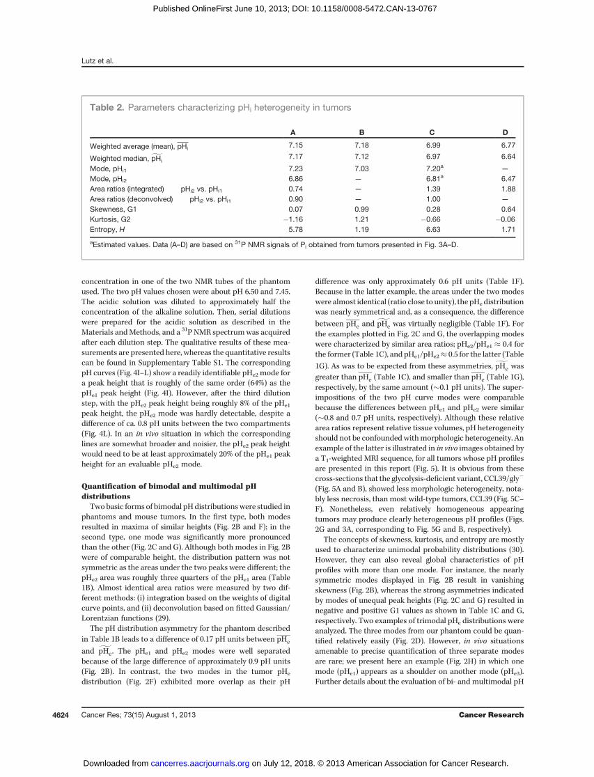
distribution was more leptokurtic (increased G2) than thatof Table 1A, it also had a higher entropy reflecting a more evendistribution. In tumors, the intracellular pH is generally morehomogeneous than the extracellular pH (16); pHi values aredistributed over a considerably smaller range than pHe values.Bi- or multimodal pHi distributions are rare (Fig. 3A), whereasmost tumors exhibit more or less asymmetric unimodal pHi
distributions (Fig. 3B), occasionally presenting a shoulder (Fig.3C and D). The narrowest unimodal pHi distribution (Fig. 3B)was themost leptokurtic and the least smooth pHi distribution(Table 2B). Owing to its high asymmetry reflected by its
skewness, the differences between pHi1, pHi , and gpHi wererather large. In summary, all six quantitative parametersapplicable to unimodal distributions describe the behavior ofthe underlying phantom and tissue samples very well, for bothpHe and pHi.
The number of distinct pH environments in a sample canbe modeled by phantom 31P NMR experiments
In addition to the universally accessible parametersdescribed in the preceding paragraph, some 31P NMR–basedpH profiles reveal a finite number of distinct pH environments.Note that no assumptions aremadewith respect to the size andspatial distribution of the underlying tissue volume elements.The capability of 3-APP 31P NMR spectra to reveal multimodalpH heterogeneity was tested by studying phantoms that con-tained one to three compartments filled with 3-APP solutionsof varying pH. As a starting point, a nondecoupled high-resolution 31P NMR spectrum of an 3-APP solution wasobtained (Fig. 4A). Then, a 3-APP solution contained in aspherical phantom was measured in a small-animal NMRspectrometer/imager. Under these conditions, the 31P NMR
Multiparametric Study of Tumor pH Heterogeneity
www.aacrjournals.org Cancer Res; 73(15) August 1, 2013 4621
on July 12, 2018. © 2013 American Association for Cancer Research. cancerres.aacrjournals.org Downloaded from
Published OnlineFirst June 10, 2013; DOI: 10.1158/0008-5472.CAN-13-0767

peaks were broadened such that only five broad 3-APP peakscould be distinguished (Fig. 4B). Subsequently, a glass spherephantom similar to the phantom used for Fig. 4B (with pH
adjusted to pH 7.0) was imaged (Fig. 4C). The quality of the 3-APP spectrum from the selected volume presented in Fig. 4Cwas comparable with the quality of the spectrum shown
pHe1 pHe1
pHe1
pHe1
pHe1
pHe1
pHe2
pHe2
pHe2
pHe2pHe2pHe2
pHe3
pHe3
8.57.56.55.5 8.57.56.55.5 8.57.56.55.5 8.5pH7.56.55.5
8.57.56.55.5 8.57.56.55.5 8.57.56.55.5 8.5pH7.56.55.5
A B C D
E F G H
I J K L
Figure2. Series of pHprofiles derived from3-APP 31PNMR resonancesof phantomsandmouse tumors for varying numbers of characteristic pHe values. A–D,typical shapes of pH profiles of 3-APP phantoms with one to three characteristic pHe values. E–H, typical shapes of pHe profiles from 3-APP–injected mousetumors with one to three characteristic pHe values. Vertical dashed lines in pH curves indicate the borderline between distinct pH regions within each pHprofile used for the integration of separate areas for the pHe1, pHe2, and pHe3 regions. I–L, representativeMRI cross-sections of the tumors presenting the pHprofiles given in E–H. Shaded areas in images indicate regions saturated by OVS during subsequently conducted localized 31P NMR spectroscopy.
Table 1. Parameters characterizing pHe heterogeneity, as obtained for well-defined test samples (A–D) andtumors (E–H), presented in Fig. 2A–D and E–H, respectively
A B C D E F G H
Weighted average (mean), pHe 7.02 7.15 7.19 7.03 6.76 7.06 6.71 6.95
Weighted median, gpHe7.02 7.32 7.30 6.99 6.79 7.03 6.61 6.89
Mode, pHe1 7.02 7.48 7.38 7.42 — 7.29 7.18 7.6a
Mode, pHe2 — 6.59 6.56 6.64 6.83 6.72 6.48 6.72Mode, pHe3 — — — 6.99 — — — 7.20Area ratios pHe2 vs. pHe1 — 0.75 0.38 1.06 — 0.97 1.85 7.42(integrated) pHe2 vs. pHe3 — — — 0.69 — — — 2.18Area ratios pHe2 vs. pHe1 — 0.72 0.37 1.05 — 0.99 1.98 4.02a
(deconvolved) pHe2 vs. pHe3 — — — 0.77 — — — 1.25a
Skewness, G1 0.15 0.01 �0.44 0.41 �0.57 0.39 0.34 0.58Kurtosis, G2 0.13 �0.78 �0.22 0.33 0.49 �0.19 �0.38 �0.38Entropy, H 3.95 4.81 5.00 5.09 5.86 5.05 6.14 4.66
aEstimated values; five modes were needed for reasonable fit; fitted areas were combined for comparison with integrated areas.
]]
Lutz et al.
Cancer Res; 73(15) August 1, 2013 Cancer Research4622
on July 12, 2018. © 2013 American Association for Cancer Research. cancerres.aacrjournals.org Downloaded from
Published OnlineFirst June 10, 2013; DOI: 10.1158/0008-5472.CAN-13-0767

in Fig. 4B. However, major filtering had been applied toincrease the final linewidth of the pH curve to a value closeto what is achievable in vivo, such that the multiplet was nolonger resolved (Fig. 4F). Next, phantoms containing two andthree NMR tubes were imaged (Fig. 4D and E, respectively).These tubes were immersed in saline, and contained 3-APPsolutions at about pH 6.50 and 7.45 (Fig. 4D), and at about pH6.50, 7.00, and 7.45 (Fig. 4E). All 3-APP concentrationswere keptat roughly the same order of magnitude to prevent the bases oflarger peaks from hiding considerably smaller peaks. Thecorresponding spectra exhibited two (Fig. 4G) and three (Fig.4H) components (modes), respectively. This measurementseries shows that under ideal conditions, up to three distinct
regions of similar size can be identified on the basis of differingpH values, for a pH range close to physiologic values. SeeSupplementary Section S1.2.8 for further details.
The sensitivity of in vivo 31P NMR spectroscopy inidentifying multiple distinct pH environments can bemodeled by phantom experiments
The ability of 3-APP 31P NMR spectra to identify distinctsample regions by their pH values not only depends on thenumber of such regions and on the pH differences betweenthese regions, but also on the relative intensities of the pHmodes associated with these regions. We mimicked variationsin relative volumes within a tissue region by varying the 3-APP
8.57.5 8.07.06.0 6.55.5 8.57.5 8.07.06.0 6.55.5
8.57.5 8.07.06.0 6.55.5 8.57.5 8.07.06.0 6.55.5
pHi1
A B
C DpHi1
pHi1
pHi1
pHi2
pHi2pHi2
pH
pH
Figure 3. Representative MRIcross-sections and associated pHprofiles derived from Pi
31P NMRresonances of mouse tumors forvarying numbers of characteristicpHi regions. A–C, in almost alltumors, pHi valueswere distributedover a considerably smaller pHrange than pHe values. Bi- ormultimodal distributions as shownin A were rare. D, atypical case ofa tumor pHi distribution with arelatively heavy positive tail.Vertical dashed lines in pH curvesindicate the borderlines betweendistinct pH regions within each pHprofile used for the integration ofseparate areas under the pHi1 andpHi2 regions. Shaded areas inimages indicate regions saturatedby OVS during subsequentlyconducted localized 31P NMRspectroscopy.
Multiparametric Study of Tumor pH Heterogeneity
www.aacrjournals.org Cancer Res; 73(15) August 1, 2013 4623
on July 12, 2018. © 2013 American Association for Cancer Research. cancerres.aacrjournals.org Downloaded from
Published OnlineFirst June 10, 2013; DOI: 10.1158/0008-5472.CAN-13-0767
concentration in one of the two NMR tubes of the phantomused. The two pH values chosen were about pH 6.50 and 7.45.The acidic solution was diluted to approximately half theconcentration of the alkaline solution. Then, serial dilutionswere prepared for the acidic solution as described in theMaterials andMethods, and a 31P NMR spectrumwas acquiredafter each dilution step. The qualitative results of these mea-surements are presented here, whereas the quantitative resultscan be found in Supplementary Table S1. The correspondingpH curves (Fig. 4I–L) show a readily identifiable pHe2 mode fora peak height that is roughly of the same order (64%) as thepHe1 peak height (Fig. 4I). However, after the third dilutionstep, with the pHe2 peak height being roughly 8% of the pHe1
peak height, the pHe2 mode was hardly detectable, despite adifference of ca. 0.8 pH units between the two compartments(Fig. 4L). In an in vivo situation in which the correspondinglines are somewhat broader and noisier, the pHe2 peak heightwould need to be at least approximately 20% of the pHe1 peakheight for an evaluable pHe2 mode.
Quantification of bimodal and multimodal pHdistributions
Twobasic forms of bimodal pHdistributionswere studied inphantoms and mouse tumors. In the first type, both modesresulted in maxima of similar heights (Fig. 2B and F); in thesecond type, one mode was significantly more pronouncedthan the other (Fig. 2C and G). Although both modes in Fig. 2Bwere of comparable height, the distribution pattern was notsymmetric as the areas under the two peaks were different; thepHe2 area was roughly three quarters of the pHe1 area (Table1B). Almost identical area ratios were measured by two dif-ferent methods: (i) integration based on the weights of digitalcurve points, and (ii) deconvolution based on fitted Gaussian/Lorentzian functions (29).
The pH distribution asymmetry for the phantom describedin Table 1B leads to a difference of 0.17 pH units between pHe
and gpHe. The pHe1 and pHe2 modes were well separatedbecause of the large difference of approximately 0.9 pH units(Fig. 2B). In contrast, the two modes in the tumor pHe
distribution (Fig. 2F) exhibited more overlap as their pH
difference was only approximately 0.6 pH units (Table 1F).Because in the latter example, the areas under the two modeswere almost identical (ratio close to unity), the pHe distributionwas nearly symmetrical and, as a consequence, the difference
between pHe and gpHe was virtually negligible (Table 1F). Forthe examples plotted in Fig. 2C and G, the overlapping modeswere characterized by similar area ratios; pHe2/pHe1 � 0.4 forthe former (Table 1C), and pHe1/pHe2� 0.5 for the latter (Table
1G). As was to be expected from these asymmetries, gpHe wasgreater than pHe (Table 1C), and smaller than pHe (Table 1G),respectively, by the same amount (�0.1 pH units). The super-impositions of the two pH curve modes were comparablebecause the differences between pHe1 and pHe2 were similar(�0.8 and 0.7 pH units, respectively). Although these relativearea ratios represent relative tissue volumes, pH heterogeneityshould not be confoundedwithmorphologic heterogeneity. Anexample of the latter is illustrated in in vivo images obtained bya T1-weighted MRI sequence, for all tumors whose pH profilesare presented in this report (Fig. 5). It is obvious from thesecross-sections that the glycolysis-deficient variant, CCL39/gly�
(Fig. 5A and B), showed less morphologic heterogeneity, nota-bly less necrosis, than most wild-type tumors, CCL39 (Fig. 5C–F). Nonetheless, even relatively homogeneous appearingtumors may produce clearly heterogeneous pH profiles (Figs.2G and 3A, corresponding to Fig. 5G and B, respectively).
The concepts of skewness, kurtosis, and entropy are mostlyused to characterize unimodal probability distributions (30).However, they can also reveal global characteristics of pHprofiles with more than one mode. For instance, the nearlysymmetric modes displayed in Fig. 2B result in vanishingskewness (Fig. 2B), whereas the strong asymmetries indicatedby modes of unequal peak heights (Fig. 2C and G) resulted innegative and positive G1 values as shown in Table 1C and G,respectively. Two examples of trimodal pHe distributions wereanalyzed. The three modes from our phantom could be quan-tified relatively easily (Fig. 2D). However, in vivo situationsamenable to precise quantification of three separate modesare rare; we present here an example (Fig. 2H) in which onemode (pHe1) appears as a shoulder on another mode (pHe3).Further details about the evaluation of bi- and multimodal pH
Table 2. Parameters characterizing pHi heterogeneity in tumors
A B C D
Weighted average (mean), pHi 7.15 7.18 6.99 6.77
Weighted median, gpHi7.17 7.12 6.97 6.64
Mode, pHi1 7.23 7.03 7.20a —
Mode, pHi2 6.86 — 6.81a 6.47Area ratios (integrated) pHi2 vs. pHi1 0.74 — 1.39 1.88Area ratios (deconvolved) pHi2 vs. pHi1 0.90 — 1.00 —
Skewness, G1 0.07 0.99 0.28 0.64Kurtosis, G2 �1.16 1.21 �0.66 �0.06Entropy, H 5.78 1.19 6.63 1.71
aEstimated values. Data (A–D) are based on 31P NMR signals of Pi obtained from tumors presented in Fig. 3A–D.
Lutz et al.
Cancer Res; 73(15) August 1, 2013 Cancer Research4624
on July 12, 2018. © 2013 American Association for Cancer Research. cancerres.aacrjournals.org Downloaded from
Published OnlineFirst June 10, 2013; DOI: 10.1158/0008-5472.CAN-13-0767

distributions are given in Supplementary Section S1.2.10. Aflowchart displaying all basic steps required for our pH param-eter calculations is given in Supplementary Fig. S6.
DiscussionThis report describes the first method providing quantita-
tive heterogeneity parameters that characterize the statisticaldistribution of tissue pH values. The approach presented herecan be extended to further statistical parameters describingthe shape of pH distributions (pH profiles). In fact, besidesskewness, kurtosis, and entropy, statistics provides a numberof parameters that characterize distribution functions, eachone presenting specific advantages and disadvantages (31). Anoriginal characteristic of our approach is that it providesmultiple quantitative parameters describing global features
of pH heterogeneity within a selected tissue volume. Theseparameters describe details about the exact shape of pHdistributions. None of the current methods designed to assessspatial pH differences in tissues in vivo provides such para-meters. For instance, fluorescence imagingmicroscopy using apH-sensitive fluorophore (32) is able to detect pH variationswith submillimeter resolution (1, 33), but as an optical methodthis approach is restricted to tissue surfaces. Proton-basedNMR methods such as chemical-shift imaging (CSI) of exog-enous pHe (but not pHi) markers are well established (34, 35);however, their applications have essentially been limited todistinguishing pHe values in a viable tumor rim from those oftumor tissue close to necrosis in two-dimensional pH charts.More recent pHe mapping techniques based on pH-sensitiverelaxation (36), chemical exchange saturation transfer (CEST;
Figure 4. 31PNMRspectra, pHprofiles, and 1Hmagnetic resonance images of 3-APP solutions. A, a high-resolution spectrumof an aqueous 3-APP solution ina 5-mmNMR tube obtained at 160MHz and Fourier-transformed without filtering. The triple triplet and its associated proton-phosphorus coupling constantsare indicated above thespectrum.B, spectrumof thesame3-APPsolutionas forA, but contained ina1-cmdiameter glass sphere andacquiredat 80MHzona4.7 T magnetic resonance imager/spectrometer. The chemical-shift range shown was adjusted such that the multiplet of this spectrum is alignedwith the multiplet from A. C, image of the phantom used for spectrum (B) and pH profile (F) with the pH of the 3-APP solution indicated inside the image.D, imageof thephantomused for pHprofile (G)with thepHof the two3-APPsolutions indicated for theNMR tubes immersed in saline. E, imageof thephantomused for pH profile (H) with the pH of the three 3-APP solutions indicated in analogy to image (D). All phantom images were acquired with coronal sliceorientation. F–H, pH profiles of phantoms described in C–E. I–L, pHprofiles based on twoNMR tubes filled with 3-APP solutions at different pH and immersedin saline (phantom design as shown in D). The 3-APP concentration of the low-pH solution decreases from left to right. The vertical dashed linesindicate the borderlines between the low- and high-pH regions within each pH profile used for the integration of separate areas for the pHe1 and pHe2 regions.Shaded areas in images indicate regions saturated by OVS during subsequently conducted localized 31P NMR spectroscopy.
Multiparametric Study of Tumor pH Heterogeneity
www.aacrjournals.org Cancer Res; 73(15) August 1, 2013 4625
on July 12, 2018. © 2013 American Association for Cancer Research. cancerres.aacrjournals.org Downloaded from
Published OnlineFirst June 10, 2013; DOI: 10.1158/0008-5472.CAN-13-0767

ref. 37), or hyperpolarized 13C CSI (38) offer somewhat betterspatial resolution than 1H CSI. However, besides being ratherintricate, requiring special pulse sequences, and/or lackingvalidated robustness, these techniques do not offer quantita-tive parameters characterizing the distribution of pH values.They may thus be considered complementary to the 31P NMRmethod presented here. Our technique results in pH profilesindistinguishably taking into account both macroscopic andmicroscopic pH heterogeneity; therefore, the detection of pHheterogeneity is not limited to differences between tissueregions above a critical size. In contrast, magnetic resonanceimages are voxel-based, and for this reason pH differencesexistingwithin a voxel (i.e., between tissue regions smaller thana voxel) are averaged and cannot be represented by image-based pH maps.
The method presented here allows simultaneous noninva-sive characterization of complex in vivo pHi and pHe distribu-tions. It opens important perspectives in many areas of bio-medical research, either in animal models or in humans. Both
intra- and extracellular pH heterogeneity can be determinedsimultaneously in animal models, based on the 31P NMRresonance of endogenous Pi and exogenous 3-APP, respective-ly. As for other measured biologic quantities, the pH para-meters suggested here need to be evaluated in comparisonsbetween groups of animals or humans to determine theirrespective roles in biologic research. Although at the presenttime human applications are limited to intracellular pH anal-ysis (based on the Pi signal), future development of extracel-lular phosphonated pH markers that are safe for human useshould lead to an extension of thismethod to patient studies. Inaddition, the pH profiles presented in this report can beobtained with spatial resolution in cases in which the use ofappropriate 31P NMR sequences (e.g., CSI) is feasible (2).Finally, metabolic events frequently coupled with pH changes[notably hypoxia-induced energy (39) and phospholipidmetabolism (40)] can be analyzed from the very same 31PNMR spectrum obtained for pH analysis, and can be directlycorrelated with pH heterogeneity to analyze the metabolic
A
B
C
D
E
F
G
Figure 5. MRI cross-sections for mouse tumor xenografts. A, glycolysis-deficient CCL39/gly� tumor (see Fig. 3B). B, glycolysis-deficient CCL39/gly� tumor(see Fig. 3A). C, wild-type CCL39 tumor (see Fig. 2H). D, wild-type CCL39 tumor (see Fig. 3C). E, wild-type CCL39 tumor (see Figs. 2F and 3D). F, wild-typeCCL39 tumor (see Fig. 2E). G, wild-type CCL39 tumor (see Figs. 1A and 2G). The cross-sections (1-mm slice thickness) cover the tumor volumes usedfor themeasurements of pHheterogeneity by localizedT1-weighted 31PNMRspectroscopy.Shadedareas in images indicate regions saturatedbyOVSduringsubsequently conducted localized 31P NMR spectroscopy. Dark-appearing tumor areas stem from (hemorrhagic) necrosis.
Lutz et al.
Cancer Res; 73(15) August 1, 2013 Cancer Research4626
on July 12, 2018. © 2013 American Association for Cancer Research. cancerres.aacrjournals.org Downloaded from
Published OnlineFirst June 10, 2013; DOI: 10.1158/0008-5472.CAN-13-0767

underpinnings of variations in the pH parameters introducedhere. In addition, the 31P NMRmeasurement can be combinedseamlessly with other in vivo experiments in the same exam-ination for further research into the physiopathologic basis ofpH heterogeneity, including both in vivo and in vitro metabo-lomics (41). For instance, the total pH heterogeneity deter-mined by our new method (i.e., macroscopic and microscopicpH heterogeneity combined) may be used in conjunction withdiffusion-weightedMRI results and spatially resolved pHmapsto clarify to what extent structures underlying macroscopicdiffusion tensor anisotropy (42) and pH variations explain thetotal pH heterogeneity found in tumors. Histologicmicroscopyand other ex vivomicroscopymethodsmay reveal microscopicstructures potentially contributing to the total tumor pHheterogeneity quantifiable by the method presented here.
Disclosure of Potential Conflicts of InterestNo potential conflicts of interest were disclosed.
Authors' ContributionsConception and design: N.W. Lutz, P.J. CozzoneDevelopment of methodology: N.W. Lutz, P.J. CozzoneAcquisition of data (provided animals, acquired and managed patients,provided facilities, etc.): N.W. Lutz, J. Chiche, J. Pouyss�egurAnalysis and interpretation of data (e.g., statistical analysis, biostatistics,computational analysis): N.W. Lutz, P.J. CozzoneWriting, review, and/or revision of themanuscript:N.W. Lutz, P.J. CozzoneAdministrative, technical, or material support (i.e., reporting or orga-nizing data, constructing databases): N.W. Lutz, Y. Le FurStudy supervision: N.W. Lutz, P.J. Cozzone
Grant SupportThis study was supported by funding from Centre National de la Recherche
Scientifique (CNRS; UMR 6612, 7339, and 6543).The costs of publication of this article were defrayed in part by the
payment of page charges. This article must therefore be hereby markedadvertisement in accordance with 18 U.S.C. Section 1734 solely to indicate thisfact.
Received March 15, 2013; revised April 26, 2013; accepted May 7, 2013;published OnlineFirst June 10, 2013.
References1. Helmlinger G, Yuan F, Dellian M, Jain RK. Interstitial pH and pO2
gradients in solid tumors in vivo: high-resolution measurements reveala lack of correlation. Nat Med 1997;3:177–82.
2. Kobus T, Bitz AK, vanUdenMJ, LagemaatMW,RothgangE,OrzadaS,et al. In vivo 31P MR spectroscopic imaging of the human prostate at7 T: safety and feasibility. Magn Reson Med 2012;68:1683–95.
3. Vandenborne K, McCully K, Kakihira H, Prammer M, Bolinger L, DetreJA, et al.Metabolic heterogeneity in humancalfmuscle duringmaximalexercise. Proc Natl Acad Sci U S A 1991;88:5714–8.
4. Rossiter HB, Ward SA, Howe FA, Kowalchuk JM, Griffiths JR, WhippBJ. Dynamics of intramuscular 31P-MRS P(i) peak splitting and theslowcomponents of PCr andO2 uptake during exercise. J Appl Physiol2002;93:2059–69.
5. Ackerman JJ, Soto GE, Spees WM, Zhu Z, Evelhoch JL. The NMRchemical shift pHmeasurement revisited: analysis of error and model-ing of a pH dependent reference. Magn Reson Med 1996;36:674–83.
6. Moon RB, Richards JH. Determination of intracellular pH by 31Pmagnetic resonance. J Biol Chem 1973;248:7276–8.
7. Griffiths JR, Stevens AN, Iles RA, Gordon RE, Shaw D. 31P-NMRinvestigation of solid tumours in the living rat. Biosci Rep 1981;1:319–25.
8. Gillies RJ, Liu Z, Bhujwalla ZM. 31P-MRS measurements of extracel-lular pH of tumors using 3-aminopropylphosphonate. Am J Physiol1994;36:C195–203.
9. Raghunand N. Tissue pHmeasurement bymagnetic resonance spec-troscopy and imaging. Methods Mol Med 2006;124:347–64.
10. MaddenA,GlaholmJ, LeachMO.An assessment of the sensitivity of invivo 31P nuclear magnetic resonance spectroscopy as a means ofdetecting pH heterogeneity in tumours: a simulation study. Br J Radiol1990;63:120–4.
11. Viola A, Lutz NW, Maroc C, Chabannon C, Julliard M, Cozzone PJ.Metabolic effects of photodynamically induced apoptosis in an ery-throleukemic cell line. A (31)P NMR spectroscopic study of Victoria-Blue-BO-sensitized TF-1 cells. Int J Cancer 2000;85:733–9.
12. Sokal RR, Rohlf FJ. Biometry, 3rd ed. New York: Freeman; 1995.13. Doane DP, Seward LE. Measuring skewness: a forgotten statistic?
J Stat Educ 2011;19:1–18.14. Cover TM, Thomas JA. Elements of information theory. New York:
Wiley; 1991.15. Lesne A. Statistical entropy: at the crossroads between probability,
information theory, dynamical systems and statistical physics. MathStruct Comp Sci. In press.
16. Chiche J, Fur YL, VilmenC, Frassineti F, Daniel L, Halestrap AP, et al. Invivo pH inmetabolic-defective Ras-transformed fibroblast tumors: key
role of the monocarboxylate transporter, MCT4, for inducing an alka-line intracellular pH. Int J Cancer 2012;130:1511–20.
17. Sauter R, Mueller S, Weber H. Localization in in vivo 31P NMR spec-troscopy by combining surface coils and slice-selective saturation.J Magn Reson 1987;75:167–73.
18. Lutz NW, Cozzone PJ. Multiparametric optimization of (31)P NMRspectroscopic analysis of phospholipids in crude tissue extracts. 1.Chemical shift and signal separation. Anal Chem 2010;82:5433–40.
19. Lutz NW, Cozzone PJ. Multiparametric optimization of (31)P NMRspectroscopic analysis of phospholipids in crude tissue extracts. 2.Line width and spectral resolution. Anal Chem 2010;82:5441–6.
20. Lutz NW, Cozzone PJ. Phospholipidomics by phosphorus nuclearmagnetic resonance spectroscopy of tissue extracts. In:Lutz NW,Sweedler JV, Wevers RA, editors. Methodologies for metabolomics.New York: Cambridge University Press; 2013. p. 377–414.
21. Le Fur Y, Nicoli F, Guye M, Confort-Gouny S, Cozzone PJ, Kober F.Grid-free interactive and automated data processing for MR chemicalshift imaging data. Magma 2010;23:23–30.
22. He K, Meeden G. Selecting the number of bins in a histogram: adecision theoretic approach. J Stat Plan Inference 1997;61:49–59.
23. Hargas L, Koniar D, Stofan S. Sophisticated biomedical tissue mea-surement using image analysis and virtual instrumentation. In:Folea S,editor. Practical applications and solutions using Labview� software.Rijeka, Croatia: InTech; 2011.
24. Kohler U, Kreuter F. Data analysis using stata. College Station, TX:Stata Press; 2005.
25. Frigg R, Werndl C. Entropy: a guide for the perplexed. In:Beisbart C,Hartmann S, editors. Probabilities in physics. Oxford, UK: OxfordUniversity Press; 2011.
26. G€ucl€u B. Maximizing the entropy of histogram bar heights to exploreneural activity: a simulation study on auditory and tactile fibers. ActaNeurobiol Exp 2005;65:399–407.
27. Park HM. Univariate analysis and normality test using SAS, Stata, andSPSS. Bloomington: the University Information Technology Services(UITS) Center for Statistical and Mathematical Computing, IndianaUniversity, 2008.
28. Graham RA, Taylor AH, Brown TR. A method for calculating thedistribution of pH in tissues and a new source of pH error from the31P-NMR spectrum. Am J Physiol 1994;266:R638–45.
29. Marshall I, Higinbotham J, Bruce S, Freise A. Use of Voigt lineshape forquantification of in vivo 1H spectra. Magn Reson Med 1997;37:651–7.
30. Jensen PA, Bard JF. Operations research models and methods. Vol.Supplementary material. Hoboken, NJ: Wiley; 2003. Available from:http://www.me.utexas.edu/~jensen/ORMM.
Multiparametric Study of Tumor pH Heterogeneity
www.aacrjournals.org Cancer Res; 73(15) August 1, 2013 4627
on July 12, 2018. © 2013 American Association for Cancer Research. cancerres.aacrjournals.org Downloaded from
Published OnlineFirst June 10, 2013; DOI: 10.1158/0008-5472.CAN-13-0767

31. DeCarlo LT. On the meaning and use of kurtosis. Psychol Methods1997;2:292–307.
32. Lin Y, Wu TY, Gmitro AF. Error analysis of ratiometric imaging ofextracellular pH in a window chamber model. J Biomed Opt 2012;17:046004.
33. Hak S, Reitan NK, Haraldseth O, de Lange Davies C. Intravital micros-copy in window chambers: a unique tool to study tumor angiogenesisand delivery of nanoparticles. Angiogenesis 2010;13:113–30.
34. Garcia-Martin ML, Herigault G, RemyC, Farion R, Ballesteros P, ColesJA, et al. Mapping extracellular pH in rat brain gliomas in vivo by 1Hmagnetic resonance spectroscopic imaging: comparisonwithmapsofmetabolites. Cancer Res 2001;61:6524–31.
35. Provent P, Benito M, Hiba B, Farion R, Lopez-Larrubia P, Balles-teros P, et al. Serial in vivo spectroscopic nuclear magnetic reso-nance imaging of lactate and extracellular pH in rat gliomas showsredistribution of protons away from sites of glycolysis. Cancer Res2007;67:7638–45.
36. Garcia-Martin ML, Martinez GV, Raghunand N, Sherry AD,Zhang S, Gillies RJ. High resolution pH(e) imaging of rat glioma
using pH-dependent relaxivity. Magn Reson Med 2006;55:309–15.
37. Liu G, Li Y, Sheth VR, Pagel MD. Imaging in vivo extracellular pHwith asingle paramagnetic chemical exchange saturation transfer magneticresonance imaging contrast agent. Mol Imaging 2012;11:47–57.
38. Gallagher FA, Kettunen MI, Brindle KM. Imaging pH with hyperpolar-ized 13C. NMR Biomed 2011;24:1006–15.
39. Vaupel P, Kallinowski F, Okunieff P. Blood flow, oxygen and nutrientsupply, and metabolic microenvironment of human tumors: a review.Cancer Res 1989;49:6449–65.
40. Glunde K, Shah T, Winnard PT Jr, Raman V, Takagi T, Vesuna F, et al.Hypoxia regulates choline kinase expression through hypoxia-induc-ible factor-1 alpha signaling in a humanprostate cancermodel. CancerRes 2008;68:172–80.
41. Lutz NW, Sweedler JV, Wevers RA, editors. Methodologies for meta-bolomics. New York: Cambridge University Press; 2013.
42. Lope-Piedrafita S, Garcia-Martin ML, Galons JP, Gillies RJ, TrouardTP. Longitudinal diffusion tensor imaging in a rat brain glioma model.NMR Biomed 2008;21:799–808.
Lutz et al.
Cancer Res; 73(15) August 1, 2013 Cancer Research4628
on July 12, 2018. © 2013 American Association for Cancer Research. cancerres.aacrjournals.org Downloaded from
Published OnlineFirst June 10, 2013; DOI: 10.1158/0008-5472.CAN-13-0767

Correction
Correction: Quantitative In VivoCharacterizationof Intracellular and Extracellular pH Profiles inHeterogeneous Tumors: A Novel MethodEnabling Multiparametric pH Analysis
In this article (Cancer Res 2013;73:4616–28), which was published in the August1, 2013 issue of Cancer Research (1), Fig. 4 was incorrectly labeled because of aproduction error. The online version has been corrected and no longer matchesthe print version.
Reference1. Lutz NW, Le Fur Y, Chiche J, Pouyss�egur J, Cozzone PJ. Quantitative in vivo characterization
of intracellular and extracellular pH profiles in heterogeneous tumors: a novel method enablingmultiparametric pH analysis. Cancer Res 2013;73:4616–28.
Published OnlineFirst September 6, 2013.doi: 10.1158/0008-5472.CAN-13-2314�2013 American Association for Cancer Research.
CancerResearch
www.aacrjournals.org 5845

2013;73:4616-4628. Published OnlineFirst June 10, 2013.Cancer Res Norbert W. Lutz, Yann Le Fur, Johanna Chiche, et al. Method Enabling Multiparametric pH AnalysisExtracellular pH Profiles in Heterogeneous Tumors: A Novel
Characterization of Intracellular andIn VivoQuantitative
Updated version
10.1158/0008-5472.CAN-13-0767doi:
Access the most recent version of this article at:
Material
Supplementary
http://cancerres.aacrjournals.org/content/suppl/2013/06/10/0008-5472.CAN-13-0767.DC1
Access the most recent supplemental material at:
Cited articles
http://cancerres.aacrjournals.org/content/73/15/4616.full#ref-list-1
This article cites 32 articles, 8 of which you can access for free at:
E-mail alerts related to this article or journal.Sign up to receive free email-alerts
Subscriptions
Reprints and
To order reprints of this article or to subscribe to the journal, contact the AACR Publications Department at
Permissions
Rightslink site. Click on "Request Permissions" which will take you to the Copyright Clearance Center's (CCC)
.http://cancerres.aacrjournals.org/content/73/15/4616To request permission to re-use all or part of this article, use this link
on July 12, 2018. © 2013 American Association for Cancer Research. cancerres.aacrjournals.org Downloaded from
Published OnlineFirst June 10, 2013; DOI: 10.1158/0008-5472.CAN-13-0767


