Quality Initiative Recommendation in The Provision of ... · PDF fileCentral Renal Committee...
104
Hong Kong College of Physicians & Central Renal Committee (Hospital Authority) Quality Initiative Recommendation in The Provision of Renal Services Prepared by the Working Group on Quality Assurance in Renal Services
Transcript of Quality Initiative Recommendation in The Provision of ... · PDF fileCentral Renal Committee...

Hong Kong College of Physicians
&
Central Renal Committee
(Hospital Authority)
Quality Initiative Recommendation
in
The Provision of Renal Services
Prepared by the Working Group on Quality Assurance in Renal Services

1
1
FOREWARD
Quality Assurance is the kingpin of any professional service, in particular
health care service, an essential service that the populace cannot do without. It
must be on the basis of Quality Assurance that the time honoured doctor-
patient relation is brought to fruition, it is the basis by which the health care
profession pledge our responsibility to the public we serve, it is the basis on
which our profession wins respect from the public and it must be the basis for
the health care profession to stand high above others as the profession that
truly cares!
The Academy of Medicine was established on the same principle and
institution. As a statutory body, the role of the Academy is to set and
execute standard guidelines to assure the public that only those who have
undergone the rigorous training that the public expects will be able to be
registered as a specialist and that each will have to stay at cutting edge of
medical science through compulsory life long learning.
Yet any Fellows of the Academy, any specialists irrespective of his/her
quality will never be able to exhibit fully his/her efficacy, nor demonstrate
his/her work unless he/she works with a team who is not only as dedicated and
similar quality assured. He/she must work in an institution, a service
provision, a medical service unit whose quality in all aspects must be
stringently assured under set categories.
Who then should set these guidelines and categories and monitor these
services. The various Academy Colleges should take such a leadership role.
As "standards" bearers the Colleges could and must set the standards criteria
and guidelines not only for specialist service provider but also for institutions
and service units independent of influence either by the public or, private
sectors, nor give way to political pressures. The College of Physicians
proposal to set guidelines for quality assurance in Renal Service units is
therefore a step in the right direction and must be applauded!
Let me however sound a word of caution. Any reform, and
2
2
implementation of quality assurance guidelines will not and cannot be easy.
Resistant to change, and fear that stringent guidelines could not be kept, will
fuel many existing service units to raise oppositions. Yet these are NOT
insurmountable, with proper education to promote confidence, and with an
adequate grace period for them to adapt, the very much needed reform based
on quality assurance must prevail at the end of the day.
Dr. CH Leong
President
Hong Kong Academy of Medicine
3
3
PREFACEPREFACEPREFACEPREFACE
Dialysis whether peritoneal or haemodialysis is now the established form
of treatment in end-stage renal disease. This modality of treatment is widely
available in all acute general hospitals in the Hospital Authority and in most
private hospitals. With rapid advances in dialysis technology and technique
in the past 20 years patient survival and outcome has improved enormously.
There is a wide variation in the procedure and standard from one unit to
another. As there are guidelines and standard criteria available in most of the
developed countries such as in North America, Australasia, United Kingdom
and Singapore, it is now timely that in Hong Kong SAR we must set the
guidelines for quality initiative in the provision of renal service.
The purpose of these guidelines is to ensure a certain standard criteria for
institutions and dialysis centres in both pubic and private sectors. This will in
no way affect the practice in renal medicine individually nor collectively but to
ensure that hospital management and the service provider will provide a
minimum standard in their renal dialysis units for the safety and efficacy of this
mode of treatment. I would strongly urge visiting nephrologists to form
Advisory Renal Committees in their respective centres to advise hospital
management in providing the necessary standard as outlined in this document,
bearing in mind that it is the patients who will be benefited by better treatment.
I would like to express my sincere thanks and gratitude to Dr CS Li,
Chairman of the Specialty Board in Nephrology of the Hong Kong College of
Physicians for having undertaken this enormous task of producing the
guideline. He was very ably supported by representatives drawn from the
Specialty Board in Nephrology, the Society of Nephrology, the Central Renal
Committee and Quality Assurance (Renal) Nursing Subcommittee, Working
Group on Quality Assurance in Renal Services and nephrologists from the
private sector who had dedicated so much of their valuable time and effort to
make this very important document possible. Lastly to Dr Ko Wing Man,
Chairman of the Central Renal Committee of the Hospital Authority for his

4
4
continuous advice and support.
Professor Richard Y H Yu
President
Hong Kong College of Physicians
5
5
Contents
Membership of the Working Group on Quality Assurance of Renal Services 7
Membership of the QA (Renal) Nursing Subcommittee 8
Acknowledgement 11
Chapter 1 Quality Assurance in Renal Services 12
Chapter 2 Haemodialysis 17
Chapter 3 Peritoneal Dialysis 36
Chapter 4 Renal Nursing Practice 49
Chapter 5 Infection Control and Surveillance in Renal Units 78
Chapter 6 Kidney Transplantation 87
Chapter 7 General Nephrology 92
Chapter 8 Renal Registry 101
All Copyrights reserved by the Hong Kong College of Physicians and the
Central Renal Committee and no part of this document can be reproduced
without the prior approval from the Hong Kong College of Physicians and the
Central Renal Committee

6
6
Supported by a donation from the Li Shu Fan Education Foundation

7
7
Membership of the Working Group on Quality Assurance of Renal Services
Representing
Specialty Board in Nephrology, Hong Kong College of Physicians
Dr. Li, Chun Sang (Chairman), Queen Elizabeth Hospital
Dr. Lui, Siu Fai, Prince of Wales Hospital
Representing
Hong Kong Society of Nephrology
Dr. Cheng, Kum Po Ignatius, private practice
Dr. Li, Kam Tao Philip, Prince of Wales Hospital
Representing
Central Renal Committee, Hospital Authority
Dr. Tong, Kwok Lung, Princess Margaret Hospital
Dr. Wong, Kui Man Andrew, Kwong Wah Hospital
Representing
Renal Nursing
Ms. Kong, Lim Lim Irene, Princess Margaret Hospital
Ms. Man, Bo Lin Manbo, Hong Kong Sanatorium and Hospital
8
8
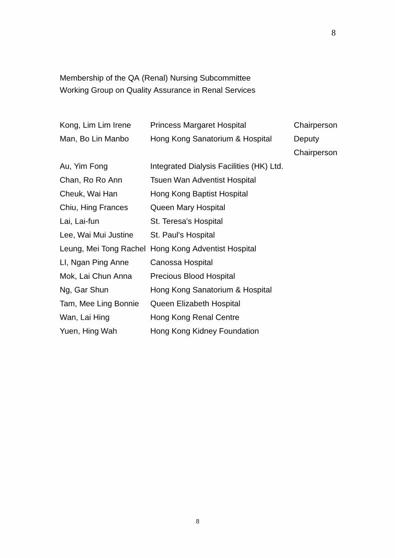
Membership of the QA (Renal) Nursing Subcommittee
Working Group on Quality Assurance in Renal Services
Kong, Lim Lim Irene Princess Margaret Hospital Chairperson
Man, Bo Lin Manbo Hong Kong Sanatorium & Hospital Deputy
Chairperson
Au, Yim Fong Integrated Dialysis Facilities (HK) Ltd.
Chan, Ro Ro Ann Tsuen Wan Adventist Hospital
Cheuk, Wai Han Hong Kong Baptist Hospital
Chiu, Hing Frances Queen Mary Hospital
Lai, Lai-fun St. Teresa's Hospital
Lee, Wai Mui Justine St. Paul's Hospital
Leung, Mei Tong Rachel Hong Kong Adventist Hospital
LI, Ngan Ping Anne Canossa Hospital
Mok, Lai Chun Anna Precious Blood Hospital
Ng, Gar Shun Hong Kong Sanatorium & Hospital
Tam, Mee Ling Bonnie Queen Elizabeth Hospital
Wan, Lai Hing Hong Kong Renal Centre
Yuen, Hing Wah Hong Kong Kidney Foundation
9
9
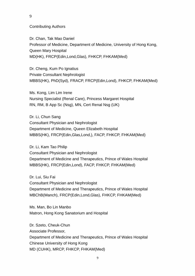
Contributing Authors
Dr. Chan, Tak Mao Daniel
Professor of Medicine, Department of Medicine, University of Hong Kong,
Queen Mary Hospital
MD(HK), FRCP(Edin,Lond,Glas), FHKCP, FHKAM(Med)
Dr. Cheng, Kum Po Ignatius
Private Consultant Nephrologist
MBBS(HK), PhD(Syd), FRACP, FRCP(Edin,Lond), FHKCP, FHKAM(Med)
Ms. Kong, Lim Lim Irene
Nursing Specialist (Renal Care), Princess Margaret Hospital
RN, RM, B App Sc (Nsg), MN, Cert Renal Nsg (UK)
Dr. Li, Chun Sang
Consultant Physician and Nephrologist
Department of Medicine, Queen Elizabeth Hospital
MBBS(HK), FRCP(Edin,Glas,Lond,), FACP, FHKCP, FHKAM(Med)
Dr. Li, Kam Tao Philip
Consultant Physician and Nephrologist
Department of Medicine and Therapeutics, Prince of Wales Hospital
MBBS(HK), FRCP(Edin,Lond), FACP, FHKCP, FHKAM(Med)
Dr. Lui, Siu Fai
Consultant Physician and Nephrologist
Department of Medicine and Therapeutics, Prince of Wales Hospital
MBChB(Manch), FRCP(Edin,Lond,Glas), FHKCP, FHKAM(Med)
Ms. Man, Bo Lin Manbo
Matron, Hong Kong Sanatorium and Hospital
Dr. Szeto, Cheuk-Chun
Associate Professor,
Department of Medicine and Therapeutics, Prince of Wales Hospital
Chinese University of Hong Kong
MD (CUHK), MRCP, FHKCP, FHKAM(Med)
10
10
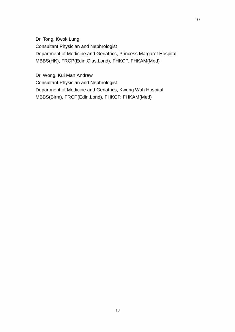
Dr. Tong, Kwok Lung
Consultant Physician and Nephrologist
Department of Medicine and Geriatrics, Princess Margaret Hospital
MBBS(HK), FRCP(Edin,Glas,Lond), FHKCP, FHKAM(Med)
Dr. Wong, Kui Man Andrew
Consultant Physician and Nephrologist
Department of Medicine and Geriatrics, Kwong Wah Hospital
MBBS(Birm), FRCP(Edin,Lond), FHKCP, FHKAM(Med)
11
11
ACKNOWLEDGEMENT
On behalf of the Working Group on Quality Assurance in Renal Services, I
would like to acknowledge the support of the following parties in the production
of this report.
We are grateful to the Li Shu Fan Education Foundation for financially
supporting this project. We would like to thank the Hong Kong College of
Nursing (Specialty Standard Subcommittee – Renal Nursing) for permitting us
to reprint the “17 statements of the Standards for Renal Nursing Practice”.
We would express our gratitude to the Coordinating Committee in Nursing
(Hospital Authority) and the Working Group on Guidelines for Specialty
Nursing Services for permitting us to reprint the 10 Nursing Standards from the
“Guidelines for Specialty Nursing Services (Renal Care)”.
During the preparation of this report, the Working Group has organized
two forums to solicit feedback from medical and nursing professionals on the
report. Their enthusiastic and constructive response contributed towards its
refinement and finalization.
The Working Group takes this opportunity to thank Dr. CH Leong,
President of the Academy of Medicine, for his encouragement and writing a
foreword for this report and Dr Richard Yu, President of the College of
Physicians, for his advice.
12
12
Quality Assurance in Renal ServicesQuality Assurance in Renal ServicesQuality Assurance in Renal ServicesQuality Assurance in Renal Services
Dr CS Li
(I) Introduction
1. The Specialty Board in Nephrology is assigned by the College to look into areas
of quality assurance in the delivery of renal services. Through quality
assurance, healthcare providers may minimize variation in standards of clinical
practices and ensure acceptable patient outcomes.
2. The core activities of nephrologists and renal nurses include care of patients on
renal replacement therapy. Practices of haemodialysis, peritoneal dialysis and
kidney transplantation had been refined throughout the years. By standardizing
these practices, quality of patient care can be upheld.
(Standards are categorized as “Recommended” and denoted (R) in the following
chapters of this document if they are based on strong evidence. They are
categorized as “Desirable” and denoted (D) if the evidence is not as strong.)
3. Implementation of these practices requires the necessary organization structure
and supporting systems, and close attention to the key processes that have an
impact on the patient outcomes plus regular monitoring of the quality of care
provided.
(II) Organization Structure
1. A person with experience in running a renal unit should take charge of the day-
to-day operation of the unit. (R)
2. This person should be responsible for (R)
2.1 Directing the resources, which include the human resources, equipment and
consumables required for smooth running of the unit.
2.2 Planning for the expansion and growth of the unit in response to the
13
13
changing need of the patient population.
2.3 Continuing development of staff to accommodate the technological
advances.
2.4 Monitoring the performance of the unit and ensuring that this meets or
exceeds the standard accepted by the community.
2.5 Representing the unit to liaise with other organizations.
3 Depending on the size of the unit, a committee or board may be needed to govern
the performance of the unit. (R) This body will be responsible for setting
policies over various areas:
3.1 Admission and rejection criteria to new patients for joining the treatment
programme.
3.2 Clinical privilege for practicing nephrologists.
3.3 Human resources management including recruitment, promotion, and
remuneration.
3.4 Issues with far-reaching impact such as occupation safety, environment
protection.
3.5 And any other matters that may have a major financial impact on the unit.
4. The medical and nursing staff of the renal unit should (R)
4.1 Have acquired the necessary skills and knowledge required for taking
care of renal patients .
4.2 Have either been accredited by the respective professional bodies as
specialists or specialty nurses or been accepted as trainees in the specialty
to practice under supervision of the specialists or specialty nurses in the
unit.
4.3 Undertake continuing medical and nursing education to keep in pace with

14
14
the challenges of new development.
(III) Policies, Guidelines and Protocols
1. A renal unit should establish various mechanisms to facilitate its operation.
Policies should be in place to guide the decisions and actions of staff. (R) These
are particularly important in
1.1 Admission of patients into and discharge of patients from the unit.
1.2 Transferal of patients to acute hospital.
1.3 Referral of patients among different dialysis units
1.4 Resuscitation.
1.5 Infection control.
1.6 Waste disposal.
1.7 Risk management.
1.8 Contingency plan.
1.9 Handling of medical information.
1.10 Compliance with legal requirement
2. Guidelines and protocols should be in place (R) to standardize
2.1 The initiation and termination of hemodialysis procedure.
2.2 The monitoring of progress of patients during dialysis.
2.3 The operation of the water treatment system and the reprocessing
15
15
machines
2.4 The disinfection and rinsing of these equipment.
3. A system for record storage, maintenance and retrieval should be in place. (R).
Emphasis should be made on accurate documentation of information and good
keeping of medical record.
4. Channels should be established for communication among staff, between staff
and patients and allow feedback from patients to the renal unit. (R) A hot line
should be in place to facilitate patients to seek advice from staff. Staff of the unit
should provide relevant and comprehensive information to their patients
concerning their care (which include the cost of treatment if the patients are
seeking treatment in the private sector) before accepting them and later when such
need arises.
5. Referral Guidelines (R)
5.1 All patients who are referred for dialytic treatment and who are currently
undergoing treatment in another dialysis unit especially locally must be
referred by the nephrologist or the renal team in charge of the patient.
Except for patients referred to H.A. dialysis units, all referred patients
should be followed up by the referring nephrologist or renal team unless
otherwise requested by the patient. The unit should at the same time
make a request on the patient's behalf for a summary of his/her past
medical histories especially those which may affect the dialytic treatment.
If the in-charge nephrologist or renal team refuses to do so, after full
discussion with the patient, the unit may accept the patient for treatment
but only after informing the nephrologist and renal team concerned of the
intention of the patient to receive continuing treatment in the dialysis unit.
5.2 All units have the responsibility to inform the patients seeking dialytic
treatment in their units of the costs involved. These should include cost
incurred in the dialytic treatment itself including all consumables and the
estimated costs incurred in the blood tests, drugs if these are included in a
package and consultative charge by the attending nephrologist.
16
16
(IV) Audit of processes and outcomes
1. The ultimate indicators of performance of a renal unit are the clinical outcome of
patients. The actions recommended for achieving optimal patient outcome are
detailed in the subsequent chapters.
2. Among the standard raised for the various performances, they can be categorized
into:
2.1 recommended – which is commendable practice based on evidence that
adherence to standard will benefit patients;
2.2 desirable – where the strength of evidence is variable or low.
3. In haemodialysis, peritoneal dialysis and transplantation, the steps for carrying
out some of the procedures have been clearly delineated. Such steps, if
followed observantly, have been shown to reduce risk and enhance safety.
Accurate documentation is crucial for fostering a proper working habit and for
allowing later verification.
4. Many of the clinical outcomes have now been quantified and are measurable.
Benchmarks have been put up for comparison against. Deviation from the
agreed standard should raise concern over the quality of service and be followed
by investigation and appropriate remedial actions. Sometimes clinician should
pay attention to the trend of performance, which may be as important, if not
more, than a single result.
5. All renal units should devise effective mechanism for monitoring their day-to-
day operation, administrative capabilities, and standards of patient care. They
should implement timely corrective measures whenever downward trend in their
performance was noticed. An external peer review of performance against
these standards can be undertaken for accreditation purpose.
17
17
Consensus Guidelines in Renal Services Haemodialysis
Dr KL Tong
1.1 Introduction
Currently there are 12 Hospital Authority hospital renal units, 2 attached satellite
centers, 7 private hospitals, 5 charitable and 1 private center providing
haemodialysis (HD) service for the End Stage Renal Failure (ESRF) patients.
For the public sector, most of the ESRF patients requiring dialysis are put on the
peritoneal dialysis (PD) program. Those patients with contraindication for PD
will be treated with HD. On the other hand, most ESRD patients in the private
sector received HD as the first line of treatment.
1.2 Contraindication for Peritoneal Dialysis
Previous extensive abdominal surgery
Previous pelvic surgery or irradiation
Previous generalized or pelvic peritonitis
Severe chronic obstructive airway disease
Known peritoneopleural communication
Failed peritoneal function as a result of repeated peritonitis or sclerosing
peritonitis associated with previous CAPD.
Failed peritoneal function as a result of loss of ultrafiltration/urea clearance
associated with previous CAPD.
2. Institution Based Haemodialysis
Hospital dialysis
Satellite/limited-care/self-care dialysis
3. Staffing
3.1 Nephrologist ( R )
Patients undergoing dialysis treatment must be under the care of a qualified
nephrologist. The nephrologist or the trainees under his/her direct supervision
should pay regular visits and must be kept informed of any complications which
occur during treatment. A qualified nephrologist must be a Fellow of the Hong
Kong College of Physician or equivalent and has full specialist accreditation in

18
18
nephrology by the Specialty Board in Nephrology of the Hong Kong College of
Physician and registered as a specialist in Nephrology with the Hong Kong
Medical Council.
3.2 Renal Nurses ( R )
Patients undergoing dialysis treatment must be under the care of the qualified
renal nurses. For definition of renal nurse, please refer to Nursing
Subcommittee report on Quality Assurance in Renal Service.
Recommended Nurse : patient ratio
Critically ill patients 1 : 1
Hospital HD 1 : 3
Satellite HD 1 : 4 to 5
3.3.1 Technical staff (D)
1 – 2 technical staff depending on the size of the dialysis unit, with special
training in handling the HD equipment and reverse osmosis units, to assist the
renal nurses to carry out the daily operation of the dialysis unit.
4. Water Treatment System, HD/HDF machines, Dialyser Reprocessing
Machines
Safety Procedure Guidelines
4.1 Water treatment system and distribution loop
4.1.1 Disinfection procedure guidelines for Reverse Osmosis Machine and loop
(as recommended by manufacturer) ( R )
4.1.2 Written documentation of absence of disinfectant for RO and loop post
disinfection ( R )
4.1.3 Daily recording of pressure gauge reading of either resistivity or conductivity
of the RO machine if pressure gauge available ( D )
4.1.4 Central station monitor or alarm system for water treatment plant ( D )
4.1.5 3-monthly checking of rejection rate of RO water and accuracy of timer of the
pre RO System e.g. water softener and charcoal filter ( D )
4.1.6 At least 6-monthly checking of inorganic contaminants in RO system ( D )
4.1.7 At least monthly microbial count of Treated water ( R )
4.2 Dialyzer Reuse
4.2.1 Procedure guidelines for dialyzer reprocessing ( R )
19
19
4.2.2 Written documentation of presence of disinfectant by appropriate test before
rinsing ( R )
4.2.3 Procedure guidelines for rinsing out reprocessed dialyzer and documentation
of the whole process ( R )
4.2.4 Written documentation of absence of disinfectant by appropriate test after
rinsing ( R )
4.3 Haemodialysis machine
4.3.1 Procedure guidelines on Preparation of haemodialysis machine for
haemodialysis ( R )
4.3.2 Procedure guidelines for putting patient on haemodialysis ( R )
4.3.3 Procedure guidelines for taking patient off haemodialysis ( R )
4.3.4 Guidelines on disinfection and aftercare of haemodialysis machine ( R )
4.3.5 Documentation for absence of residual disinfectants for machines requiring
manual chemical disinfection. ( R )
4.4 On-line Haemodiafiltration (HDF)
4.4.1 Documentation of water quality according to the European Guideline for
on-line HDF before direct IV infusion into patient’s circulation at least
monthly ( R )
microbial count < 10 -1 cfu/ml and endotoxin (LAL) < 0.03 EU/ml)
4.4.2 Procedure guideline for preparation and after care of machine and equipment
for the procedure ( R )
4.5 Occupational Safety
4.5.1. Infection control guidelines regarding handling of body fluids, handling of
spills and decontamination procedures, sharps disposal and contingency plan
on exposure of needle stick injury. ( R )
4.5.2. Guidelines on proper handling of disinfectants and decontamination facilities
for accidental spills. ( R )
4.5.3. Appropriate Personal Protect Equipment (PPE) should be provided for staff
handling the disinfectants. (D)
4.6 Contingency
4.6.1 Contingency guidelines for suspension of water, electricity supply and fire
hazard. ( R )
4.6.2 Clinical guidelines for patient’s management on exhibition of the symptoms
of disinfectant toxicity ( R )

20
20
4.6.3 Resuscitation guidelines ( R )
4.7 Maintenance and Repair Work
4.7.1 Guidelines on repair of RO ( R )
4.7.2 Notification and written documentation on the completion of maintenance /
repair work of RO ( R )
4.7.3 Service / maintenance record of all electronic / electric dialysis equipment
( R )
5. Quality of water for dialysis
5.1 Microbiological contaminants ( R )
Microbial countHD Online HDF
< 200 cfu /ml < 10-1 cfu/ml before IV infusion into the patient’s circulation
Test should be done at least monthly
5.2 Endotoxin contaminants ( D )
There is no international recommendation regarding endotoxin testing on RO water
used for routine HD/HDF. It is up to the discretion of the individual dialysis center
to decide whether to peform the testing on a regular basis. For units practising on
line HDF, endotoxin (LAL) should preferably be done at least monthly.
Endotoxin (LAL)
HD Online HDF< 0.25Eu/ml <0.03 EU/mlLAL: Limulus amoebocyte lysate test
5.3 Inorganic Contaminants ( D )
The adoption of Association for Advanced of Medical Instrumentation (AAMI)
Standard is recommended (Annex 1). The water checking either by EMSD, Water
Supply Department, Local laboratories should preferably be done at least once every
six months.
21
21
For details on safe haemodialysis practice, please refer to the
RECOMMENDATIONS ON SAFE HAEMODIALYSIS PRACTICE IN HA
HOSPITALS prepared by the Central Renal Committee HA as annex 2.
6. Biomedical Equipment
6.1 Haemodialysis Machines
Equipment should have facilities for producing bicarbonate-based dialysate and for
volumetric control of ultrafiltration. Each dialysis unit should use similar brands /
models of HD machines from the same manufacturer to facilitate maintenance,
smooth dialysis operation, and to avoid confusion in the stock of different varieties of
dialysis consumables ( D ). It is also desirable to acquire the water treatment system
and the HD machines supplied by the same manufacturer to facilitate auto-
disinfections of the distribution system and HD machines (D).
6.2 Reprocessing / Reuse
Although commercial dialyzers are intended for single use, the reprocessing of
dialyzers for reuse has been quite popular among the dialysis units. This is
particularly important for the expensive high flux dialyzers because of implication on
economic saving. There are no standards for the number of reuse but the following
precautions should be taken.
Quality of water used for reprocessing the blood compartment should be as pure
as for the dialysis itself (microbial count should be < 200 cfu/ml ( R ) .
Demonstration that the blood compartment has been washed free of the
sterilizing agent before reuse ( R ).
Confirmation of the efficiency of the dialyser by checking volume of dialyser or
other direct or indirect means to check urea clearance at regular interval (R).
7. Biocompatibility Issues
7.1 Bicarbonate Dialysis Fluid
Although no increment in patient survival has yet been demonstrated using
bicarbonate rather than acetate, bicarbonate dialysis has been accepted as the
buffer of choice because of improved cardiovascular stability in acute patients,

22
22
chronic patients with cardiovascular complications, for high efficiency and high
flux dialysis ( D ).
7.2 Dialysis Membranes
There are some potential beneficial effect for using the synthetic membranes (e.g.
polysulphone, polyamide, polyacrylonitrile) including enhanced
biocompatibility with less accumulation of beta-2 microglobulin, less severe
interdialytic symptoms and better nutritional indices. However, patient
survival on long term dialysis has not been improved compared with cellulose
membranes. At this stage, it would be inappropriate to set the standard of the
dialysis membrane types but because of the many potential advantages, it is
desirable to dialyse patients with the dialyzers with synthetic membranes (D).
7.3 On-line Haemodiafiltration
This treatment combines convection and diffusion process in the removal of
solutes. High molecular weight solutes e.g. β2M are better removed.
Studies have shown that patients on long-term hemodialfiltration have less
dialysis related amyloidosis. The problem of high cost, bacteremia and
endotoxemia have limited its use as a first line treatment of patients requiring
HD. It may be useful for patients who have been on hemodialysis for more
than 8-10 years (D).
8. Clinical Standard and Targets
8.1.1 Monitor Adequacy of Dialysis
Monitoring the adequacy of dialysis treatment involves a global assessment
which includes clinical assessment and objective measurement, including
weight, blood pressure and laboratory investigations, together with some
measurement of the amount of solute cleared during the dialysis process.
( R )
Methods
8.1.2 For patient on thrice weekly haemodialysis
Either stable URR > 65% ( D )
23
23
Or stable Kt/V > 1.2 (dialysis and residual renal function) ( D )
i) URR = 100 x (1- Ct/CO)
URR is simple but has limitations
- Does not account for the effect of intradialytic urea generation
- Does not account for the effect of ultrafiltration on urea clearance
- Errors in the delivered dose of HD may be difficult to detect in the target
range of URR of ≥65%, where a curvilinear relationship exists between
URR and Kt/V.
ii) Kt/V
The method to calculate Kt/V should be stated
Establish a unit policy to implement a uniform method of
measuring the adequacy of haemodialysis
8.1.3 For patient on twice weekly haemodialysis
Either stable URR > 80% ( D )
Or stable Kt/v > 1.8 (dialysis and residual renal function) ( D )
The minimal standards for twice weekly dialysis are theoretical and not based on
published data. These may be difficult to achieve in many patients.
8.1.4 Frequency of Monitoring
At least once every 6 months ( R )
If a patient is found to be receiving less than the target amount of dialysis, steps
should be taken to increase this by the duration of dialysis, or increasing the
dialyser surface area, or increasing blood flow.
8.2 Correction of Anaemia
Anaemia of chronic renal failure can be corrected by recombinant human
erythropoietin (EPO) or repeated blood transfusion. EPO decreases the
likelihood of transfusion associated infections iron overload, and avoids
sensitization before renal transplant. It improves quality of life, cognitive
function, cerebral blood flow, cardiac function and exercise capacity. However
the use of EPO carries with it financial implications.
A target haemoglobin concentration of ≅ 10g/dl (haematocrit ≅ 30%) should be
24
24
achieved in patients who has been stabilized on HD ( D ). Other common causes of
anaemia (e.g. blood loss, hemolysis and iron deficiency) have to be ruled out and
corrected before considering EPO. The use of EPO depends on many factors
including financial resources and rehabilitation potential of the patients.
Monitoring of treatment should include Hb/Hct concentration, iron stores by
measurement of serum ferritin and iron supply by transferring saturation ( R ).
8.3 Nutritional Status
Poor nutrition with low serum albumin is a powerful predictor of mortality in dialysis
patients. Patient should have a dietary protein intake of 1.0g/kg ideal body weight
intake of together with caloric intake of at least 35 Kcal/kg ideal body weight per day.
Regular assessment by a dietitian is desirable (D). A target serum albumin above
35g/L is recommended for patients stable on HD ( D ).
8.4 Blood Pressure
Hypertension is a predictor for cardiovascular mortality in dialysis patients. Control of
BP is important.
Target pre-dialysis blood pressure ( D )
Age < 60 – BP < 140/90 mmHg
Age >60 – BP < 160/90 mmHg
8.5 Biochemical / Bone Profiles
Target pre-dialysis values
Potassium – 3.5 – 6.5 mmol ( D )
Phosphate – 1.2 – 1.8 mmol/L ( D )
Calcium – normal total calcium corrected for serum albumin concentration or normal
ionized calcium ( D )
iPTH – 2 to 3x the normal range ( D )

25
25
9. Vascular Access
9.1 Acute Haemodialysis Vascular Access – Noncuffed catheters
9.1.1 Internal jugular or subclavian noncuffed catheters should generally not be used
for more than 6 weeks ( D ).
9.1.2 The subclavian insertion site should not be used in a patient who may need
permanent vascular access owing to the risk of central venous stenosis ( D ).
9.1.3 Femoral catheters should preferably not be left in place longer than 7 days
( D ).
9.1.4 The catheter exit site should be examined at each hemodialysis treatment for
signs of infection ( D ).
9.1.5 Catheter exit site dressings should be changed at each hemodialysis treatment
( D ).
9.2 Permanent Vascular Access (Primary AV Fistulae and AV Grafts)
9.2.1 Arm veins suitable for placement of vascular access should be preserved,
particularly the cephalic veins of the non-dorminant arm ( D ).
9.2.2 A primary AV fistula is mature and suitable for use when the vein’s diameter
is sufficient to allow successful cannulation, but preferably no sooner than 1
month ( D ).
9.2.3 Polytetrafluoroethylene (Gore-tex) AV grafts should not routinely be used
until 14 days after placement ( D ).
26
26
9.3 Tunneled Cuffed Catheter
9.3.1 Tunneled cuffed venous catheters are the method of choice for temporary
access of longer than 6 weeks’ duration and those patients who have
exhausted all other access options ( D ).
9.3.2 The preferred insertion site for tunneled cuffed venous catheters is the right
internal jugular vein ( D ).
9.4 Monitoring and Surveillance of Permanent Vascular Access (Primary
AV Fistulae and AV Grafts)
Prospective surveillance of permanent vascular access for hemodynamically
significant stenosis, when combined with correction improves patency and
reduces the incidence of thrombosis ( D ).
Techniques that can be used, in order of decreasing preference:
9.4.1 Dynamic venous pressures
The threshold that indicates elevated pressure (and therefore the presence of a
hemodynamically significant venous outlet stenosis) is 150 mmHg at a blood
flow rate of 200 ml/min during the first 2 to 5 minutes of hemodialysis using 15-
gauge needles.
9.4.2 Measurement of access recirculation using urea concentrations
Recirculation exceeding 10 % should prompt investigation of the presence of
stenosis
9.4.3 Unexplained decreases in hemodialysis adequacy (URR, Kt/V)
9.4.4 Physical findings of persistent swelling of the arm, clotting of the vascular
access, prolonged bleeding after needle withdrawal, or altered characteristics
of pulse or thrill in a vascular access

27
27
9.4.5 Elevated negative arterial pre-pump pressures that prevent increasing to
acceptable blood flow
9.4.6 Doppler Ultrasound
Persistent abnormalities in any of these parameters should prompt referral for
further management.
9.5 Management of Permanent Vascular Access Stenosis
Appropriate intervention should be initiated when there is hemodynamically
significant stenosis, which is defined as a ≥ 50 % reduction of the lumen diameter
accompanied by the following clinical/physiologic abnormalities: ( D )
Previous thrombosis in the access
Elevated venous dialysis pressure
Abnormal urea recirculation measurements
Unexplained decrease in URR or Kt/V
Haemodynamically significant stenosis of permanent vascular access should be
treated with percutaneous transluminal angioplasty (PTA) or surgical revision. Each
dialysis center should determine which procedure is the best for the patient based on
the expertise at that center.
If PTA is required more than 2 times within 3 months, the patient should be
referred for surgical revision.
Items for Audit (D)
Demographic dataAge distribution of patients receiving haemodialysisPrimary dialysis treatment or transferred from CAPDNumbers on thrice vs twice weekly dialysis
Technique: numbers of patients usingBicarbonate vs acetate dialysisCellulosic vs synthetic membranes

28
28
Standard dialysis vs high flux dialysis vs haemofiltraion
Correction of anaemiaPercentage of patients receiving erythropoietinHaemoglobin frequency distribution (all patients)Percentage of patients with Hb < 10g/dl
Dialysis adequacy and nutritionKt/V or URR frequency distribution in dialysis populationPre-dialysis serum albumin frequency distribution
Blood pressure controlSystolic, diastolic and mean arterial pressure (MAP) frequency distribution
Biochemical profilesPre-dialysis: potassium, calcium, phosphate, serum albumin, iPTH frequencydistribution
Water treatment system, HD/HDF procedure, Dialyser ReuseSafety procedure check list for water treatment system and dialysis equipment
Water qualityBacterial counts +/- endotoxin levels : test frequency and results
Access for dialysisTiming of access creation in relation to start of dialysisProportion of access by :Radiocephalic or brachiocephalic A-V fistulaPTFE or other prosthetic fistulaCentral venous line or similar access (eg Permcath)Duration of function of access procedure
29
29
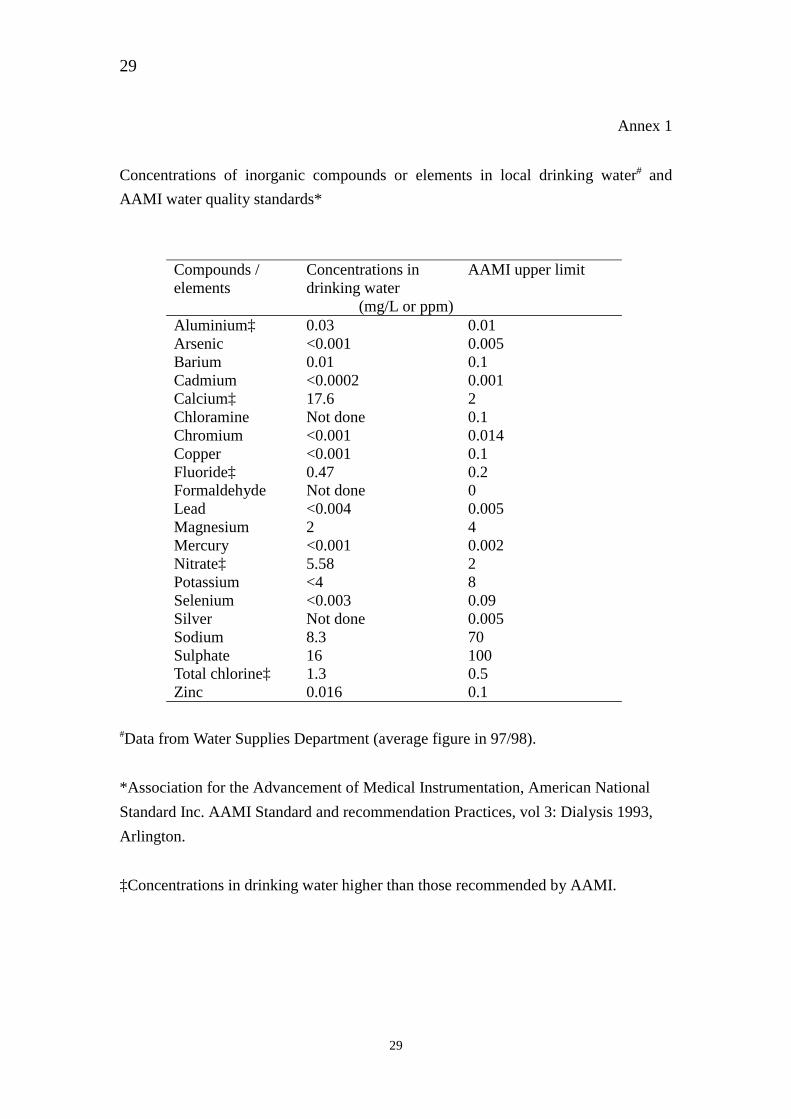
Annex 1
Concentrations of inorganic compounds or elements in local drinking water# and
AAMI water quality standards*
Compounds /elements
Concentrations indrinking water
AAMI upper limit
(mg/L or ppm)Aluminium‡ 0.03 0.01Arsenic <0.001 0.005Barium 0.01 0.1Cadmium <0.0002 0.001Calcium‡ 17.6 2Chloramine Not done 0.1Chromium <0.001 0.014Copper <0.001 0.1Fluoride‡ 0.47 0.2Formaldehyde Not done 0Lead <0.004 0.005Magnesium 2 4Mercury <0.001 0.002Nitrate‡ 5.58 2Potassium <4 8Selenium <0.003 0.09Silver Not done 0.005Sodium 8.3 70Sulphate 16 100Total chlorine‡ 1.3 0.5Zinc 0.016 0.1
#Data from Water Supplies Department (average figure in 97/98).
*Association for the Advancement of Medical Instrumentation, American National
Standard Inc. AAMI Standard and recommendation Practices, vol 3: Dialysis 1993,
Arlington.
‡Concentrations in drinking water higher than those recommended by AAMI.

30
30
Annex 2
RECOMMENDATIONS ON SAFE HAEMODIALYSIS PRACTICEIN HA HOSPITALS
Prepared by the Special Working Group* on Haemodialysis safety
Central Renal Committee, HA
* The Working Groups is composed of Dr F S LUI, Cons(Med), PWH, Dr K L Tong,Cons(Med), PMH,
Dr C S LI, Cons(Med), QEH and Dr Albert LO, EM(PS)3, HAHO
1. The design of water treatment plant/reverse osmosis (RO) system1.1 Back up RO system (2 or more central RO systems for each centre) is
preferable.
1.2 In dual RO systems, each system should be used on alternative days.While in the stand by mode, the system should preferably be flushedonce every 24 hours. Auto-flush function is usually in place for newerRO models. Dialysis centres should check whether this procedure isbeing carried out according to the instructions of the manual. Forsystem requiring manual operation, flushing can be carried out for theidling system before closure of the dialysis centre so that the systemwill be ready for use the next day.
1.3 All dialysis units should carefully examine the structure of the ROsystems and loops with the suppliers. Potential dead space in thesystem must be clearly identified and documented.
2. Disinfection procedure for RO system2.1 Disinfectant should be used as recommended by the manufacturers.
In view of the occupation hazard of using formaldehyde, it is desirablefor new dialysis centres to use non-formaldehyde based disinfectant toclean the RO system.
2.2 Only well trained nurses and /or laboratory attendants in the dialysiscentres are allowed to carry out the disinfection procedure.
2.3 Disinfectant should preferably be done while the dialysis centre is notin service (at Saturday pm or Sunday). For centres who are carrying outthe process during service hours, the director of the dialysis unit shouldbe completely satisfied with himself that the disinfectant cannot go intothe operating system under any circumstances. If in doubt, theworkflow has to be rearranged for staff to perform disinfectionprocedures after service hours.
31
31
2.4 For centres with a single RO system, it is mandatory that disinfectionprocedures be carried out outside dialysis hours.
2.5 The dwell time recommended by the manufacturers should befollowed.
2.6 Dialysis centres should note whether backwash and regenerationmechanism (automatic or manual) for the pre-treatment filters andsofteners are in place. RO system with backwash mechanism ispreferable because it increases the durability of the system.
2.7 The disinfection procedure should preferably be done once every 1 to 2weeks or as recommended by the manufacturer.
2.8 The servicing personnel should be informed by the staff regarding thepresence of disinfectant in the RO system.
3. Rinsing Process
3.1 Dialysis centres should ensure that the testing methods for residualdisinfectant are appropriate and the sensitively of the tests conformwith the international recommendations. The validity of newlyintroduced testing methods should preferably be determined by theGovernment Laboratory.
3.2 Written documentation upon completion of testing residual disinfectantis mandatory.
4. Performance of RO system
4.1 Pressure gauge reading, resistivity / conductivity or other relevantmeasurement should be recorded daily by trained LA/Nurse.
4.2 Staff of dialysis centre can be alerted immediately on abnormalperformance of RO system if the system alarm can be connected to thecentral station/monitor. Possibility of such an arrangement should beexplored.
4.3 Rejection ratio should be checked and recorded during routinemaintenance 3-monthly.
4.4 All essential performance parameters (including sensor function andrejection ratio) of the RO system should be checked 6-monthly.
5. Detection of inorganic contaminants in RO system
5.1 While water conductivity can give a general idea on the RO water

32
32
quality, it should be supplemented by determining the concentrations ofspecific inorganic compounds or elements in post-RO water. Theadoption of Association for the Advancement of MedicalInstrumentation (AAMI) standard is recommended (appendix 1).
5.2 There is no international standard on what inorganic compounds orelements should be tested because of the variations in the quality ofinfluent water in different countries / cities. In Hong Kong, theconcentrations of several compounds or elements in drinking water arehigher than those recommended by AAMI (appendix 1). It isconsidered by the working group that the concentrations of thesecompounds or elements should be determined regularly. Commercialtest kits are available to test these compounds/elements at a reasonableprice.
5.3 There is no established recommendation regarding the frequency of thetests. It is considered by the working group that the frequency of notless than once every 6-month is an acceptable practice.
5.4 To protect the RO system from being damaged by the contaminants inthe influent water, chlorine, nitrates and hardness in pre-RO watershould be determined regularly.
6. Detection of microbiological contaminants in RO system
6.1 Tests should be done at least monthly. The colony forming unit (CFU)should be less than 200 /ml and no special culture medium is required.Sample procedural guidelines for getting water sample from the ROsystem is set out in appendix 2.
There is no international recommendation regarding endotoxin testingon RO water used for routine haemodialysis. It is up to the discretionof individual dialysis center to decide whether to perform the testingon a regular basis. The working group considered that monthlytesting will be adequate and the endotoxin level should be less than0.25EU/ml. Sample procedural guidelines for the collection andtransport of water samples are set out in appendix.
6.2 The sites of water sampling depend on the system structure. For closedloop system, pre- and post-RO water should be sampled. For open loopsystem, water at post-RO site and each open end should be sampled.
6.3 For units practising haemodiafiltration, CFU should be less than 1000 /1000 ml for samples taken at post pre-filter site and 100 / 1000 ml atinfusion port. Special culture medium should be used to increase theculture sensitivity. Sample procedural guidelines for sampling andculture is set out in appendix 3.
7. Piping system

33
33
7.1 The disinfection procedures should follow the recommendation of themanufacturer.
7.2 While 4% formaldehyde is commonly used for disinfecting the pipingsystem, it is noted that the occupational hazard is unduly high undersuch circumstances. Possibility of using lower concentration offormaldehyde or other types of disinfectant should be explored. Newermode of disinfection (e.g., heat or other types of disinfectant) should beconsidered in the installation of new RO and piping system.
7.3 Testing of residual disinfectant is mandatory. For open loop system,water at each and every end of the system tributary should be tested.For close loop system, water at both ends of the loop should be tested.
7.4 Method of testing should follow the same principle as stated in 3.1.
8. Re-use of dialyser
8.1 It is desirable to use disinfectants other than formaldehyde.
8.2 The presence of disinfectant must be confirmed before rinsing byappropriate test.
8.3 The absence of disinfectant must be documented by appropriate testafter rinsing and the completion of the whole process should bedocumented in written form.
9. Haemodialysis machine
9.1 Chemical disinfection carries the risk of having residual disinfectant inthe machines. Other mode of disinfection (e.g., heat) is preferred.While total avoidance of chemical disinfection is not possible, it isadvisable that non-formaldehyde based disinfectant be used to lowerthe staff and patients’ risk of exposure to formaldehyde.
9.2 For machines requiring chemical disinfection, appropriate test must bedone immediately before the start of haemodialysis to ensure noresidual disinfectant remains in the machine after disinfection process.
10. Occupational hazard of disinfectant
10.1 Guidelines should be in place to ensure proper handling of disinfectant,including decontamination facilities for accidental “ spills”.
10.2 Staff handling formaldehyde must be provided with the appropriatemasks.
10.3 Innovative methods to minimize occupational exposure toformaldehyde are being explored. One example from QEH is set out inappendix 4.
34
34
11. Contingency
11.1 Guidelines should be in place to ensure patient safety in case of suddencut off of water or electricity supply and fire hazard to the dialysiscentres.
12. Medical emergency
12.1 Staff should be informed of the symptoms of disinfectant toxicity.Clinical guidelines for managing such conditions should be in place.
12.2 Resuscitation guidelines should be in place and staff of the dialysiscentres should participate in regular CPR drills.
13. Water supply
13.1 Separate twin tank system is preferable to avoid sudden cut off of watersupply to the dialysis centre and to facilitate cleansing of the watertank.
13.2 Alarm should be installed for low level warning.
14. Procedural guidelines
14.1 Guidelines must be in place for all procedures including disinfectionand rinsing of RO systems, checking performance of RO and quality ofwater, and checking residual disinfectant in the system.
14.2 All guidelines (and relevant procedural checklist) must have Chineseversion and both English and Chinese versions must be shown to thesupplier/manufacturer for their endorsement in writing.
14.3 Any amendments to the procedural guidelines have to go through andbe agreed upon by the head of the dialysis centre as well as theequipment manufacturer when appropriate.
14.4 The procedural guidelines must be strictly adhered to by all staff.Deviations from the guidelines without sound reasons and priorapproval from the head of the dialysis centre is not allowed.
15. Maintenance and repair work
15.1 The RO machine under repair should never be switched on followingservicing when patients are receiving haemodialysis.
15.2 Staff should confirm with the servicing personnel that themaintenance/repair work has been completed, preferably by writtendocumentation.
15.3 Guidelines must in place to ensure that the RO system can only be re-
35
35
started after servicing when adequate rinsing and confirmatory testingfor residual disinfectant have been carried out.
15.4 For centres with a single RO system, it is mandatory that maintenanceand repair work be carried out outside dialysis hours.
February 2000
36
36
PERITONEAL DIALYSIS
Dr Philip Li, Dr CC Szeto
(I) Introduction
1. Although peritoneal dialysis (PD) is a relatively simple technique, it should be
performed in the right setting, with appropriate staff and facilities, and be
integrated into a renal replacement program.
2. Doctors, nurses and paramedical staff should work together as a multi-disciplinary
team.
3. A unit offering PD should provide not only continuous ambulatory peritoneal
dialysis (CAPD) but also automated peritoneal dialysis (APD). It should have
adequate access to back-up hemodialysis (HD) facilities and renal transplantation.
(II) Structural requirement
1. Space requirement for PD unit
1.1 PD unit should encompass dedicated places, including: PD training rooms,
store rooms, clean and dirty utility rooms, clinic area, access to emergency
beds and hemodialysis, toilet and showers, office for nurse, doctors, clerical
and administrative staff. (D)
1.2 PD training room should include the following equipment: comfortable
chair and bed, wash basin, surface or trolley, weighing scales, drip stand or
hook, shelving for consumables, bag-warming equipment, ambulatory PD
machine, clock and sphygmomanometer. (D)
2. Biomedical standard of equipment and PD solutions
2.1 All equipment used in the delivery and monitoring of therapies should
comply with the relevant standards for medical electrical equipment. All
electromechanical equipment used to undertake PD should comply with
international standards for electromechanical safety. Fluids for peritoneal

37
37
dialysis should satisfy the current international quality standards. (R)
3. Human resource
3.1 The PD team should include physicians, nurses, dieticians and social worker.
The team should also work closely with other clinical specialists, such as
surgeon, microbiologist, psychologist and rehabilitation specialists. (D)
3.2 Ideally there should be 24-hour on-call service. (D)
3.3 At least some formal structure is required to promote optimal function of
the team. Definition of roles and job responsibilities is essential. Regularly
scheduled team meetings such as patient-care conferences and quality
improvement meetings are recommended. (D)
3.4 The PD unit should provide adequate training for medical and nursing staff.
There should be written protocols for standard procedures in caring PD
patients. Continuing education program in the unit is strongly recommended.
(R)
(III) Protocols
1. Each unit needs to have protocols for various procedures to ensure safe and
consistent care. It is essential that all members of the team are aware of those, and
any amendments that are made. Protocols should be developed by the nursing staff
and the medical director and should be reviewed on a regular basis. Protocols
should be based on sound scientific principles and research findings. (R)
2. Protocols recommended for a peritoneal dialysis program
2.1 CAPD exchange procedure (for each system)
2.2 Cycler set-up procedure (for each cycler)
2.3 Dialysate and urine collections for adequacy assessment
2.4 Intermittent PD regimens, eg IPD, CCPD
2.5 Exit site care (post-implantation and chronic)
2.6 Administration of intra-peritoneal medication
2.7 Transfer set change procedure
2.8 Peritoneal equilibration test
2.9 Treatment of infections: peritonitis, exit site

38
38
2.10 Managing complications, eg poor outflow-inflow, crack in catheter
(IV) Recommended Standard
1. PD systems
1.1 The use of disconnect systems should be standard unless contraindicated. (1st)
(D)
1.2 Rationale
1.2.1 Disconnect, 'flush before fill' CAPD systems are superior to earlier
systems. Peritonitis is significantly less using the Y-set or modified Y-
set compared to the standard spike system and the use is cost-effective
[1-4]. Such systems should be standard for all patients, unless they are
incapable of managing the technique.
1.2.2 APD should be available for selected patients. Monitoring of the dose
of dialysis delivered is important in APD. Automated systems are more
expensive than standard CAPD; their extra costs will need to be
considered in the selection of the mode of therapy.
2. General clinical standards
2.1 The standards listed below apply equally to PD and HD:
Correction of anaemia
Control of blood pressure
Prevention of transmissible infections to patients and staff
3. Nutritional status
3.1 A protein intake greater than 1.2 g/kg/day, and a calorie intake, including
glucose absorption from the dialysate, of above 35 kcal/kg/day should be
attained by all patients. (2nd) (D)
3.2 Serum albumin should be targetted at the lower limit of the local normal
range. (2nd) (D)
39
39
3.3 Rationale
3.3.1 PD patients lose protein 0.12 g/kg/day and amino acids to the
equivalent of protein 0.2 g/kg/day in the dialysate. Protein malnutrition
with low serum albumin is a powerful predictor of mortality in dialysis
patients [5,6]. Therefore, effort should be made to ensure adequate
protein and caloric intake in PD patients. Education by dietician is
advisable. Special attention should be paid to vegetarians.
3.3.2 There is no one parameter that ideally measures nutritional status.
Malnutrition can be recognised by a reduced serum albumin, actual
body weight below 90% of ideal body weight for height, and estimated
protein intake below 0.8 g/kg/day [7].
3.3.3 Since the concentration of serum albumin varies substantially with the
method employed, the technique of measurement should be recorded in
audit data [8,9].
4. Biochemical profiles
4.1 Potassium 3.5-5.5 mmol/l
Phosphate 1.1-1.6 mmol/l
Calcium within normal limits for local laboratory, corrected for serum
albumin concentration. (2nd) (D)
4.2 Intact PTH (iPTH) should be maintained at between 2 and 3 times the upper
limit of the local normal range. (2nd) (D)
4.3 The serum bicarbonate level should not fall below the local normal range, or
rise more than 3 mmol/l above it. (2nd) (D)
4.4 Rationale
4.4.1 The ideal target concentration of calcium has not been established
firmly. However, it is desirable to avoid the use of aluminium
containing phosphate-binding agents.
4.4.2 Currently the estimation of iPTH by an intact hormone assay is the best
40
40
non-invasive method for assessing parathyroid activity and renal bone
disease. Values in excess of three times the upper limit of the normal
range usually indicate parathyroid over-activity, whilst values below
the upper limit of local normal range suggest the presence of adynamic
bone. The latter finding is associated with metastatic calcification and a
relative inability to dispose of a calcium load [10].
5. Peritoneal equilibration tests
5.1 Peritoneal equilibration test (PET) should be performed after 4 to 8 weeks on
dialysis, and when clinically indicated, or when therapy is changed to APD.
(3rd) (D)
5.2 Rationale
5.2.1 PET measures two aspects of membrane function: low molecular
weight solute transport (expressed as the dialysate-to-plasma ratio of
creatinine at 4 hours), and the ultrafiltration capacity [11,12].
5.2.2 Membrane function takes 4 to 6 weeks after starting dialysis to stabilise
[13]. The initial CAPD regimen should be prescribed assuming normal
transport characteristics. Subsequently, PD regimen should be adjusted
to meet targets of solute clearance and fluid removal by changes in
dwell times, fill volumes, glucose concentration and so forth.
5.2.3 The clinical values of assessing membrane function are:
Allow optimisation of both solute clearance and ultrafiltration
because solute transport rates vary considerably in the PD
population.
In CAPD patients, high solute transport is associated with reduced
technical and patients survival [14,15].
5.2.4 The methods of performing PET are well described in the literature
[16]. The following points should be remembered in the interpretation
of results:
High concentrations of glucose interfere with many assays for
creatinine. It is important to work with the local biochemists to
ensure that the appropriate correction for measurement of creatinine

41
41
in dialysate has been taken into account.
The patient should follow their usual dialysate regime, draining out
as completely as possible before the test dwell. Large residual
volume will affect the results.
Intra-patient variability of the ultrafiltration capacity (around 20%)
is greater than for the solute transport (less than 10%). Results of
the PET, in particular the ultrafiltration capacity, should always be
interpreted in the light of additional exchanges performed during
the same 24 to 48 hour period (usually collected to assess solute
clearance).
PET is not a surrogate for measuring solute clearance.
5.2.5 Using a standard PET, an ultrafiltration capacity of below 200 ml is
associated with a 50% risk of achieving less than 1-L ultrafiltration in
anuric patients [17]. The additional measurement of the sodium D/P
ratio at one hour of PET gives an estimation of sodium sieving across
the peritoneal vasculature, which if absent indicated poor ultrafiltration.
5.2.6 The standard peritoneal permeability analysis (SPA) can be an
alternative to PET for investigation of possible ultrafiltration failure. It
uses a 3.86% glucose dwell (as opposed to the PET which uses 2.27%)
over 4 hours and defines ultrafiltration failure as below 400 ml
ultrafiltration capacity in the absence of fluid leak or catheter
malfunction [18].
6. Adequacy of CAPD
6.1 A total weekly creatinine clearance (CCr) above 50 L/week/1.73m2 and/or a
weekly Kt/V urea above 1.7 are recommended. Higher targets with weekly
Kt/V of 1.9 are desirable, especially for high transporters and APD patients.
(2nd) (D)
6.2 Achieving either Kt/V or CCr target is acceptable. (3rd) (D)
6.3 Adequacy studies should be repeated at least annually, and more frequently if
clinically indicated, particularly if suspicion arises that residual function has
declined more rapidly than expected. (3rd) (R)
42
42
6.4 Careful attention to fluid balance, especially in anuric patient, is essential.
(2nd) (R)
6.5 If an inadequate Kt/V or CCr is found, it is important to identify the cause so
that appropriate action can be taken. Some patients may need to be
transferred from PD to HD if they remain inadequately dialysed despite
exhaustive measures. (D)
6.6 Rationale
6.6.1 Adequacy is a global concept, which includes clinical assessment of
well-being and physical measurements, measures of small molecule
solute clearance and fluid removal. It is important that clinical aspects
be taken into consideration.
6.6.2 A weekly Kt/V below 1.65 was reported to be associated with poor
outcome [19,20]. It must be emphasised that most of the studies are
observational ones, and that there is no final proof that achieving these
targets will result in improved outcome [21,22]. Nevertheless, there is
some evidence that increasing delivered dialysis dose can improve
nutritional status and reduce hospitalisation rates [23-25]. The
recommended standard given above should be regarded as approximate
targets for which to aim, that need to be refined when more data are
available. Since there is evidence that Chinese PD patients require
lower adequacy targets [26], the standards given above are lower than
those recommended by the DOQI guideline of USA [27].
6.6.3 The influence of dialysis adequacy on survival could be attributed to
the effect of residual renal function [28]. PD patients who have lost
residual renal function are at increased risk, due to a combination of
reduced clearance and fluid removal. Residual renal function should be
carefully monitored in all PD patients (see below).
6.6.4 There is some evidence that CAPD patients are chronically fluid
overloaded [29], and this impacts on cardiovascular outcome [30].
Particular care should be taken in anuric patients treated with APD, due
to the risk of fluid re-absorption during the daytime dwell. There is no
simple, direct way of assessing fluid status in PD patients, and clinical

43
43
judgement is important. Management guidelines recently published by
the ISPD [31], which outline the approach to managing a PD patient
with fluid overload, should be referred to.
6.6.5 In measuring solute clearance and planning changes to the dialysis
regime, there are a number of commercial computer programs that are
designed to aid dialysis prescription. Nevertheless, a change in dialysis
prescription should be checked in its efficacy by repeating clearance
studies.
6.6.6 Details of recommended methods for the estimation of Kt/V, CCr and
protein nitrogen appearance (PNA) are given the guidelines by DOQI
[27] and the Renal Association [32].
7. Residual renal function
7.1 In patients with urine output, residual renal function should be measured at
least annually. (2nd) (R)
7.2 Rationale
7.2.1 Decline in residual renal function has an important bearing on the
adequacy of dialysis and nutritional intake [33,34]. Consequently,
residual renal function should be assessed at least yearly as part of the
assessment of adequacy, or whenever under-dialysis is suspected [35].
Recommended methods for measurement of residual renal function are
described in the DOQI guideline.
7.2.2 There is evidence that the regular use of a loop diuretic can maintain
urine volume, although whether this will affect outcome is unknown.
Prescription of loop diuretic should be considered in PD patients with
urine output.
8. Outpatient monitoring of patients during CAPD
8.1 This should include:
Assessment of weekly Kt/V and/or creatinine clearance
Reassessment of prescription in the event of excessive weight gain
44
44
Collection of biochemical data
Assessment of residual renal function annually and as clinically
indicated
PET measurement as indicated clinically
9. Peritonitis
9.1 Peritonitis rates should be less than 1 episode per 18 patient-months. (1st)
(D)
9.2 The negative peritoneal fluid culture rate in patients with clinical peritonitis
should be less than 20%. (1st) (D)
9.3 The initial cure rate of peritonitis should be more than 80%. (1st) (D)
9.4 Rationale
9.4.1 Despite its theoretical and practical disadvantages, the number of
episodes per patient month is recommended for convenience
expression and comparison of peritonitis rate.
9.4.2 Peritonitis rates are improving with the introduction of disconnect
systems (1-4). The successful diagnosis and management of peritonitis
requires high quality microbiological facilities and close liaison with
the microbiology department. Protocols for managing peritonitis
episodes have been published [36,37]. It must be noted that the use of
vancomycin as a first-line antibiotic has been curtailed recently
because of the emergence of vancomycin resistant organism [38].
9.4.3 Methods for the culture of PD fluid as described in the Renal
Association guideline should be referred to [32].
9.4.4 Guidelines for insertion of peritoneal access catheters and their
subsequent care have been published [39] and should be referred to.
The following points should be noted:
The insertion must be done by a competent and experienced
operator.
No catheter appears to be superior to the standard double cuff
45
45
Tenckhoff catheter.
A downward directed exit site decreases the incidence of catheter
related infections.
9.4.5 Prevention catheter-related infection (exit-site, tunnel) is important.
Nasal carriage of Staph aureus is strongly linked with exit-site infection
[40]. Antibiotic prophylaxis in carriers may help to reduce catheter-
related infections [41]. However, it is not yet clear whether prolonged
usage of mupirocin is necessary or desirable.
(V) Possible items for audit of PD
1. Demographic data
− age distribution of patients receiving PD
2. Technique
− number of patients on disconnect systems
− number on CAPD and APD
− immediate catheter non-function or leak
− catheter survival rate
3. Dialysis adequacy and nutrition
− see the DOQI guideline for details of methodology
− Kt/V; weekly CCr
− normalized protein nitrogen appearance
− serum albumin
4. Correction of biochemical parameters
− serum potassium frequency distribution
− serum bicarbonate frequency distribution
− serum albumin frequency distribution
5. Peritonitis

46
46
− peritonitis rate – episode per patient-month of therapy
− primary cure rate – percentage
− culture negative rate – percentage
6. Exit site infection
− rate – episode per patient-month of therapy
7. Temporary transfer (< 2 months duration) to hemodialysis
− number and rate
8. Correction of anemia )
9. Blood pressure control )
10. Cardiovascular disease ) as for hemodialysis
11. Transmissible disease )
12. Hospitalization )
13. Outcome
− actuarial patient survival
− technique survival
(VI) Reference
1. MacLeod A, Grant A, Donaldson C, et al. Effectiveness and efficiency of methodsof dialysis therapy for end-stage renal disease: systematic reviews. Health TechnolAssess 1998; 2:1-166.
2. Harris DC, Yuill EJ, Byth K, Chapman JR, Hunt C. Twin- versus single-bagdisconnect systems: infection rates and cost of continuous ambulatory peritonealdialysis. J Am Soc Nephrol 1996; 7:2392-8.
3. Li PKT, Chan TH, So WY, Wang AYM, Leung CB, Lai KN. Comparisons of Y-set disconnect system (Ultraset) versus conventional spike system in CAPDpatients: outcome and cost analysis. Perit Dial Int 1996; 16: S368-S370
4. Li PKT, Szeto CC, Law MC, Chau KF, Fung KS, Leung CB, Li CS, Lui SF, TongKL, Tsang WK, Wong KM, Lai KN. Comparison of Double-Bag and Y-Setdisconnect systems in continuous ambulatory peritoneal dialysis – A randomisedprospective multi-center study. Am J Kidney Dis 1999; 33: 535-40
5. Lowrie EG, Lew NL. Death risk in hemodialysis patients: the predictive value ofcommonly measured variables and an evaluation of death rate differences betweenfacilities. Am J Kidney Dis 1990; 15: 458-482.
6. Owen WF Jr, Lew NL, Liu Y, Lowrie EG, Lazarus JM. The urea reduction ratioand serum albumin concentration as predictors of mortality in patients undergoinghemodialysis. N Engl J Med 1993; 329: 1001-1006.
7. Kopple JD, Jones MR, Keshaviah P, et al. A proposed glossary for dialysis
47
47
kinetics. Am J Kidney Dis 1995; 26: 963-981.8. Blagg CR, Liedtke RJ, Batjer JD, et al. Serum albumin concentration-related
Health Care Financing Administration quality assurance criterion is method-dependent: revision is necessary. Am J Kidney Dis 1993; 21: 138-144.
9. Joseph R, Tria L, Mossey RT, et al. Comparison of methods for measuringalbumin in peritoneal dialysis and hemodialysis patients. Am J Kidney Dis 1996;27: 566-572.
10. Kurz A. Calcium homeostasis in adynamic bone lesion. Kidney Int 1994; 46: 855-860.
11. Twardowski ZJ, Nolph KD, Khanna R, et al. Peritoneal Equilibration Test. PeritDial Bull 1987; 7:138-147.
12. Korbet SM, Roxby RM. Peritoneal membrane failure: differential diagnosis -evaluation and treatment. Semin Dial 1994; 7:128-137.
13. Rocco MV, Jordan JR, Burkart JM. Changes in peritoneal transport during the firstmonth of peritoneal dialysis. Perit Dial Int 1995; 15:12-7.
14. Churchill DN, Thorpe KE, Nolph KD, Keshaviah PR, Oreopoulos DG, Page D.Increased peritoneal membrane transport is associated with decreased patient andtechnique survival for continuous peritoneal dialysis patients. J Am Soc Nephrol1998; 9:1285-1292.
15. Davies SJ, Phillips L, Russell GI. Peritoneal solute transport predicts survival onCAPD independently of residual renal function. Nephrol Dial Transplant 1998;13:962-968.
16. Twardowski ZJ, Nolph KD, Prowant B, et al. Peritoneal equilibration test.Peritoneal Dial Bull 1987; 7: 138-147.
17. Davies SJ. Montoring of long-term membrane function. Perit Dial Int 2001; 25:225-230.
18. Ho-dac-Pannekeet MM, Atasever B, Struijk DG, Krediet RT. Analysis ofultrafiltration failure in peritoneal dialysis patients by means of standardperitoneal permeability analysis. Perit Dial Int 1997; 17:144-150.
19. Li PKT, Szeto CC. Adequacy targets of peritoneal dialysis in the Asian population.Perit Dial Int (in press)
20. Blake PG. Problems predicting continuous ambulatory peritoneal dialysisoutcomes with small solute clearances. Perit Dial Int 1993; 13 Suppl 2:S209-11.
21. Genestier S, Hedelin G, Schaffer P, Faller B. Prognostic factors in CAPD patients:a retrospective study of a 10-year period. Nephrol Dial Transplant 1995; 10:1905-11.
22. Gokal R, Harty J. Are there limits for CAPD? Adequacy and nutritionalconsiderations [editorial]. Perit Dial Int 1996; 16:437-41.
23. Lo WK, Cheng IK, Ho YW, Tsoi KS, Chan TM, Wong KS, Yu AW. Effect of Kt/Von CAPD patient survival in a prospective randomized study. Perit Dial Int 2001;21 (Suppl 2): S17.
24. Davies SJ, Phillips L, Griffiths AM, Naish PF, Russell GI. Analysis of the effectsof increasing delivered dialysis treatment to malnourished peritoneal dialysispatients. Kidney Int 2000; 57:1743-54.
25. Mak SK, Wong PN, Lo KY, Tong GM, Fung LH, Wong AK. Randomizedprospective study of the effect of increased dialytic dose on nutritional and clinicaloutcome in continuous ambulatory peritoneal dialysis patients. Am J Kidney Dis2000; 36:105-14.
26. Szeto CC, Wong TY, Leung CB, Wang AY, Law MC, Lui SF, Li PKT. Importanceof dialysis adequacy in mortality and morbidity of Chinese CAPD patients.Kidney Int 2000; 58: 400-407.
27. NKF-DOQI Clinical practice guidelines for peritoneal dialysis adequacy. Am JKidney Dis 1997; 30 (Suppl 2): S69-S133.
28. Blake PG. A critique of the Canada/USA (CANUSA) Peritoneal Dialysis Study.Perit Dial Int 1996; 16: 243-245.
29. Coles GA. Have we underestimated the importance of fluid balance for thesurvival of PD patients? [editorial]. Perit Dial Int 1997; 17:321-6.
30. Lamiere NH, Vanholder R, Van Loo A, et al. Cardiovascular diseases in peritoneal
48
48
dialysis patients: the size of the problem. Kidney Int 1996; 50:S28-S36.31. Mujais S, Nolph K, Gokal R, et al. Evaluation and management of ultrafiltration
problems in peritoneal dialysis. International Society for Peritoneal Dialysis AdHoc Committee on Ultrafiltration Management in Peritoneal Dialysis [In ProcessCitation]. Perit Dial Int 2000; 20 Suppl 4:S5-21.
32. The Renal Association. Treatment of adult patients with renal failure.Recommended standards and audit measures, 2nd ed. Royal College of Physiciansof London, 1997.
33. Szeto CC, Lai KN, Wong TYH, Law MC, Leung CB, Yu AWY, Li PKT.Independent effects of residual renal function and dialysis adequacy on nutritionalstatus and patient outcome in continuous ambulatory peritoneal dialysis. Am JKidney Dis 1999; 34: 1056-1064
34. Wang AY, Sea MM, Ip R, Law MC, Chow KM, Lui SF, Li PKT, Woo J.Independent effects of residual renal function and dialysis adequacy on actualdietary protein, calorie and other nutrients intake in patients on continuousambulatory peritoneal dialysis. J Am Soc Nephrol 2001; 12: 2450-7.
35. Tattersall JE, Doyle S, Greenwood RN, Farrington K. Kinetic modelling andunderdialysis in CAPD patients. Nephrol Dial Transplant 1993; 8:535-8.
36. Keane WF, Alexander SR, Bailie GR, et al. Peritoneal dialysis-related peritonitistreatment recommendations: 1996 update. Perit Dial Int 1996; 16:557-73.
37. Keane WF, Bailie GR, Boeschoten E, et al. Adult peritoneal dialysis-relatedperitonitis treatment recommendations: 2000 update. Perit Dial Int 2000; 20: 396-411
38. Golper TA, Tranaeus A. Vancomycin revisited. Perit Dial Int 1996; 16:116-7.39. Gokal R, Alexander S, Ash S, et al. Peritoneal catheters and exit-site practices
toward optimum peritoneal access: 1998 update. (Official report from theInternational Society for Peritoneal Dialysis) Perit Dial Int 1998; 18:11-33.
40. Luzar MA, Coles GA, Faller B, et al. Staphylococcus aureus nasal carriage andinfection in patients on continuous ambulatory peritoneal dialysis. N Engl J Med1990; 322:505-9.
41. Piraino B. Staphylococcus aureus infections in dialysis patients: focus onprevention. ASAIO J 2000; 46:S13-7.

49
49
Renal Nursing Practice
Ms Irene Kong, Ms Manbo Man
(I) Introduction
1. The need to define quality in Renal Nursing care and to evaluate the
outcomes of care delivered has become the norm of professional growth in
nursing. In Hong Kong, nursing practice is recognised for its high standards
with the available resources in meeting the needs of the community. With an
aim to ensure uniform quality service throughout the community and across
the public and private sector, a working group was formed to conduct a
systematic collaborative review and document the schema of Renal Patient
Care in the local context, with specific reference to the international standards.
This document serves as a reference in meeting the required standards of
service provided to renal patients in Hong Kong.
2. Professional nurses are accountable for their independent patient assessment,
care planning, implementation and evaluation of interventions, in providing
the holistic care to achieve the best possible rehabilitation. Renal nurses in
Hong Kong are committed to provide quality care to our clients. Hence,
continuous effort to evaluate professional competencies and to maintain up-to-
date knowledge is deemed necessary to pledge for the high standard of service.
3. The publications of “Guidelines for Specialty Nursing Service – Renal
Nursing" (Hospital Authority, 2001), “Standards for Renal Nursing Practice”
(College of Nursing, Hong Kong 2001) and “Standards and Guidelines of
Clinical Practice for Nephrology Nursing” (American Nephrology Nurses
Association, 1999) are important references to guide local renal nursing
practices.
4. This paper aims to describe renal nursing practice in Hong Kong. The
three specific objectives include: (1) to provide guidelines for practice and
policy making within the specialty, (2) to identify areas for evaluation of care
and practice standards and (3) to facilitate quality assurance in renal patient

50
50
services.
(II) Structural Requirement
1. The nurse-in-charge of the renal unit is responsible for:
1.1. directing the resources including human and material resources required
for the smooth running of the unit;
1.2. planning for the expansion and growth of the unit in response to the
changing needs of the community;
1.3. promoting continuous nursing development to accommodate the
technological advancement;
1.4. monitoring the performance of the staff and ensuring provision of
quality care to renal patients;
1.5. ensuring the availability of emergency services such as laboratory
service and acute haemodialysis service can be provided in designated /
affiliated institutions;
1.6. representing the unit to liaise with other institutions / organizations.
2. The nurse-in-charge of an accredited renal centre should be a registered nurse
(general) at the Nursing Council of Hong Kong and has completed a post
registration renal nursing program. The nursing staff working in the centre is
specialty trained either through on the job training or a formal structured
program. All new comers should undergo a structured and comprehensive
orientation program. Hospital/ organisational policies, nursing practices
requirement, renal specialty standards and guidelines should be in place.
3. The renal nurse works collaboratively with other health care professionals to
provide safe, competent and high quality care in a cost-effective manner. All
nurses working in renal centers should attend relevant courses, seminars or
conference to update their knowledge and to keep pace with the advance in
care and technology. Reference materials should be available in the renal
centre.
4. A renal nurse is recommended to attain “Continuous Nursing Education”
points (45 points in 3 years) in line with the policy of the Nursing Council of
Hong Kong, whereas a minimum of 30% annual CNE points should relate to
renal specialty.

51
51
(III) Renal Specialty Training Program
1. All post-graduate nursing programs should be conducted in a recognised
training institution with the relevant expertise. There should be significant
input from qualified nurses experienced in renal specialty in the design of
curriculum, the teaching and the evaluation of the course. The majority of
the lectures and theoretical input should be related to renal nursing
competencies, and there should be at least 50% nursing input in the theory
section.
2. The renal nurse caring for haemodialysis patients is recommended to attend a
course that consists of a minimum 28 hours theoretical input and 2 weeks of
practical haemodialysis training through clinical attachment to an accredited
renal center. An additional 2 weeks of CAPD practicum will be required for
nurses working with peritoneal dialysis.
3. Recommended renal specialty courses:
3.1 Post-registration Certificate Course in Renal Nursing organized by
Institute of
Advanced Nursing Studies, Hospital Authority.
3.2 Certificate Renal Course organized by The Hong Kong Kidney
Foundation, Hong Kong Baptist Hospital & The Lions Kidney
Educational Centre (L.K.E.C.) & The Integrated Dialysis Facilities
(H.K.) Ltd.
3.3 Certificate In Renal Nursing Organized by SPACE, University of
Hong Kong.
3.4 English National Board Course No. 136, Renal Nursing for RGNs, or
3.5 Course(s) approved by the accredited body that is recognised by the
Hospital Authority/ Subspecialty Board in Nephrology, the Hong Kong
College of Physicians.
(IV) Manpower Requirement
The Nurse: Patient Ratio will depend on the patient’s dependency level. The
recommended ratio is 1:1 for patient requiring acute haemodialysis with high
dependency care, 1:3 in hospital haemodialysis setting, and 1: 4 -5 in satellite
centres.
52
52
Standards and Guidelines
1. “Standards are authoritative statements by which the nursing profession
describes the responsibilities for which its practitioners are accountable” as
stated by the American Nurse Association (1998). As professional nursing
evolves, standards of renal nursing are involved to serve as continuous
reinforcement and build upon the foundation of renal nursing practice.
2. Guideline is a statement that describes a process of care that has the potential
to improve clinical outcomes and patient decision-making. Guidelines for
renal nursing practices are systematically developed statements that address
the care of renal patients or phenomena. The contents are based on the best
available scientific evidence and / or expert opinion (ANNA, 1999).
3. Standards and guidelines in renal specialty provide a guide to enable nurses to
deliver safe, efficient and cost-effective care. However, provision of
competent and high quality care to clients requires the stringent control within
the profession.
4. The availability of standards serves as an objective tool for the evaluation of
competency. Standards for practice also establish measures for determining
the quality of renal nursing care and provide a means for judging the
competence of renal nurses (Guidelines for Specialty Nursing Service – Renal
Nursing, 2001).
(V) Renal Specialty Standards
1. Renal nurses should acquire the necessary knowledge and skills in order to
provide quality care to the clients and their family. The 17 statements of
Care Standard for practice and the 10 Standards of Renal Nursing Procedures
(Appendices 1-10) are important established standards to provide a basis to
guide nursing practice in renal specialty. The procedure standards include
care of patient on (a) haemodialysis, (b) haemofiltration and
haemodiafiltration, (c) peritoneal dialysis, (d) renal transplantation, and (e)
special procedures e.g. care of patient on plasma exchange.
2. The full text of the following 17 standards could be found in the “Standards
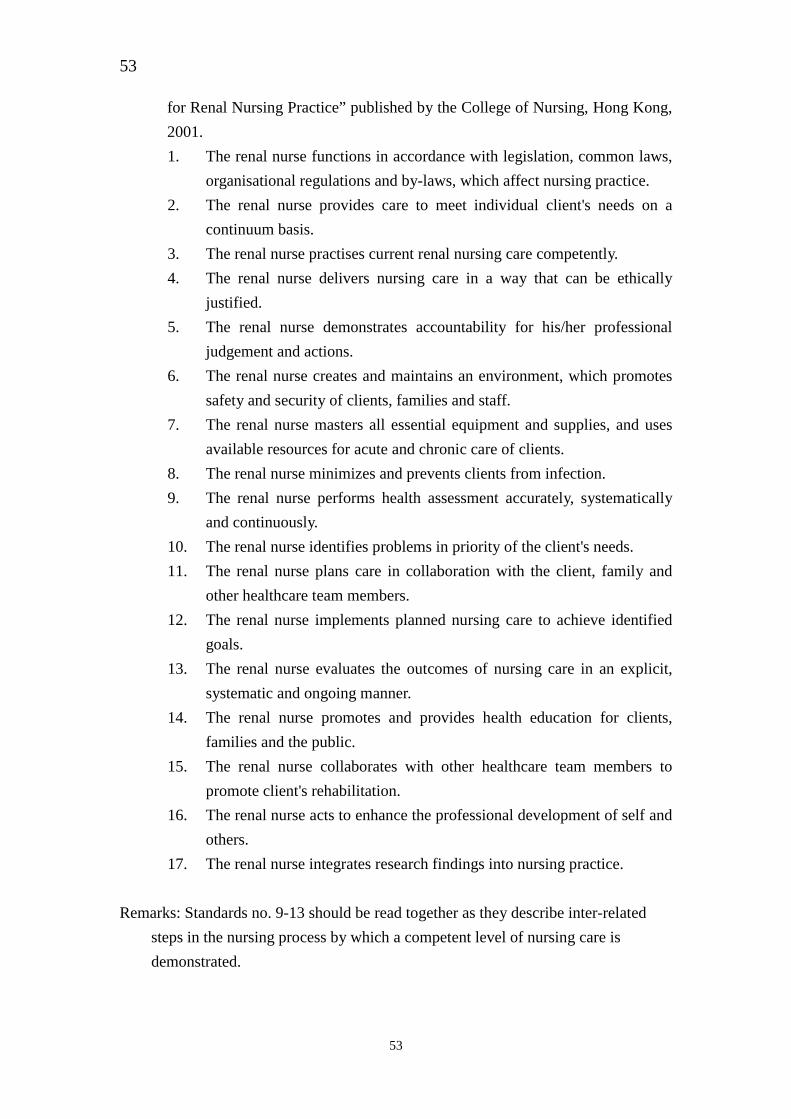
53
53
for Renal Nursing Practice” published by the College of Nursing, Hong Kong,
2001.
1. The renal nurse functions in accordance with legislation, common laws,
organisational regulations and by-laws, which affect nursing practice.
2. The renal nurse provides care to meet individual client's needs on a
continuum basis.
3. The renal nurse practises current renal nursing care competently.
4. The renal nurse delivers nursing care in a way that can be ethically
justified.
5. The renal nurse demonstrates accountability for his/her professional
judgement and actions.
6. The renal nurse creates and maintains an environment, which promotes
safety and security of clients, families and staff.
7. The renal nurse masters all essential equipment and supplies, and uses
available resources for acute and chronic care of clients.
8. The renal nurse minimizes and prevents clients from infection.
9. The renal nurse performs health assessment accurately, systematically
and continuously.
10. The renal nurse identifies problems in priority of the client's needs.
11. The renal nurse plans care in collaboration with the client, family and
other healthcare team members.
12. The renal nurse implements planned nursing care to achieve identified
goals.
13. The renal nurse evaluates the outcomes of nursing care in an explicit,
systematic and ongoing manner.
14. The renal nurse promotes and provides health education for clients,
families and the public.
15. The renal nurse collaborates with other healthcare team members to
promote client's rehabilitation.
16. The renal nurse acts to enhance the professional development of self and
others.
17. The renal nurse integrates research findings into nursing practice.
Remarks: Standards no. 9-13 should be read together as they describe inter-related
steps in the nursing process by which a competent level of nursing care is
demonstrated.

54
54
(VI) Nursing Audit
Clinical audits provide an official means of checking the process of care delivery in
an objective manner and in accordance to the established standards and
guidelines. Audit checklists can incorporate individual unit's protocol into the
checklist to enable comprehensiveness in the audit process. Compliance and
non-compliance data can be utilised in quality improvement projects and serve as
benchmark of service provision. The 10 Standards of Renal Nursing
Procedures are the items for audit in renal nursing. It is recommended to
conduct nursing audit at least once every 12 months.
Standard No. 1 Care of Patient for Insertion of Percutaneous Catheter ( D )
Standard No. 2 Care of Patient on Haemodialysis ( R )
Standard No. 3 On-line Haemodiafiltration ( D )
Continuous Renal Replacement Therapy ( D )
- Continuous Arterio-Venous Haemofiltration (CAVH)
- Continuous Arterio-Venous Haemodiafiltration (CAVHD)
- Continuous Veno-Venous Haemofiltration (CVVH)
- Continuous Veno-Venous Haemodiafiltration (CVVHD)
Standard No. 4 Care of Patient for Insertion of Peritoneal Catheter (D )
Standard No. 5 Care of Patient on Peritoneal Dialysis ( R )
Standard No. 6 Care of Patient with Peritoneal Dialysis Access ( D )
Standard No. 7 Pre-operative Care of Patient for Renal Transplantation
(D )
Standard No. 8 Immediate Post-operative Care of Patient after Renal
Transplantation ( D )
Standard No. 9 Care of Patient on Charcoal Perfusion (Charcoal
Haemoperfusion) ( D )
Standard No. 10 Care of Patient on Plasma Exchange ( D )
R = Recommended D = Desirable
(VII) Important Patient-Focused Outcomes Indicators (refer to
Haemodialysis and Peritoneal Dialysis Chapters)
1. Haemodialysis
1.1 Vascular access infection rate

55
55
- A-V fistula/ graft
- Percutaneous catheter-related infection
1.2 Adequacy of dialysis
2. Peritoneal Dialysis
2.1 Exit site infection rate
2.2 Peritonitis rate
2.3 Adequacy of dialysis
(VIII) Guidelines for Renal Nursing Services
1. Haemodialysis
1.1 Guidelines on water treatment system, dialysers reuse, and contingency
management - refer to 'Haemodialysis Chapter'. ( R )
1.2 'Recommendations on Safe Haemodialysis Practice in HA Hospitals'.
Prepared by Special Working Group on Haemodialysis Safety, Central
Renal Committee, HA. ( R )
1.3 Guidelines for Nursing Management of Patient on Haemodialysis. ( R )
1.4 Procedure guidelines on preparation and aftercare of haemodialysis
machine for haemodialysis. ( R )
1.5 Written documentation of absence of residual disinfectant for machines
requiring manual/programmed/central chemical disinfectant during the
preparation of machine for haemodialysis. ( R )
1.6 Procedure guidelines for priming of new dialysers (different types).
( R )
1.7 Procedure Guidelines for dialyser re-used. ( R )
1.8 Written documentation of presence of disinfectant by appropriate test
before rinsing the reused dialyser. ( R )
1.9 Written documentation of absence of disinfectant by appropriate test
after rinsing the reused dialyser. ( R )
1.10 Procedure guidelines for putting patient on haemodialysis. ( R )
1.11 Procedure guidelines for putting patient off haemodialysis. ( R )
1.12 Guidelines for patient education on vascular access care, fluid and
dietary compliance. ( D )
2. Haemofiltration/ Haemodiafiltration/ On-line Haemodiafiltration/
Charcoal Perfusion/ Plasma Exchange ( R )

56
56
2.1 Guidelines for nursing management of patient on haemofiltration/
haemodiafiltration/ on-line haemodiafiltration/ charcoal perfusion/
plasma exchange. ( R )
2.2 Procedure guideline for preparation and aftercare of machine and
equipment for the special procedures. ( R )
3. Peritoneal Dialysis ( R )
3.1 'Guidelines for Ambulatory Peritoneal Dialysis Service in Hong Kong'.
( R )
3.2 Guidelines for nursing management of patient on peritoneal dialysis.
( R )
3.3 Procedure guidelines for peritoneal catheter and exit site care. ( R )
3.4 Procedure guidelines for peritoneal dialysis bag exchange (different
systems) and change of transfer set. ( R )
3.5 Guidelines for CAPD/APD training and patient education on peritoneal
dialysis. ( R )
4. Renal Transplantation ( R )* / ( D )
4.1 Guidelines on nursing management and preparation of patient before
renal transplantation (living-related and cadaveric transplant).
4.2 Guidelines on nursing management of patient post renal transplantation.
4.3 Guidelines for patient education pre and post renal transplantation.
( R )* for renal transplant centres in HA Hospitals
5. Resuscitation Guidelines ( R )
6. Guidelines for Occupational Safety ( R )
( X ) Conclusion
The renal dialysis centres in Hong Kong should conform to the agreed health
policies and safety guidelines. The 17 statements of Care Standard for
practice and the 10 Standards of Renal Nursing Procedures are important
establishment to guide renal nursing practice. They are measures to determine
the quality of renal nursing care and provide a framework to judge the
competence of renal nurses (Guidelines for Specialty Nursing Service- Renal
Nursing, 2001), the 10 Standards are the proposed items for audit in nursing.
57
57
The auditing items are categorised as recommended and desirable. Guidelines
for Renal Nursing Services are also recommended. In summary, the adoption
of the Standards and Guidelines can facilitate quality assurance in renal
nursing practice. All renal centres should strive to implement quality
assurance program so as to enhance nurses to provide safe, efficient, cost-
effective and high quality renal services.
( XI ) Acknowledgement
The QA (Renal) Nursing Subcommittee members wish to thank Dr. Susie Lum
(Senior Executive Manager, Nursing), and Dr. Eric Chan (Executive Manager,
Nursing) for their tremendous support in the project. A special "thank you"
is directed to the Working Group on Guidelines for Specialty Nursing Service
(Renal Nursing) for permitting us to adopt and reprint the 10 Standards of
Renal Nursing Procedures. We would like to acknowledge the Specialty
Standard Subcommittee (Renal Nursing) of the College of Nursing, Hong
Kong for permitting us to reprint the 17 statements of the Standards for Renal
Nursing Practice.
We also wish to express our gratitude and deep appreciation to Dr. Richard Yu,
Dr. W. M. Ko, Dr. C. S. Li , Ms. Adela Lai and Ms. Dora Leung for their
expert advice and support. Special thanks to all nurses, doctors and
administrative personnel working in Renal Units (Hospital Authority, private
and charity) for their support and precious comments on the paper.

58
58
Bibliography
American Nephrology Nurses’ Association (1989) Quality Assurance for
Nephrology Nursing. Pitman, NJ: American Nephrology Nurses’ Association.
American Nephrology Nurses’ Association (2001) Core Curriculum for Nephrology
Nursing. Pitman, NJ: American Nephrology Nurses’ Association
Burrows-Hudson, S. (Ed.). (1999) Standards and Guidelines of Clinical Practice for
Nephrology Nursing. Pitman, NJ: Burrows-Hudson, S.
Daugirdas, J.T., Ing, T.S. (2000) Handbook of Dialysis. (3rd Ed.). Massachusetts:
Lippincotts.
Girouard, S.A. (1996) Evaluating Advanced Nursing Practice. In A.B. Hamric, J.A.
Spross, C.M. Hanson (Eds.). Advanced Nursing Practice: an Integrative Approach.
p. 569-600. Philadelphia: W.B. Saunders Company.
Hong Kong College of Nursing (2001) Standards for Renal Nursing Practice.
Hong Kong: Hong Kong College of Nursing.
Hospital Authority (2001) Guidelines for Specialty Nursing Services: Renal Care.
Hong Kong: Hospital Authority
Levy, J., Morgan, J., & Brown, E. (2001) Oxford Handbook of Dialysis. New York:
Oxford University Press.
Appendices 1-10 (The 10 Standards of Renal Nursing Procedures are reprinted with permission from
Hospital Authority, Hong Kong)
59
59
Category A – Haemodialysis
Standard No. 1
Care of Patient for Insertion of Percutaneous Catheter
Standard Statement
The patient receives the insertion of percutaneous catheter safely.
Process Standards
The nurses should:
1. identify the patient for insertion of percutaneous catheter;
2. assess patient’s physical and psychological status;
3. explain the reasons and the procedure to patient/family;
4. obtain valid consent;
5. assist patient in an optimal position for insertion of percutaneous catheter;
6. administer prophylactic antibiotics and analgesic according to prescription;
7. prepare the environment for insertion of percutaneous catheter;
8. ensure all the required accessories and equipment are available;
9. adhere to the established infection control guidelines;
10. monitor and document the progress and response of the patient;
11. ensure the site, patency and security of percutaneous catheter are maintained;
12. put patient in comfortable and appropriate position after completion of the
procedure;
13. act promptly according to the unit guidelines if any complications
encountered;
14. encourage patient to report any discomfort experienced;
14. educate the patient/family on the care of the percutaneous catheter; and
15. complete the nursing documentation accurately.

60
60
Outcome Standards
1. The patient states satisfaction with the care given.
2. Complications are identified early and managed promptly.
3. The patient’s safety is ensured.
4. Accurate records are maintained.

61
61
Category A – Haemodialysis
Standard No. 2
Care of Patient on Haemodialysis
Standard Statement
The patient requiring haemodialysis treatment has the accumulated waste product
reduced and acid-base/electrolytes/fluid balance restored effectively and safely.
Process Standards
The nurse should:
1. identify the patient for haemodialysis;
2. assess patient’s physical (pre dialysis condition) and psychosocial status;
3. explain the procedure of haemodialysis and the potential risks of the treatment
to patient/family;
4. obtain valid consent;
5. ensure the standard of water quality is achieved;
6. prepare and test the proper functioning of the haemodialysis machine, the
selected dialyser and appropriate accessories according to the unit guidelines;
7. ensure proper functioning of the vascular access;
8. follow the unit procedure guidelines when putting patient on haemodialysis;
9. implement the prescribed regimen according to the treatment plan;
10. ensure treatment parameters are accurately set and monitored;
11. encourage patient to report any discomfort experienced;
12. monitor and document the progress and patient’s response to treatment;
13. act promptly according to unit guidelines if any complications arise;
14. adhere to infection control guidelines;
15. educate patient/family on care of vascular access according to unit guidelines;
16. educate patient/family on the expected compliance with renal replacement
therapy;
17. evaluate the efficiency of haemodialysis (post dialysis condition); and
18. report on the effectiveness of treatment.
62
62
Outcome Standards
1. The patient states satisfaction with the care given.
2. Complications are identified early and managed promptly.
3. Balance of azotemia, acid-base, electrolyte and fluid are maintained.
4. The patient’s safety is ensured.
5. Accurate records are maintained.
63
63
Category B - Haemofiltration & Haemodiafiltration
Standard No. 3
3.1 On-line Haemodialfiltration
3.2 Continuous Renal Replacement Therapy
- Continuous Arterio-Venous Haemofiltration (CAVH)
- Continuous Arterio-Venous Haemodiafiltration (CAVHD)
- Continuous Veno-Venous Haemofiltration (CVVH)
- Continuous Veno-Venous Haemodialfiltration (CVVHD)
Standard Statement
The patient receives on-line haemodiafiltration/continuous renal replacement therapy
safely and effectively.
Process Standards
The nurse should:
1. identify the patient for on-line haemodiafiltration/continuous renal
replacement therapy;
2. assess the patient’s physical condition and psychosocial status;
3. explain the rationale of the procedure and necessary preparation, course of
event to patient/family with appropriate support and health education;
4. obtain a valid consent from patient/family;
5. prepare appropriate machine, equipment and accessories according to
prescriptions and unit guidelines;
6. ensure the quality of treated water for on-line haemodiafiltration;
7. ensure proper functioning of the haemofiltration/haemodiafiltration circuit;
8. ensure proper functioning of the vascular access;
9. implement the prescribed regimen according to the treatment plan and
response of patient in terms of anticoagulation regime, desired volume of
ultrafiltrate, and the desired type and volume of replacement fluid and
dialysate;
10. ensure treatment parameters are accurately set and monitored;
11. monitor and document the progress and response of patient to treatment;

64
64
12. act promptly in carrying out modifications in therapy according to patient’s
response and prescription;
13. provide post treatment vascular access care;
14. adhere to established infection control guidelines;
15. ensure patient’s comfort;
16. evaluate the effectiveness and any complication of the treatment; and
17. educate patient/family on subsequent care after receiving haemofiltration/
haemodiafiltration.
Outcome Standards
1. The patient states satisfaction and understanding of the need and purpose
regarding the procedure.
2. The patient experiences minimal complication of
haemofiltration/ haemodiafiltration.
3. The patient’s haemodynamic status is improved.
4. The patient’s safety is ensured.
5. Accurate records are maintained.
65
65
Category C - Peritoneal Dialysis
Standard No. 4
Care of Patient for Insertion of Peritoneal Catheter
Standard Statement
The patient requiring peritoneal catheter insertion undergoes the procedure safety.
Process Standards
The nurse should:
1. identify the patient for insertion of peritoneal catheter;
2. assess the patient’s physical and psychological status;
3. explain reasons, the procedure and its potential complications to
patient/family;
4. obtained valid consent;
5. prepare clean environment for the procedure;
6. ensure all necessary instrument and accessories are available;
7. follow the unit protocol for pre-operative care of the procedure;
8. prepare patient in a supine position to facilitate insertion;
9. administer prophylactic antibiotics and analgesic according to prescription;
10. assist doctor in performing the procedure;
11. ensure asepsis throughout the procedure;
12. provide psychological support to the patient;
13. observe and take appropriate actions for any signs and symptoms of
complications during and after the procedure;
14. follow the unit protocol for post-operative care of the procedure; and
15. complete the nursing documentation accurately.
Outcome Standards
1. The patient understands the need and purpose of the insertion of peritoneal
catheter.
66
66
2. The patient states satisfaction with explanations.
3. The patient experiences minimal complication after insertion of peritoneal
catheter.
4. The patient’s safety is maintained and ensured.
5. Accurate records are maintained.
67
67
Category C – Peritoneal Dialysis
Standard No. 5
Care of Patient on Peritoneal Dialysis
Standard Statement
The patient requiring peritoneal dialysis treatment has the accumulated waste product
reduced and acid-base/electrolytes/fluid balance restored effectively and safely.
Process Standards
The nurse should:
1. identify the patient for peritoneal dialysis;
2. assess patient’s physical and psychological status;
3. explain the procedure to patient/family;
4. obtain valid consent;
5. ensure proper functioning of the peritoneal dialysis machine and its
accessories;
6. ensure proper functioning of the peritoneal access;
7. implement the prescribed treatment and regimen according to the treatment
plan and regimen;
8. ensure treatment parameters are accurately set and monitored if peritoneal
dialysis machine is used;
9. encourage patient to report any discomfort experienced;
10. ensure that the peritoneal catheter and exit site is observed for complications
and dressed properly;
11. monitor and document the progress and patient’s response to dialysis
treatment;
12. report and act promptly according to unit guidelines if any complication
arises;
13. follow the unit procedure guidelines for putting patient on and off peritoneal
dialysis;
14. adhere to infection control guidelines;
68
68
15. educate patient/family on the compliance with renal replacement therapy;
16. evaluate the effectiveness of the peritoneal dialysis treatment; and
17. complete the nursing documentation accurately.
Outcome Standards
1. The patient states satisfaction with care given.
2. Complications are identified early and managed promptly.
3. Balance of azotemia, acid-base, electrolyte and fluid are maintained.
4. The patient’s safety is maintained and ensured.
5. Accurate records are maintained..

69
69
Category C – Peritoneal Dialysis
Standard No. 6
Care of Patient with Peritoneal Dialysis Access
Standard Statement
The patient receives appropriate care on the peritoneal catheter and the exit site.
Process Standards
The nurse should:
1. assess patient’s/helper’s physical and psychosocial status;
2. assess exit site and abdominal wound for any complications;
3. observe integrity and proper functioning of the peritoneal catheter;
4. provide catheter and exit site care according to unit guidelines/protocol;
5. explain importance of care of the peritoneal dialysis (PD) catheter and the exit
site to the patient/family members;
6. assess patient’s/helper’s learning ability in taking care of the peritoneal
catheter and the exit site;
7. educate the patient and/or helper on the care of peritoneal catheter and the exit
site by using teaching methods and teaching aids for individual patient as
appropriate;
8. observe any abnormalities and intervene accordingly; and
9. complete the nursing documentation accurately.
Outcome Standards
1. The patient/helper understands the importance of peritoneal catheter and the
exit site care.
2. The patient/helper states satisfaction with the care given.
3. The patient/helper performs the peritoneal catheter and the exit site care.
4. The patient experiences minimal PD catheter-related complication.
5. Accurate records are maintained.
70
70
Category D – Renal Transplantation
Standard No. 7
Pre-operative Care of Patient for Renal Transplantation
Standard Statement
The patient receives safe and effective pre-operative care before renal transplantation.
Process Standards
The nurse should:
1. identify the patient for renal transplant;
2. prepare patient for renal transplantation according to unit guidelines;
3. assess the physical and psychosocial status of the patient;
4. explain the reasons and the procedures to patient/family with appropriate
reassurance provided;
5. encourage verbalization of anxieties, fears, and questions;
6. provide specific pre-operative renal transplantation education prior to
operation according to unit guidelines;
7. obtain a written consent from patient/family;
8. obtain a valid legal approval from appropriate authority* according to
corporate guidelines;
9. ensure the specific pre-operative screening and work up are implemented as
required;
10. prepare patient to meet the preparatory requirements to maintain optimal
haemodynamic and systemic status;
11. ensure general and specific pre-operative nursing care is provided;
12. ensure the prescribed treatment is performed;
13. ensure the specific nursing care is carried out;
14. administer the prescribed immunosuppressants and pre-medication as
required;
15. monitor patient’s response to treatment regime and care provided; and
16. complete the nursing documentation accurately.
71
71
Outcome Standards
1. The patient states satisfaction with pre-operative care.
2. The patient demonstrates understanding of the education and explanation
provided.
3. The patient experiences minimal discomfort in pre-operative care period.
4. The patient attains and maintains optimal haemodynamic and systemic
stability for renal transplantation.
5. A comprehensive and accurate documentation is maintained.
Remarks:
* Includes the administrative guidelines in the Human Organ Transplant Ordinance
(Chap. 465) published by Human Organ Transplant Board (1998).

72
72
Category D – Renal Transplantation
Standard No. 8
Immediate Post-operative Care of Patient after Renal Transplantation
Standard Statement
The patient receives safe and effective immediate post-operative care after renal
transplant.
Process Standards
The nurse should:
1. prepare environment for immediate post-operative care for renal transplant
according to unit guidelines;
2. minimize opportunity of infection;
3. assess the vital signs and general condition of the patient;
4. provide psychological support and counseling to patient and family as
required;
5. monitor the renal function;
6. maintain proper respiratory function;
7. ensure haemodynamic stability and cardiovascular stability;
8. ensure electrolyte and fluid balance;
9. report signs and symptoms of potential renal transplant related complications;
10. teach patient to detect the signs and symptoms of complications;
11. implement specific post-operative treatment according to unit guidelines;
12. administer medications according to prescribed treatment regime;
13. observe and monitor patient’s response to immunosuppressants and other
treatment regime;
14. educate patient to care for himself/herself after renal transplantation as per
established unit protocols/guidelines;
15. evaluate effectives of patient education on post renal transplantation care; and
16. complete the nursing documentation accurately.
73
73
Outcome Standards
1. The patient states satisfaction with immediate post-operative care.
2. The patient demonstrates that appropriate health education and explanation are
received.
3. The patient experiences minimal discomfort after renal transplantation.
4. The patient experiences minimal complication after renal transplantation.
5. Accurate records are maintained.
74
74
Category E – Special Procedures
Standard No. 9
Care of Patient on Charcoal Perfusion (Charcoal Haemoperfusion)
Standard Statement
The patient has the charcoal perfusion performed effectively and safely.
Process Standards
The nurse should:
1. identify the patient for charcoal perfusion;
2. assess patient’s physical and psychological status;
3. explain reasons and the procedure to patient/family;
4. obtain informed consent;
5. prepare the environment for charcoal perfusion;
6. ensure the correct machine and charcoal filter are prepared accordingly to unit
guidelines;
7. ensure aseptic technique in handling the accessories for charcoal perfusion;
8. prime the charcoal perfusion extracorporeal circuit to remove air and residual
sterilant according to unit guidelines;
9. implement the prescribed regimen according to the treatment plan;
10. ensure proper functioning of the vascular access;
11. ensure treatment parameters are accurately set and monitored;
12. monitor and document patient’s response to treatment;
13. follow the unit procedure guidelines for charcoal perfusion and act promptly if
any complications arise;
14. adhere to infection control guidelines;
15. encourage patient to report any discomfort experienced;
16. evaluate the efficiency of charcoal perfusion;
17. report the effectiveness of treatment;
18. provide appropriate care to the vascular access after the procedure; and
19. complete the nursing documentation.
75
75
Outcome Standards
1. The patient states satisfaction with the care given.
2. The unwanted substances (e.g. drugs and intoxicants) are removed from the
body effectively.
3. Complications are identified early and managed promptly.
4. The patient’s safety is ensured.
5. Accurate records are maintained.

76
76
Category E – Special Procedures
Standard No. 10
Care of Patient on Plasma Exchange
Standard Statement
The patient receives the plasma exchange procedure effectively and safely.
Process Standards
The nurses should:
1. identify the patient for plasma exchange;
2. assess patient’s physical and psychological status;
3. explain reasons and the procedure to patient/family;
4. obtain an informed consent;
5. prepare the environment for plasma exchange;
6. ensure the correct machine, plasma filter and substitution fluid are prepared
according to prescription;
7. prepare and test the proper functioning of machine and equipment;
8. ensure aseptic technique in handling the accessories for plasma exchange;
9. ensure effective removal of air and residual sterilant from the plasma exchange
extracorporeal circuit according to unit guidelines;
10. implement the prescribed regimen according to the treatment plan;
11. ensure proper functioning of the vascular access;
12. ensure treatment parameters are accurately set and monitored;
13. monitor and document the progress and patient’s response to treatment;
14. follow the unit procedure guidelines for plasma exchange and act promptly if
any cations complications arise;
15. adhere to infection control policies during the procedure;
16. encourage patient to report any discomfort experienced;
17. evaluate the efficiency of plasma exchange after completion of the procedure;

77
77
18. report the effectiveness of treatment after completion of plasma exchange;
19. provide appropriate care to the vascular access after the procedure; and
20. complete the nursing documentation.
Outcome Standards
1. The patient states satisfaction with the care given.
2. The patient’s haemodynamic status is maintained stable..
3. Complications are identified early and managed promptly.
4. The patient’s safety is ensured.
5. Accurate records are maintained.

78
78
Infection Control and Surveillance in Renal UnitsInfection Control and Surveillance in Renal UnitsInfection Control and Surveillance in Renal UnitsInfection Control and Surveillance in Renal Units
Prof. TM Chan
(1) Foreword
Information, guidelines, and recommendations presented in this chapter take reference
from current guidelines from the Centers for Disease Control and Prevention, U.S.A.,
(CDC, April 27, 2001), the U.K. Renal Association (Standards Document, Renal
Association and Royal College of Physicians, U.K., 2nd Edition, November 1997), the
Consensus Statement (2001) of the Australian and New Zealand Society of
Nephrology, data from the current literature, local infection data, and local clinical as
well as practical circumstances.
(II) Introduction
The increased opportunities for exposure to blood and\or body fluid during dialysis
and the immunosuppressed state of patients with renal failure are unique features in
renal units, which predispose towards blood borne infections, in particular nosocomial
cross-infections among patients and staff. These quality assurance guidelines on
infection control and surveillance aim to prevent infections, especially blood borne
viral infections, in patients on dialysis as well as in staff members of renal units.
Examples of blood borne viruses include hepatitis B virus (HBV), hepatitis C virus
(HCV), human immunodeficiency virus (HIV), and hepatitis G virus (HGV).
Infection by these viruses may be acquired as a result of percutaneous contact with
blood or body fluids, either directly or indirectly through droplets, the surfaces of
equipments, and the hands\forearms of patients or staff. The guidelines have taken
into account the higher infectivity of HBV compared to HCV or HIV.
79
79
(III) Guidelines and Recommendations
[R]: recommended [D]: desirable
1. The renal unit should have ready access to microbiological services [R].
Liaison with the hospital infection control team is desirable [D].
2. The significance of blood borne viral infections and the procedures to prevent
nosocomial transmission of these infections should be core components in
renal staff training programmes [R].
3. Surveillance programmes for HBV, HCV, and HIV in patients on dialysis
The renal unit should have an established programme for regular surveillance
of blood borne viral infections in dialysis patients –
˙̇̇̇ HBsAg, anti-HBs, anti-HCV, anti-HIV, and alanine aminotransferase
level to be tested at baseline [R].
˙̇̇̇ Testing for HBV and\or HCV infection (and other microbiological
agents as clinically indicated) should be performed in susceptible
individuals with clinical evidence of hepatitis [R].
3.1 HBV
• In patients susceptible to HBV infection (HBsAg and anti-HBs both
negative)[these patients to be considered for HBV vaccination also] – HBsAg
to be tested every 6 months in patients on haemodialysis [R], and annually
in patients on peritoneal dialysis [D].
• In immune patients (anti-HBs-positive) – anti-HBs to be tested annually [D].
• In chronic HBV carriers (HBsAg-positive) – annual testing of HBsAg can be
considered to detect the small proportion of patients with spontaneous sero-
conversion.
• Patients with acute hepatitis B should be followed to determine whether they
have developed immunity or have become chronic HBV carriers [R].
3.2 HCV
• In patients susceptible to HCV infection (anti-HCV-negative) – anti-HCV (by
enzyme immunoassay) and alanine aminotransferase levels to be tested every
6 months in patients on haemodialysis [D], and annually in patients on
peritoneal dialysis [D].
80
80
• In chronic HCV carriers (anti-HCV-positive) – anti-HCV to be tested annually
[D].
• Doubtful results with enzyme immunoassays for anti-HCV can be
supplemented or confirmed with strip immunoblot assays or testing for HCV
RNA if necessary [D].
• In anti-HCV-negative patients with clinical evidence of non-A non-B hepatitis
not related to drug hepatotoxicity, testing for HCV RNA is advisable [D].
3.3 HIV and HGV
• In anti-HIV-negative patients – apart from the baseline anti-HIV test, routine
regular testing of HIV infection status is not necessary unless clinically
indicated.
• The consequences and long-term implications of HGV infection have not been
fully established; testing for HGV infection in renal units is presently of
research interest.
3.4 New infections
˙ In the event of a new case of blood borne virus infection in a dialysis unit,
testing for the respective viral infection is recommended in other susceptible
patients who have shared the dialysis sessions and\or machines with the index
patient [R].
˙̇̇̇ Susceptible patients at risk of contracting HBV from the newly
infected individual should be monitored for sero-conversion to become
HBsAg-positive over a period of 3 months [R], at intervals not longer than
monthly [D]. It is recommended that these patients receive a booster dose
of HBV vaccine [R]. The administration of HBV immune globulin is
desirable [D].
˙̇̇̇ Susceptible patients at risk of contracting HCV from a newly infected
individual should be monitored for sero-conversion to become anti-HCV-
positive over a period of 6 months [R], at intervals not longer than 3-
monthly [D]. Testing for HCV RNA may be considered.
3.5 Patients who have received haemodialysis or blood product transfusion elsewhere
• Susceptible patients who have returned to the renal unit after receiving
haemodialysis sessions or blood product transfusion in high- or unknown-
risk areas elsewhere should be monitored for infection by HBV for 3
months and for infection by HCV for 6 months as recommended under
Section 3.4 [R].
81
81
• Before the results are known, it is recommended that these patients be
managed as potentially infected with the use of dedicated haemodialysis
machines until the risk is discounted [D].
4. HBV and HCV status in staff
˙̇̇̇ It is desirable that staff members be tested for HBsAg and anti-HBs before
joining the renal unit [D].
˙̇̇̇ Individuals susceptible to HBV infection (HBsAg and anti-HBs both
negative) are recommended to undergo vaccination [R].
˙̇̇̇ Annual testing for HBsAg is desirable in staff who remain sero-negative
for anti-HBs after vaccination [D].
˙̇̇̇ It is preferable that HBsAg-positive staff refrain from carrying out
invasive procedures in susceptible patients [D].
˙̇̇̇ Testing for anti-HCV in staff need not be routine in our locality, which
has a low HCV carrier rate in the general population, but is recommended in
individuals with identifiable risk factors for HCV infection [D], or a history of
non-A non-B hepatitis [R].
˙̇̇̇ It is preferable that staff members who are HCV carriers refrain from
carrying out invasive procedures in susceptible patients [D].
˙̇̇̇ Confidentiality of personal data must be respected in this regard [R].
5. Recommended good clinical practice
5.1 Organizational aspects
• The space allocation, lighting, layout, staff duty assignment, and working
environment of the renal unit should be conducive towards good infection
control practices [D].
• Gloves, aprons, face-masks, goggles, and sharps containers should be
readily available [R].
• There should be one hand-washing basin for each segregated area of dialysis
[D].
• Staff members should have designated area(s) to eat and drink [R].
• Aprons and gloves should be worn by domestic staff when they are
exposed to potentially infectious material, and these should be discarded
before leaving the unit [R].
• It is desirable that a staff member should care for either infected or un-infected
patients within the same shift of duty, but not both [D].
• It is desirable that HBsAg-positive haemodialysis patients be taken care of by

82
82
staff members who are immune against HBV infection (anti-HBs-positive)
[D].
5.2 It is recommended that all patients with unknown status with regard to
individual blood borne viral infections should be managed as if they are viral
carriers until their infection status have been clarified [R].
5.3 Standard ‘universal precautions’ must be rigorously followed, which include
barrier procedures to prevent exposure to blood borne micro-organisms, such
as:
• Washing hands after contact with potentially infectious surface or
material [R].
• Wearing gloves when contacting potentially infectious surface or material
[R].
˙̇̇̇ Wearing goggle\face-mask and apron when exposure to blood or body
fluids is expected [R].
5.4 In addition, measures specific to haemodialysis units are also important to
prevent nosocomial blood borne virus infection. These include:
• Wearing disposable gloves and aprons when caring for the patient or
touching the patient’s dialysis equipments; changing gloves between
different patients, and apron after taking care of an infected patient [R].
• Equipments used by patients (e.g. sphygnomanometer) to be segregated
according to infection status, and disposed of or disinfected before taken
to a central area or used in another group of patients [R].
• Routine cleaning and disinfection procedures – the dialysis circuit should
be disinfected by standard procedures, and the surfaces of dialysis
machines should be cleaned with appropriate agent(s) after each session
[R].
• Preparing and distributing medications from a centralized area; moving
the medication supply cart from patient to patient is prohibited [R].
• When multi-dose medication or diluent vials are used, individual patient
doses should be prepared in an area away from the dialysis stations and
delivered to each patient separately; carrying a vial from one patient to
another patient is prohibited [R].
5.5 Dialysis equipments
• The manufacturer’s recommendations on the assembly and handling of
83
83
dialysis equipments should be readily available and strictly followed, and
any doubt must be clarified with the manufacturer [R].
• The dialysis circuit should be decontaminated by standard recommended
methods after each session or after repair work [R].
5.6 Blood spillages
˙ Blood spillage should be handled by trained staff [R].
˙ Small spillages should be cleaned with a chlorine-based disinfectant (10,000
ppm chlorine) using a paper towel. Large spillages should either be covered
with dichloroisocyanate granules for 2 minutes before cleaning with paper
towels, or gently flooded with hyperchlorite solution for 2 minutes before
cleaning with water and detergent [R].
5.7 Clinical waste should be put into a standard garbage bag before disposal
[R].
5.8 Used haemodialysis and peritoneal dialysis fluids should be disposed directly
to a drain or sluice [R].
6. Segregation and dialyzer re-use
6.1 HBV carriers should have dedicated haemodialysis machines and be
dialyzed in segregated areas [R]. Failure to segregate and use dedicated
haemodialysis machines for HBsAg-positive patients has been associated with
an increased incidence of HBV infection.
6.2 Both horizontal transmission (between patients in the same unit not sharing
haemodialysis machines) and vertical transmission (between patients sharing
haemodialysis machines) have been reported. However, inadequate infection
control practices rather than machine or space segregation were the main reasons
for these outbreaks. HCV-positive patients do not require dedicated
haemodialysis machines. While the CDC does not recommend spatial
segregation of HCV-positive patients, the Renal Association (upon
recommendation by the Public Health Laboratory Service of England and Wales)
is advising spatial segregation of HCV-positive patients during haemodialysis,
though recognizing the practical difficulty to achieve this in many renal units.
Similarly, the Australian and New Zealand Society of Nephrology is advising
spatial with or without machine isolation of HCV-positive patients if feasible,
84
84
especially in centers with high prevalence rates.
The HCV-carrier rates among renal units in Hong Kong are mostly below 15-
20% for patients on haemodialysis. If circumstances allow, it is desirable to
practice spatial segregation for HCV-carriers during haemodialysis. Under ideal
conditions, using dedicated haemodialysis monitors can also be considered.
6.3 Spatial or machine segregation is not necessary for haemodialysis patients with
HIV infection. They can also participate in dialyzer re-use programmes.
6.4 Carriers with vancomycin-resistant enterococcus should be strictly isolated,
and advice from the infection control team sought to prevent spread of the
micro-organism [R].
6.5 It is recommended that dialyzers from HBsAg-positive patients be excluded
from re-use programs [R]. While the CDC does not stipulate the exclusion of
dialyzers from anti-HCV-positive or anti-HIV-positive patients from re-use
programmes, the Renal Association advises against such practice. We take a
more cautious stance and advise that HCV-positive or anti-HIV-positive patients
be excluded from dialyzer re-use programmes if possible [D].
7. Vaccination against HBV
7.1 Immunization against HBV is recommended in susceptible patients and staff
[D].
7.2 It is prudent that dialysis patients with a potential for kidney transplantation,
who have tested negative for both HBsAg and anti-HBs, receive HBV
vaccination [R]. In non-endemic areas, hepatitis B vaccination has been shown
to reduce the risk of HBV infection in hemodialysis patients by up to 70
percent. Vaccination is important both to prevent susceptible patients from
acquiring HBV and to reduce the pool of HBV infected patients. Due to their
immunosuppressed state, up to 80 percent of patients with renal failure become
chronic HBV carriers after acute hepatitis B.
7.3 Patients with progressive renal failure should be considered for hepatitis B
vaccination early, since the antibody response after standard hepatitis B
vaccination (20 µg Engerix B given intramuscularly at 0, 1, and 6 months) is
reduced in patients on long-term dialysis compared to the general population.
85
85
7.4 The efficacy of HBV vaccination can be improved in dialysis patients by
doubling the dosage, increasing the number of doses, or adopting the intradermal
instead of the conventional intramuscular route. Fortnightly intradermal
vaccination with 5 µg Engerix B (Smith Kline Beecham Pharma Inc.) has been
compared to 40 µg Engerix B given intramuscularly at 0, 1, 2, and 6 months in
haemodialysis patients. Both regimes achieved high response rates exceeding 90
percent, with similar sero-conversion times at 4-6 months and immunity durations
of 24-31 months.
7.5 Since spurious sero-positivity for HBsAg may occur shortly after vaccination, it
is recommended that testing for HBsAg be avoided within 3 weeks of
vaccination.
7.6 Anti-HBs should be checked 2-4 months after the last dose of vaccine. A booster
dose of vaccine every 5 years is desirable in patients who have responded to HBV
vaccination.
7.7 While the clinical significance of measuring anti-HBs levels remains
controversial, it has been advised that patients with sub-optimal response (anti-
HBs levels 10-100 mIU/ml) be given an extra booster dose of vaccine at 1 year
after vaccination [D].
7.8 Susceptible patients who have inadvertent exposure to HBV should receive
hepatitis B immune globulin and vaccination [R].

86
86
Reference:
1. Recommendations for Preventing Transmission of Infections Among Chronic
Hemodialysis Patients, Centers for Disease Control and Prevention, U.S.A., April 27,
2001.
2. Standards Document, Renal Association and Royal College of Physicians, U.K.,
2nd Edition, November, 1997.
3. Consensus Statement, Australian and New Zealand Society of Nephrology,
2001.
87
87
Kidney TransplantationKidney TransplantationKidney TransplantationKidney Transplantation
Dr CS Li, Dr IKP Cheng
(I) Introduction
1. Transplantation is currently the best treatment for patients with end stage renal
failure (ESRF). All renal units should refer their patients on dialysis for kidney
transplantation, provided that the patients do not suffer from other medical
conditions that would adversely affect the clinical outcome of the transplantation
significantly. (R) They should provide these patients with comprehensive
information on renal transplantation, including mortality, morbidity, and data
concerning sources of kidneys. (D)
(II) Allocation of cadaveric kidneys
1. The Central Renal Committee of the Hospital Authority has set the criteria for
including dialysis patients into the transplant waiting list. All renal units should
follow this set of selection criteria. (R) All potential recipients on the transplant
waiting list should be assessed regularly for their suitability as transplant
candidates. (R)
2. A system should be in place for allocating cadaveric kidneys among the potential
recipients. Recipients should be selected based on a scoring system that takes
into account tissue type matching and time waiting for transplant. (R)
3. Tissue typing must be performed in a tissue-typing laboratory that meets the
requisite standards. Staff of the laboratory must be available for contact outside
normal working hours. Efficient communication system between the laboratory
and clinical staff, particularly on patients’ relevant medical information, is
crucial to the success of a transplant programme. (R)
(III) Preparation for transplant
1. Identity or compatibility of ABO blood groups between donor and recipient
must be ascertained. A cross-match between recipient serum and donor

88
88
lymphocytes must be performed immediately before transplantation. (R)
Selected stored sera from the patient should be tested against donor’s
mononuclear cells prepared from blood, spleen or lymph node. A screening
programme to detect HLA-specific antibodies in potential recipients should be in
place. (R)
(IV) Organ procurement
1. Intensive care units are the main sources of kidney grafts. Transplant
coordinators should be employed to coordinate organ procurement and to take
part in education on organ donation. Organs must only be procured from donors
that satisfy certain inclusion criteria. (R)
2. A clear protocol should be available for the management of living kidney donor.
Before being selected as a ‘living donor’, careful information should be provided to
the potential donor and he/she should undergo a careful medical and physical
evaluation. (R) After complete evaluation of the donor, formal written consent
must be obtained from the donor. (R) The ‘living donor’ should be offered long
term follow-up at regular intervals. (D)
3. Living related (genetic or by marriage) transplant should be explored first in all
cases. Living non-related transplantation should only be practiced under strict
regulation and in accordance with the legal and ethical requirement. The
Human Organ Transplant Ordinance was in place in Hong Kong to prohibit
organ trade and to regulate living non-related transplantation. Prior approval
from the Human Transplant Organ Board (HOTB) is not required for living
related transplant but this must be obtained in living non-related transplant. (R)
In both cases, a report should be filed to the HOTB after the transplant has taken
place. (R)
(V) Institutional requirement
1. Renal units providing transplantation service should be staffed with medical and
nursing personnel experienced in kidney transplantation. Support services
should include laboratory facilities providing full haematology, biochemistry,
blood bank, renal histopathology services. There must be access to virology
and bacteriologic services, including screening for CMV antibody and antigen,
HIV, hepatitis B and C. (R)
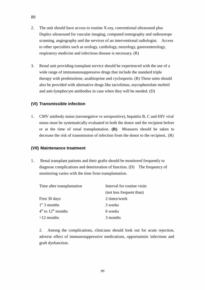
89
89
2. The unit should have access to routine X-ray, conventional ultrasound plus
Duplex ultrasound for vascular imaging, computed tomography and radiosotope
scanning, angiography and the services of an interventional radiologist. Access
to other specialties such as urology, cardiology, neurology, gastroenterology,
respiratory medicine and infectious disease is necessary. (R)
3. Renal unit providing transplant service should be experienced with the use of a
wide range of immununosuppressive drugs that include the standard triple
therapy with prednisolone, azathioprine and cyclosporin. (R) These units should
also be provided with alternative drugs like tacrolimus, mycophenolate mofetil
and anti-lymphocyte antibodies in case when they will be needed. (D)
(VI) Transmissible infection
1. CMV antibody status (seronegative vs seropositive), hepatitis B, C and HIV viral
status must be systematically evaluated in both the donor and the recipient before
or at the time of renal transplantation. (R) Measures should be taken to
decrease the risk of transmission of infection from the donor to the recipient.. (R)
(VII) Maintenance treatment
1. Renal transplant patients and their grafts should be monitored frequently to
diagnose complications and deterioration of function. (D) The frequency of
monitoring varies with the time from transplantation.
Time after transplantation Interval for routine visits
(not less frequent than)
First 30 days 2 times/week
1st 3 months 3 weeks
4th to 12th months 6 weeks
>12 months 3 months
2. Among the complications, clinicians should look out for acute rejection,
adverse effect of immunosuppressive medications, opportunistic infections and
graft dysfunction.
90
90
(VIII) Management of hepatitis virus infection in transplant patients
1. All transplant candidates should be tested for HBV and HCV status. A
transplant unit should have a clear policy in managing patients with HbsAg or
anti-HCV positivity. (R)
2. Transplant recipients positive for hepatitis B surface antigen (HbsAg) should be
carefully followed after transplantation with monitoring of liver function (R),
serum α-fetal protein level (R) and serum markers for viral replication (HBV-
DNA). (D) Tailored immunosuppression and specific anti-viral therapy may be
recommended in these patients. (D)
3. HCV antibody positive patients should be carefully followed after
transplantation with monitoring of liver function (R), serum α-fetal protein level
(R) and serum markers for viral replication (HCV-RNA). (D)
(I) Clinical audit
1. Transplant unit should conduct clinical and medical audit to monitor the
performance of the unit. (D)
2. The items of clinical outcome indicators for medical audit are as follows:
- Proportions of patients on dialysis entered on to the transplant waiting list (%)
- Waiting time of patients on dialysis (mean ± S.D.)
- Cold ischaemic time of transplanted kidneys (% <30 hours)
- Proportion of cadaver transplant recipients with immediate function, delayed
function and failure of function (%)
- Number of days of hospitalization in the first and subsequent years after
transplantation
- Proportion of patients with urological problems after grafting (%)
- Proportion of patients with renal vascular problems after grafting (%)
- Number of serious infections in the post-operative period and later (abscesses,
septicaemia, serious fungal or viral disease)
- Proportion of patients with one or more histologically or clinically diagnosed
rejection episodes in the first 3 months (%)
- Percentage of these episodes that were resistant to corticosteroid treatment (%)
- Incidence of graft loss from acute rejection in the first 3 months
- Plasma creatinine concentration over time in those with functioning grafts (mean
91
91
± S.D.)
- Incidence of death with a functioning graft in the first 3 months
- Frequency and causes of death
- Frequency and attributed causes of graft failure
- Prevalence of malignant disease of all types
References
The Renal Association. Treatment of adult patients with renal failure: Recommended
standards and audit measures, 2nd edition, Nov. 1997,
Kasiske et. al. Recommendations for the Outpatient Surveillance or Renal Transplant
Recipients. JASN. 11:S1-S86, 2000
The EBPG Expert Group on Renal Transplantation European Best Practice
Guidelines for Renal Transplantation. NDT 15:S7:1-85, 2000
92
92
General Nephrology General Nephrology General Nephrology General Nephrology
Dr Andrew Wong
1. Introduction
General nephrology encompasses the prevention, early diagnosis andprompt treatment of renal diseases. The provision of this aspect of renalservices is particularly important with a rising population of patients withend stage renal failure and limited health care resources. It is thusessential to maintain a high standard of care for the large number ofpatients with progressive renal diseases such as diabetic nephropathy, andthose with chronic and acute renal failure.
2. Institutional Requirement
2.1 The general nephrology team should comprise of a core group ofphysicians and nurses led by a qualified nephrologist. The lattermust be a Fellow of the Hong Kong College of Physicians orequivalent and has full specialist accreditation in nephrology by theSpecialty Board in Nephrology of the Hong Kong College ofPhysicians and registered as a Specialist in Nephrology with theHong Kong Medical Council. (D)
2.2 Designated in-patients beds and outpatients clinics for the generalnephrology service is desirable for the effective management ofsuch patients. (D)
2.3 A special low clearance clinic for patients with serum creatinine ∼300-400 µmol/L has also been suggested to be of value. (D)
2.4 A full range of supportive services is essential, this includes theavailability of dietitians, medical social workers, a full complementof diagnostic and therapeutic radiological services, and a fullyequipped laboratory with comprehensive histopathological,microbiological, immunological and biochemical capabilities. (D)
2.5 Support from other hospitals specialists and community physiciansare equally important, and liaison with colleagues such as theurologists, critical care physicians, rheumatologists, obstetricians,cardiologists, infectious diseases and rehabilitation experts etc.should be routine. (D)
2.6 In view of the urgency of the many acute nephrological problems,
93
93
out of hours availability of many of the above services is highlydesirable. (D)
3. Referral Guidelines
3.1 In order to ensure that the general nephrology service is madeavailable to those in need, clear recommendations for referrals ofpatients with symptoms or signs of renal diseases should bedisseminated to the referring sources. (D)
3.2 Bearing in mind that any list could never be exhaustive, it should at least include thefollowing:
- Patients with repeated serum creatinine ≥ 150 µmol/L- Acute Renal Failure- Nephrotic syndrome- Persistent proteinuria and microhaematuria- Frank haematuria in patients <40-45 years of age- Adult males, pregnancy associated, or frequently recurrent
urinary tract infections- Diabetic nephropathy- Patients with polycystic kidneys or other inherited renal
diseases and their relatives- Renal disease in pregnancy
4. Diagnostic Procedures
4.1 The diagnosis of various renal diseases demands the availability ofhighly sophisticated equipment and laboratories supervised byexperts in the various fields of pathology and radiology.
4.2 Renal biopsy is an important tool in general nephrology, and itsperformance and interpretation must be closely monitored.
4.2.1 Renal biopsies are now best performed under directradiological visualization, and preferably using disposablespring-loaded needles or mechanical tools such as theBiopty TM gun. (D)
4.2.2 Regardless of whether the radiologists are routinelyperforming the biopsies for the renal unit, the nephrologytrainees should receive such instruction and appropriatetraining under expert guidance. Less experiencedgeneral medical staff should not normally be involved inperforming biopsies. (R)
94
94
4.2.3 An experienced pathologist who is a specialist in this fieldshould perform renal biopsies interpretation. Facilitiesfor electron microscopy and immunohistological stainingshould be available. (R)
4.3 For better patient care, quality control and medical education,regular clinicopathological and radiology meetings should be held.(D)
5. Therapeutic Interventions
5.1 Specific and supportive treatment for primary and secondary renaldiseases should be under the supervision of a qualifiednephrologist. (D)
5.2 Such a physician should be familiar with the use of the variousimmunosuppressive therapies including steroids, cytotoxics andplasmapheresis, and the different modes of renal replacementtherapy. (D)
5.2.1 Such therapies should only be instituted with informedconsent, and both the physicians and the patients shouldbe aware of the effectiveness and side effects involved.(D)
5.2.2 Appropriate measures for monitoring and prevention ofinfections, malignancies and infertility should be routine.(D)
5.3 Chronic Renal Diseases
5.3.1 Reno-protective measures
5.3.1.1 Blood pressure control is of paramount importance.Threshold for treatment should be lower than for thegeneral population, and treatment should be considered ifthe blood pressure is consistently above 140/90.The target blood pressure should be <130/80, and if thereis significant proteinuria >1g/24 hours, it should be<125/75. ACE inhibitors should be considered as thedrug of choice because of its proven efficacy in retardingrenal failure, but close monitoring with its use is essential.Angiotensin II antagonists are best regarded as a secondline therapy because of their costs. (R)
95
95
5.3.1.2 As there is an important correlation between urinaryprotein excretion and the rate of glomerular filtration ratedecline in diabetic and non-diabetic patients, measures toreduce it should be considered whenever appropriate.Again ACE inhibitors and Angiotensin II antagonists haveproved to be of value in this respect. (R)
5.3.1.3 Very low protein diet (<0.6g protein/Kg/day) may beuseful in some, but malnutrition, poor compliance andcosts are perhaps insurmountable problems. Dietitiansshould be involved early to assess optimal minerals,vitamins, carbohydrates, proteins and fat intake. Andwith a 0.8-1g protein/Kg/day diet of high biological valuestarted when the GFR <25 mls/min, symptomatic benefitscould be achieved. (R)
5.3.1.4 Diabetic nephropathy patients deserve a special mention,as it has become the leading cause (25-44%) of end stagerenal failure in the developed world. Intensive glycaemiccontrol has proved to be beneficial to the risk ofnephropathy in diabetic patients. A HbA1c target of <7%is widely accepted. (R)
5.3.2 Cardiovasculr risk factors control
5.3.2.1 Patients with chronic renal diseases should be consideredin the highest risk group for subsequent cardiovasculardisease events. Smoking cessation, moderation inalcohol consumption, weight control and moderate levelof physical activity should be recommended. (R)
5.3.2.2 Lipids should be closely monitored and aggressivelytreated according to international guidelines. (R)
5.3.3 Biochemical control
5.3.3.1 Control of serum bicarbonate within the normal range isadvocated. The possibility of this resulting in furthersodium and fluid retention has to be borne in mind. (R)

96
96
5.3.3.2 Manipulation of the calcium and phosphate levels towithin the normal range, and adjusting the iPTHmeasurement to 2-3 times normal by the judicial use ofphosphate binders, calcium salts and one alphahydroxycholecalciferol / calcitriol should be attempted.(R)
5.3.4 Correction of anaemia
5.3.4.1 Provided there is no other cause apart from uraemia isidentified for the anaemia, treatment should becontemplated when haematocrit <30% or haemoglobin <10 g/dl by using erythropoietin. This has been shown toimprove the quality of life and attenuates cardiovascularproblems. A haemoglobin target of 11-12 g/dl or ahaematocrit of 33-36% has been advocated. Ironsupplements are frequently required to keep the serumferritin > 100 µg/L and transferrin saturation >20%. (D)
5.3.5 Renal Replacement Therapy
5.3.5.1 Initiation of renal replacement therapy should be basedon clinical status rather than on laboratory values only,and should be considered before the GFR falls below 10-15 ml/min/1.73m2. Earlier intervention should beconsidered in diabetics because of polyneuropathy, fluidretention and compromised cardiovascular status. (R)
5.3.5.2 Preparation and assessment of the patient’s suitability forany form of renal replacement therapy should beindividualized and based on a team approach. Input fromthe patient and his/her family, nurses, medical socialworkers, physicians and, clinical psychologists etc. areessential. (D)
5.3.5.3 The patients should be explicitly informed of the pros andcons of treatment, the long term and irreversible nature ofthe problem and the resources implication associated withsuch therapies. (D)
5.3.5.4 Dialysis access creation should be planned well before thepatient has reached end stage renal failure, particularlywhen the need for long-term dialysis is anticipated tooccur within 6 months. (D)
97
97
5.4 Acute Renal Failure
5.4.1 This medical emergency affects almost all medicalspecialties, and is a syndrome with many causes. Itsassociated consequences affect all organ systems.
5.4.2 Rapid diagnosis, specific treatment of the underlyingcauses and supportive therapy should be based on astructured approach to the evaluation of the presentingproblem.
5.4.3 Exclusion of urinary tract obstruction, identification ofsepsis, and avoidance of nephrotoxins should be theroutine practice. Appreciation for the multiple drugs thataffect renal function is especially important. (D)
5.4.4 Correcting the underlying causes should be without delay.Treatment such as steroids, immunosuppressives, surgeryor antibiotics should be instituted once the cause has beenelucidated. (D)
5.4.5 Therapies that maintain renal perfusion should be giventop priority, and these must include an adequate bloodpressure, cardiac output and ventilatory support, inaddition to an optimal blood volume and haematocrit. (D)
5.4.6 As a rule it should be assumed that all such patients arehypercatabolic. Maintaining nutrition is a centralrequirement for the successful management, and abalanced and adequate intake of carbohydrates andproteins is recommended. (D)
5.4.7 Renal replacement therapy should be started once it isobvious that acute renal failure is established. Initiationof such therapy before severe pathologic derangementoccurs has been suggested to favour a better outcome.Emergency treatment is sometimes necessary forhyperkalaemia, gross acidosis, pulmonary oedema andmarked uraemia. (D)
5.4.8 Facilities and expertise on peritoneal dialysis,haemodialysis, and continuous renal therapies should beavailable in units providing care for such patients. (D)

98
98
5.4.9 Acute peritoneal dialysis can be carried out quickly andrequires little specialist support. However, it is generallyless efficient and could not be relied upon to maintainadequate biochemical control in hypercatabolic patients.
5.4.10 Continuous renal replacement therapies are generallywell-tolerated haemodynamically, fluid balance could bebetter managed, and the dialytic dose delivery could beeasily optimized. It is particularly useful for patientswith unstable haemodynamic status, or those with cerebraloedema. (D)
5.4.11 Patients who are haemodynamically stable can also bemanaged by regular intermittent haemodialysis. This isperhaps less labour intensive, but the availability ofrelevant expertise and equipment is prerequisite.Bicarbonate is now the dialysate of choice. (D)
5.4.12 In patients with acute renal failure as part of multipleorgan failure, biocompatible membranes should be usedfor renal replacement therapy. These have been shown tobe of value in improving mortality and recovery of renalfunction. (D)
5.4.13 Patients with acute renal failure and multiple organsfailure should be managed in an intensive care setting withmultidisciplinary expertise. Critical care physicians andnephrologists should jointly manage the renal dysfunction.All ICUs without the benefit of an in house renal unitshould have nephrologists back up from another hospitalfor consultation and advice. (D)
Possible Items for Audit
Referrals- Appropriateness of referrals- New cases waiting time to first appointment- Level of creatinine when first seen- Number requiring dialysis immediately and within 3-6 months
99
99
Renal biopsies- No. of renal biopsies- Appropriateness and techniques of biopsies- Complications – rate and type- Adequacies of biopsies obtained (8-10 glomeruli)
Chronic Renal Disease- Blood pressure control- Glycaemic control in diabetic nephropathy patients- Biochemical control – Bicarbonate
--
Calcium/phosphateiPTH
- Appropriate use of ACEI and AIIA in diabetics and non-diabetics- Correction of anaemia and iron status- Modifications of cardiovascular risks
Acute Renal Failure- No. of patients requiring temporary renal support- Causes of acute renal failure- APACHE II score at admission, start of dialysis and 7-10 days later- No. of organs failing with the kidneys- Site of management- Techniques and complications of renal replacement therapy used- Type of membrane/dialysate used.- Type and efficacy of anticoagulation used in extracorporeal
circulation.
- Outcome – Percentage leaving ICU Percentage discharge from hospital
Percentage surviving 6-12 months after onset Percentage requiring long term renal replacement within 3-6 months.
References
1. Bonventre JV. Daily haemodialysis – will treatment each day improvethe outcome in patients with acute renal failure? N Engl J Med, 2002;340: 362-364.
2. Druml W. Non-dialytic management of the patient with acute renalfailure. Nephrol Dial Transplant, 1996; 11: 1517-1520.
3. Hostetter TH. Prevention of end-stage renal disease due to type 2diabetes. N Engl J Med, 2001; 345: 910-912.
100
100
4. Levey AS, Adler S, Caggiula AW et al. Effects of dietary proteinrestriction on the progression of advanced renal disease in theModification of Diet in Renal Disease (MDRD) Study. Am J KidneyDis, 1996; 27: 652-663.
5. Levey AS, Eknoyan G. Cardiovascular disease in chronic renal disease.Nephrol Dial Transplant, 1999; 14: 828-833.
6. Macdougall IC. Present and future strategies in the treatment of renalanaemia. Nephrol Dial Transplant, 2001; 16 (Suppl 5): 50-55.
7. Mackenzie HS, Brenner BM. Current strategies for retardingprogression of renal disease. Am J Kidney Dis, 1998; 31: 161-170.
8. Meyer MM. Renal replacement therapies. Crit Care Clin, 2000; 16:29-56.
9. Morgan AG. The management of acute renal failure. Br J Hosp Med,1996; 55(4): 167-170.
10. Parving H, Hovind P, Rossing K, Audersen S. Evolving strategies forrenoprotection: diabetic nephropathy. Curr Opin Nephrol Hypertens,2001; 10: 515-522.
11. The Renal Association. Treatment of adult patients with renal failure.Recommended standards and audit measures, 2nd ed. Royal College ofPhysicians of London, 1997.
12. Ruggenenti P, Schieppati A, Remuzzi G. Progression, remission,regression of chronic renal disease. Lancet, 2001; 357: 1601-1608.
13. Taal MW, Brenner BM. Evolving strategies for renoprotection: non-diabetic chronic renal disease. Curr Opin Nephrol Hypertens, 2001; 10:523-531.
14. Thadhani R, Pascual M, Bonventre JV. Acute renal failure. N Engl JMed, 1996; 334: 1448-1460.
15. Turney JH. Acute renal failure – Some progress? N Engl J Med, 1994;331: 1372-1374.

101
101
Renal Registry: Application in QARenal Registry: Application in QARenal Registry: Application in QARenal Registry: Application in QA
Dr. SF Lui
PURPOSES
To provide indicators on the provision of renal services
- quantity / quality / outcome
Data to be interpreted in the context of types of patient, confounding factors,
resource limitation, restriction, etc.
AIMS
To capture essential / meaningful data (compulsory items)
To minimise the workload require to input the data
- make use of existing data in renal registry
- single input screen / form
DATA COLLECTION / ANALYSIS
Data collection - annually as of 31st March
(of available data closest to the date)
Data analysis - annually on 30th June
COMPULSORY DATA
(A) DEMOGRAPHICS DATA PD
HD TX
ID y y yName y y y
DOB y y y
Sex y y y
Diagnosis y y y
Comorbidity - IHD y y y
Comorbidity - vascular y y y
Comorbidity - DM y y y
HBV y y y
HCV y y y

102
102
existing data being collected via Renal Registry
(B) MODE OF RRT / DEATH
PD HD TX
RRT start date y y y
RRT mode (HD, PD, TX, EPO) y y y
RRT end date y y y
Date of death y y y
Cause of death y y y
existing data being collected via Renal Registry
(C) CLINICAL DATA
PD HD TX
Hb * y y y
Urea * y y yCreatinine * y y y
Albumin * y y y
Calcium * y y y
Phosphate * y y y
Adequacy * Kt/V
Wk CrCl
Kt/V
URR
-
* Additional compulsory data to be collected via Renal Registry
(D) OUTCOME INDICATORS
PD HD TX
Complicationaperitonitis
raterejection
% on EPOaX X X
Patient survival ** 1,3,5 y 1,3,5 y 1,3,5 y
Technique survival ** 1,3,5 y 1,3,5 y 1,3,5 ya existing data being collected via Renal Registry
** (calculated data)
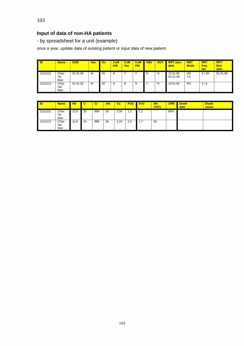
103
103
Input of data of non-HA patients
- by spreadsheet for a unit (example)
once a year, update data of existing patient or input data of new patient
ID Name DOB Sex Dx CoMIHD
CoMVas
CoMDM
HBV HCV RRT startdate
RRTMode
RRTfreqdur
RRTEnddate
11111111 ChanTaiMan
01.01.50 M 81 N Y Y N N 13.01.9002.01.99
HDTX
3 x 5h 01.01.99-
11111112 ChanSaiMan
01.01.52 M 82 N N N Y N 13.01.90 PD 3 / d
ID Name Hb U Cr Alb Ca PO4 Kt/V WkCRCL
URR Deathdate
Deathcause
11111111 ChanTaiMan
12.0 25 999 35 2.34 1.2 1.2 65%
11111112 ChanSaiMan
11.8 24 888 36 2.24 2.0 1.7 50