Phosphatidylserine Decarboxylase 1 Autocatalysis and Function ...

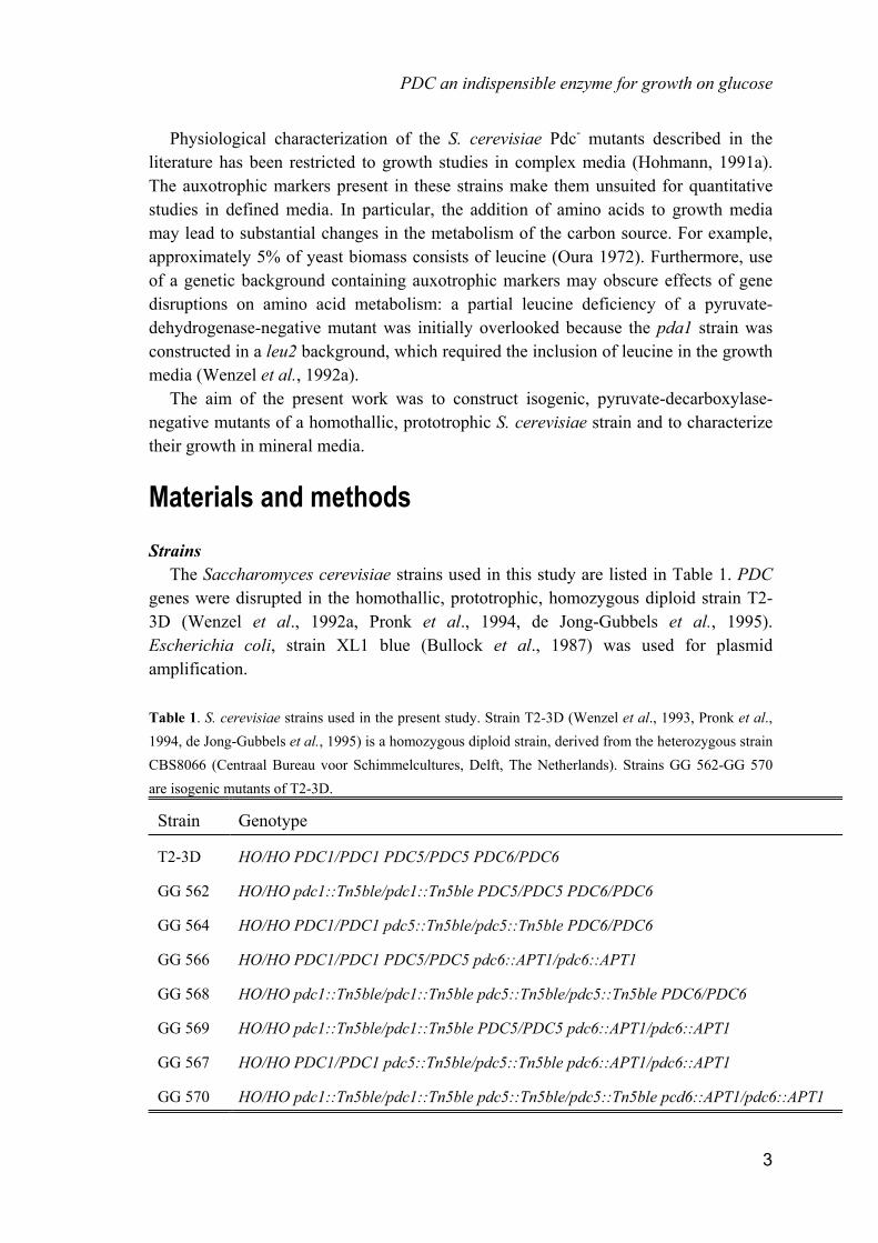
PDC an indispensible enzyme for growth on glucose
Chapter 2
Pyruvate decarboxylase: an indispensable enzyme for growth of Saccharomyces cerevisiae
on glucose In Saccharomyces cerevisiae, the structural genes PDC1, PDC5 and PDC6 each encode an active pyruvate decarboxylase. Replacement mutations in these genes were introduced in a homothallic wild-type strain, using the dominant marker genes APT1 and Tn5ble. A pyruvate-decarboxylase-negative (Pdc-) mutant lacking all three PDC genes exhibited a three-fold lower growth rate in complex medium with glucose than the isogenic wild-type strain. Growth in batch cultures on complex and defined media with ethanol was not impaired in Pdc- strains. Furthermore, in ethanol-limited chemostat cultures, the biomass yield of Pdc- and wild-type S. cerevisiae were identical. However, Pdc- S. cerevisiae was unable to grow in batch cultures on a defined mineral medium with glucose as the sole carbon source. When aerobic, ethanol-limited chemostat cultures (D = 0.10 h-1) were switched to a feed containing glucose as the sole carbon source, growth ceased after approximately 4 hours and, consequently, the cultures washed out. The mutant was, however, able to grow in chemostat cultures on mixtures of glucose and small amounts of ethanol or acetate (5 % on a carbon basis). No growth was observed when such cultures were used to inoculate batch cultures on glucose. Furthermore, when the mixed-substrate cultures were switched to a feed containing glucose as the sole carbon source, wash-out occurred. It is concluded that the mitochondrial pyruvate-dehydrogenase complex cannot function as the sole source of acetyl-CoA during growth of S. cerevisiae on glucose, neither in batch cultures nor in glucose-limited chemostat cultures.
1

Chapter 2
Introduction In yeasts, respiratory dissimilation of pyruvate is initiated by its conversion into acetyl-CoA. This can occur in two ways: via a direct reaction catalysed by the mitochondrial pyruvate-dehydrogenase complex or via an indirect route, involving pyruvate decarboxylase, acetaldehyde dehydrogenase and acetyl-coenzyme A synthetase (Fig.1; Holzer and Goedde, 1957, Pronk et al., 1994). Experiments with isogenic S. cerevisiae mutants defective in the synthesis of an active pyruvate-dehydrogenase complex have demonstrated that, during glucose-limited aerobic growth of wild-type cells, this enzyme is predominantly or even exclusively responsible for respiratory pyruvate dissimilation. Under these conditions, the indirect route apparently does not play an important role in respiratory pyruvate metabolism (Pronk et al., 1994). To study the metabolic significance of the pyruvate-dehydrogenase bypass route, it is of interest to investigate the physiology of mutants affected in pyruvate decarboxylase (EC 4.1.1.1). S. cerevisiae contains three structural genes that each encode an active pyruvate decarboxylase; PDC1, PDC5 and PDC6 (Hohmann, 1991a). Strains in which PDC1 and PDC5 or all three PDC genes have been disrupted lack pyruvate-decarboxylase activity. Such pyruvate-decarboxylase-negative (Pdc-) mutants showed a reduced growth rate in complex (yeast extract-peptone) media supplemented with glucose (Hohmann, 1991a). Although, under all growth conditions tested, expression of PDC6 was either very low or absent, revertants of pdc1-pdc5 double mutants have been isolated, in which a recombination event had caused a fusion of the PDC1 promotor and the PDC6 open-reading frame (Hohmann et al., 1991b). Therefore, physiological studies on Pdc- S. cerevisiae mutants should preferably be performed with stable strains in which all three PDC genes are disrupted. Fig. 1. Enzymes of pyruvate metabolism in Saccharomyces cerevisiae. Numbered reactions are catalysed by the following enzymes: 1, pyruvate decarboxylase; 2, pyruvate dehydrogenase complex; 3, acetaldehyde dehydrogenase; 4, acetyl-coenzyme A synthetase; 5, alcohol dehydrogenase.
2
PDC an indispensible enzyme for growth on glucose
Physiological characterization of the S. cerevisiae Pdc- mutants described in the literature has been restricted to growth studies in complex media (Hohmann, 1991a). The auxotrophic markers present in these strains make them unsuited for quantitative studies in defined media. In particular, the addition of amino acids to growth media may lead to substantial changes in the metabolism of the carbon source. For example, approximately 5% of yeast biomass consists of leucine (Oura 1972). Furthermore, use of a genetic background containing auxotrophic markers may obscure effects of gene disruptions on amino acid metabolism: a partial leucine deficiency of a pyruvate-dehydrogenase-negative mutant was initially overlooked because the pda1 strain was constructed in a leu2 background, which required the inclusion of leucine in the growth media (Wenzel et al., 1992a). The aim of the present work was to construct isogenic, pyruvate-decarboxylase-negative mutants of a homothallic, prototrophic S. cerevisiae strain and to characterize their growth in mineral media.
Materials and methods Strains The Saccharomyces cerevisiae strains used in this study are listed in Table 1. PDC genes were disrupted in the homothallic, prototrophic, homozygous diploid strain T2-3D (Wenzel et al., 1992a, Pronk et al., 1994, de Jong-Gubbels et al., 1995). Escherichia coli, strain XL1 blue (Bullock et al., 1987) was used for plasmid amplification. Table 1. S. cerevisiae strains used in the present study. Strain T2-3D (Wenzel et al., 1993, Pronk et al., 1994, de Jong-Gubbels et al., 1995) is a homozygous diploid strain, derived from the heterozygous strain CBS8066 (Centraal Bureau voor Schimmelcultures, Delft, The Netherlands). Strains GG 562-GG 570 are isogenic mutants of T2-3D.
Strain Genotype
T2-3D HO/HO PDC1/PDC1 PDC5/PDC5 PDC6/PDC6
GG 562 HO/HO pdc1::Tn5ble/pdc1::Tn5ble PDC5/PDC5 PDC6/PDC6
GG 564 HO/HO PDC1/PDC1 pdc5::Tn5ble/pdc5::Tn5ble PDC6/PDC6
GG 566 HO/HO PDC1/PDC1 PDC5/PDC5 pdc6::APT1/pdc6::APT1
GG 568 HO/HO pdc1::Tn5ble/pdc1::Tn5ble pdc5::Tn5ble/pdc5::Tn5ble PDC6/PDC6
GG 569 HO/HO pdc1::Tn5ble/pdc1::Tn5ble PDC5/PDC5 pdc6::APT1/pdc6::APT1
GG 567 HO/HO PDC1/PDC1 pdc5::Tn5ble/pdc5::Tn5ble pdc6::APT1/pdc6::APT1
GG 570 HO/HO pdc1::Tn5ble/pdc1::Tn5ble pdc5::Tn5ble/pdc5::Tn5ble pcd6::APT1/pdc6::APT1
3

Chapter 2
Maintenance of strains Wild-type S. cerevisiae and pdc mutants were grown to stationary phase in shake-flask cultures on complex medium containing 2 % (v/v) ethanol. After adding glycerol (15 % v/v), 2-ml aliquots were stored in sterile vials at -70 oC. Prior to growth experiments, samples from a frozen stock culture were streaked on complex medium-ethanol agar plates. Precultures were inoculated directly from these plates. Recombinant-DNA techniques Standard protocols were followed for plasmid isolation, restriction, ligation, Southern blotting, hybridization and gel electrophoresis (Maniatis et al., 1982). Yeast chromosomal DNA was isolated by the method of Holm et al. (1986). S. cerevisiae and E. coli strains were transformed with a Bio-Rad gene pulser (Dower et al., 1988). Sporulation, dissection and mating of S. cerevisiae strains was performed according to published procedures. Construction of pdc mutants Subclones of PDC1, PDC5 and PDC6 in pUC vectors were kindly provided by Dr. S. Hohmann. The one-step gene-disruption method (Rothstein 1983) was used to inactivate the PDC1, PDC5 and PDC6 genes in S. cerevisiae T2-3D (Fig. 2). PDC1 was disrupted by replacing an internal 1058 bp KpnI-BglII fragment with a 1.35 kb KpnI-BglII fragment from the plasmid pUT332 (Gatignol et al., 1990) containing the marker gene Tn5ble under the control of the S. cerevisiae TEF1 promoter and CYC1 terminator. A 1.15 kb HindIII-KpnI fragment from pUT332, carrying the same marker gene, was used to replace an internal 691 bp HindIII-KpnI fragment of PDC5. PDC6 was disrupted by replacing an internal 1190 bp BclI fragment with a 3.2 kb BamHI-BclI fragment from the plasmid pBEJ24 (Hadfield et al., 1990) containing the marker gene APT1 under the control of the S. cerevisiae PGK1 promoter and CYC1 terminator. After transformation of S. cerevisiae T2-3D with linear restriction fragments containing the disrupted genes, transformants were selected on YPD plates containing either phleomycin (strains expressing Tn5ble) or G418 (transformants expressing APT1), as described by Wenzel et al. (1992b) and Hadfield et al. (1990), respectively. Since the strains are homothallic, spore-to-spore matings were used to obtain strains in which two or three PDC genes were disrupted. The following combinations were used: GG 562 x GG 564; GG 562 x GG 566, GG 564 x GG 566, and GG 562 x GG 567. The resulting heterozygous diploid strains were again sporulated and dissected to obtain the homozygous strains. Spore-to-spore matings were performed on CY plates with 2 % (v/v) ethanol instead of glucose. Spore viability was low, probably due to the pdc mutations (Hohmann 1991a). Dissection on glucose or galactose media did not significantly improve spore viability. The genotype of all strains containing single or multiple disrupted PDC genes was confirmed by Southern analysis (Fig. 3).
4

PDC an indispensible enzyme for growth on glucose
Fig. 2. Schematic representation of the gene disruptions in PDC1, PDC5 and PDC6. Restriction sites are indicated by the following abbreviations: B = BamHI, Bc = BclI, Bg = BglII, H = HindIII, K = KpnI, P = PstI. Fig. 3. Southern analyses of genomic DNA restriction digests. Panel A: HindIII digests. The probe contained the TEF1 promoter and the Tn5ble gene from the phleomycin-resistance cassette. The largest hybridizing fragment in lanes 1-5 contains the native TEF1 promoter. Panel B: PstI digests. The probe contained the 5' region of the PDC6 gene. Relevant restriction sites are indicated in Figure 1. Lane 1: S. cerevisiae T2-3D (wild-type), lane 2: GG 562 (pdc1::Tn5ble), lane 3: GG 564 (pdc5::Tn5ble), lane 4: GG 568 (pdc1::Tn5ble pdc5::Tn5ble), lane 5: GG 570 (pdc1::Tn5ble pdc5::Tn5ble pdc6::APT1), lane 6: T2-3D (wild-type), lane 7: GG 566 (pdc6::APT1), lane 8: GG 570 (pdc1::Tn5ble pdc5::Tn5ble pdc6::APT1).
5

Chapter 2
Media The mineral medium contained per litre of demineralized water: (NH4)2SO4, 5 g; KH2PO4, 3 g; MgSO4.7H2O, 0.5 g; EDTA, 15 mg; ZnSO4.7H2O, 4.5 mg; CoCl2.6H2O, 0.3 mg; MnCl2.4H2O, 1 mg; CuSO4.5H2O, 0.3 mg; CaCl2.2H2O, 4.5 mg; FeSO4.7H2O, 3.0 mg; Na2MoO4.2H2O, 0.4 mg; H3BO3, 1.0 mg; KI, 0.1 mg and silicone antifoam (BDH); 0.05 ml. After heat sterilization (120 �C) of the medium, filter-sterilized vitamins were added, to final concentrations per litre of: biotin, 0.05 mg; calcium pantothenate, 1.0 mg; nicotinic acid, 1.0 mg; inositol, 25.0 mg; thiamin HCl, 1.0 mg; pyridoxine HCl, 1.0 mg and para-aminobenzoic acid, 0.2 mg. The concentration of ethanol or glucose in the reservoir medium was 5.75 g.l-1 or 7.5 g.l-1 respectively (0.25 Cmol.l-1). Complex medium contained per litre: yeast extract (Difco), 10 g; peptone from casein (Merck), 20 g; and 2 % (v/v) ethanol (YPE) or 20 g D-glucose (YPD). CY plates contained per litre: yeast extract (Difco), 5 g; bactopeptone (Difco), 5 g; agar (Difco), 20 g; and glucose, 20 g. Shake-flask cultivation Precultures were prepared by inoculating 100 ml YPE (2% ethanol) with a few colonies from a plate. Cultures were incubated on an orbital shaker (200 rpm) at 30 �C for two days. For growth curves, 1 ml of the preculture was inoculated in a 500 ml erlenmeyer with 100 ml YPE (2% ethanol) or 100 ml YPD (2% glucose) and then shaken (200 rpm) at 30 �C. Optical-density measurements were performed at appropriate intervals as described by Weusthuis et al. (1994). For induction of pyruvate decarboxylase, 10 ml of a preculture was inoculated in a 100 ml shake flask with either 50 ml YPE (2% ethanol) or 50 ml YPD (8% glucose) and shaken for 6 hours at 30 �C (Hohmann, 1991a). Batch cultivation in fermenters Batch cultivation was performed at 30 �C in laboratory fermenters (Applikon, Schiedam The Netherlands) with a working volume of 1.5 litre. The pH was controlled at 5.0 ± 0.1 by automatic addition of 2 mol.l-1 KOH and 1 mol.l-1 H2SO4. The fermenter was flushed with air at a flow rate of 1.5 l.min-1 and stirred at 800 rpm. The dissolved-oxygen concentration was continuously monitored with an oxygen electrode (Ingold, 34 100 3002) and remained above 60% of air saturation. Cultures were grown on the mineral medium described above, with glucose (25 g.l-1 initial concentration) or ethanol (7.9 g.l-1 initial concentration) as the sole carbon source. 25 ml samples were withdrawn at appropriate intervals for determination of dry weight and metabolite concentrations. Chemostat cultivation in fermenters Aerobic chemostat cultivation was performed at 30 �C in laboratory fermenters (Applikon, Schiedam, The Netherlands), at a stirrer speed of 750 rpm and at a dilution rate of 0.10 h-1. The working volume of the cultures was kept at 1.0 l by a peristaltic
6

PDC an indispensible enzyme for growth on glucose
effluent pump coupled to an electrical level sensor. This set-up ensured that under all growth conditions, biomass concentrations in samples taken directly from the cultures differed by less than 1% from biomass concentrations in samples taken from the effluent line (Noorman et al., 1991). The pH was kept constant at 5.0 by an ADI 1020 biocontroller, via the automatic addition of 2 mol.l-1 KOH. The fermenter was flushed with air at a flow rate of 0.7 l.min-1 using a Brooks 5876 mass-flow controller. The dissolved-oxygen concentration was continuously monitored with an oxygen electrode (Ingold, 34 100 3002) and remained above 50% air saturation. Steady-state data refer to cultures without detectable oscillations. Chemostat cultures were checked for purity using phase-contrast microscopy. Determination of culture dry weight The dry weight of washed culture samples was determined using 0.45 �m membrane filters and a microwave oven as described by Postma et al. (1989). Parallel samples varied by less than 1%. Metabolite analysis Organic acids, ethanol and glycerol in culture supernatants were determined by HPLC analysis using a Phenomenex column (Rezex ROA Organic acid 00H-0138-KO) at 60 �C. The column was eluted with 0.5 g.l-1 sulphuric acid at a flow rate of 0.5 ml.min-1. Organic acids were detected by a Waters 441 UV-meter at 214 nm coupled to a Waters 741 Data module. Ethanol and glycerol were detected by an Erma ERC 7510 refractive-index detector coupled to a Hewlett Packard 3390A RI integrator. 20 �l samples were injected using a Hamilton syringe. Glucose in reservoir media and supernatants was determined enzymically using the GOD-PAP method (Merck Systems kit 14144; detection limit ca. 5�M). Ethanol was assayed colorimetrically with an alcohol oxidase/peroxidase kit (Leeds Biochemicals; detection limit ca. 100 �M). Preparation of cell-free extracts For preparation of cell-free extracts, culture samples were harvested by centrifugation, washed twice with 10 mM potassium-phosphate buffer, pH 7.5, containing 2 mM EDTA, concentrated 4-fold and stored at -20 �C. Before assaying, the samples were thawed at room temperature, washed and resuspended in 100 mM potassium phosphate buffer, pH 7.5, containing 2 mM MgCl2 and 1 mM dithiothreitol (DTT). Extracts were prepared by sonification with 0.7 mm diameter glass beads at 0 �C for 2 min. at 0.5 min. intervals with an MSE sonicator (150 W output, 7 �m peak-to-peak amplitude). Unbroken cells and debris were removed by centrifugation at 4 �C (20 min. at 36,000 � g). The supernatant was used as the cell-free extract. Pyruvate-decarboxylase assays Pyruvate-decarboxylase activity was assayed at 30 �C immediately after preparation of the extracts, using a Hitachi model 100-60 spectrophotometer set at 340
7
Chapter 2
nm. Reaction rates were linearly proportional to the amount of cell-free extract added. The assay mixture consisted of: 40 mM imidazole-HCl buffer (pH 6.5), 0.2 mM thiamine pyrophosphate (TPP), 0.15 mM NADH, alcohol dehydrogenase 88 U.ml-1 (Boehringer), 5 mM MgCl2, and cell-free extract. The reaction was started with 50 mM pyruvate. Protein determination Protein concentrations in cell-free extracts were determined by the Lowry method. Bovine-serum albumin (BSA; fatty-acid-free, Sigma Chemical Co.) was used as a standard. The protein content of whole cells was determined by a modified biuret method (Verduyn et al., 1990).
Results Specific activities of pyruvate decarboxylase in pdc mutants Effects of the gene disruptions on pyruvate-decarboxylase expression were investigated by measuring enzyme activities in cell-free extracts of wild-type S. cerevisiae T2-3D and in homozygous mutant strains containing one, two or three disrupted PDC genes. To discriminate between constitutive and glucose-inducible pyruvate-decarboxylase activity, cells were pregrown in complex medium with ethanol as the carbon source and then either incubated in the ethanol medium used for growth or induced by incubation in glucose medium (Hohmann, 1991a). The wild-type strain T2-3D exhibited a high pyruvate-decarboxylase activity after induction in complex medium with glucose (ca. 3 U.mg protein-1, Table 2). An approximately three-fold lower activity was measured in extracts from non-induced wild-type cells grown on ethanol. When strain GG562 carrying the pdc1::Tn5ble mutation was induced with glucose, its pyruvate-decarboxylase activity, determined in cell-free extracts, was only ca. 30 % lower than that of glucose-induced wild-type cells (Table 2). In non-induced cells, disruption of PDC1 resulted in a ten-fold reduction of the pyruvate-decarboxylase activity in comparison with the wild-type strain. Single gene disruptions in either PDC5 or PDC6 did not significantly affect enzyme activities, neither in induced nor in non-induced cells (Table 2). Pyruvate-decarboxylase activities in strains which, in addition to a disrupted PDC1 or PDC5 gene, contained a disruption in PDC6, were not significantly different from the activities in strains carrying the corresponding single gene disruptions (Table 2). When both PDC1 and PDC5 were disrupted, and PDC6 was the only remaining intact PDC gene, no enzyme activity was detected in cell-free extracts prepared from induced or non-induced cells. A complete absence of pyruvate-decarboxylase activity was also observed in extracts of a triple mutant (strain GG 570), in which all three PDC genes had been disrupted (Table 2).
8

PDC an indispensible enzyme for growth on glucose
Table 2. Specific pyruvate-decarboxylase activity and growth rates of wild-type (T2-3D) and pdc mutant strains. For enzyme activity assays, cells pregrown on complex medium with ethanol were induced on either 8% (w/v) glucose or 2% (v/v) ethanol in complex medium. Growth rates were determined in complex medium containing either 2% (v/v) ethanol or 2% (w/v) glucose.
Strain Genotype Ethanol Glucose
PDC activity U.(mg prot)-1
µmax (h-1)
PDC activity U.(mg prot)-1
µmax (h-1)
T2-3D PDC1 PDC5 PDC6 1.0 ± 0.10 0.30 ± 0.02 3.1 ±0.55 0.54 ±0.02
GG 562 �pdc1 PDC5 PDC6 0.1 ± 0.05 0.30 ± 0.01 2.1 ±0.15 0.53 ±0.02
GG 564 PDC1 �pdc5 PDC6 1.0 ± 0.20 0.29 ± 0.01 2.9 ±0.20 0.55 ±0.02
GG 566 PDC1 PDC5 �pdc6 1.0 ± 0.25 0.30 ± 0.01 2.9 ±0.35 0.56 ±0.02
GG 568 �pdc1 �pdc5 PDC6 < 0.01 0.29 ± 0.01 <0.01 0.15 ±0.01
GG 569 �pdc1 PDC5 �pdc6 0.1 ± 0.05 0.29 ± 0.01 2.0 ±0.15 0.54 ±0.02
GG 567 PDC1 �pdc5 �pdc6 1.2 ± 0.10 0.29 ± 0.02 2.7 ±0.00 0.55 ±0.02
GG 570 �pdc1 �pdc5 �pdc6 < 0.01 0.27 ± 0.00 < 0.01 0.15 ±0.01
Growth rates in complex medium For an initial physiological characterization, and to enable comparison with pdc mutations introduced in a different S. cerevisiae genetic background (Hohmann 1991a), growth rates of the PDC mutant strains were determined in shake-flask cultures on complex media with glucose or ethanol. In complex medium with ethanol, growth rates of strains carrying one, two or three disrupted PDC genes did not differ significantly from those of the isogenic wildtype (Table 2). This result is consistent with the fact that pyruvate decarboxylase is not involved in ethanol metabolism. Nevertheless, it differs from the observation of Hohmann (1991a) that strains in which both PDC1 and PDC5 had been disrupted showed a 20 - 25 % reduction of the specific growth rate on ethanol. Disruption of any single PDC gene did not affect the growth rate in complex medium with glucose. In double mutants, growth rates on glucose were not significantly reduced when combinations of PDC6 and either PDC1 or PDC5 were disrupted (Table 2). However, disruption of both PDC1 and PDC5 resulted in a 70 % decrease of the specific growth rate on glucose. This negative effect on growth rate was not enhanced by the additional disruption of PDC6 (Table 2).
9

Chapter 2
Our results confirm the conclusion of Hohmann (1991a) that, during growth in ethanol- or glucose-containing media, PDC6 expression is either very low or absent. However, it has been demonstrated that recombination events may lead to the activation of PDC6 (Hohmann, 1991b). Since such instability is not desirable in physiological studies, it was decided to use the triple mutant strain GG 570 for further physiological investigations on the effects of pyruvate-decarboxylase deficiency during growth of S. cerevisiae in mineral media. Batch cultivation in defined mineral medium Quantitative analysis of yeast physiology requires the use of defined mineral media. Therefore, aerobic growth of wild-type S. cerevisiae T2-3D in a defined mineral salts medium supplemented with vitamins was compared with growth of the isogenic pyruvate-decarboxylase-negative strain GG 570, using pH-controlled fermenter cultures. When grown on ethanol, there was no difference in growth rate between the wild-type strain and the pyruvate-decarboxylase-negative mutant: both strains grew exponentially with a specific growth rate of 0.13 ± 0.01 h-1. The wild-type strain grew exponentially on glucose, with a specific growth rate of 0.45 ± 0.01 h-1 (Fig. 4). Growth on glucose was accompanied by the formation of ethanol and small amounts of pyruvate (3 mmol.l-1) and glycerol (0.3 mmol.l-1). In contrast to the wild-type strain, the Pdc- strain GG 570 did not exhibit exponential growth on glucose. Instead, growth ceased after less than one biomass doubling (Fig. 4). No ethanol or acetate was detected, but concentrations of pyruvate (8 mM) and glycerol (2 mM) attained higher values than in wild-type cultures, even though the biomass concentrations in mutant cultures were much lower. Since a Pdc- mutant can not grow fermentatively, respiration is essential for its growth on glucose. In S. cerevisiae, many enzyme activities involved in respiratory sugar metabolism are subject to glucose catabolite repression (Gancedo 1992). To investigate whether glucose repression of respiration might be responsible for the mutant's impaired growth on glucose in batch cultures, it was subsequently attempted to establish glucose-limited chemostat cultures. Growth of pyruvate-decarboxylase-negative S. cerevisiae in chemostat cultures The pyruvate-dehydrogenase complex, rather than the bypass via pyruvate decarboxylase, is the predominant route of respiratory pyruvate metabolism during glucose-limited growth at D=0.10 h-1 (Pronk et al., 1994). Furthermore, many key enzymes of glucose metabolism, including the pyruvate-dehydrogenase complex, are expressed constitutively during growth of S. cerevisiae T2-3D on ethanol (Wenzel et al., 1993; Pronk et al., 1994; de Jong-Gubbels et al., 1995). It was therefore anticipated that steady-state chemostat cultures growing on ethanol would readily adapt to growth on glucose under glucose limitation.
10

PDC an indispensible enzyme for growth on glucose
Fig. 4. Growth of wild-type S. cerevisiae T2-3D (�) and Pdc- triple mutant GG 570 (pdc1::Tn5ble pdc5::Tn5ble pdc6::APT1; �) triple mutant on a defined mineral medium containing 25 g.l-1 glucose as the sole carbon source. Batch cultivation was performed in pH-controlled, aerobic fermenters. In ethanol-limited chemostat cultures (D=0.10 h-1) grown on a defined medium, the biomass yield of the pyruvate-decarboxylase-negative triple mutant GG 570 was not significantly different from that of the isogenic wild-type strain T2-3D (Table 3). In an attempt to avoid glucose repression, ethanol-limited chemostat cultures (D = 0.10 h-1) of the pyruvate-decarboxylase-negative triple mutant were switched to a medium containing glucose as the sole carbon source. During the first 4 h after the switch, the biomass concentration remained approximately constant and the glucose concentration in the culture remained below 0.2 g.l-1 (Fig. 5). This suggested that indeed, the culture rapidly adapted from ethanol-limited to glucose-limited growth. However, after this initial period, the biomass concentration in the culture decreased and glucose accumulated (Fig. 5). The observed decrease of the biomass concentration was consistent with wash-out kinetics, indicating that growth had ceased completely. The wash-out of biomass and the accumulation of glucose was accompanied by the transient accumulation of pyruvate in the culture to a maximum concentration of 7 mM (Fig. 5). The observation that, both in batch and chemostat cultures, growth of the Pdc- strain on glucose continued for a number of hours before growth ceased, can in theory be caused by a bottleneck in a biosynthetic pathway that requires pyruvate decarboxylase. This would be consistent with the ability of Pdc- strains to grow, albeit poorly, in complex media with glucose (Table 2), in which precursors for biosynthesis can be
11

Chapter 2
obtained from yeast extract and/or peptone. Since growth of the mutant strain on mineral medium with ethanol appeared normal, formation of biosynthetic intermediates from ethanol was apparently not affected. To study whether growth was possible on mixtures of glucose and C2-compounds, ethanol-limited chemostat cultures were switched to mineral medium containing a mixture of glucose (237.5 mmol C.l-1) and ethanol (12.5 mmol C.l-1). This approach resulted in steady-state cultures, in which no residual glucose or acetate could be detected. Enzyme assays in cell-free extracts confirmed the absence of pyruvate decarboxylase activity (Table 3). The biomass concentration in the cultures did not differ significantly from that in similar cultures of the wild-type strain (Table 3). The same results were obtained when, instead of acetate, low concentrations of ethanol were added to the reservoir media (data not shown).
Table 3. Steady-state biomass yields (YSX, g biomass.[mol substrate carbon]-1), protein contents and pyruvate- decarboxylase activities in ethanol- and glucose-limited, aerobic chemostat cultures of wild-type (T2-3D) and Pdc- (GG 570) S. cerevisiae. Relative concentrations of glucose and acetate in mixed-substrate cultures are presented as a percentage of the total carbon concentration (0.25 mol C.l-1) in the feed. Growth conditions: D = 0.10 h-1, pH 5, T = 30 oC, dissolved-oxygen concentration > 50 % air saturation (n.d.: not determined).
Strain Carbon source Ysx
g.Cmol-1 Protein content (%)
PDC-activity U.mg protein-1
T2-3D (wildtype) ethanol 14.4 ± 0.4 41 ± 2 0.7 ± 0.3
GG 570 (Pdc-) ethanol 14.3 ± 0.3 42 ± 2 < 0.01
T2-3D (wildtype) glucose 16.0 ± 0.3 40 ± 2 0.7 ± 1
T2-3D (wildtype) 95 % glucose-5 % acetate
16.5 ± 0.1 40 ± 2 n.d.
GG 570 (Pdc-) 95 % glucose- 5 % acetate
16.2 ± 0.4 40 ± 1 < 0.01
In the mixed-substrate cultures, glucose made up 95 % of the substrate carbon fed to the cultures and was completely consumed. Nevertheless, when samples from such glucose-limited cultures were used to inoculate batch cultures on mineral medium with glucose, no growth was observed. The inability to grow on glucose in batch cultures could not be relieved by the addition of low concentrations of acetate or ethanol to the mineral media. Attempts to change the medium feed of chemostat cultures from a 95 % glucose/5 % acetate mixture to glucose as the sole carbon source reproducibly resulted in wash-out of the cultures (data not shown). This indicated that even cells utilizing high glucose-to-acetate ratios could not be readily adapted to growth on glucose as the sole carbon source.
12

PDC an indispensible enzyme for growth on glucose
Discussion Growth of PDC mutants in batch cultures In batch cultures grown on complex media with ethanol or glucose, PDC1 was expressed constitutively, whereas expression of PDC5 appeared to be induced by glucose. PDC6 did not contribute significantly to the overall level of pyruvate decarboxylase. The pattern of pyruvate-decarboxylase (Table 2) was generally consistent with the pattern observed by Hohmann (1991a), who studied the effect of PDC gene disruptions in a different S. cerevisiae genetic background. It should be borne in mind, however, that the differential regulation of the three PDC genes has so far only been studied during growth on complex media in shake-flask cultures. The possibility that, under appropriate growth conditions, PDC6 is transcribed at significant levels, can therefore not be excluded. Growth experiments in defined and complex media with ethanol as the carbon source gave no indications for pleiotropic effects of the PDC mutations. This is in contrast with the results of Hohmann (1991a), who found a significantly reduced growth rate of Pdc- strains on ethanol. Although, based on established metabolic pathways, no effect of pyruvate-decarboxylase deficiency on ethanol metabolism is to be expected, this discrepancy deserves further attention. In the literature, the effect of a Pdc- phenotype, established either by random mutagenesis (Schmitt & Zimmermann 1982) or by gene disruption (Hohmann 1991a) has only been studied in cultures grown on complex media. These studies invariably demonstrated a reduction of the specific growth rate in glucose-containing media. The residual growth rates in the mutant strains were consistently at least 25 % of the wild-type rate (Schmitt & Zimmermann 1982; Hohmann 1991a). A similar effect was observed in the present study (Table 2). In the absence of pyruvate-decarboxylase activity, growth of S. cerevisiae becomes critically dependent on respiration. Indeed, Hohmann (1991a) demonstrated that growth of Pdc- mutants on glucose was completely arrested in the presence of the respiratory inhibitor antimycin A. It is well-known that in S. cerevisiae, many respiratory enzymes are repressed in the presence of excess glucose (Entian 1986, Gancedo & Serrano 1989). However, glucose catabolite repression of respiratory enzymes is generally not complete (Gancedo 1992). This is consistent with the observed growth, albeit at a reduced rate, of the Pdc- strains in complex medium with glucose (Table 2). Surprisingly, Pdc- S. cerevisiae completely failed to grow in batch cultures on a mineral medium with glucose as the sole carbon source. Clearly, if glucose repression of respiratory enzymes were the sole factor affecting the growth rate of Pdc- mutants, a residual growth rate similar to that observed in complex medium would be expected. Pdc- strains retained the ability to grow in mineral medium with ethanol and were apparently able to convert glucose into pyruvate (Fig. 5). It is therefore conceivable that the absence of growth on glucose in a defined medium is due to a shortage of acetyl-CoA.
13

Chapter 2
Fig. 5. Concentrations of biomass, glucose and pyruvate after switching a chemostat culture (D = 0.10 h-
1) of the Pdc- triple mutant S. cerevisiae GG 570 (pdc1::Tn5ble pdc5::Tn5ble pdc6::APT1) from growth on a mineral medium with ethanol (0.25 Cmol.l-1) to a medium containing glucose (0.25 Cmol.l-1) as the sole carbon source. The dashed line drawn through biomass data points represent wash-out kinetics, assuming a zero growth rate.
pyruvate acetaldehyde
acetyl-CoA
acetate
Lipids
acetatepyruvate
acetyl-CoA
glucose
CO2
mitochondrion
cytosol
glucose
ethanol
41
ethanol
23
8
5
6
7
Xpyruvate acetaldehyde
acetyl-CoA
acetate
Lipids
acetatepyruvate
acetyl-CoA
glucose
CO2
mitochondrion
cytosol
glucose
ethanol
41
ethanol
23
8
5
6
7
X
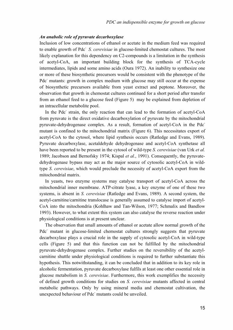
Fig. 6. Hypothetical scheme of subcellular compartmentation of pyruvate and acetyl-CoA metabolism in Saccharomyces cerevisiae, explaining the requirement of a Pdc- mutant for C2-compounds. If acetyl-CoA export from the mitochondria is restricted, glucose-grown cells depend on a source of cytosolic acetyl-CoA. In the absence of pyruvate decarboxylase, cytosolic acetyl-CoA cannot be synthesized from glucose, resulting in a requirement for exogenous C2-compounds. Numbered arrows indicate the following pathways or enzymes: 1, glycolysis; 2, pyruvate dehydrogenase complex; 3, TCA cycle; 4, pyruvate decarboxylase; 5, acetaldehyde dehydrogenase; 6, acetyl-coenzyme A synthetase; 7, lipid synthesis.
14
PDC an indispensible enzyme for growth on glucose
An anabolic role of pyruvate decarboxylase Inclusion of low concentrations of ethanol or acetate in the medium feed was required to enable growth of Pdc- S. cerevisiae in glucose-limited chemostat cultures. The most likely explanation for this dependency on C2-compounds is a limitation in the synthesis of acetyl-CoA, an important building block for the synthesis of TCA-cycle intermediates, lipids and some amino acids (Oura 1972). An inability to synthesize one or more of these biosynthetic precursors would be consistent with the phenotype of the Pdc- mutants: growth in complex medium with glucose may still occur at the expense of biosynthetic precursors available from yeast extract and peptone. Moreover, the observation that growth in chemostat cultures continued for a short period after transfer from an ethanol feed to a glucose feed (Figure 5) may be explained from depletion of an intracellular metabolite pool. In the Pdc- strain, the only reaction that can lead to the formation of acetyl-CoA from pyruvate is the direct oxidative decarboxylation of pyruvate by the mitochondrial pyruvate-dehydrogenase complex. As a result, formation of acetyl-CoA in the Pdc- mutant is confined to the mitochondrial matrix (Figure 6). This necessitates export of acetyl-CoA to the cytosol, where lipid synthesis occurs (Ratledge and Evans, 1989). Pyruvate decarboxylase, acetaldehyde dehydrogenase and acetyl-CoA synthetase all have been reported to be present in the cytosol of wild-type S. cerevisiae (van Urk et al. 1989; Jacobson and Bernofsky 1974; Kispal et al., 1991). Consequently, the pyruvate-dehydrogenase bypass may act as the major source of cytosolic acetyl-CoA in wild-type S. cerevisiae, which would preclude the necessity of acetyl-CoA export from the mitochondrial matrix. In yeasts, two enzyme systems may catalyse transport of acetyl-CoA across the mitochondrial inner membrane. ATP-citrate lyase, a key enzyme of one of these two systems, is absent in S. cerevisiae (Ratledge and Evans, 1989). A second system, the acetyl-carnitine/carnitine translocase is generally assumed to catalyse import of acetyl-CoA into the mitochondria (Kohlhaw and Tan-Wilson, 1977; Schmalix and Bandlow 1993). However, to what extent this system can also catalyse the reverse reaction under physiological conditions is at present unclear. The observation that small amounts of ethanol or acetate allow normal growth of the Pdc- mutant in glucose-limited chemostat cultures strongly suggests that pyruvate decarboxylase plays a crucial role in the supply of cytosolic acetyl-CoA in wild-type cells (Figure 5) and that this function can not be fulfilled by the mitochondrial pyruvate-dehydrogenase complex. Further studies on the reversibility of the acetyl-carnitine shuttle under physiological conditions is required to further substantiate this hypothesis. This notwithstanding, it can be concluded that in addition to its key role in alcoholic fermentation, pyruvate decarboxylase fulfils at least one other essential role in glucose metabolism in S. cerevisiae. Furthermore, this work exemplifies the necessity of defined growth conditions for studies on S. cerevisiae mutants affected in central metabolic pathways. Only by using mineral media and chemostat cultivation, the unexpected behaviour of Pdc- mutants could be unveiled.
15

Chapter 2
Acknowledgements We thank Dr. Stefan Hohmann for providing us with subclones of the PDC genes and our colleagues Gijs Kuenen, Lex Scheffers and Mike Jetten for critical reading of the manuscript. J.T. P. thanks Marco van den Berg and Thibaut Wenzel for their help and advice during the molecular genetic part of this study. This research was carried out in the framework of the ABON program supported by the Dutch Ministry of Economic Affairs.
References Bullock, W.O., Fernandez, J.M. and Short, J.M. (1987) A high efficiency plasmid transforming recA
Escherichia coli strain with �-galactosidase selection. BioTechniques 5, 376-380 Dower, W.J., Miller, J.F. and Ragsdale, C.W. (1988). High efficiency transformation of E. coli by
high voltage electroporation. Nucleic Acids Res. 16, 6127-6132 Entian, K.-D. (1986). Glucose repression: a complex regulatory system. Microbiol. Sci. 3, 366-371 Gancedo, C. and Serrano, R. (1989). Energy-yielding metabolism, In: Rose, A.H. and Harrison, J.S.
(Eds). The yeasts, 2nd ed., Vol. 3. Academic Press, New York, pp. 205-259 Gancedo, J.M. (1992) Carbon catabolite repression in yeast. Eur. J. Biochem. 206, 297-313 Gatignol, A., Dassain, M. & Tiraby, G. 1990. Cloning of Saccharomyces cerevisiae promoters using a
probe vector based on phleomycin resistance. Gene 91, 35-41 Hadfield, C., Jordan, B.E., Mount, R.C., Pretorius, G.H.J. and Burak, E. 1990. G418 resistance as a
dominant marker and reporter for gene expression in Saccharomyces cerevisiae. Curr. Genet. 18, 303-313
Hohmann, S. 1991a. Characterization of PDC6, a third structural gene for pyruvate decarboxylase in Saccharomyces cerevisiae. J. Bacteriol. 173, 7963-7969
Hohmann, S. 1991b. PDC6, a weakly expressed pyruvate decarboxylase gene from yeast, is activated when fused spontaneously under the control of the PDC1 promoter. Curr. Genet. 20, 373-378
Holm, C., Meeks-Wagner, D.W., Fangman, W.L. and Botstein, P. 1986. A rapid, efficient method for isolating DNA from yeast. Gene 42, 169-173
Holzer, H. and Goedde, W.H. 1957. Zwei Wegen von Pyruvat zu Acetyl-Coenzym A in Hefe. Biochem. Z. 329, 175-191
Jacobson, M.K. and Bernofsky, C. (1974) Mitochondrial acetaldehyde dehydrogenase from Saccharomyces cerevisiae. Biochim. Biophys. Acta 350: 277-291
de Jong-Gubbels, P., Vanrolleghem, P., Heijnen, S., van Dijken, J.P. and Pronk, J.T. (1995). Regulation of carbon metabolism in chemostat cultures of Saccharomyces cerevisiae grown on mixtures of glucose and ethanol. Yeast 11: 407-418
Kispal, G., Cseko, J., Alkonyi, I. and Sandor, A. (1991) Isolation and characterization of carnitine acetyltransferase from Saccharomyces cerevisiae. Biochim. Biophys. Acta 1085: 217-222
16

PDC an indispensible enzyme for growth on glucose
Kohlaw, G.B. and Tan-Wilson, A. (1977) Carnitine-acetyltransferase: candidate for the transfer of acetyl groups through the mitochondrial membrane of yeast. J. Bacteriol. 129: 1159-1161
Maniatis, T., Fritsch, E.F. and Sambrook, J. 1982. Molecular cloning: a laboratory manual. Cold Spring Harbor Laboratory Press, Cold Spring Harbor, USA
Noorman, H.J., Baksteen, J., Heijnen, J.J. and Luyben, K.Ch.A.M. 1991. The bioreactor overflow device: an undesired selective separator in continuous cultures? J. Gen. Microbiol. 137, 2171-2177
Oura, E. 1972. The effect of aeration on the growth energetics and biochemical composition of baker's yeast. PhD Thesis, University of Helsinki, Finland
Postma, E., Verduyn, C., Scheffers, W.A. and van Dijken, J.P. 1989. Enzymic analysis of the Crabtree effect in glucose-limited chemostat cultures of Saccharomyces cerevisiae. Appl. Environ. Microbiol. 55, 468-477
Pronk, J.T., Wenzel, T.J., Luttik, M.A.H., Klaassen, C.C.M., Scheffers, W.A., Steensma, H.Y. and van Dijken, J.P. (1994). Energetic aspects of glucose metabolism in a pyruvate-dehydrogenase-negative mutant of Saccharomyces cerevisiae. Microbiology 140, 601-610
Ratledge, C. and Evans, C.T. (1989) Lipids and their metabolism. In: Rose, A.H. and Harrison, J.S. (Eds). The Yeasts Vol. 3: Metabolism and physiology of Yeasts (2nd Edition). Academic Press, New York, pp. 367-455
Rothstein, R.J. 1983. One-step gene disruption. Methods Enzymol. 101, 202-211 Schmalix, W. and Bandlow, W. (1993) The ethanol-inducible YAT1 gene from yeast encodes a
presumptive mitochondrial outer carnitine acetyltransferase. J. Biol. Chem. 268, 27428-27439 Schmitt, H.D. & Zimmermann, F.K. 1982. Genetic analysis of the pyruvate decarboxylase reaction in
yeast glycolysis. J. Bacteriol. 151, 1146-1152 Van Urk, H., Schipper, D., Breedveld, G.J., Mak, P.R., Scheffers, W.A. and van Dijken, J.P. (1989)
Localization and kinetics of pyruvate-metabolizing enzymes in relation to aerobic alcoholic fermentation in Saccharomyces cerevisiae CBS 8066 and Candida utilis CBS 621. Biochim. Biophys. Acta 992, 78-86
Verduyn, C., Postma, E., Scheffers, W.A. and van Dijken, J.P. 1990. Physiology of Saccharomyces cerevisiae in anaerobic glucose-limited chemostat cultures. J. Gen. Microbiol. 136, 395-403
Wenzel, T.J., van den Berg, M.A., Visser, W., van den Berg, J.A. and Steensma, H.Y. (1992a). Characterization of Saccharomyces cerevisiae mutants lacking the E1� subunit of the pyruvate dehydrogenase complex. Eur. J. Biochem. 209, 697-705
Wenzel, T.J., Migliazza, A., Steensma, H.Y. and van den Berg, J.A. 1992b. Efficient selection of phleomycin-resistant Saccharomyces cerevisiae transformants. Yeast 8, 667-668
Wenzel, T.J., Luttik, M.A.H., van den Berg, J.A. and Steensma, H.Y. 1993. Regulation of the PDA1 gene encoding the E1� subunit of the pyruvate dehydrogenase complex from Saccharomyces cerevisiae. Eur. J. Biochem. 218, 405-411
Weusthuis, R.A., Luttik, M.A.H., Scheffers, W.A., van Dijken, J.P. and Pronk, J.T. (1994). Is the Kluyver effect in yeasts caused by product inhibition? Microbiology 140, 1723-1729
17