Pyoverdine and histicorrugatin-mediated iron acquisition ... 2016.pdf · Pyoverdine and...
19
Pyoverdine and histicorrugatin-mediated iron acquisition in Pseudomonas thivervalensis Sandra Matthijs . Nathalie Brandt . Marc Ongena . Wafa Achouak . Jean-Marie Meyer . Herbert Budzikiewicz Received: 20 November 2015 / Accepted: 19 March 2016 / Published online: 23 March 2016 Ó Springer Science+Business Media New York 2016 Abstract The genome of Pseudomonas thivervalen- sis LMG 21626 T has been sequenced and a genomic, genetic and structural analysis of the siderophore mediated iron acquisition was undertaken. Pseu- domonas thivervalensis produces two structurally new siderophores, pyoverdine PYO thi which is typical for P. thivervalensis strains and a closely related strain, and the lipopeptidic siderophore histicorrugatin which is also detected in P. lini. Histicorrugatin consists out of an eight amino acid long peptide which is linked to octanoic acid. It is structurally related to the siderophores corrugatin and ornicorrugatin. Analysis of the proteome for TonB-dependent recep- tors identified 25 candidates. Comparison of the TonB-dependent receptors of P. thivervalensis with the 17 receptors of its phylogenetic neighbor, P. brassicacearum subsp. brassicacearum NFM 421, showed that NFM 421 shares the same set of receptors with LMG 21626 T , including the histicorrugatin receptor. An exception was found for their cognate pyoverdine receptor which can be explained by the observation that both strains produce structurally different pyoverdines. Mass analysis showed that NFM 421 did not produce histicorrugatin, but the analogue ornicorrugatin. Growth stimulation assays with a variety of structurally distinct pyoverdines produced by other Pseudomonas species demonstrated that LMG 21626 T and NFM 421 are able to utilize Electronic supplementary material The online version of this article (doi:10.1007/s10534-016-9929-1) contains supple- mentary material, which is available to authorized users. S. Matthijs (&) N. Brandt Institut de Recherches Microbiologiques-Wiame, Campus du CERIA, 1 avenue Emile Gryson, ba ˆt 4B, 1070 Brussels, Belgium e-mail: [email protected] M. Ongena Walloon Center for Industrial Biology, University of Lie `ge/Gembloux Agro-Bio Tech, Gembloux, Belgium W. Achouak Laboratory of Microbial Ecology of the Rhizosphere and Extreme Environments (LEMIRE), Aix-Marseille Universite ´, CEA, CNRS, UMR 7265 Biosciences and biotechnology Institute of Aix-Marseille (BIAM), ECCOREV FR 3098, CEA/Cadarache, St-Paul-Lez-Durance, France J.-M. Meyer De ´partement Ge ´ne ´tique Mole ´culaire, Ge ´nomique, Microbiologie, UMR, 7156, CNRS/Universite ´ Louis- Pasteur, 28 rue Goethe, 67000 Strasbourg, France H. Budzikiewicz Institut fu ¨r Organische Chemie, Universita ¨t zu Ko ¨ln, Greinstrasse 4, 50939 Cologne, Germany 123 Biometals (2016) 29:467–485 DOI 10.1007/s10534-016-9929-1
Transcript of Pyoverdine and histicorrugatin-mediated iron acquisition ... 2016.pdf · Pyoverdine and...

Pyoverdine and histicorrugatin-mediated iron acquisitionin Pseudomonas thivervalensis
Sandra Matthijs . Nathalie Brandt .
Marc Ongena . Wafa Achouak .
Jean-Marie Meyer . Herbert Budzikiewicz
Received: 20 November 2015 / Accepted: 19 March 2016 / Published online: 23 March 2016
� Springer Science+Business Media New York 2016
Abstract The genome of Pseudomonas thivervalen-
sis LMG 21626T has been sequenced and a genomic,
genetic and structural analysis of the siderophore
mediated iron acquisition was undertaken. Pseu-
domonas thivervalensis produces two structurally
new siderophores, pyoverdine PYOthi which is typical
for P. thivervalensis strains and a closely related
strain, and the lipopeptidic siderophore histicorrugatin
which is also detected in P. lini. Histicorrugatin
consists out of an eight amino acid long peptide which
is linked to octanoic acid. It is structurally related to
the siderophores corrugatin and ornicorrugatin.
Analysis of the proteome for TonB-dependent recep-
tors identified 25 candidates. Comparison of the
TonB-dependent receptors of P. thivervalensis with
the 17 receptors of its phylogenetic neighbor, P.
brassicacearum subsp. brassicacearum NFM 421,
showed that NFM 421 shares the same set of receptors
with LMG 21626T, including the histicorrugatin
receptor. An exception was found for their cognate
pyoverdine receptor which can be explained by the
observation that both strains produce structurally
different pyoverdines. Mass analysis showed that
NFM 421 did not produce histicorrugatin, but the
analogue ornicorrugatin. Growth stimulation assays
with a variety of structurally distinct pyoverdines
produced by other Pseudomonas species demonstrated
that LMG 21626T and NFM 421 are able to utilizeElectronic supplementary material The online version ofthis article (doi:10.1007/s10534-016-9929-1) contains supple-mentary material, which is available to authorized users.
S. Matthijs (&) � N. BrandtInstitut de Recherches Microbiologiques-Wiame, Campus
du CERIA, 1 avenue Emile Gryson, bat 4B,
1070 Brussels, Belgium
e-mail: [email protected]
M. Ongena
Walloon Center for Industrial Biology, University of
Liege/Gembloux Agro-Bio Tech, Gembloux, Belgium
W. Achouak
Laboratory of Microbial Ecology of the Rhizosphere and
Extreme Environments (LEMIRE), Aix-Marseille
Universite, CEA, CNRS, UMR 7265 Biosciences and
biotechnology Institute of Aix-Marseille (BIAM),
ECCOREV FR 3098, CEA/Cadarache,
St-Paul-Lez-Durance, France
J.-M. Meyer
Departement Genetique Moleculaire, Genomique,
Microbiologie, UMR, 7156, CNRS/Universite Louis-
Pasteur, 28 rue Goethe, 67000 Strasbourg, France
H. Budzikiewicz
Institut fur Organische Chemie, Universitat zu Koln,
Greinstrasse 4, 50939 Cologne, Germany
123
Biometals (2016) 29:467–485
DOI 10.1007/s10534-016-9929-1

almost the same set of pyoverdines. Strain NFM 421 is
able utilize two additional pyoverdines, pyoverdine of
P. fluorescens Pf0–1 and P. citronellolis LMG
18378T, these pyoverdines are probably taken up by
the FpvA receptor of NFM 421.
Keywords Histicorrugatin � Pyoverdine � TonB-dependent receptor � Pseudomonas thivervalensis �Pseudomonas brassicacearum � Genome
Introduction
Most microorganisms produce siderophores when
they are faced with iron-limiting conditions. Fluores-
cent pseudomonads produce a yellow–green, fluores-
cent siderophore, called pyoverdine (Meyer and
Abdallah 1978). The structures of pyoverdines pro-
duced by different strains of fluorescent pseudomon-
ads have been characterized. They are made of three
distinct structural parts: a small peptide chain of L- and
D-amino acids (6–14 amino acids depending on the
producing strain), linked to a yellow–green chro-
mophore group and a small dicarboxylic acid or its
monoamide connected amidically to the NH2-group of
the chromophore (Budzikiewicz 1997). Pyoverdines
contain catechol, hydroxamic and/or b-hydroxy amino
acid groups that are participating in Fe3? binding, the
chromophore contains one catechol group, and the
peptide chain the hydroxamic and b-hydroxy amino
acid groups.
Besides pyoverdine, other secondary siderophores,
which have a relatively lower affinity for iron, have
been identified in fluorescent pseudomonads (Cornelis
and Matthijs 2002). These include pyochelin, pseu-
domonine and thioquinolobactin (Cox and Graham,
1979; Mercado-Blanco et al. 2001; Matthijs et al.
2004, 2007). In addition the non-fluorescent P.
corrugata has been reported to produce the lipopep-
tidic siderophore corrugatin (Risse et al. 1998).
Corrugatin consists out of an eight amino acid long
peptide which is linked to octanoic acid, an eight-
carbon saturated fatty acid (Risse et al. 1998). Ten
years after the description of the structure of corru-
gatin an analogue of corrugatin has been identified and
annotated as ornicorrugatin (Matthijs et al. 2008) since
the second diaminobutyric acid monomer of corru-
gatin is replaced by ornithine. Ornicorrugatin was
isolated from Pseudomonas sp. AF76 (Matthijs et al.
2008), a Pseudomonas strain belonging to the P.
fluorescens group. This siderophore is also produced
by P. fluorescens SBW25 (Cheng et al. 2013) and a
putative ornicorrugatin gene cluster has been proposed
to be present in Pseudomonas sp. CH-C52 and P.
fluorescens Q8r1-96 (Van Der Voort et al. 2015).
Siderophores trap traces of Fe3? under the form of
very stable complexes. After these siderophore-Fe
complexes have been formed, they are internalized
into the cells by specific TonB-dependent membrane
receptors. Pseudomonas strains are able to use a
variety of different siderophores produced by other
micro-organisms, including fungi and bacteria (Poole
et al. 1990; Jurkevitch et al. 1992; Raaijmakers et al.
1995; Hartney et al. 2011). They can also use
heterologous pyoverdines, i.e. structurally different
pyoverdines produced by other fluorescent pseu-
domonads. Growth stimulation assays with heterolo-
gous pyoverdines demonstrated that P. entomophila
L48T, P. protegens Pf-5 and P. fluorescens ATCC
17400 are able to utilize a large variety of structurally
distinct pyoverdines (Matthijs et al. 2009; Hartney
et al. 2011; Ye et al. 2014a). In contrast, P. putida
KT2440 is able to utilize only its own pyoverdine and
the pyoverdine produced by P. syringae (Matthijs
et al. 2009).
In this study we show that under iron limiting
conditions P. thivervalensis LMG 21626T produces
histicorrugatin, a siderophore structurally related to
corrugatin and ornicorrugatin. In addition the structure
elucidation of a new pyoverdine, PYOthi, is described.
Analysis of the draft genome revealed the putative
siderophore biosynthetic and uptake gene clusters
which were confirmed through the construction and
characterization of mutants. The ability to take up
exogenous siderophores was also investigated.
Screening of the genome sequence of LMG 21626T
for genes coding for TonB-dependent receptors iden-
tified 25 candidates. Growth stimulation assays with
heterologous pyoverdines demonstrated that P. thiver-
valensis is able to utilize a variety of structurally
distinct pyoverdines produced by other Pseudomonas
species. The TonB-receptors and the pyoverdine
uptake profiles were compared to the ones of the
phylogenetically closely related strain P. brassi-
cacearum subsp. brassicacearum NFM 421. These
were surprisingly similar, reflecting their genetic
similarities.
468 Biometals (2016) 29:467–485
123

Materials and methods
Bacterial strains and growth conditions
The Pseudomonas strains (Table 1) were routinely
grown at 28 �C on an in house medium, medium 853
(Matthijs et al. 2013) or casamino acids (CAA). When
required CAA was supplemented with FeCl3 to a final
concentration of 50 lM.Escherichia coliwas grown in
853 at 37 �C. The antibiotics chloramphenicol (Cm),
kanamycin (Km) and tetracycline (Tc) were used at a
concentration of 25, 50 and 20 lg ml-1, respectively.
Sequencing and annotation of the P. thivervalensis
LMG 21626T genome
Illumina paired-end sequencing was performed by
Beckman Coulter Genomics (UK). The 100 bp reads
were de novo assembled using Velvet (Zerbino and
Birney 2008) using a minimum contig length cut-off
of 1 kb and various k-mer lengths. This resulted in 59
contigs with in total 58 gaps between scaffolded
contigs. The gaps were re-amplified by PCR using
Fermentas DreamTaqGreen PCR enzyme. To assem-
ble some of the 59 contigs together, outgoing primers
were designed and a PCR was carried out (Fermentas
DreamTaqGreen). The amplicons were sequenced
using Sanger sequencing technique at Beckman
Coulter Genomics (UK). During this work the genome
sequence of P. thivervalensis DSM 13194T was
deposited (accession umber NZ_LHVE00000000.1),
some of the contigs were assembled together (order of
the contigs) based on that sequence. The draft genome
sequence of P. thivervalensis LMG 21626T has been
deposited at DDBJ/EMBL/GenBank under accession
number LRSO00000000. Annotation of open reading
frames (ORF) was performed with the NCBI Prokary-
otic Automatic Annotation Pipeline (PGAAP).
In silico analyses of siderophore gene clusters
and TonB-receptors of P. thivervalensis LMG
21626T
The gene clusters for pyoverdine and histicorrugatin
biosynthesis and transport, and the TonB-receptors of
P. thivervalensis were found by BLAST homology
and search by keywords. The program NRPSpredic-
tor2 (http://www.nrps.informatik.uni-tuebingen.de)
which uses the methods of Stachelhaus et al. (1999)
and Rausch et al. (2005) was used to predict the pep-
tide backbone of the pyoverdine and histicorrugatin.
Construction of 21626TDpvdL, 21626TDhcsA,21626TDhcsEF, 21626TDpvdLDhcsEF in-frame
deletion mutants of P. thivervalensis LMG 21626T
A pyoverdine-negative deletion mutant was con-
structed in P. thivervalensis LMG 21626T by deleting
a 10.9 internal portion of the 13.0 kb pvdL gene.
Therefore, 1 kb fragments from each of the 50- andthe 30-end of pvdL were PCR amplified from LMG
21626T using the primer pairs pvdL-AF and pvdL-
AR, and pvdL-BF and pvdL-BR (Table 2), respec-
tively. The resulting 50- and the 30-ends were cut withthe restriction enzymes HindIII and XbaI and with
XbaI and EcoRI, respectively, and cloned by triple
ligation into EcoRI and HindIII-digested pUK21
(KmR) (pUK-pvdL21626T). The 2055-bp EcoRI–
HindIII insert was verified by sequencing and re-
cloned into the suicide plasmid pME3087 (TcR)
(pME-pvdL21626T). The resulting plasmid was then
integrated into the chromosome of strain LMG
21626T by triparental mating using E. coli HB101/
pME497 as the mobilizing strain, with selection for
Tc- and Cm resistant recombinants. Excision of the
vector by a second crossing-over occurred after
enrichment for Tc-sensitive cells (Schnider-Keel
et al. 2000). The obtained mutant was verified by
PCR and checked for loss of fluorescence in the iron-
limiting media CAA.
Using the same approach in-frame deletion mutants
were constructed for the histicorrugatin biosynthetic
genes hcsEF (21626TDhcsEF) and the histicorrugatin
receptor hcsA (21626TDhcsA), with the difference thatthe upstream and downstream flanking regions of the
gene(s) to be deleted were amplified and not the 50 and30 end (Table 2). For the double mutant 21626T-
DpvdLDhcsEF the pvdL gene was deleted in the
21626TDhcsEF mutant.
Chrome azurol S assay
Siderophore production was detected by the chrome-
azurol S (CAS) assay (Schwyn and Neilands 1987).
On CAS-agar, siderophores remove iron from CAS,
resulting in a blue to yellow–orange color change in
zones surrounding the colonies. Therefore, 10 ll of anovernight culture adjusted to OD600 = 0.5 was plated
Biometals (2016) 29:467–485 469
123

on CAA–CAS agar, kept at 28 �C for 48 h and the size
of the halo was measured.
Large scale purification of PYOthi
and histicorrugatin of P. thivervalensis LMG
21626T
Pyoverdine and histicorrugatin were purified from
72 h old culture supernatant of respectively P.
thivervalensis LMG 21626T and the pyoverdine-
negative mutant 21626TDpvdL, grown at 28 �C in
three 4 l Erlenmeyer flasks containing 1 l of iron-poor
CAA medium at 200 rpm. Bacterial cells were
removed by centrifugation at 10,000g during 15 min.
After filtration the supernatant was passed on a C-18
column that was activated with methanol and washed
with distilled water. Elution was done with acetoni-
trile/H2O (70/30 %). Most of the acetonitrile was
Table 1 The strains used in this study
Strain/plasmid Relevant genotype/characteristics References
P. thivervalensis
LMG 21626T Wild type, isolated from rhizoplane of Brassica napus (Nancy, France),
produces PYOthi and histicorrugatin
Achouak et al.
(2000)
21626TDpvdL Pyoverdine-negative deletion mutant of P. thivervalensis LMG 21626T,
produces histicorrugatin
This study
21626TDhcsA Histicorrugatin TonB-receptor (hcsA)-negative deletion mutant of P.
thivervalensis LMG 21626T, produces PYOthi and histicorrugatin
This study
21626TDhcsEF Histicorrugatin-negative hcsEF deletion mutant of P. thivervalensis LMG
21626T, produces PYOthi
This study
21626TDpvdLDhcsEF Pyoverdine/histicorrugatin-negative deletion mutant of P. thivervalensis LMG
21626TThis study
P. thivervalensis DR5 Endophyte isolated from field-grown Solanum nigrum, produces PYOthi and
histicorrugatin
Long et al.
(2008)
P. thivervalensis MLG19 Isolated from Arabidopsis thaliana (Yvelines Thiverval Grignon, France),
produces PYOthi and histicorrugatin
Achouak et al.
(2000)
P. thivervalensis MLG39 Isolated from Arabidopsis thaliana (Yvelines Thiverval Grignon, France),
produces PYOthi and histicorrugatin
Achouak et al.
(2000)
P. thivervalensis MLG45 Isolated from Arabidopsis thaliana (Yvelines Thiverval Grignon, France),
produces PYOthi and histicorrugatin
Achouak et al.
(2000)
P. thivervalensis R1-4 Rhizosphere oilseed rape, produces PYOthi Gasser et al.
(2009)
P. brassicacearum subsp.
brassicacearum NFM 421
Isolated from Arabidopsis thaliana plants growing in soils from Mereville,
France. Produces pyoverdine (PYOATCC39167) and ornicorrugatin
Achouak et al.
(2000)
NFM421-4D1 Pyoverdine-negative Tn5 mutant of P. brassicacearum subsp. brassicacearum
NFM 421, TcR, produces ornicorrugatin
This study
P. brassicacearum BGCR2-9(1) Endophyte from field-grown Solanum nigrum, produces PYOthi and
ornicorrugatin
Long et al.
(2008)
P. lini LMG 21625T Isolated from rhizosphere soil of Dijon (Linum usitatissinum), produces
pyoverdine and histicorrugatin
Delorme et al.
(2002)
Pseudomonas sp. GM50 Isolated from root of Populus deltoides Brown et al.
(2012)
pUK21 Cloning vector; lacZa; KmR Vieira and
Messing
(1991)
pME3087 Suicide vector, ColE1 replicon, RK2-Mob, TcR Voisard et al.
(1994)
pME-pvdL pME3087 containing the 50 and 30 end of pvdL, TcR This study
pME-hcsA pME3087 containing the 50 and 30 flanking regions of hcsA, TcR This study
pME-hcsEF pME3087 containing the 50 and 30 flanking regions of hcsEF, TcR This study
470 Biometals (2016) 29:467–485
123

evaporated with a rotavapor and the samples were
lyophilized.
Preparative-scale purification was done on a Prep
150 LC system (Waters). A SunFire Prep C18 column
(C-18, 19 9 250 mm, 5 lm particle size) was used
with a flow rate of 20 ml/min and a gradient going
from H2O/CH3CN 9:1 containing 0.1 % CF3COOH to
H2O/CH3CN 2:8 containing 0.1 % CF3COOH in
30 min. From the extract CH3CN was evaporated in
vacuo and the sample was lyophilized.
LC/MS analyses were performed on a Kontron 325
system, coupled to the mass spectrometer and
equipped with a UV detector (model 322), an
automatic injector (model 465) and LC-6A type
pumps. The column used was a Vydac 218TP54 RP
column (C18, 5 lm, d = 0.46 cm, l = 25 cm) and a
flow rate of 1 ml/min was maintained. Mass spectral
data (MS) were recorded on a VG Quattro II
spectrometer (ESP ionization, cone voltage 70 V,
capillary voltage 3.5 kV, source temperature 80 �C).Data collection was done with Masslynx software.
Amplification and sequencing of housekeeping
genes of non type P. thivervalensis strains
The almost complete 16S rRNA gene sequence
(position 29–1522 in E. coli) of non type P. thiver-
valensis strains and P. brassicacearum BGCR2-9(1)
was amplified with primers pA and pH (Edwards et al.
1989). Part of the housekeeping genes gyrB and rpoD
was amplified using primers UP1E/APrU (Yamamoto
et al. 2000) and PsEG30F/PsEG790R (Mulet et al.
2009), respectively. The PCR was carried out in a final
volume of 25 ll containing PCR buffer (Qiagen) with
Table 2 The primers, and
their sequence, used for the
amplification of fragment A
and B for the construction
of plasmid pUK-
pvdL21626T, pUK-
hcsA21626T and pUK-
hcsEF21626T are given
Restriction enzymes sites
are italics. In addition the
primers, and their sequence,
used to confirm the obtained
deletions DpvdL, DhcsAand DhcsEF (verification
deletion) are given
Primer name Primer sequence
Plasmid pUK-pvdL21626T
Amplification of fragment A (AF and AR) and B (BF and BR):
pvdL-AF 50-GTGAAGCTTGACCGACGCGTTCGAACT-30
pvdL-AR 50-GTGTCTAGAACCCGCAACGAAGGAATG-30
pvdL-BF 50-GTGTCTAGAAGCGATGCCACCGGAACT-30
pvdL-BR 50-GTGGAATTCCCTCCAACTCCGCCATCA-30
Verification primers of deletion
pvdL-delF 50-CACGATCTGGAACAGGTAACT-30
pvdL-delR 50-TGGCCTTGGTCCAGGTAGT-30
Plasmid pUK-hcsA21626T
Amplification of fragment A (AF and AR) and B (BF and BR):
hcsA-AF 50-GTGAAGCTTCACTGGCCTTGATGCAATG-30
hcsA-AR 50-GTGTCTAGAGCCAACGCAATTGTGTTCT-30
hcsA-BF 50-GTGTCTAGACCTGCGCTACGACATGTAA-30
hcsA-BR 50-GTGGAATTCCGACAGGTTGCCGCTGAT-30
Verification deletion
hcsA-delF 50-GTCCTGGCAGTAGCGTTCA-30
hcsA-delR 50-CGTTCAATGTGACCGCTTGA-30
Plasmid pUK-hcsEF21626T
Amplification of fragment A (AF and AR) and B (BF and BR):
hcsEF-AF 50-GTGAAGCTTGCAAGGCATGACCTTGTTCA-30
hcsEF-AR 50-GTGCTGCAGCACGATAGACATAATGGCATC-30
hcsEF-BF 50-GTGCTGCAGGACGAGGCCAACCTGTGAA-30
hcsEF-BR 50-GTGTCTAGACTGCTCGTCGAGGTGAATC-30
Verification deletion
hcsEF-delF 50-GCAGCATGACCTGCTGTTGA-30
hcsEF-delR 50-GTTGATGAACAGGCCGATCA-30
Biometals (2016) 29:467–485 471
123

0.625 U Taq DNA polymerase (Qiagen), 5 ll Q-so-lution (Qiagen), the deoxynucleotide mixture at
100 lM (Fermentas), each of the primers at 0.5 lM(Sigma) and 1–2 ll template DNA. Template DNA
for PCR reaction was purified using the DNeasy Blood
and Tissue kit of Qiagen. The PCR program used was
an initial denaturation of 2 min at 94 �C, followed by
40 cycles of denaturation at 94 �C for 30 s, annealing
at 55 �C for 30 s and extension at 72 �C for 90 s for
16S rRNA, and 50 s for gyrB and rpoD, followed by
an incubation for 10 min at 72 �C. The obtained PCR
fragments were purified and sequenced at Beckman
Coulter Genomics (UK) using amplification and
internal primers (for 16S rRNA).
Phylogenetic analysis
Phylogenetic analysis based on concatenated almost
complete 16S rRNA gene sequences and partial gyrB
and rpoD genes was performed using CLUSTALX
and MEGA v5.0 (Tamura et al. 2011). The neighbor-
joining method was used with the Jukes-Cantor model
and topological robustness was evaluated by bootstrap
analysis based on 1.000 replicates. The 16S rRNA,
gyrB and rpoD gene sequences of the type strains were
taken from GenBank.
Using neighbor-joining analysis an unrooted tree
was generated from the amino acid sequences of the
TonB-dependent receptors of P. thivervalensis LMG
21626T and P. brassicacearum subsp. brassicacearum
NFM 421, with bootstrap support from 500 resampled
datasets. The TonB-dependent receptors were identi-
fied by means of search by keywords including TonB,
receptor, ligand gated channel and siderophore, and
blast analysis.
Histicorrugatin gene cluster of Pseudomonas sp.
GM50
The putative histicorrugatin gene cluster of Pseu-
domonas sp. GM50 was located on two different
contigs, AKJK00000000 (50 part) and AKJK01000000(30 part). By means of PCRwith primers GM50-F1 (50-CCGCTGTTCCAGGTGATGT-30) and GM50-R3
(50-GCTTCGTCGAGGATGTACA-30) using Dream-
TaqGreen (Fermentas) an amplicon with the sequence
linking both contigs together was obtained. The
amplicon was sequenced (accession number
KT748760 gives the sequence of the fragment after
removal of the overlapping sequence of the contigs)
and the fragments AKJK00000000, KT748760 and
AKJK01000000 were concatenated.
Plasposon mutagenesis of P. brassicacearum
NFM 421
The plasposon mutagenesis method (Dennis and
Zylstra 1998) was used to generate transposon
insertions in the chromosome of P. brassicacearum
NFM 421. Mid-log phase cultures of E. coli SM10
(kpir), the host of the plasposon pTnmod-OTc, was
mixed with strain NFM 421 in a 1:1 ratio. P.
brassicacearum was kept at 37 �C for 1 h just
before mixing of both strains in order to inactive its
restriction system. After overnight incubation on
853 at 28 �C, transposon insertions in strain P.
brassicacearum were selected on CAA supple-
mented with 50 lg ml-1 Tc and 20 lg ml-1 Cm.
A bank of 728 transconjugants was screened for
mutants with a reduced or loss of the ability to
produce pyoverdine as detected by reduction or loss
of fluorescence. The mutants were characterized
molecularly as described in (Matthijs et al. 2009).
Pyoverdine typing through IEF and mass analysis
The pyoverdine of the Pseudomonas strains (Table 3)
was purified using the medium scale method as
described in Matthijs et al. (2009). Pyoverdine-
isoelectrofocusing was done with the Clean Gel IEF
from GE Healthcare. For ampholines Pharmalyte pH 3
till 10 was used (GE Healthcare). The gel was
prepared according the manufactor instructions with
following minor modifications; the rehydradation of
the gel was done with double volume and the
ampholines concentration was increased by 25 %.
Utilization of exogenous pyoverdines by P.
thivervalensis LMG 21626T and P.
brassicacearum subsp. brassicacearum NFM 421
Twenty ml CAA agar plates containing 400 lM of
2,2-dipyridyl were overlaid with 5 9 107 cells of a
fresh culture of the mutant 21626TDpvdLDhcsEF or
NFM421-4D1, and filter-paper disks impregnated
with 20 ll of 8 mM or 5 ll of 32 mM purified
pyoverdine were placed on the agar. The plates were
subsequently incubated at 28 �C and scored for the
472 Biometals (2016) 29:467–485
123

Table 3 List of strains with the type of pyoverdine produced
Strain pyoverdine isolated
from
Cross-feeding Composition of pyoverdine peptide
chain or siderotype
References or source
21626TDpvdLDhcsEF NFM421–4D1
Six amino acids
P. koreensis LMG 21318T - - Ala-Lys-Thr-Ser-AOHOrn-
cOHOrnaThis study,
Budzikiewicz et al.
(1992)
P. lini W2Aug36 ? ? eLys-OHAsp-Ala-aThr-Ala-
cOHOrn
Matthijs et al. (2009)
Seven amino acids
P. aeruginosa 7NSK2 ? ? Ser-FOHOrn-Orn-Gly-aThr-Ser-
cOHOrna
(Type II pyoverdine)
This study, Tappe
et al. (1993)
P. libanensis LMG 21606T - - Ala-Orn-OHAsp-Ser-Orn-Ser-
cOHOrn
Meyer et al. (2008)
P. putida BTP2 - - Ser-Val-OHAsp-Gly-Thr-Ser-
cOHOrn
Ongena et al. (2001)
P. putida KT 2440 - - Asp-Orn-(OHAsp-Dab)-Gly-Ser-
cOHOrn
Matthijs et al. (2009)
Pseudomonas sp.
W15Feb38
? ? Ser-AOHOrn-Ala-Gly-aThr-Ala-
cOHOrn
Matthijs et al. (2009)
Eigth amino acids
P. aeruginosa PAO1 - - Ser-Arg-Ser-FOHOrn-(Lys-
FOHOrn-Thr–Thr)
(Type I pyoverdine)
Briskot et al. (1989)
P. brassicacearum subsp.
brassicacearum NFM
421
± ? Ser-AOHOrn-Ala-Gly-(Ser-Ala-
OHAsp-Thr)aThis study, Urıa-
Fernandez et al.
(2003)
P. chlororaphis D-TR133 ? ? Asp-FOHOrn-Lys-(Thr-Ala-Ala-
FOHOrn-Ala)
Barelmann et al.
(2003)
P. fluorescens Pf-5 ? ? Asp-FOHOrn-Lys-(Thr-Ala-Ala-
FOHOrn-Lys)
Matthijs et al. (2009)
P. salomonii LMG 22120T ? ? Ser-Orn-FOHOrn-Ser-Ser-(Lys-
FOHOrn-Ser)aThis study, Schlegel
et al. (2001)
Nine amino acids
P. fluorescens Pf0-1 - ? Ala-AcOHOrn-Orn-Ser-Ser–Ser-
Arg-OHAsp-Thr
Meyer et al. (2008)
P. lurida LMG 21995T ? ? Ser–Ser-FOHOrn-Ser-Ser-(Lys-
FOHOrn-Lys-Ser)aThis study, Sultana
et al. (2000)
P. costantinii LMG
22119T? ? Ser-AOHOrn-Gly-aThr-Thr-Gln-
Gly-Ser-cOHOrnaThis study,
Fernandez et al.
(2001)
P. thivervalensis LMG
21626T? ? Ala-AcOHOrn-Gly-Thr-Thr-Gln-
Gly-Ser-cOHOrn
This study
Ten amino acids
P. brenneri LMG 23068T - - Ser-Dab-Gly-Ser-OHAsp-Ala-Gly-
Ala-Gly-cOHOrn
Matthijs et al. (2009)
P. entomophila L48T - - Ala-Asn-Dab-OHHis-Gly–Gly-Ala-
Thr-Ser-cOHOrn
Matthijs et al. (2009)
P. fluorescens DSM 50106 - - Ser-Lys-Gly-FOHOrn-Ser–Ser-
Gly-(Orn-FOHOrn-Ser)
Meyer et al. (2008)
Biometals (2016) 29:467–485 473
123

presence of detectable growth of the pyoverdine-
negative mutant after 1 and 2 days.
Results
P. thivervalensis LMG 21626T produces a new
pyoverdine PYOthi
From P. thivervalensis MLG45 a pyoverdine was
isolated for which only the molecular mass, 1261
determined by mass spectrometry, has been reported
(Meyer et al. 2008). A detailed study of the electro-
spray mass spectra (Budzikiewicz et al. 2007) allows
to present the amino acid sequence of the pyoverdine:
Ala-AcOHOrn-Gly-(a)Thr-(a)Thr-Gln-Gly-Ser-
cOHOrn. Two isoforms were observed, for one of
themwith a succinamide (Suca) residue attached to the
chromophore (Chr) the typical pattern for a Suca-Chr-
Ala N-terminus was obtained by collision induced
fragmentation of [M ? 2H]2? as well as the sequence
of b-ions (cleavage of the amide bonds) (Table 4). A
differentiation between Thr and aThr is not possible,
nor can the chirality of the amino acids be determined.
A second isoform with malamide (Mala) attached to
Chr yields [M ? H]? with a mass of 1277 and a1shifted to 416 and losing 115 Da (Mala) instead of
99 Da (Suca). Through IEF and mass analysis the
pyoverdine of LMG 21626T was compared to the one
of MLG45. Based on these results the pyoverdine
structures of MLG45 and LMG 21626T are identical.
To verify whether this pyoverdine is typical for P.
thivervalensis, i.e. do they belong to the same
siderotype (Meyer et al. 2002), 3 additional P.
thivervalensis strains (Table 1), including 2 isolates
(MLG19 and MLG39) from Arabidopsis thaliana
Table 3 continued
Strain pyoverdine isolated
from
Cross-feeding Composition of pyoverdine peptide
chain or siderotype
References or source
21626TDpvdLDhcsEF NFM421–4D1
P. rhodesiae LMG 17764T ? ? Ser-Lys-FOHOrn-Ser-Ser-Gly-
(Lys-FOHOrn-Ser–Ser)aThis study,
Budzikiewicz
(2004)
Twelve amino acids
Pseudomonas sp.
W15Oct28
- - Asp-Ala-AOHOrn-Thr-Gly-c(Thr-
(O)-Hse-Hya-Ser-Orn-Hse-Ser-O)
Ye et al. (2013)
Unknown structures
P. agarici LMG 2112T ? ?
P. asplenii LMG 21749T - -
P. cedrina LMG 23661T - -
P. chlororaphis W2Apr9 ? ?
P. citronellolis LMG
18378T- ?
P. fluorescens LMG 14562 - -
P. mosselii LMG 21539T - -
Pseudomonas sp.
W2Jun14
- -
Pseudomonas sp.
W15Apr2
? ?
Pseudomonas sp.
W15Oct11
- -
Abbreviations used for uncommon amino acids, three letter code; aThr allo-Thr, eLys Lys linked by its e-NH2, AOHOrn dN-acetyl-dN-hydroxy-ornithine, FOHOrn dN-formyl-dN-hydroxy-ornithine, cOHOrn cyclo-hydroxy-ornithine (3-amino-1-hydroxy-
piperidone-2), OHHis threo-b-hydroxy-histidine, OHAsp threo-b-hydroxy-aspartic acid, Dab diamino-butanoic acid. Cyclic
pyoverdines are indicated by parenthesesa The structure was deduced from the observation that the IEF profile and the mass of the pyoverdine was identical to the relevant
reference strain
474 Biometals (2016) 29:467–485
123

(Achouak et al. 2000) and a rhizosphere isolate (R1–4)
from oilseed rape (Gasser et al. 2009), were analyzed
by means of IEF and LC–MS. All these strains
produced PYOthi indicating that this pyoverdine is
typical for P. thivervalensis. Sequencing of the almost
complete 16S rRNA gene and partial gyrB and rpoD
genes and subsequent phylogenetic analysis with the
type strains of the P. corrugata subgroup (Mulet et al.
2010) confirmed for all strains, except strain R1–4,
their identification as P. thivervalensis (Fig. 1).
Screening of an in house IEF data set of pyoverdine
samples of a large collection of more than 450 strains
revealed an additional strain, the endophyte P. bras-
sicacearumBGCR2-9(1) from Solanum nigrum (Long
et al. 2008), with the same IEF profile. LC–MS
analysis confirmed that this strain also produces
PYOthi. P. brassicacearum BGCR2-9(1) clusters with
strain P. thivervalensis R1-4. These results indicate
that the ability to produce PYOthi is specific for P.
thivervalensis and a closely related species repre-
sented by P. brassicacearum BGCR2-9(1) and P.
thivervalensis R1-4.
PYOthi is structurally almost identical to the
pyoverdine of P. costantinii (PYOcos) (Fernandez
et al. 2001) (previously P. tolaasii-like isolate), the
first amino acid of the peptide chain of the pyoverdine
of P. costantinii is serine instead of alanine which
gives mass differences of 16 for the B-series (Table 3).
The structural similarity is reflected in the IEF profile
of both pyoverdines (Fig. 2) which are highly similar.
Pyoverdine-mediated 59Fe-uptake showed that
PYOcos was taken up at similar levels (95 %, value
Table 4 Mass spectrometry data allowing a peptide chain
structure proposal for PYOthi of P. thivervalensis MLG45
m/z value –NH3 –H2O –CH2CO
A1 400 383 382
B1 428 410
B2 600 583 582 558
B3 657 639
B4 758 740
B5 859 841
B6 987 969
B7 1044 1026
B8 1131 1113
M ? H 1261 1243
M ? H
–NH3
–H2O
1226 1184
Suca-Chr-Ala-AcOHOrn-Gly-(a)Thr-(a)Thr-Gln-Gly-Ser-
cOHOrn
An isoform with Mala gives M ? H 1277 and A1 416
P. brassicacearum subsp. brassicacearum CFBP 11706T (AF100321-AM084675-AM084334)
P. brassicacearum subsp. neoaurantiaca CIP 109457T (EU391388-KT358209-KT358216)
P. kilonensis LMG 21624T (AJ292426-AM084677-AM084336)
P. thivervalensis R1-4 (KT358221-KT358207-KT358214)
P. brassicacearum BGCR2-9(1) (KT358222-KT358208-KT358215)
P. thivervalensis DR5 (KT358220-KT358206-KT358213)
P. thivervalensis LMG 21626T (AF100323-AM084679-AM084338)
P. thivervalensis MLG19 (KT358217-KT358203-KT358210)
P. thivervalensis MLG39 (KT358218-KT358204-KT358211)
P. thivervalensis MLG45 (KT358219-KT358205-KT358212)
P. corrugata NCPPB 2445T (D84012-AB039460-AB039566)
P. mediterranea CFBP 5447T (AF386080-AM084678-AM084337)
P. aeruginosa LMG 1242T (AF094713-AJ633104-AJ633568)
100
100
99
100
49
100
0.1
100
Fig. 1 Neighbor-joining tree based on concatenated almost
complete 16S rRNA gene sequence and partial gyrB and rpoD
gene sequences showing the position of P. thivervalensis strains
LMG 21626T (in bold), DR5, MLG19, MLG39 and MLG45.
Distance matrices were calculated by the Jukes-Cantor method.
Pseudomonas aeruginosa was used as outgroup. The bar
indicates sequence divergence. Bootstrap values are indicated
at branch points. Genbank accession numbers are given in
parentheses
Biometals (2016) 29:467–485 475
123

is expressed in percentage of incorporation compared
to the homologous system) as PYOthi by P. thiver-
valensis wild type strain.
A search of the draft genome sequence of P.
thivervalensis LMG 21626T revealed 25 genes
involved in pyoverdine-mediated iron acquisition,
transport and regulation which are distributed across
three different loci of the genome. Based on in silico
analysis of the 3 non-ribosomal peptide synthetases
(NRPS) genes (APS14_17615-APS14_17630)
(Fig. 3) a nine amino acid long peptide backbone
was predicted. As observed for other Pseudomonas
species, the chromophore NRPS gene pvdL
(APS14_09630) is located at a separate locus (gener-
ally together with one enzyme involved of the
modification of the peptide chain, and the sigma
factor regulator PvdS) while the receptor gene fpvA
(APS14_17610) is found at the same locus as the
peptide chain biosynthesis genes (Moon et al. 2008).
Deletion of pvdL in the mutant 21626TDpvdL led to
complete loss of pyoverdine production as confirmed
by loss of fluorescence and loss of the pyoverdine pics
in HPLC chromatogram. The pyoverdine-negative
mutant was unable to grow on CAA supplemented
with the strong iron chelator EDDHA.
P. thivervalensis LMG 21626T produces
the secondary siderophore histicorrugatin
The pyoverdine-negative mutant 21626TDpvdL was
still able to decolorize chrome azurol S, a large halo
was observed after 48 h, suggesting the production of
a second siderophore. The halo observed for the
21626TDpvdL mutant is slightly reduced in compar-
ison with the halo of the wild type.
The CAS-positive fraction of the pyoverdine-neg-
ative mutant 21626TDpvdL was collected and purified
by preparative HPLC. A siderophore, designated
histicorrugatin, structurally related to corrugatin and
ornicorrugatin was isolated. The mass of the
[M ? H]? ion was found to be 1034.5016 corre-
sponding to a molecular formula of C43H68N15O15
(calc’d. 1034.5018). The mass difference relative to
ornicorrugatin 2 ([M ? H]? 1012) of 22 Da suggests
that one of the b-hydroxyaspartic acid (OHAsp) units
of the latter is replaced by b-hydroxyhistidine(OHHis). The lower mass region up to B3 (m/z 476)
corresponds to that of ornicorrugatin (see discussion
Matthijs et al. 2008) indicating identical partial
structures of the two compounds up to the third
N-terminal amino acid (Orn)—except for m/z 485 for
1 corresponding to m/z 463 in the spectrum of 2. This
ion represents Y005 after the loss of 74 Da (Supple-
mentary Fig S1, loss of the side chain of OHAsp) from
m/z 559, a mass increment of 22 Da in agreement with
the exchange of one OHAsp by OHHis in the
C-terminal part.
In the upper mass range besides [M ? H]? (m/z
1034) elimination of the side chain of OHAsp
(-74 Da, m/z 960), of OHHis (-96 Da, m/z 938)
and of both (-170 Da, m/z 864)—see Supplementary
Fig S1—is observed. All these ions are accompanied
by the loss of one and two molecules of H2O. Ions
indicating the combined loss of H2O and CO2 (62 Da)
from various precursors can be seen, viz.m/z 972 from
1034, 876 from 938, and the most pronounced one at
m/z 802 arising from m/z 864. Of importance for the
localization of the new OHHis unit (exchange of the
first or third OHAsp starting from the C-terminus) is
the ion m/z 711. It is shifted to m/z 697 for corrugatin
and it is observed also at m/z 711 for ornicorrugatin.
1 2 Fig. 2 Isolectrofocusing
patterns of PYOthi produced
by P. thivervalensis LMG
21626T (1) and pyoverdine
produced by P. costantinii
LMG 22119T (2)
476 Biometals (2016) 29:467–485
123

Therefore it contains the N-terminal parts of the
molecules. It stems from the combined elimination
(loss of 170 Da) of the side chains of OHAsp and
OHHis from m/z 881 (this ion is more pronounced in
the MS2-spectrum) which in turn is formed by the loss
of the respective C-terminal amino acid with back-
transfer of the OH group, in this case by the loss of
153 Da corresponding to OHHis (Fuchs and Budzi-
kiewicz 2001). Kept in the solution obtained by
elution of the sample from the column during purifi-
cation histicorrugatin 1 decomposes after some time
by elimination of the OHHis residue (96 Da), as
observed in the mass spectra. The mass of the
[M ? H]? ion of the resulting species m/z 938.4692
corresponds to C39H64N13O14 (calc’d. 938.4695). Its
mass spectrometric fragmentation corresponds to that
discussed for the parent compound 1. Of importance is
again m/z 711, here due to the loss of the OHAsp side
chain only from m/z 785 which is formed by the
elimination of 153 Da (see above).
A gene cluster for histicorrugatin biosynthesis and
transport was found by searching the P. thivervalensis
genome and BLAST homology. The gene cluster
resides on a 42.1 kb fragment and includes 12 open
reading frames (ORFs). The predicted function for
each ORF was assigned by comparing the translated
product with known proteins in public databases. Four
non-ribosomal peptide synthetases (NRPS), desig-
nated HcsF, HcsG, HcsH, and HcsI (APS14_13800-
APS14_13815) assemble the 8-residue histicorrugatin
peptide backbone. The organization of the modules
and domains in these four proteins is shown in Fig. 4.
These eight modules adhere to the colinearity rule,
specifying a colinear relationship between number and
type of amino acids and the number and sequence of
modules. In addition a thioesterase (TE) domain which
catalyzes the release of NRPS product was found in
the last protein HcsI (Fig. 4).
Several non-proteinogenic amino acids were found
in the peptide chain of histicorrugatin (Fig. 4). There
are 2 L-Dab (2,4-diaminobutanoic acid) residues.
These arise probably from HcsB (APS14_13840), a
diaminobutyrate-2-oxoglutarate transaminase that cat-
alyzes the reversible conversion of L-aspartate
pvdG
Chromophore NRPS
pvdZ Z pvdS pvdL 21626T∆pvdL
Pep�de NRPS Membrane proteins RND, membrane
efflux
dipep�dase
fpvA pvdO
alpha/beta hydrolase
cyclic pep�de transporter
pvdP
amino transferase
Fig. 3 The organization of the pyoverdine biosynthetic uptake and regulation gene cluster of P. thivervalensis LMG 21626T. Double
vertical lines represent intervening DNA
Fig. 4 A schematic presentation of the structure of histicorru-
gatin and the histicorrugatin biosynthetic and uptake gene
cluster of P. thivervalensis LMG 21626T. The organization of
the modules and domains in these four proteins is shown above
the arrows. Below the gene cluster the similarities of the
proteins of P. thivervalensis LMG 21626T with translated gene
products of Pseudomonas sp. GM50, Pseudomonas sp.
45MFCol3.1, P. lini LMG 21625T and P. lini ZBG1 are shown.
Similarities below 50 % are shown in red. The locus tags of the
proteins are given in Supplementary Table S1
Biometals (2016) 29:467–485 477
123

4-semialdehyde ? L-glutamate to L-2,4-diaminobu-
tanoate ? 2-oxoglutarate. In addition one ornithine
residue was found in the peptide chain of histicorru-
gatin (Fig. 4). Genes directing the synthesis of this
amino acid were not expected since ornithine is
normally found in the bacterial cell as intermediate in
arginine metabolism.
Several hydroxylated amino acids were found in the
histicorrugatin backbone, at the first and last position
an OHHis and at the sixth position an OHAsp (Fig. 4).
These residues are most likely hydroxylated by the
SyrP proteins HcsC (APS14_13830) and HcsE
(APS14_13820) (Singh et al. 2008). Both SyrP
proteins showed an identity of 43 % in a 294 residues
overlap. At the N-terminal of the peptide chain of
histicorrugatin a fatty acid is found (Fig. 4). HcsD
(APS14_13825), similar to acyl-CoA synthetases
(AMP-forming)/AMP-acid ligases, is predicted to act
on the octanoid acid to synthesize an acyl-CoA
derivate. The role of the gene cluster in histicorrugatin
biosynthesis was confirmed through the construction
of deletion mutants, deletion of 2 biosynthetic genes
(hcsEF) in 21626TDhcsEF and 21626TDpvdLDhcsEFresulted in complete loss of histicorrugatin production.
Export of the siderophore from the cell is likely to
involve HcsJ, HcsK and HcsL (APS14_13785-13795)
predicted to function as components of ABC trans-
porters (Fig. 4). Uptake of histicorrugatin probably
occurs through the TonB-dependent receptor HcsA
(APS14_13835) (Fig. 4). Deletion of the receptor in
mutant 21626TDhcsA resulted in a decreased produc-
tion of histicorrugatin.
The double mutant 21626TDpvdLDhcsEF, unableto produce PYOthi and histicorrugatin, showed a weak,
diffuse CAS discoloration activity. If there is an
alternative third iron uptake system it should have a
rather low activity for iron.
By LC–MS analysis histicorrugatin was also
detected in the P. thivervalensis strains DR5,
MLG19, MLG39 and MLG45. Histicorrugatin was
not detected in the closely related strain P. brassi-
cacearum BGCR2-9(1), but the siderophore ornicor-
rugatin was found in its supernatant. By searching
public databases a putative histicorrugatin biosyn-
thetic and uptake gene cluster was found in Pseu-
domonas lini DSM 16768T, Pseudomonas lini ZBG1,
Pseudomonas sp. 45MFCol3.1 and Pseudomonas sp.
GM50 (Fig. 4). Histicorrugatin was indeed detected in
the culture supernatant of P. lini LMG 21625T, but
under the conditions used, histicorrugatin was not
detected in the supernatant of strain GM50 (nor
ornicorrugatin or corrugatin). The other 2 strains were
not available and their siderophores produced could
therefore not be verified.
In Pseudomonas sp. GM50 and 45MFCol3.1 the
last two genes, a putative siderophore-iron reductase
and a gene involved in transport of histicorrugatin,
showed respectively no and a low similarity at the
amino acid level to the translated genes hcsK and hcsL
of P. thivervalensis LMG 21626T (Fig. 4). But they
were highly similar to each other (89 and 93 %
identity at amino acid level, respectively) indicating
that they have the same origin. Blast analysis showed
only low levels of identity of these two genes of GM50
and 45MFCol3.1 with predominantly non Pseu-
domonas strains so it is not clear as where these genes
are coming from. The presence of these ‘atypical’
genes might explain why no histicorrugatin production
was observed in Pseudomonas sp. GM50. Neverthe-
less, these results show that histicorrugatin production
is probably not rare and not limited to P. thivervalensis
strains.
Utilization of exogenous pyoverdines by P.
thivervalensis LMG 21626T and P.
brassicacearum subsp. brassicacearum NFM 421
Pseudomonas strains produce a limited number of
siderophores but they are able to use many exogenous
siderophores due to the presence of a multitude of
receptors in their genome (Cornelis and Matthijs
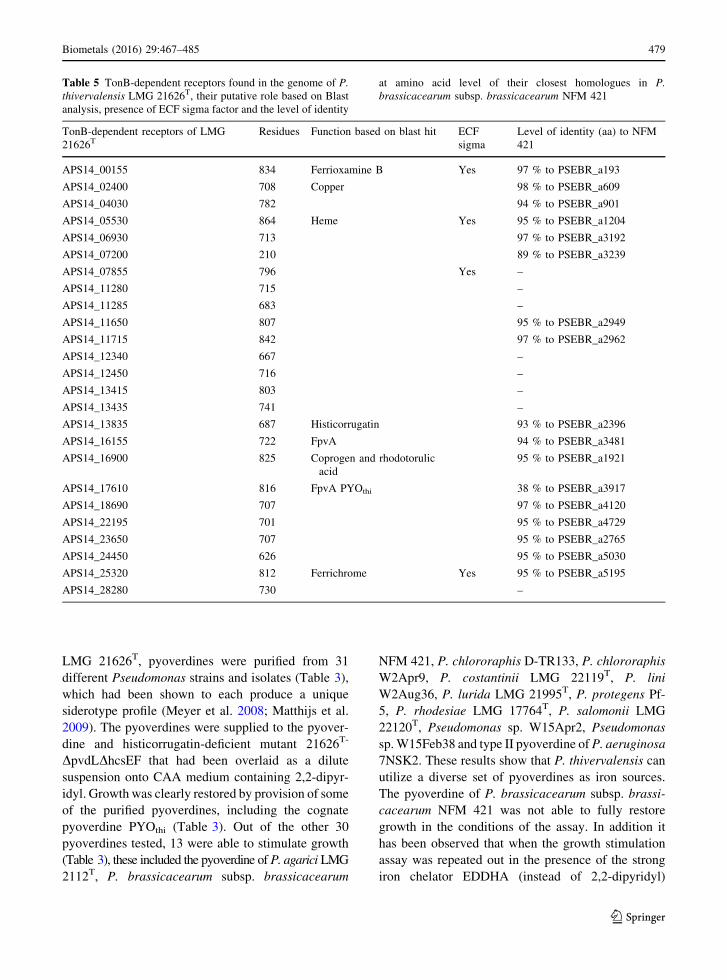
2002). In addition to the cognate pyoverdine receptor
FpvA and the histicorrugatin receptor an additional 23
TonB-dependent receptors were found in the LMG
21626T proteome. For four of these receptors gene
expression is probably controlled by ECF r factors as
indicated by the presence of extracytoplasmic sigma
factors and transmembrane anti-r factors adjacent to
the receptor (Table 5). Based on Blast analysis a
putative function could be attributed to five TonB
dependent receptors, responsible for the uptake of
ferrioxamine B, copper, heme, coprogen and rhodo-
torulic acid, and ferrichrome, respectively (Table 5).
Pseudomonas strains are also able to utilize
pyoverdine from other fluorescent pseudomonads
(Matthijs et al. 2009; Hartney et al. 2011; Ye et al.
2014a). To estimate the diversity of exogenous
pyoverdines that may be utilized by P. thivervalensis
478 Biometals (2016) 29:467–485
123
LMG 21626T, pyoverdines were purified from 31
different Pseudomonas strains and isolates (Table 3),
which had been shown to each produce a unique
siderotype profile (Meyer et al. 2008; Matthijs et al.
2009). The pyoverdines were supplied to the pyover-
dine and histicorrugatin-deficient mutant 21626T-
DpvdLDhcsEF that had been overlaid as a dilute
suspension onto CAA medium containing 2,2-dipyr-
idyl. Growth was clearly restored by provision of some
of the purified pyoverdines, including the cognate
pyoverdine PYOthi (Table 3). Out of the other 30
pyoverdines tested, 13 were able to stimulate growth
(Table 3), these included the pyoverdine of P. agarici LMG
2112T, P. brassicacearum subsp. brassicacearum
NFM 421, P. chlororaphis D-TR133, P. chlororaphis
W2Apr9, P. costantinii LMG 22119T, P. lini
W2Aug36, P. lurida LMG 21995T, P. protegens Pf-
5, P. rhodesiae LMG 17764T, P. salomonii LMG
22120T, Pseudomonas sp. W15Apr2, Pseudomonas
sp.W15Feb38 and type II pyoverdine of P. aeruginosa
7NSK2. These results show that P. thivervalensis can
utilize a diverse set of pyoverdines as iron sources.
The pyoverdine of P. brassicacearum subsp. brassi-
cacearum NFM 421 was not able to fully restore
growth in the conditions of the assay. In addition it
has been observed that when the growth stimulation
assay was repeated out in the presence of the strong
iron chelator EDDHA (instead of 2,2-dipyridyl)
Table 5 TonB-dependent receptors found in the genome of P.
thivervalensis LMG 21626T, their putative role based on Blast
analysis, presence of ECF sigma factor and the level of identity
at amino acid level of their closest homologues in P.
brassicacearum subsp. brassicacearum NFM 421
TonB-dependent receptors of LMG
21626TResidues Function based on blast hit ECF
sigma
Level of identity (aa) to NFM
421
APS14_00155 834 Ferrioxamine B Yes 97 % to PSEBR_a193
APS14_02400 708 Copper 98 % to PSEBR_a609
APS14_04030 782 94 % to PSEBR_a901
APS14_05530 864 Heme Yes 95 % to PSEBR_a1204
APS14_06930 713 97 % to PSEBR_a3192
APS14_07200 210 89 % to PSEBR_a3239
APS14_07855 796 Yes –
APS14_11280 715 –
APS14_11285 683 –
APS14_11650 807 95 % to PSEBR_a2949
APS14_11715 842 97 % to PSEBR_a2962
APS14_12340 667 –
APS14_12450 716 –
APS14_13415 803 –
APS14_13435 741 –
APS14_13835 687 Histicorrugatin 93 % to PSEBR_a2396
APS14_16155 722 FpvA 94 % to PSEBR_a3481
APS14_16900 825 Coprogen and rhodotorulic
acid
95 % to PSEBR_a1921
APS14_17610 816 FpvA PYOthi 38 % to PSEBR_a3917
APS14_18690 707 97 % to PSEBR_a4120
APS14_22195 701 95 % to PSEBR_a4729
APS14_23650 707 95 % to PSEBR_a2765
APS14_24450 626 95 % to PSEBR_a5030
APS14_25320 812 Ferrichrome Yes 95 % to PSEBR_a5195
APS14_28280 730 –
Biometals (2016) 29:467–485 479
123

pyoverdine of NFM 421 cannot stimulate at all the
growth of a pyoverdine-negative mutant of P. thiver-
valensis. Pyoverdine-mediated 59Fe-uptake showed
that the pyoverdine of NFM 421 was taken up at low
levels (35 %, value is expressed in percentage of
incorporation compared to the homologous system) by
P. thivervalensis wild type strain.
Strain LMG 21626T was not able to utilize type I
pyoverdine produced by P. aeruginosa PAO1 and
pyoverdines of strains of the P. putida group (Mulet
et al. 2010) including P. entomophila L48T, P. putida
BTP2, P. putida KT2440, P. putida W15Oct28,
Pseudomonas sp. W2Jun14 and Pseudomonas sp.
W15Oct11 which all produce structurally different
pyoverdines. In addition, LMG 21626T was not able to
utilize the pyoverdine of P. cedrina, P. citronellolis, P.
fluorescens DSM 50106, P. fluorescens LMG 14562,
and the type strain of P. asplenii, P. brenneri, P.
koreensis, P. libanensis and P. mosselii.
P. thivervalensis is phylogenetically most closely
related to P. brassicacearum. Therefore it was verified
whether their phylogenetic relationship was reflected
in the number and type of TonB-dependent receptors
and whether there are large differences in their
pyoverdine uptake profiles. IEF and LC–MS analysis
showed that NFM 421 produces the same pyoverdine
as P. putidaATCC 39167 (Urıa-Fernandez et al. 2003)
(Table 3). Sequencing of 16S rRNA of ATCC 39167
revealed that the strain received as ATCC 39167 is not
a P. putida strain, but belongs to the P. fluorescens
group and is closely related to P. brassicacearum.
Their 16S rRNAs (1278 bp) show a 99.7 % identity at
nucleotide level.
Seventeen TonB-dependent receptors of NFM 421
were extracted from its genome and a phylogenetic
tree was constructed with the 25 TonB-dependent
receptors of LMG 21626T. Interestingly, almost all the
receptors of NFM 421 clustered with a homologue of
P. thivervalensis (Fig. 5). The TonB-receptors showed
very high identity levels at the amino acid level, values
between 89 and 98 % were observed (Table 5). In fact
only the cognate pyoverdine receptor of NFM 421,
which showed only an identity of 38 % with the FpvA
receptor of P. thivervalensis (Table 5), did not cluster
with a homologue.
The histicorrugatin receptor of P. thivervalensis
showed an identity of 93 % to PSEBR_a2396 of NFM
421. Histicorrugatin was not detected in the culture
supernatant of NFM 421 but the analogue
ornicorrugatin was found. Ornicorrugatin is produced
by the gene cluster PSEBR_a2386-PSEBR_a2397 of
NFM 421, Tn5 mutants in the nrps genes PSE-
BR_a2390 and PSEBR_a2391 lead to a complete loss
of ornicorrugatin production (Matthijs S., unpublished
results).
To compare the diversity of exogenous pyoverdines
that may be utilized by both strains, growth stimula-
tion assays were repeated as described above with the
pyoverdine-negative mutant NFM421-4D1 of P. bras-
sicacearum subsp. brassicacearum NFM 421. The
pyoverdine utilization profiles of both strains are
almost identical (Table 3). In contrast to the pyover-
dine of NFM 421 which was not able to stimulate
growth of P. thivervalensis very well, clear growth
stimulation was observed of PYOthi for P. brassi-
cacearum. A difference in the utilization profiles of
both strains was observed for the pyoverdine of P.
citronellolis LMG 18378T and P. fluorescens Pf0-1,
these pyoverdines were only able to crossfeed P.
brassicacearum (Table 3).
Discussion
P. thivervalensis sp. are typically plant-associated
bacteria, they have been isolated as endophyte from
Solanum nigrum (Long et al. 2008) and from Ara-
bidopsis thaliana and Brassica napus roots (Achouak
et al. 2000). This work shows that they produce the
high affinity siderophore pyoverdine (PYOthi) and the
lipopeptidic siderophore histicorrugatin under iron
limiting conditions to fulfill their need for iron. Iron is
an essential element; it is a constituent of enzymes
with critical roles in metabolism of hydrogen, oxygen
or nitrogen, electron transfer, RNA synthesis and
dissolution of reactive oxygen intermediates (Braun
1997). Acquisition of iron is complicated by the
inherently low solubility of ferric iron, possible
adsorption of Fe3? on to colloids and precipitation of
iron with other ions result in even lower iron
concentrations in soil.
The production of iron-chelating siderophores by
bacteria can be beneficial for the plant, because they
can significantly increase the solubility of iron
(Lemanceau et al. 2009). Nutritional competition for
iron by the production of siderophores has been
extensively studied as a mechanism of biological
control. The plant growth-promoting effect of some
480 Biometals (2016) 29:467–485
123

APS14_25320
PSEBR_a5195
APS14_17610 FpvA
APS14_00155
PSEBR_a193
APS14_06930
PSEBR_a3192
APS14_22195
PSEBR_a4729
APS14_23650
PSEBR_a2765
APS14_07855
APS14_16155
PSEBR_a3481
PSEBR_a3917 FpvA
APS14_16900
PSEBR_a1921
APS14_13415
APS14_13435
APS14_28280
APS14_11650
PSEBR_a2949
APS14_18690
PSEBR_4120
APS14_24450
PSEBR_a5030
APS14_13835 histicorrugatin
PSEBR_a2396 ornicorrugatin
APS14_04030
PSEBR_a901
APS14_11280
APS14_11285
APS14_12450
APS14_12340
APS14_02400
PSEBR_a609
APS14_11715
PSEBR_a2962
APS14_05530
PSEBR_a1204
APS14_07200
PSEBR_a3239
100
100
100
100
100
100
100
100
100
100
100
100
100
100
100
100
96
94
94
92
95
92
90
76
71
97
64
100
60
58
51
77
Fig. 5 Phylogenetic relationships among amino acid sequences
of all the putative TonB-dependent receptors of P. brassi-
cacearum NFM 421 and P. thivervalensis LMG 21626T. The
locus tag of each receptor is indicated on the tree. The tree was
constructed using the NJ method. Numbers at nodes represent
levels (%) of bootstrap support from 500 resampled datasets
Biometals (2016) 29:467–485 481
123

fluorescent pseudomonads is thought to be in part due
to siderophores, including pyoverdine, pyochelin and
salicylic acid, that sequester iron in a form unavailable
to deleterious micro-organisms in the rhizosphere,
thereby preventing the pathogens’ access to the
already limited pool of soluble iron in the rhizosphere
if the latter do not possess more efficient iron uptake
systems (Kloepper et al. 1980; Hofte et al. 1991; Loper
and Henkels 1997; Duijff et al. 1999). In addition, it
has been observed that siderophores produced by
fluorescent pseudomonads can trigger defense
responses in the host (Maurhofer et al. 1994; Leeman
et al. 1996; Bakker et al. 2007; De Vleesschauwer and
Hofte, 2009).
Another trait, besides the ability to produce high
affinity siderophore(s), which is important when
microorganisms are competing for iron, is the ability
to utilise iron complexes of a variety of different
siderophores produced by other microorganisms,
including fungi and bacteria (Poole et al. 1990;
Jurkevitch et al. 1992; Raaijmakers et al. 1995). This
‘‘siderophore piracy’’ has two obvious advantages, the
microorganism does not have to use metabolic energy
for the synthesis of iron chelators, and by stealing the
siderophore of other microorganisms it deprives these
organisms of iron. It has been shown that P.
fluorescens BBc6R8, in the presence of a coelichelin
and desferrioxamines producing Streptomyces strain,
did not produce pyoverdine but relied on utilisation of
the siderophores produced by the Streptomyces strain
(Galet et al. 2015). The capacity to utilise exogenous
siderophores varies between Pseudomonas species
and probably reflects the importance of iron compe-
tition in the natural habitat of the bacteria. Based on in
silico analysis the receptors for the uptake of the
siderophores ferrioxamine B, coprogen, rhodotorulic
acid and ferrichrome could be attributed to P. thiver-
valensis LMG 21626T.
Strain LMG 21626T has in total 25 TonB-depen-
dent receptors, eigth more than its phylogenetic
neighbor P. brassicacearum. These numbers are on
the lower side, especially for P. brassicacearum
subsp. brassicacearumNFM 421 which has the lowest
number of TonB-dependent receptors reported until
now, but is in the same range as the phytopathogen P.
syringae for which 19 (B728a) to 25 (DC3000) TonB-
dependent receptors are found (Cornelis and Bodilis
2009). In contrast, the total number of genes coding for
TonB-dependent receptors in the plant-associated P.
fluorescens strains A506, SBW25 and P. protegens Pf-
5 are respectively 34, 37 and 45 (Hartney et al. 2011).
The highest number of TonB-dependent receptors has
been found in P. putida W15Oct28 isolated from a
small river (Matthijs et al. 2013), which has the
amazing number of 56 genes coding for TonB-
dependent receptors (Ye et al. 2014b). But what is
intriguing when comparing the type of TonB depen-
dent-receptors between P. thivervalensis and P. bras-
sicacearum is that NFM 421 has the same pool of
TonB-dependent receptors, except for the cognate
FpvA receptor, as P. thivervalensis. P. thivervalensis
has an additional 8 receptors, giving this strain
probably a competitive advantage over P. brassi-
cacearum. It would be interesting to study which type
of compounds can be taken up by these additional
TonB-dependent receptors.
Two pyoverdine receptors are found in P. thiver-
valensis LMG 21626T and P. brassicacearum subsp.
brassicacearum NFM 421, their cognate FpvA recep-
tor and a putative second pyoverdine receptor
(APS14_16155 and PSEBR_a3481, respectively).
These receptors do not belong to the core genome,
but to the accessory genome (Cornelis and Bodilis
2009). Pyoverdine growth stimulation assays showed
that P. thivervalensis and P. brassicacearum are able
to utilize respectively 13 and 15 structurally distinct
pyoverdines produced by other Pseudomonas sp. out
of 31 pyoverdines tested. This number is roughly
comparable to the 17 pyoverdines P. protegens Pf-5 is
able to utilize (Hartney et al. 2011). For P. fluorescens
ATCC 17400 this was 20 out of 28 pyoverdines tested
(Ye et al. 2014a) and for P. entomophila 16 out of 24
(Matthijs et al. 2009). Since sometimes different
pyoverdines have been tested for these strains these
data cannot be directly compared but they do indicate
that many pyoverdines can be utilized by P.
thivervalensis.
Despite the fact that the cognate FvpA receptors of
P. thivervalensis and P. brassicacearum did not show
large sequence similarities and did not cluster together
in the phylogenetic tree of TonB-dependent receptors
(Fig. 5), the pyoverdine utilisation profiles of P.
thivervalensis and P. brassicacearum are almost
identical. The observed differences in the uptake
profiles of both strains, whereby NFM 421 can take up
2 additional pyoverdines, can probably be attributed to
FpvANFM421. Through the construction of Fpvmutants
the specificity and the level of redundancy of each
482 Biometals (2016) 29:467–485
123

receptor can be further studied. The similarities in
their uptake profile might reflect, besides their genetic
similarities, their biological origin. Both strains were
isolated in the same study, but from different plants
and geographic environments; LMG 21626T was
isolated from Brassica napus and NFM 421 from
Arabidopsis thaliana (Achouak et al. 2000). But in the
latter study both P. thivervalensis and P. brassi-
cacearum strains have been isolated from B. napus and
A. thaliana (Achouak et al. 2000), which indicate that
they are probably able to colonize the same host plant.
Acknowledgments The authors are indebted to Pelletier Dale,
Baldwin Ian-Thomas and Gasser Ilona for generous gift of
strains.
References
AchouakW, Sutra L, Heulin T,Meyer J-M, Fromin N, Degraeve
S, Christen R, Gardan L (2000) Pseudomonas brassi-
cacearum sp. nov. and Pseudomonas thivervalensis sp.
nov., two root-associated bacteria isolated from Brassica
napus and Arabidopsis thaliana. Int J Syst Evol Microbiol
50:9–18
Barelmann I, Fernandez DU, Budzikiewicz H, Meyer JM (2003)
The pyoverdine fromPseudomonas chlororaphisD-TR133
showing mutual acceptance with the pyoverdine of Pseu-
domonas fluorescens CHA0. BioMetals 16:263–270
Bakker PAHM, Pieterse CMJ, van Loon LC (2007) Induced
systemic resistance by fluorescent Pseudomonas spp.
Phytopathol 97:239–243
Braun V (1997) Avoidance of iron toxicity through regulation of
bacterial iron transport. Biol Chem 378:779–786
Briskot G, Taraz K, Budzikiewicz H (1989) Pyoverdine-type
siderophores from Pseudomonas aeruginosa. Liebigs Ann
Chem 1989:375–384
Brown SD, Utturkar SM, Klingeman DM, Johnson CM, Martin
SL, LandML, Lu T-YS, Schadt CW, Doktycz MJ, Pelletier
DA (2012) Twenty-one genome sequences from Pseu-
domonas species and 19 genome sequences from diverse
bacteria isolated from the rhizosphere and endosphere of
Populus deltoids. J Bacteriol 194:5991–5993
Budzikiewicz H (1997) Siderophores of fluorescent pseu-
domonads. Z Naturforsch 52:713–720
Budzikiewicz H (2004) Siderophores of the Pseudomonadaceae
sensu stricto (fluorescent and non-fluorescent Pseu-
domonas spp.). Prog Chem Org Nat Prod 87:81–237
Budzikiewicz H, Schroder H, Taraz K (1992) Zur Biogenese der
Pseudomonas-siderophore: der Nachweis analoger Struk-
turen eines Pyoverdin-Desferribactin-Paares. Z Natur-
forsch C 47:26–32
Budzikiewicz H, Schafer M, Fernandez DU, Matthijs S, Cor-
nelis P (2007) Characterization of the chromophore of
pyoverdins and related siderophores by electrospray tan-
dem mass spectrometry. Biometals 20:135–144
Cheng X, de Bruijn I, van der Voort M, Loper JE, Raaijmakers
JM (2013) The Gac regulon of Pseudomonas fluorescens
SBW25. Environ Microbiol Rep 5:608–619
Cornelis P, Bodilis J (2009) A survey of TonB-dependent
receptors in fluorescent pseudomonads. Environ Microbiol
Rep 1:256–262
Cornelis P, Matthijs S (2002) Diversity of siderophore-mediated
iron uptake systems in fluorescent pseudomonads: not only
pyoverdines. Environ Microbiol 4:787–798
Cox CD, Graham R (1979) Isolation of an iron-binding com-
pound from Pseudomonas aeruginosa. J Bacteriol 137:
357–364
De Vleesschauwer D, Hofte M (2009) Rhizobacteria-induced
systemic resistance. Plant Innate Immunity 51:223–281
Delorme S, Lemanceau P, Christen R, Corberand T, Meyer JM,
Gardan L (2002) Pseudomonas lini sp. nov., a novel spe-
cies from bulk and rhizospheric soils. Int J Syst Evol
Microbiol 52:513–523
Dennis JJ, Zylstra GJ (1998) Plasposons: modular self-cloning
minitransposon derivatives for rapid genetic analysis of
gram-negative bacterial genomes. Appl Environ Microbiol
64:2710–2715
Duijff BJ, Recorbet G, Bakker PAHM, Loper JE, Lemanceau P
(1999)Microbial antagonism at the root level is involved in
the suppression of fusarium wilt by the combination of
nonpathogenic Fusarium oxysporum Fo47 and Pseu-
domonas putida WCS358. Phytopathol 89:1073–1079
Edwards U, Rogall T, Blocker H, Emde M, Bottger EC (1989)
Isolation and direct complete nucleotide determination of
entire genes, characterization of a gene coding for 16S
ribosomal RNA. Nucl Acids Res 17:7843–7853
Fernandez DU, Fuchs R, Taraz K, Budzikiewicz H, Munsch P,
Meyer J-M (2001) Structure of a pyoverdine produced by a
Pseudomonas tolaasii-like isolate. Biometals 14:81–84
Fuchs R, Budzikiewicz H (2001) Rearrangement reactions in the
electrospray ionization mass spectra of pyoverdins. Int J
Mass Spectrom 201(211):603–612
Galet J, Deveau A, Hotel L, Frey-Klett P, Leblond P, Aigle B
(2015) Pseudomonas fluorescens pirates both ferrioxamine
and ferri-coelichelin siderophores from Streptomyces
ambofaciens. Appl Environ Microbiol 81:3132–3141
Gasser I, Muller H, Berg G (2009) Ecology and characterization
of polyhydroxyalkanoate-producing microorganisms on
and in plants. FEMS Microbiol Ecol 70:142–150
Hartney SL, Mazurier S, Kidarsa TA, Quecine MC, Lemanceau
P, Loper JE (2011) TonB-dependent outer-membrane
proteins and siderophore utilization in Pseudomonas fluo-
rescens Pf-5. Biometals 24:193–213
Hofte M, Seong KY, Jurkevitch E, Verstraete W (1991) Pyo-
verdine production by the plant growth beneficial Pseu-
domonas strain 7NSK2: ecological significance in soil.
Plant Soil 130:249–257
Jurkevitch E, Hadar Y, Chen Y (1992) Differential siderophore
utilization and iron uptake by soil and rhizosphere bacteria.
Appl Environ Microbiol 58:119–124
Kloepper JW, Leong J, Teintze M, Schroth MN (1980)
Enhanced plant growth by siderophores produced by plant
growth-promoting rhizobacteria. Nature 286:885–886
Leeman M, den Ouden FM, van Pelt JA, Dirkx FPM, Steijl H,
Bakker PAHM, Schippers B (1996) Iron availability
affects induction of systemic resistance to Fusariumwilt of
Biometals (2016) 29:467–485 483
123

radish Pseudomonas fluorescens. Phytopathology 85:
149–155
Lemanceau P, Expert D, Gaymard F, Bakker P, Briat JF (2009)
Role of iron in plant-microbe interactions. Plant Innate
Immunity 51:491–549
Long HH, Schmidt DD, Baldwin IT (2008) Native bacterial
endophytes promote host growth in a species-specific
manner; phytohormone manipulations do not results in
common growth responses. PLoS One 3(7):e2702
Loper JE, Henkels MD (1997) Availability of iron to Pseu-
domonas fluorescens in rhizosphere and bulk soil evaluated
with an ice nucleation reporter gene. Appl Environ
Microbiol 63:99–105
Matthijs S, Baysse C, Koedam N, Abbaspour Tehrani K, Ver-
heyden L, Budzikiewicz H, Schafer M, Hoorelbeke B,
Meyer J-M, De Greve H, Cornelis P (2004) The Pseu-
domonas siderophore quinolobactin is synthesized from
xanthurenic acid, an intermediate of the kynurenine path-
way. Mol Microbiol 52:371–384
Matthijs S, Abbaspour Tehrani K, Laus G, Jackson RW, Cooper
RM, Cornelis P (2007) Thioquinolobactin, a Pseudomonas
siderophore with antifungal and anti-Pythium activity.
Environ Microbiol 9:425–434
Matthijs S, Budzikiewicz H, Schafer M, Wathelet B, Cornelis P
(2008) Ornicorrugatin, a new siderophore from Pseu-
domonas fluorescens AF76. Z Naturforsch C 63:8–12
Matthijs S, Laus G, Meyer JM, Abbaspour-Tehrani K, Schafer
M, Budzikiewicz H, Cornelis P (2009) Siderophore-me-
diated iron acquisition in the entomopathogenic bacterium
Pseudomonas entomophila. Biometals 22:951–964
Matthijs S, Coorevits A, Gebrekidan TT, Tricot C, Vander
Wauven C, Pirnay J-P, De Vos P, Cornelis P (2013)
Evaluation of the oprI and oprL genes as molecular
markers for the genus Pseudomonas and their use to study
the biodiversity of a small Belgian River. Res Microbiol
164:254–261
Maurhofer M, Hase C, Meuwly P, Metraux JP, Defago G (1994)
Induction of systemic resistance of tobacco to tobacco
necrosis virus by the root-colonizing Pseudomonas fluo-
rescens strain CHA0: influence of the gacA gene and of
pyoverdine production. Phytopathol 84:39–146
Mercado-Blanco J, van der Drift KMGM, Olsson PE, Thomas-
Oates JE, van Loon LC, Bakker PAHM (2001) Analysis of
the pmsCEAB gene cluster involved in the biosynthesis of
salicylic acid and the siderophore pseudomonine in the
biocontrol strain Pseudomonas fluorescens WCS374.
J Bacteriol 183:1909–1920
Meyer J-M, Abdallah MA (1978) The fluorescent pigment of
Pseudomonas fluorescens: biosynthesis, purification and
physicochemical properties. J Gen Microbiol 107:319–328
Meyer J-M, Geoffroy VA, Baida N, Gardan L, Izard D,
Lemanceau P, Achouak W, Palleroni NJ (2002) Side-
rophore typing, a powerful tool for the identification of
fluorescent and nonfluorescent pseudomonads. Appl
Environ Microbiol 68:2745–2753
Meyer J-M, Gruffaz C, Raharinosy V, Bezverbnaya I, Schafer
M, Budzikiewicz H (2008) Siderotyping of fluorescent
Pseudomonas: molecular mass determination by mass
spectrometry as a powerful pyoverdine siderotyping
method. Biometals 21:259–271
Moon CD, Zhang XX, Matthijs S, Schafer M, Budzikiewicz H,
Rainey PB (2008) Genomic, genetic and structural analysis
of pyoverdine-mediated iron acquisition in the plant
growth-promoting bacterium Pseudomonas fluorescensSBW25. BMC Microbiol 8:7
Mulet M, Bennasar A, Lalucat J, Garcıa-Valdes E (2009) An
rpoD-based PCR procedure for the identification of Pseu-
domonas species and for their detection in environmental
samples. Mol Cellul Prob 23:140–147
Mulet M, Lalucat J, Garcıa-Valdes E (2010) DNA sequence-
based analysis of the Pseudomonas species. Environ
Microbiol 12:1513–1530
Ongena M, Jacques P, Thonart P, Gwose I, Fernandez DU,
Schafer M, Budzikiewicz H (2001) The pyoverdin of
Pseudomonas fluorescens BTP2, a novel structural type.
Tetrahedron Lett 42:5849–5851
Poole K, Young L, Neshat S (1990) Enterobactin-mediated iron
transport in Pseudomonas aeruginosa. J Bacteriol
172:6991–6996
Raaijmakers JM, Van Der Sluis I, Koster M, Bakker PAHM,
Weisbeek PJ, Schippers B (1995) Utilization of heterolo-
gous siderophores and rhizosphere competence of fluo-
rescent Pseudomonas spp. Can J Microbiol 41:126–135
Rausch C, Weber T, Kohlbacher O, Wohlleben W, Huson DH
(2005) Specificity prediction of adenylation domains in
nonribosomal peptide synthetases (NRPS) using trans-
ductive support vector machines (TSVM). Nucleic Acids
Res 33:5799–5808
Risse D, Beiderbeck H, Taraz K, Budzikiewicz H, Gustine D
(1998) Corrugatin, a lipopeptide from Pseudomonas cor-
rugata. Z Naturforsch C 53:295–304
Schlegel K, Fuchs R, Schafer M, Taraz K, Budzikiewicz H,
Geoffroy V, Meyer JM (2001) The pyoverdins of Pseu-
domonas sp. 96-312 and 96-318. Z Naturforsch C
56:680–686
Schnider-Keel U, Seematter A, Maurhofer M, Blumer C, Duffy
B, Gigot-Bonnefoy C, Reimmann C, Notz R, Defago G,
Haas D, Keel C (2000) Autoinduction of 2,4-di-
acetylphloroglucinol biosynthesis in the biocontrol agent
Pseudomonas fluorescens CHA0 and repression by the
bacterial metabolites salicylate and pyoluteorin. J Bacteriol
182:1215–1225
Schwyn B, Neilands JB (1987) Universal chemical assay for the
detection and determination of siderophores. Anal Bio-
chem 160:47–56
Singh GM, Fortin PD, Koglin A, Walsh CT (2008) Beta-Hy-
droxylation of the aspartyl residue in the phytotoxin syr-
ingomycin E: characterization of two candidate
hydroxylases AspH and SyrP in Pseudomonas syringae.
Biochemistry 47:11310–11320
Stachelhaus T, Mootz HD,Marahiel MA (1999) The specificity-
conferring code of adenylation domains in nonribosomal
peptide synthetases. Chem Biol 6:493–505
Sultana R, Fuchs R, Schmickler H, Schlegel K, Budzikiewicz H,
Siddiqui BS, Geoffroy V, Meyer JM (2000) A pyoverdine
from Pseudomonas sp. CFML 95-275. Z Naturforsch C
55:857–865
Tamura K, Peterson D, Peterson N, Stecher G, Nei M, Kumar S
(2011) MEGA5: molecular evolutionary genetics analysis
using maximum like-lihood, evolutionary distance, and
484 Biometals (2016) 29:467–485
123

maximum parsimony methods. Mol Biol Evol 28:
2731–2739
Tappe R, Taraz K, Budzikiewicz H, Meyer JM, Lefevre JF
(1993) Structure elucidation of a pyoverdin produced by
Pseudomonas aeruginosa ATCC 27853. J Prakt Chem
335:83–87
Urıa-Fernandez D, Geoffroy V, Schafer M, Meyer JM, Budzi-
kiewicz H (2003) Structure revision of pyoverdines pro-
duced by plant-growth promoting and plant deleterious
Pseudomonas species. Monatsh Chem 134:1421–1431
Van Der Voort M, Meijer HJ, Schmidt Y, Watrous J, Dekkers E,
Mendes R, Dorrestein PC, Gross H, Raaijmakers JM
(2015) Genome mining and metabolic profiling of the
rhizosphere bacterium Pseudomonas sp SH-C52 for
antimicrobial compounds. Front Microbiol 6:693
Vieira J, Messing J (1991) New pUC-derived cloning vectors
with different selectable markers and DNA replication
origins. Gene 100:189–194
Voisard C, Bull C, Keel C, Laville J, Maurhofer M, Schnider U,
Defago G, Haas D (1994) Biocontrol of root diseases by
Pseudomonas fluorescens CHA0: current concepts and
experimental approaches. In: O’Gara F, Dowling D,
Boesten B (eds) Molecular ecology of rhizosphere
microorganisms. VCH, Weinheim, pp 67–89
Yamamoto S, Ksai H, Arnold DL, Jackson RW, Vivian A,
Harayama S (2000) Phylogeny of the genus Pseudomonas:
intrageneric structure reconstructed from the nucleotide
sequences of gyrB and rpoD genes. Microbiology
146:2385–2394
Ye L, Ballet S, Hildebrand F, Laus G, Guillemyn K, Raes J,
Matthijs S, Martins J, Cornelis P (2013) A combinatorial
approach to the structure elucidation of a pyoverdine
siderophore produced by a Pseudomonas putida isolate and
the use of pyoverdine as a taxonomic marker for typing P.
putida subspecies. Biometals 26:561–575
Ye L, Matthijs S, Bodilis J, Hildebrand H, Raes J, Cornelis P
(2014a) Analysis of the draft genome of Pseudomonas
fluorescens ATCC 17400 indicates a capacity to take up
iron from a wide range of sources, including different
exogenous pyoverdines. Biometals 27:633–644
Ye L, Hildebrand F, Dingemans J, Ballet S, Laus G, Matthijs S,
Berendsen R, Cornelis P (2014b) Draft genome sequence
analysis of a Pseudomonas putida W15Oct28 strain with
antagonistic activity to Gram-positive and Pseudomonas
sp. pathogens. PLoS One 9(11):e110038
Zerbino DR, Birney E (2008) Velvet: algorithms for de novo
short read assembly using de Bruijn graphs. Genome Res
18:821–829
Biometals (2016) 29:467–485 485
123


![Iron Oxide Mediated Degradation of Mutagen Pyrene and ... · been studied. Another iron oxide, goethite, has also been used for the photodegradation of pyrene by Wang et al. [18].](https://static.fdocuments.net/doc/165x107/5f7e4fa3833a6814005e25d0/iron-oxide-mediated-degradation-of-mutagen-pyrene-and-been-studied-another.jpg)

