PURIFICATION STUDIES ON THE INFECTIOUS CANINE … · Virus purification: The procedure for the...
15
Instructions for use Title PURIFICATION STUDIES ON THE INFECTIOUS CANINE HEPATITIS VIRUS : II. VIRAL PARTICLES AND THEIR CHARACTERISTICS REVEALED BY MODIFICATION OF THE PURIFICATION PROCEDURE Author(s) KUNISHIGE, Teruo; MIYATA, Yasuhisa; MIFUNE, Yoshikatsu; HIRATO, Katsushichi Citation Japanese Journal of Veterinary Research, 9(3), 137-144 Issue Date 1961-09 DOI 10.14943/jjvr.9.3.137 Doc URL http://hdl.handle.net/2115/1755 Type bulletin (article) File Information KJ00002373313.pdf Hokkaido University Collection of Scholarly and Academic Papers : HUSCAP
Transcript of PURIFICATION STUDIES ON THE INFECTIOUS CANINE … · Virus purification: The procedure for the...

Instructions for use
Title PURIFICATION STUDIES ON THE INFECTIOUS CANINE HEPATITIS VIRUS : II. VIRAL PARTICLES ANDTHEIR CHARACTERISTICS REVEALED BY MODIFICATION OF THE PURIFICATION PROCEDURE
Author(s) KUNISHIGE, Teruo; MIYATA, Yasuhisa; MIFUNE, Yoshikatsu; HIRATO, Katsushichi
Citation Japanese Journal of Veterinary Research, 9(3), 137-144
Issue Date 1961-09
DOI 10.14943/jjvr.9.3.137
Doc URL http://hdl.handle.net/2115/1755
Type bulletin (article)
File Information KJ00002373313.pdf
Hokkaido University Collection of Scholarly and Academic Papers : HUSCAP

PURIFICATION STUDIES ON THE INFECTIOUS
CANINE HEPATITIS VIRUS
II. VIRAL PARTICLES AND THEIR CHARACTERISTICS REVEALED
BY MODIFICATION OF THE PURIFICATION PROCEDURE
Teruo KUNISHIGE, Yasuhisa MIYATA, Y oshikatsu MIFUNE
and Katsushichi HIRATO
Depm·tmcnt of Hygiene and ~1icrobiology, Jiaculty of Veterinary 11;fedicine,
Hokkaido UniVl'l'sity, Sappom, Japan
(Received for publication, June 30, 1961)
INTRODUCTION
A previous publication6) has described one method of purification of the ICH
virus from infectious tissue culture fluid for electron microscopy, and demonstrated
that these particles are an average 85 m.a (non.shadow), 96 mil (Pd.shadow) In
diameter. The purpose of the present communication is to report with respect to
the fine structure of virus particles revealed by a slightly altered procedure In
comparison with the previously reported one and electron stain or formalin treat·
ment of purified virus particle forms extracted from infectious tissue culture fluid
and cells, and furthermore the several characteristics of purified virus.
MATERIALS AND METHODS
Virus: The "MATSUDA" strain of ICH virus was used throughout. It had been
transferred serially 5 to 10 times in monolayer cultures of dog kidney epithelial cells before
the yields were subjected to purification.
Virus purification: The procedure for the purification and concentration of the virus
is in part a modification of the methods used in the purification of poliomyelitis and foot-and·
mouth disease virus as was described in the previous report. That is to say, the extraction
with organic solvent was performed using chloroform or Daifuron-S 3 (Osaka Metal Industry
Co.) and the concentration of virus was accomplished by means of differential centrifugation
for 10 min. at 10,000 rpm, 90 min. at 25,000 rpm in a rotor No. 40 of the Hitachi Moc1el-40 P
ultracentrifuge as is outlined diagrammatically in table 1.
Electron staining method: Electron staining procedure based on that of BRENNER
& HORN (1959) was used. To 1.0 ml of virus suspension in 4% ammonium acetate was added
1.0 ml of 1 to 2;10 solution of phosphotungstic acid or 1;''; sodium phosphotungstate. The
solutions were then sprayed directly onto the carbon·coated collodion membrane grids with
a Vaponef6n atomiser spray gun.
Formalin treatment: The final purified virus (pellets of the centrifigation for 90 min.
JAP. ]. VET. RES., VOL. 9, No.3, 1961

138 KUNISHIGE, T. et al.
TABLE 1. Purification of feR Virus
InfeClious 1'1',11" C"lIul'e Fluid lnl"Cliou,; Tis,u" Culrure Cells
I I <I ,lUll rpm, \11 min, Washcu \-:. lim.'-< wil" PBS
~ , p I
Suspend in PBS
I Homogeniler, 2 min.
I Frozen and Ihawed 2-:1 limes
with liquid Nit rogen
I 4.lOU rpm, 10 min.
~ S P
Adjust 10 pH S.O
I Methanol, E. 'Y., H\'f1o-Super Cel
I Elute with pH 9 Saline
I Extract with Chloroform or Daifuron
I 10,O(X) rpm, 10 min.
~ S P
I 25.000 rpm, 90 min.
~ S P
I Suspend in pH 7.5-7.8 Saline
I 3,000 rpm, 5 min.
~ S P
I EleCII'on Micro<cope
at 25,000 rpm) was resuspended in 2% neutral formalin saline and was prepared for electron
microscopy by the agar pseudo-replica method.
Electron microscopy: The specimens were examined in a JEM-4CHD electron
microscope at instrumental magnifications of X 4,500 ....... X 5,000 to X 10,000 using single condenser
illumination.
Complement fixation test: The technique used was essentially identical with that of
the modification of KOLMER's method. For the determination of antigen titers, two exact units
of complement (0.5 ml) and 0.25 ml of hyperimmune dog serum (4 antibody units) were added
to the serial dilution of the antigen to be tested. Tubes containing the above mixtures were
stored at 4'C for 16. to 18 hrs. and 0.5 ml of hemolytic system was added the next day.
The tests were read after incubation in a water bath of 37°C for 30 min.
Hemagglutination test: Hemagglutination titrations were carried out in the manner
described by SHIMIZU et al. (1960).
Ultraviolet absorption: The ultraviolet absorption spectrum of .the virus suspension
before and after purification procedure was estimated with a Hitachi- EPU-2 spectrophotometer.

Infectious Canine Hepatitis Virus II 139
RESUL TS AND DISCUSSION
1. Electron Micrograph Electron micrographs of agar pseudo-replica of the vIrus
particles purified from infectious tissue culture fluid and cells revealed a uniform-sized, spherical
particle 113 mil (Pd-shadow) in diameter (Figs. 1 a, 3).
FIGURE 1. Distribution of Diameter of the Virus Particle
in Purified Sarnple
a) Pb-,;hadolV
20
to
140
b) Electroll Staill
'''I • XII 11111
~(). :l(a\lT. )
c) Formal", FixatIOn
IV ~ :
I J1-f1hn D, C""
lIIiI 1:!11
1117.6 lan'l'. )
Diaml'lt.'C of Particl,'.;(mp)
The fbttened particles containing a central protuberance revealed in a previous purification
procedure were not observed in this purified sample at all. In the purified virus suspension
derived from cells 24 hr5. after infection there were revealed a small number of intact virus
particles, smaller amorphous particles and their aggregates (Fig. 4).
With the lapse of time after infection these intact virus particles derived from cells
increased gradually in number.
By 48 hrs. the number of these intact virus particles had increased slightly, but the greater
part of the particles showed flattened and amorphous feature (Fig. 5). At 72 to 96 hrs. after
infection there could be observed only the same particles as revealed in infectious tissue culture
140 KUNISHIGE, T. et al.
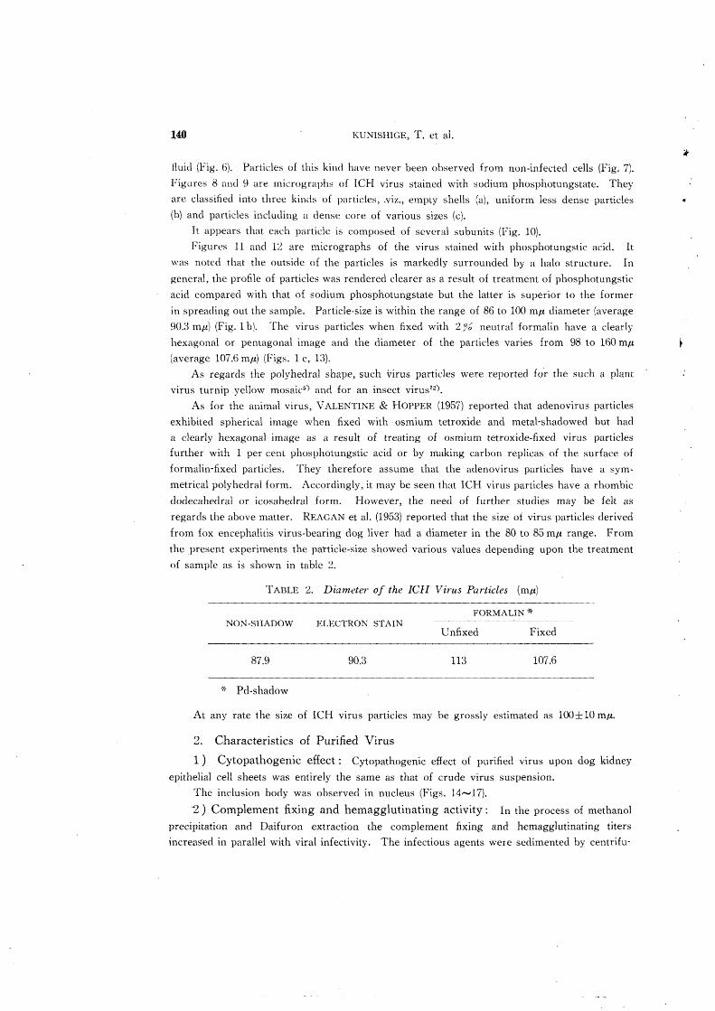
fluid (Fig. 6), Particles of this kind have never been observed from non-infected cells (Fig. 7).
Figures 8 and 9 are micrograph::; of lCI-! virus stained with sodium phosphotungstate. They
are classified into three kinds of partides, .viz., empty shells (a), uniform less dense particles
(b) and particles incluuing a dense core of various sizes (c).
It appears that each particle is composed of several subunits (Fig. 10).
Figures 11 and 12 are micrographs of the virus stained with phosphotungstic acid. It
was noted that the outside of the particles is markedly surrounded by a halo structure. In
general, the profile of particles was rendered dearer as a result of treatment of phosphotungstic
acid compared with that of sodium phosphotungstate but the latter is superior to the former
in spreading out the sample. Particle-size is within the range of 86 to 100 mtt diameter (average
90.3 mIl) (Fig. 1 b). The virus particles when fixed with 2:?G neutral formalin have a dearly
hexagonal or pentagonal image and the diameter of the particles varies from 98 to 160 mtt
(average 107.6 nw) (Figs. 1 c, 13).
As regards the polyhedral shape, such virus particles were reported for the such a plant
virus turnip yellow mosaicS) and for an insect virus12).
As for the animal virus, VALENTINE & HOPPER (1957) reported that adenovirus particles
exhibited spherical image when fixed with osmium tetroxide and metal-shadowed but had
a clearly hexagonal image as a result of treating of osmium tetroxide-fixed virus particles
further with 1 per cent phosphotungstic acid or by making carbon replicas of the surface of
formalin-fixed particles. They therefore assume that the adenovirus particles have a sym
metrical polyhedral form. Accordingly, it may be seen that ICH virus particles have a rhombic
dodecahedral or icosahedral form. However, the need of further studies may be felt as
regards the above matter. REAGAN et ai. (1953) reported that the size of virus particles derived
from fox encephalitis virus-bearing dog liver had a diameter in the 80 to 85 mtt range. From
the present experiments the particle-size showed various values depending upon the treatment
of sample as is shown in table 2.
TABLE 2. Diameter of the ICH Virus Particles (mil)
FORMALIN * NON-SHADOW ELECTRON STAIN
Unfixed Fixed
87.9 90.3 113 107.6
,'f Pd-shadow
At any rate the size of ICH virus particles may be grossly estimated as l00± 10 mIl.
2. Characteristics of Purified Virus
1) Cytopathogenic effect: Cytopathogenic effect of purifled virus upon dog kidney
epithelial cell sheets was entirely the same as that of crude virus suspension.
The inclusion body was observed in nucleus (Figs. 14,...,17).
"2) Complement fixing and hemagglutinating activity: In the process of methanol
precipitation and DaHuron extraction the complement fixing and hemagglutinating titers
increased in parallel with viral infectivity. The infectious agents were sedimented by centrifu-

Infectious Canine Hepatitis Virus II 141
gation at 25,000 rpm for 90 min. and the infectivity of supernate became very low but comple
ment fixing titer of still remained high (Table 3).
TABLE :3. Infectivity and CF and HA Titer of Each Fraction
Resultant from Purification Procedure -------~-------~------~
EXP. 1 EXP. ~ FRACTIONS
TCDso eFT HAT TCDso CFT HAT
Original TeF 4.5 12,13 256 3.7 3~ 128
Solu ble part of 5.5 51:2 ~O48 4.5 1~8 ~56 Methanol ppt.
Aqueous phase after 5.5 512 1024 :,.5 64 256 extraction with Daifurol1
Supernate < 1.0 ~56 1O~4 < 1.0 64 32
25,000 rpm 90 min.
Pellet 3.5 256 128 5.5 32 2041::\
Treatment of purified virus suspension with ether resulted in complete or almost complete
loss of infectivity. Huwever, such treatment did not decrease the cumplement fixing and
hemagglutinating titers but occasionally caused their increase over the currespunding values
of original suspension (Table 4).
TABLE 4. Infectivity and CF and HA Titer of Purified Virus b(fore
a nd after Treatment with Ethyl Ether
ETHER EXP. 1 EXP. 2 EXP. :3
TREATMENT TeD50 eFT HAT TeDso CFT HAT TCD so CFT
Before 3 r .U 256 128 6.0 US 512 5.5 512
After < 1.0 2.56 64 2.0 128 1O~4 < 1.0 2048
HAT
1024
204,13
---- -~ ... ~ ... --.. ,- -_ .... - -------,~~-~-----------.--.
Therefore, it is assumed that there is a :-Ioluble antigen 111 lCH virus as suggested by HIRATO et al. (1960). However, the present authors can not support the opinion of TAKATORI
(1960) that the virus particles are as inactive as complement fixing antigen. In this experiment,
purified infectious virus particles keep their complement fixing and hemagglutinating activities.
HOYLE (1952) has speculated on the basis of his results with ether treatment of virus suspensions
that elementary body of influenza virus has a lipid membrane envelope which contains S com
ponents and small hemagglutinating particles. Ether dissolves the membrane, releasing S
antigens and hemagglutinins. The present authors released S component and hemagglutinin
with ether treatment of purified lCI! virus using the method of HOYLE (EIS~). However, if
the time of ether treatment is too long continued, complement fixing and hemagglutinating
titers decrease in parallel with infectivity. Regarding such fact, MrzUTANI (1958) reported
a similar phenomenon in influenza virus. HENLE & WIENER (1944) estimated the particle-size

142 KUNISHIGE, T. et al.
of the soluble antigen of infl~~?za virus at 100......,150 A whilst HOYLE (1952) showed that the
soluble antigen derived from -the elementary body by disintegration with ether had a particlesize of 120 A.
In this experiment, the present authors could not observe by electron microscopy the size of S component of ICH virus.
3) Ultraviolet absorption spectrum: The ultraviolet absorption spectrum of the
suspension of ICH virus particles before and after purification procedure was as graphed in
figure 2.
FIGURE 2. Ultraviolet Absorption Spectrum of Crude
and Purified ICH Virus Suspension
1.0,
0.8
0.6
0.4
0.2
240 260 280 300 320
Wave Length (mil)
The ultraviolet spectrum of crude virus suspension before purification is a maXimum at
275 mJl and a mini~um at 250 m/-!. However, the spectrum of purified virus particles is one
characteristic of a nucleoprotein with a maximum at 258 mJl and a minimum at 240 m/.t.
SUMMARY
The picture of the infectious canine hepatitis VIrus particle derived from
infectious tissue culture fluid and cells has been clearly demonstrated by a modified
purification procedure. It was able to observe the fine structure of the particle
when the virus was stained with phosphotungstic acid or sodium phosphotungstat~.

Infectious Canine Hepatitis Virus II 143
Fixing the purified virus particles with 2% neutral formalin resulted in the revelation
of a clearly hexagonal or pentagonal image. Purified ICH virus particles keep
infectivity and complement fixing and hemagglutinating activities and can be disinte
grated by treatment with ether with the liberation of smaller particle, soluble antigen
and hemagglutinin. The ultraviolet absorption spectrum of final purified virus
particles shows a maximum at 258 mil and a minimum at 240 m/I.
The authors are greatly indebted to Assistant Professor H. SHIOKAWA, Dr. Y. INUKAI
and Dr. H. KODAMA of the Department of Biochemistry for their kind advice on the chemical
study of virus and assistance in estimate of ultraviolet absorption spectrum.
REFERENCES
1) BRENNER, S. & R W. HORNE (1959): Biochim. Biophys. Acta, 34, 103.
~) HENLE, W. & M. WIENER (1944): Proc. Soc. expo Bioi., N. Y., 57, 176.
3) HIRATO, K., Y. SHIMIZU, T. KUNISHIGE, K. YOSIIIDA, S. TANAKA, T. TOKUI,
Y. MIFUNE & T. ONU (1960): ZbZ. Vetlvfed., 7, 525.
4) HOYLE, L. (195~): J. Hyg., Camb., 50, 229.
5) KAESBERG, P. (1956): Science, 124, 6~6. 6) KUNISHIGE, T., Y. SHIMIZU & K. HIRATO (1960,: Jap. J. vet. Res., 8, 208.
7) MIZUTANI, H. (1958): Virus, 8, 195 (in Japanese with English summary).
8) REAGAN, R L., W. C. DAY, F. SANSONE & A. L. BRUECKNER (1953): Transact.
Amer. 1I1icroscop. Soc., 72, 252.
9) SHIMIZU, Y., T. KUNISHIGE & K. HIRATO(1960): Jap. J. vet. Res., 8, 27l.
10) TAKATORI, 1. (1960): Jap. J. vet. Sci., 22, 487 (in Japanese).
11 ) VALENTINE, R C. & P. K. HOPPER (1957): Nature, 180, 9~8. 12) WILLIAMS, R. C. & K. M. SMITH (1957): Ibid., 179, 119.

144 KUNISHIGE, T. et al.
EXPLANA nON OF PLATES
PLATE I.
Fig. 3. Electron micrograph of final purified ICH Virus particles.
Pd-shadow X 50,000
PLATE II.
Fig. 4. Purified sample derived from cells 24 hrs. after infection. Arrows indicate
intact virus particles. Pd-shadow X 22,500
Fig. 5. 48 hrs. after infection.
Arrows indicate intact virus particles; most of the particles show flattened
and amorphous features. Pd-shadow X 22,500
PLATE III.
Fig. 6. 72 hrs. after infection. Pd-shadow >( 22,500
Fig. 7. Purified sample derived from non-infected cells. Pd-shadow X 22,500
PLATE
Fig.
IV.
8.
Purified VIrus particles stained with sodium phosphotungstate
X 50,000
Fig. 9. a) empty shell b) uniform slightly dense particle
c) particles including dense core of various sizes X 100,000
PLATE V.
Fig. 10. Virus particles stained with sodium phosphotungstate. Arrows indicate
subunits. X 140,000
Figs. 11, 12. Purified virus particles stained with phosphotungstic acid. Arrows
indicate halo structure X 140,000
Fig. 13. Purified virus particles fixed with 2:?~ neutral formalin
a ) hexagonal form b) pentagonal form X 50,000
PLATE VI. Cytopathogenic effect of purified virus upon dog kidney epithelial
cell sheets Giemsa stain X 480
Fig. 14. Original infectious tissue culture fluid
Fig. 15. Methanol precipitation fraction
Fig. 16. Fraction after extraction with Daifuron
Fig. 17. Final purified fraction
..
..
..
..

KUr\ISHIGE, T. et al. PLATE I

KUNISHIGE, T. et al. PLATE II
..
..
..

KUNISHIGE, T. et al. PLATE III

KUNISHIGE, T. et al. PLATE IV ,
- - ---_.------

KUl':ISHIGE, T. et al. PLATE V
~. ___ ~. ___ ._. __ ~_~_~_.~ __ c ____________________ _

KUNISHIGE, T. et al. PLATE VI
~----- - - --- - ~ ---