PURIFICATION OF CYANIDE-DEGRADING NITRILASE FROM
54
APPROVED: Daniel A. Kunz, Major Professor Barney J. Venables, Committee Member Rebecca Dickstein, Committee Member Arthur J. Goven, Chair of the Department of Biological Sciences James D. Meernik, Acting Dean of the Robert B. Toulouse School of Graduate Studies PURIFICATION OF CYANIDE-DEGRADING NITRILASE FROM Pseudomonas fluorescens NCIMB 11764 Chia-Ni Chou, B.S. Thesis Prepared for the Degree of MASTER OF SCIENCE UNIVERSITY OF NORTH TEXAS December 2010
Transcript of PURIFICATION OF CYANIDE-DEGRADING NITRILASE FROM

APPROVED: Daniel A. Kunz, Major Professor Barney J. Venables, Committee Member Rebecca Dickstein, Committee Member Arthur J. Goven, Chair of the Department
of Biological Sciences James D. Meernik, Acting Dean of the
Robert B. Toulouse School of Graduate Studies
PURIFICATION OF CYANIDE-DEGRADING NITRILASE FROM
Pseudomonas fluorescens NCIMB 11764
Chia-Ni Chou, B.S.
Thesis Prepared for the Degree of
MASTER OF SCIENCE
UNIVERSITY OF NORTH TEXAS
December 2010

Chou, Chia-Ni. Purification of cyanide-degrading nitrilase from Pseudomonas
fluorescens NCIMB 11764
Cyanide is a well known toxicant that arises in the environment from both
biological and industrial sources. Bacteria have evolved novel coping mechanisms for
cyanide and function as principal agents in the biosphere for cyanide recycling. Some
bacteria exhibit the unusual ability of growing on cyanide as the sole nitrogen source.
One such organism is Pseudomonas fluorescens NCIMB 11764 (Pf11764) which
employs a novel oxidative mechanism for detoxifying and assimilating cyanide. A
unique complex of enzymes referred to as cyanide oxygenase (CNO) is responsible for
this ability converting cyanide to ammonia which is then assimilated. Because one
component of the four member CNO complex was previously shown to act on cyanide
independent of the other members, its characterization was sought as a means of
gaining a better understanding of the overall catalytic mechanism of the complex.
Preliminary studies suggested that the enzyme belonged to a subset of nitrilase
enzymes known as cyanide dihydratases (CynD), however, a cynD-like gene in
Pf11764 could not be detected by PCR. Instead, a separate nitrilase (Nit) linked to
cyanide metabolism was detected. The corresponding nit gene was shown to be one of
a conserved set of nit genes traced to a unique cluster in bacteria known as Nit1C. To
determine whether the previously described CynD enzyme was instead Nit, efforts were
undertaken to isolate the enzyme. This was pursued by cloning and expressing the
. Master of Science (Molecular Biology), December 2010, 44
pp., 5 tables, 7 figures, references, 44 titles.

recombinant enzyme and by attempting to isolate the native enzyme. This thesis is
concerned with the latter activity and describes the purification of a Nit-like cyanide-
degrading nitrilase (NitCC) from Pf11764 to ~95% homogeneity. Purification was greatly
facilitated by the discovery that fumaronitrile, as opposed to cyanide, was the preferred
substrate for the enzyme (20 versus 1 U/mg protein, respectively). While cyanide was
less effective as a substrate, the specificity for cyanide far outweighed that (10,000 fold)
of the recombinant enzyme (NitPG) implying that the native NitCC protein purified in this
work is different from that of the cloned recombinant. Further evidence of this was
provided by molecular studies indicating that the two proteins differ in mass (34.5 and
38 kDa, respectively) and amino acid sequence. In summary, two different Nit enzymes
are encoded by Pf11764. While the two share greater than 50% amino acid sequence
identity, the results suggest that the native NitCC enzyme purified in this work functions
better as a cyanide-degrading nitrilase and is one of four enzyme components
comprising CNO required for Pf11764 cyanide assimilation.

ii
Copyright 2010
by
Chia-Ni Chou

iii
ACKNOWLEDGEMENTS
I would like to express my gratitude to my professor, Dr. Daniel Kunz, whose
expertise, understanding, and patience, added considerably to my graduate experience.
I am grateful for the opportunity to work in his laboratory and his encouragement,
guidance, and vast help during the development of this study.
I would like to thank Dr. Barney Venables for generously investing his time and
his assistance in mass spectrometry analysis. I would also like to thank my final
committee member, Dr. Rebecca Dickstein, for helpful discussions and review of the
manuscript. I owe a special thanks to follow students Pallab Ghosh and Trevor Burton
for their advice, help, and friendship.
Finally, I thank my family and friends for the support and understanding they
have provided me throughout my entire life.

iv
TABLE OF CONTENTS
Page
ACKNOWLEDGEMENTS……………………………………………………………………...iii
LIST OF TABLES ………………….…………………………………………………………..vi
LIST OF FIGURES…………………………………………………………………………… vii
LIST OF ABBREVIATIONS…………………………………………………………………..viii
Chapter
I. INTRODUCTION……………………………………………………………...………..…1
Physicochemical Properties of Cyanide and Its Occurrence………….…...……………....1
Organisms Detoxification………………………………………..………………....................2
CNO Properties…………………………………………………………..……………………..5
Nit1C Cluster in Pf11764………………………….…………………………………………....8
II. MATERIALS AND METHODS…………………………………………………..….….11
Organism and Cultivation Conditions……………………………………………..…………11
Enzyme Assays……………………………………………………………………..…..……..14
Substrate Disappearance and Product Analysis……………………………………...……15
Enzyme Purification …………………………………………………………………...…...…15
Gel Electrophoresis……………………………………………………………..……………..18
In-Gel Trypsin Digestion and Peptide Extraction……………………….………………….19
Electro-Spray Tandem Mass Spectrometry (ESI-MS/MS)………………………..………20
III. RESULTS……………………………………………………………………….………..22
Induction of Cyanide-Degrading Nitrilase………………………………..…………….……22
Purification of Cyanide-Degrading Nitrilase……………………………………….………..24

v
Comparative Analysis of Recombinant and Native Isolated Pf11764
Nit Enzymes ...................................................................................................................32
IV. DISCUSSION…………………….…………………………………………………....…36
REFERENCES……………………………………………………..…………………….……41

vi
LIST OF TABLES
Page
1. Composition of glucose-ammonia minimal medium (GAM) for cultivation
of P.fluorescens NCIMB 11764………………………………………………...…….13
2. Induction of cyanide-degrading enzyme activities in P. fluorescens
NCIMB 11764…………………………………………………………………………..23
3. Copurification of fumaronitrile- and cyanide-degrading nitrilase (Nit) activities
from P. fluorescens NCIMB 11764 induced with cyanide…………………..….....29
4. Indentity matches for proteins recovered after gel-filtration chromatography
from Pf11764 by electrospray ionization mass spectrometry (ESI-MS/MS)….....31
5. Comparative properties of native and recombinant Pf11764 Nit enzymes...……35

vii
LIST OF FIGURES
Page
1. Hypothetical model of interacting proteins identified in previous studies
as being required for cyanide oxygenase (CNO) enzyme activity form
Pseudomonas fluorescens NCIMB 11764……………………................................6
2. Organization of conserved Nit1C cluster found in various…………...……………10
3. Steps used in isolation of cyanide-degrading nitrilase (Nit)…..………………......17
4. Time-course of fumaronitrile conversion to ammonia by cell extracts
prepared from cyanide-exposed and unexposed cells of Pf11764………..……..25
5. Elution of proteins and Nit activity present in cell extracts of P. fluorescens
NCIMB 11764 following anion-exchange and gel-filtration chromatography…....28
6. SDS-PAGE analysis of Pf11764 proteins recovered in fractions exhibiting
Nit activity…………………..…………………………………………………………..30
7. Alignment of Nit enzyme protein sequences from P. fluorescens Pf11764
(PfPG), Burkholderia phymatum STM815 (Bup), and Burkholderia xenovorans
LB400 (Bux)………………………………………...…………..................................33

viii
LIST OF ABBREVIATIONS
CNO Cyanide oxygenase
CynD Cyanide dihydratase
FMN Fumaronitrile
h Hour
KCN Potassium cyanide
kDa Kilodalton
min Minute
MS Mass spectrometry
Na-K Sodium potassium buffer
Nit Nitrilase
PAGE Poly acrylamide gel electrophoresis
Pf Pseudomonas fluorescens
SDS Sodium dodecyl sulfate

1
CHAPTER I
INTRODUCTION
Cyanide is a potent biological inhibitor and toxic poison. It inhibits cytochrome
oxidases and other metallo enzymes thus preventing respiration and other vital
biological functions (Solomonson, 1981; Knowles, 1988). Cyanide arises in the
environment from both biological and abiological sources. Abiological sources
include wastes generated in the electroplating, coal processing and mining industries.
It’s occurrence in industrial fire smoke, automobile exhaust, and even cigarette smoke
has also been reported (Homan, 1988; Agency for Toxic Substances and Disease
Registry, 1993). Cyanide is also produced biologically as a natural product of
metabolism by so-called cyanogenic organisms. Cyanogenic species include various
plants, arthropods, bacteria and fungi (Conn, 1980; Castric, 1981; Knowles and
Bunch, 1986; Poulton, 1988; Blumer and Haas, 2000; Zagrobelny, 2008). The classic
cyanide poisoning from over consumption of various plant materials in the human diet
(e.g., cassava roots, lima beans, and almonds) is well documented (Kojima et al.,
1983; Frehner et al., 1990; Dicenta et al., 2002).
Chemically, cyanide exists either in its free form (CN-/HCN) or complexed to
various acceptors. It is the free form of cyanide that is most toxic because of its
tendency to add as a nucleophile to various acceptor molecules. The most notorious
of these are metal ions such as iron present as a prosthetic group in cytochrome
oxidases. At pH 7.0 the free form exists predominantly as the protonated species
(HCN), which is a weak acid (pKa 9.3). HCN is also quite volatile and boils at room

2
temperature. In its complexed form cyanide is generally much less toxic because
the free form is unavailable. Cyanide readily forms complexes with various metals,
particularly of the transition series, including Fe, Ni and Zn. It also adds readily to
various organic acceptors, including for example, α-keto acids (Kunz et al., 1998).
The presence of cyanide in nature implies that their likely exist mechanisms that
organisms have evolved to survive in its presence. It is well known, for example, that
bacteria and plants adapt to cyanide by making alternative cytochrome oxidases that
are less sensitive to inhibition (Bendall and Bonner, 1971; Grant and Hommersand,
1974; Rhoads, 1998). This permits respiration to occur at concentrations that
otherwise would be inhibitory. Another way to overcome the toxic effects of cyanide is
to modify it in some way chemically. A number of enzymes with this ability are known.
In animals, for example, cyanide is detoxified by the enzyme rhodanese (thiosulfate
cyanide sulfurtransferase, EC 2.8.1.1) (equation 1), which catalyzes its conversion to
less toxic thiocyanate (equation 1)(Sorbo, 1953; Silver and Kelly, 1976; Ryan and
Tilton, 1977). In contrast, the enzyme β-cyanoalanine synthetase is believed to be
the principal enzyme involved in cyanide detoxification in plants (Castric, 1981; Jenrich
et al., 2007). It catalyzes the condensation of cyanide with cysteine as shown in
equation 2.
S2O32- + HCN
Rhodanese HSCN + SO3
2-
Thiocyanate
(1)
COOH | HCN + HSCH2 – C –H | NH2
NH2
| COOH–C–CH2CN + SO3
2-
|
H
β-Cyanoalanine Synthetase
(2)

3
Fungi detoxify cyanide mainly by way of cyanide hydratase yielding formamide as a
less toxic product (equation 3). It is unclear what role, if any, cyanide detoxifying
enzymes play in bacteria since mutants lacking related enzymes have not been
described. An additional cyanide-modifiying enzyme described thus far only in
bacteria is cyanide dihydratase (CynD) (also referred to as cyanide nitrilase or
cyanidase). This enzyme is unique in that it catalyzes the one step hydrolysis of
cyanide to less toxic formate and ammonia (equation 4). Although a physiological role
in cyanide detoxification for this enzyme has been suggested, the rather high
concentrations of cyanide required (Km values ranging from ~2-7 mM have been
reported) (Raybuck, 1992; Jandhyala et al., 2005) make it difficult to rationalize a
primary role for this enzyme as one of detoxification. This is particularly so when one
considers that most bacteria will not grow above cyanide concentrations of ~0.5 mM
(Silva-Avalos et. al, 1990). Both cyanide hydratase and CynD belong to the large
superfamily of enzymes known as nitrilases (E. C. 3.5.5.3) a characteristic feature of
which is a conserved glutamate-lysine-cysteine catalytic triad (Pace and Brenner,
2001; O’Reilly and Turner, 2003; Martinkova and Kren, 2010). An additional feature
is a tendency to form large oligomeric structures from component monomers
averaging ~40 kDa in mass (Thuku et al., 2008). In our laboratory (Kunz et al., 2001,
Fernandez et. al.,2004a, Fernandez and Kunz, 2005; Dolghih, 2004) it has been
HCN + H2O HCONH2 (3) Cyanide hydratase
Formamide
Formic acid HCN + H2O
Cyanide dihydratase (CynD)
) HCO2H + NH3 (4)

4
shown that certain bacteria make a novel enzyme that detoxifies cyanide at
concentrations as low as < 5µM. This is far below the concentration reported for any
other known cyanide detoxifying enzyme. The enzyme responsible has been
described as cyanide oxygenase (CNO) because molecular oxygen is also required as
a substrate. This mechanism is novel in biochemistry because nitriles, of which
cyanide is the simplest example, are generally metabolized hydrolytically not
oxygenatively.
The biochemical properties of CNO have thus far been described in one
organism only namely, Pseudomonas fluorescens NCIMB 11764 (Pf11764). Pf11764
was originally isolated for its ability to use cyanide as the sole nitrogen source in Great
Britain (Harris and Knowles, 1983a). The responsible enzyme was first proposed to
be an oxygenase because the degradation of cyanide by Pf11764 cell-extracts was
linked to the consumption of molecular oxygen and NADH (Harris and Knowles
(1983b). A body of studies in our laboratory, including those demonstrating that
isotopic oxygen-18 was incorporated when cyanide was degraded (Wang et al., 1996),
confirmed the original proposal that an oxygenase-mediated mechanism was involved.
Further work by Dr. Kunz and students which showed that mutants that failed to make
CNO could no longer grow (Kunz et al., 1994; Fernandez et al., 2001) provided strong
evidence that CNO was needed for growth on cyanide. Additional studies aimed at
characterizing the CNO enzyme have shown it to be fairly complex. For example,
rather than a single enzyme, studies have shown that CNO is comprised of several
enzymes identified as: NADH oxidase (Nox), NADH peroxidase (Npx), an enzyme
with properties similar to those of CynD enzymes, and carbonic anhydrase (CA)

5
(Fernandez and Kunz, 2005). A diagram depicting the four as components of CNO is
illustrated in Fig 1. Evidence that all four are needed for enzyme activity comes from
studies which showed that if any one of the four was not included in reactions no
activity was observed (Fernandez and Kunz,2005). Ironically, CNO generates the
same reaction products as those formed by CynD enzymes namely, formate and
ammonia (equation 4), however, instead of using water to accomplish substrate
cleavage, CNO utilizes molecular oxygen and NADH (equation 5). The ammonia
thus formed is assimilated and helps to explain how cells acquire nutritional nitrogen
from cyanide. The formate is further oxidized to carbon dioxide by an enzyme
separate from CNO previously identified as formate dehydrogenase (FDH) (equation
6.)(Kunz et al, 2004). Together, the CNO and FDH enzymes mediate the complete
oxidation of cyanide to carbon dioxide and ammonia recycling one NADH equivalent in
the process.
HCN + O2 + 2NADH + H+ HCO2H + NH3 +2NAD+ CNO
HCO2H + NAD+ CO2 + NAD+ FDH
(5)
(6)

6
Nox Npx
CA CynD
FIG 1. Hypothetical model of interacting proteins identified in previous studies as being
required for cyanide oxygenase (CNO) enzyme activity form Pseudomonas fluorescens
NCIMB 11764. Nox, NADH oxidase; Npx, NADH peroxidase; CynD, cyanide
dihydratase; CA, carbonic anhydrase.

7
How the four enzymes of CNO accomplish cyanide conversion is unknown. A
perplexing issue has been the discovery that one of its members, CynD, is capable on
its own of converting cyanide to formate and ammonia. However, as demonstrated
for other CynD enzymes, the concentration of cyanide required for optimal activity is
much higher than that displayed by CNO (> 5 mM vs < 5 µM, respectively). Because
CynD is the only member of CNO for which there is evidence that cyanide is a
substrate there is strong interest in characterizing this enzyme. Thus, acquiring
information on this enzyme is believed to shed light on and how the four enzymes
accomplish catalysis.
Other than a demonstrated ability to convert cyanide to formate and ammonia with
the parallel appearance on SDS gels of a polypeptide with a mass of approximately
38kDa (Fernandez, 2005), no further molecular information on the identity of the CynD
component has since been acquired. Working from the hypothesis that the enzyme
was a likely CynD homolog, considerable effort was put forward in both Dr. Kunz’s
laboratory and that of his collaborator, Dr. Michael Benedik (Texas A&M University) to
amplify and clone a CynD gene from Pf11764 but these efforts were unsuccessful.
One PCR product annotated as a nitrilase enzyme (Nit) was obtained, but since the
translated amino acid sequence shared only 31% amino acid sequence identity with
CynD enzymes it was concluded that this most likely was not a CynD-like enzyme.
However, recent studies in our laboratory have given cause to think that this Nit
enzyme may be more important to cyanide metabolism than initially thought. The
basis for this reasoning comes from the discovery of a gene in Pf11764 coding for a
small protein (17.8 kDa) of unknown function (Hyp1) the expression of which was

8
found to increase when cells were exposed to cyanide (Ghosh, 2009). Moreover, an
examination of bacterial genomes revealed that the hyp1 gene is well conserved
among bacteria, and almost invariably resides immediately adjacent to a gene coding
for Nit enzymes that show high homology to the nit discovered in Pf11764. Indeed,
the conserved hyp1 and nit genes reside in a conserved cluster of seven genes
referred to as Nit1C the physical arrangement of which is shown in Fig 2. This cluster
was so named because a survey of bacterial genomes for nitrilase-encoding genes
showed that a high number of such genes were located among similar genes.
Because the clustering of genes in bacteria is frequently suggestive of a related
metabolic function, it was thought that this might be the case for Nit1C but so far its
function remains unknown. However, we hypothesize that these genes may have a
physiological role to play in cyanide assimilation as a nutritional nitrogen source based
on (i) findings that the expression of Hyp1 in Pf11764 is elevated in response to
cyanide, (ii) in addition to hyp1 the genome of Pf11764 also contains a gene for nit,
and (iii) preliminary laboratory studies conducted in Dr. Kunz’s laboratory having
shown that several other known Nit1C-containing bacteria also exhibit the ability to
grow on cyanide as the sole nitrogen source. Because the Nit enzyme is considered
to be a key enzyme in allowing bacteria to grow on cyanide (and as already discussed
is believed to be related to or one and the same as CynD), a coordinated effort to
isolate and fully characterize this enzyme was undertaken. One part of this effort
was to try and clone the gene and over-express the enzyme which Pallab Ghosh, a
fellow graduate student in the laboratory concentrated on and succeeded at to some
measure. This thesis concentrates on the native enzyme which was purified to

9
approximately 95% homogeneity. The properties of both enzymes are compared in
the Results section leading to the conclusion that the two enzymes are not the same.
10
FPHyp2AIRsynthaseGNATSAMNitHyp1
FIG 2. Organization of conserved Nit1C cluster found in various (Podar et al., 2005):
Hyp1, hypothetical protein; Nit, Nitrilase; SAM, radical S-adenosyl methionine
superfamily member; GNAT, acetyltransferase; AIRs, AIR synthase; Hyp2, hypothetical
protein; FP, predicted flavoprotein. Adapted from Podar et. al, 2005.

11
CHAPTER II
MATERIAL AND METHODS
Organism and Cultivation Conditions
Pseudomonas fluorescens NCIMB 11764 (Pf11764) was originally isolated in
Great Britain in 1983 and deposited in the National Collection of Industrial and Marine
Bacteria (NCIMB) (Torrey, Scotland) (Harris and Knowles, 1983a). It was acquired
24 years ago from NCIMB and has been maintained in the Kunz laboratory ever since.
The complete medium used for growing cells on agar plates was Lennox
medium (LB) whose components have previously been described (Lennox, 1995;
Fernandez, 2004a). The minimal medium used for growth of cells (referred to from
here on forward as GAM), is shown in Table 1. Cultivation of cells for enzyme
isolation were routinely cultivated in 2L Erlenmeyer flasks supplied GAM and
inoculated (10% v/v) with a 48 hour-old starter culture grown on the same medium.
After 24 h incubation at 30oC on a gyratory shaker, cells where indicated were
exposed to cyanide by supplying KCN at 0.1 mM. After 3 h of further incubation (total
27 h) the cells were harvested and stored at -80oC until use. For cells not exposed to
cyanide, cells were grown similarly in GAM and harvested after 27 h. For the bulk
of the enzyme isolation experiments reported herein, cells were grown in large quantity
(400 L fermenter) at the USDA Forest Products Laboratory (Madison, Wi) employing
the same cyanide-exposure and harvesting-timed protocol. Cells were recovered by
centrifugation at 10,000 x g (or continuously with a Sharples centrifuge) and washed
12
twice in Na2HPO4-KH2PO4 (Na-K) phosphate buffer pH 7.0 before freezing the wet cell
pellet at -80oC (approximately 0.9~1.0 gram wet weight/liter).

13
TABLE 1. Composition of glucose-ammonia minimal medium (GAM)
for cultivation of P. fluorescens NCIMB 11764
Component Concentration
KH2PO4 (pH 7.0) 67 mM
MgSO4. 7H2Oa 400 µg ml-1
FeSO4. 7H2Oa 10 µg ml-1
Glucose 10mM
NH4Cl 1mM
a Added aseptically to sterilized KH2PO4 buffer (P1X) at a 200-fold
dilution from a sterile stock solution (R-salts) prepared by mixing 400
ml of MgSO4. 7H2O with 100 ml of FeSO4
. 7H2O to which was added
2 ml of concentrated HCl. Glucose and NH4Cl were also added
aseptically after autoclaving to sterile P1X. Adapted from Kunz et
al., (1998).

14
Enzyme Assays
Nitrilase. Nitrilase (Nit) activity was assayed either with cyanide (CN, supplied as
KCN) or the dinitrile, fumaronitrile (FMN), as substrates. For cyanide-dependent Nit
activity (also referred to as cyanide dihydratase [CynD] activity) reaction mixtures
contained 20 mM KCN in 50 mM Na-K phosphate buffer (pH 8.0) and protein (3 µg –
1.5 mg). Reactions were conducted in sealed vials incubated without shaking at
30oC, and at various times, samples were removed with a syringe and the amount of
remaining cyanide (or ammonia formed) determined colorimetrically. In cases where
it was preferred to have oxygen eliminated from reaction, vials were purged with argon
before reactions were initiated. Fumaronitrile-dependent Nit activity was determined
by following the formation of ammonia from FMN (10 mM). For each measured
assay the change in substrate disappearance (or product formed) was corrected for
changes observed in the absence of added protein by conducting simultaneous
incubations under identical conditions but without added protein.
Cyanide oxygenase (CNO). CNO activity was assayed as described previously
(Fernandez et al., 2004a; Fernandez and Kunz, 2005) by measuring the
disappearance of cyanide (or formation of ammonia) in reaction mixtures (100 µl) that
contained 50 mM Na-K phosphate buffer (pH 7.0), 100-200 µM KCN, 500 µM NADH
and protein (1-10 mg/ml). Reactions were incubated at 30oC with shaking (200 rpm)
and conducted in sealed vials to prevent cyanide escape.

15
Substrate Disappearance and Product Analysis
Cyanide was quantified colorimetrically by the Lambert method as described
previously (Lambert et al., 1975; Kunz et al., 1992). For this purpose, 10µl of sample
was added to a solution containing 700µl of oxidizing reagent (1 g succinimide and 0.1
g N-chlorosuccinimide in 100ml diH2O) and 300µl of diH2O. After mixing, 50µl of the
above solution was added to
1.1 ml of water containing 50µl of oxidizing reagent, followed immediately by the
addition of 50µl of barbituric acid reagent (60 mg barbituric acid in 0.3ml pyridine and
0.7ml diH2O). Color
was allowed to develop for 15 minutes after which the optical density of the solution
was read at 580 nm and cyanide quantified from a standard curve.
Ammonia was determined by modification of the indophenol method of Fawcett
and Scott (1960). For this purpose, 5 µl of reaction mixture was added into 120 µl of
diH2O, followed by 250µl of sodium phenate (0.25 g phenate and 0.78 ml of 4N NaOH
in 10ml diH2O), 375µl of 0.01% of sodium nitroprusside, and 375µl of 0.02N sodium
hypochlorite. Reactions were allowed to incubate for 30 minutes at room
temperature and the optical density read 630nm. Ammonia was quantified from a
standard curve.
Enzyme Purification
The basic protocol for enzyme purification is shown in Fig 3. Individual steps
are discussed in order.

16
Preparation of cell extracts. Frozen cell pellets were suspended in Na-K phosphate
buffer (pH 7.0 (2 ml/1 g cells) containing 1mM dithiothreitol, 1% glycerol and 50µg/ml
DNase. Cells were broken at 4oC in a French Press (20,000 psi) followed by low-
speed centrifugation at 30,000 x g. The resulting supernatant was then subjected to
ultracentrifugation at 150,000 x g for 90 minutes and the supernatant (HSS, high-
speed supernatant) retained.
Anion exchange chromatography. A Source 30Q (GE healthcare) anion exchange
column (bed volume 55 ml) was equilibrated with 20 mM Piperazine buffer (pH 10.0)
(running buffer) for 3 column volumes (flow rate 3 ml per min) before applying
approximately 200 mg of HSS protein (~ 13 ml HSS equilibrated with 2 equal volumes
of running buffer). Non-binding proteins were removed by applying an additional 3
column volumes of running buffer before eluting bound proteins with a linear gradient
of 0-0.5 M Na2SO4 applied over five column volumes. The 2nd ion-exchange step
was performed similarly except that a smaller amount of protein (about 25 mg) was
applied. Also, prior to initiating the 2nd round of Source30Q chromatography the
column was reversed in orientation and cleaned successively with 2M NaCl (3 ml/min
30-60 min), 1 N NaOH (3 ml/min 1-2 hours) followed by 70% ethanol (1ml/min 1-2
hours). Following chromatography, proteins recovered in fractions (6 mL) were tested
for Nit activity with FMN. For this purpose, 90µl of each fraction was placed in 2 ml
sealed HPLC vial to which 10 mM fumaronitrile was added. Vials were incubated for
90 min to 2 h and analyzed for evidence of ammonia formation by visible inspection.
Those fractions exhibiting activity were collected and desalted with Na-K buffer (3
times original volume) (50 mM, pH 7.0) containing 1 mM dithiothreitol, and 1% glycerol

17
Cell extracts
150,000 x g high-speed supernatant (HSS)
1st anion exchange chromatography (30Q column)
2nd anion exchange chromatography (30Q column)
Gel-filtration chromatography
FIG 3. Steps used in isolation of cyanide-degrading nitrilase (Nit)

18
in Amicon Ultra 10K centrifugal filters (Millipore).
Gel filtration chromatography. Fractions exhibiting Nit activity with FMN following the
second round of anion exchange chromatography were pooled and desalted prior to
being loaded (approximately 5 mg) onto Superdex 200 column (bed volume 24 ml).
The column was equilibrated in 20mM NaH2PO4-Na2HPO4 buffer (Na-Pi) (pH 7.0), and
proteins eluted at a linear flow rate of 0.5 ml/min. Determination of the approximate
molecular weight of eluting proteins was accomplished by comparing the elution
volumes (times) to that of standard proteins (apoferritin, 443 kDa; alcohol
dehydrogenase, 150 kDa; albumin, 66 kDa; carbonic anhydrase, 29 kDa; cytochrome
C, 12.4 kDa) eluted under identical conditions. To those fractions exhibiting Nit
activity, dithiothreitol (1 mM) and glycerol (1%) were added prior to concentration (~
40X) in Amicon Ultra 10K centrifugal filters (Millipore). Concentrated proteins were
stored at -80oC.
Gel Electrophoresis
Discontinuous SDS-PAGE (12 or 16%) was carried out with a Mini-Protean
(Bio-Rad) apparatus using either 12 or 16% acrylamide gels supported with a 5%
stacking gel. A 12% gel (8.4 x 5.3 cm), was prepared by mixing (in order) 4 ml of 30%
acrylamide/bis stock solution, 2.5 ml of 1.5 M Trizma buffer (pH 8.8), 100 µl of 10%
SDS, 2.05 ml diH2O, 50 µl of 10% ammonium persulfate, and 5µl TEMED (N,N,N,N-
tetramethylenediamine). The gel was covered with isopropyl alcohol and the gel
allowed to polymerize for 45 minutes before washing off the isopropyl alcohol and
layering the separating gel with a 5% stacking gel. The stacking gel was prepared by

19
mixing 0.65 ml of acrylamide stock solution, 1.25 ml of 0.5 M Trizma (pH 6.8), 50 µl of
10% w/v SDS, 3.05 ml of ddH2O, 25 µl of ammonium sulfate (10 % w/v) and 5 µl of
TEMED. The gel was allowed to polymerize for 45 min and placed in an
electrophoresis tank containing running buffer (25 mM Trizma, 192 mM glycine and
0.1% w/v SDS, pH 8.3). The samples were mixed with sample buffer (150mM
dithiothreitol, 150mM Trizma pH 6.8, 21% glycerol, 6% lithium dodecyl sulfate, and
0.003% bromophenol blue) in the ratio of 2:1 and then were denatured by heating at
95 oC for 10 minutes. Samples were cooled, loaded onto the gel, and
electrophoresis performed at 4 oC for 60 minutes at a constant voltage of 200 V.
The gel was stained with Coomassie Blue staining solution (40% methanol, 10%
glacial acetic acid, 0.1% Coomassie Brilliant Blue R-250 in diH2O) for 30 minutes and
destained with 40% methanol and 10% glacial acetic acid in diH2O for 90 minutes.
The gel was stored in 10% glacial acetic acid.
In-Gel Trypsin Digestion and Peptide Extraction
Protein bands from SDS-PAGE gels were excised and subjected to overnight
trypsin digestion as described by Vergote et al., (2006). Protein bands were cut out (1
mm2 gel slice) and placed into a microcentrifuge tube, and the gel contents washed 3
times with 0.5 ml water shaking the microcentrifuge tube continuously for 5 min. The
contents were taken up in 0.5 ml 25 mM ammonium bicarbonate (ABC) and the
microcentrifuge tube agitated for 10 minutes (2 times). In order to remove the blue
Coomassie-stained, 0.2 ml of destaining solution (25 mM ABC in 50% acetonitrile

20
(ACN)) was added and agitated at least 20 minutes. The gel slice was washed with 0.2
ml of ABC in 50% ACN until the blue color of Coomassie color disappeared.
The gel slice was dried in speed vacuum for 15 to 20 minutes. After the gel slice
was dry, the dried gel slice was treated with 15 to 25 µl of 33 mM dithiothreitol (DTT) in
25 mM ABC and incubated at 60oC for 45 minutes. Then, the gel slice was reacted in
dark at room temperature with 50 µl of 75 mM iodoacetic acid in 25 mM ABC for 50
minutes. The gel slice was washed and agitated with 0.1 ml of 25mM ABC for 15
minutes (2 times). The supernatant was discarded, and the gel slice was dried for 30
min in speed vacuum. The dried gel slice was treated with 10µl of 12.5 ng/µl trypsin in
10µl of 50mM ABC and incubated in ice about 45 min, and then incubated overnight
(12~16 h) at 37oC.
The supernatants from the trypsin-digested mixtures were collected in a new
microcentrifuge tube. The gel slice was subjected to vortex for 10 min, sonicated for 15
min (65 rpm) and centrifuged for 5 min in 25µl of 25 mM ABC, 25µl of 50% ACN/ 5%
formic acid (twice), 25µl of 50% ACN/ 50% isopropanol/ 2% formic acid, and 25µl of
95% ACN/ 5% formic acid. The supernatants were dried to the volume of about 15µl
using speed vacuum, and stored at -80oC.
Electro-Spray Tandem Mass Spectrometry (ESI-MS/MS)
The identity of proteins was verified by mass spectrometry on an Agilent 100
LC/MS (ESI-MS/MS) in collaboration with Dr. Berney Venables of the UNT Proteomics
Laboratory. Peptides formed following trypsin digestion were separated by reverse-
phase chromatography by injecting 8 µl of trypsinized sample onto a C18 capillary

21
column (0.3 mm x 150 mm). The mobile phase consisted of solvent A (H2O/ 0.1%
formic acid) and solvent B (acetonitrile/ 0.1 % formic acid) programmed to achieve a
0-100% gradient (solvent A to B) for 60 min at a flow rate of 4 µl/min. Separated
peptides were introduced accordingly into the mass spectrometer with the most
intense MS/MS peaks automatically selected after defining an intensity threshold.
The mass spectral profile was then compared to those in available databases using
the Mascot search tool from Matrix Science (www.matrixscience.com).

22
CHAPTER III
RESULTS
Induction of Cyanide-Degrading Nitrilase
As shown in Table 1, cells of Pf11764 exposed to cyanide under minimal
nutrient conditions induced two types of cyanide-degrading enzyme activities, one
oxygen-dependent (aerobic) and the other oxygen-independent (anaerobic). The
aerobic activity, as previously established (Kunz et al., 1994, Kunz et al., 2001), is
conferred by CNO while the oxygen-independent (anaerobic) activity, was
hypothesized to arise from Nit. Because the hyp1 gene was shown in our laboratory
to be upregulated by cyanide (Ghosh, 2009) it was hypothesized that the same would
be true for Nit given the thought that the two reside next to each other in a probable
Nit1C operon. However, to better evaluate the level of expression of Nit an improved
method over assaying enzyme activity with cyanide as a substrate was needed given
that the activity exhibited by cell extracts towards cyanide was quite low (~0.005 U/mg,
see Table 2). An alternative substrate was therefore sought to better assay the
enzyme. The literature was examined for compounds that might serve this purpose by
searching for reports specifically describing the substrate requirements of Nit1C-
derived Nit enzymes. Only one such report, that being of the characterization of a Nit
enzyme from the blue-green bacterium Synechocystis sp. (strain PCC6803), was
found (Heinemann et. al., 2003). Although it was not known at the time that this was
a Nit1C enzyme, verification that the enzyme is truly Nit1C origin was confirmed by
consulting the now available Synechocystis genome (Heinemann et. al., 2003) (locus
s110784) (merR nitrilase)(http//ncbi.nlm.nih.gov). The best substrate found was the

23
TABLE 2. Induction of cyanide-degrading enzyme activities in P. fluorescens NCIMB 11764
Specific Activity (nmol min-1 mg-1) measured
with cyanide:
Cyanide exposure
during cultivation
Aerobicallya
Anaerobicallyb
Yes
8 + 4
5 + 2
No < 0.5 < 0.5
aFormally representing CNO measured by following the disappearance of cyanide (and/or
appearance of ammonia) as described in the Materials and Methods.
bAssays conducted either anaerobically or aerobically by measuring cyanide (20 mM)
disappearance (or ammonia formed) in the absence of added NADH.

24
dinitrile, fumaronitrile (1,2-dicyanoethene). For over-expressed enzyme was reported
to have a specific activity of 2 U/mg. This is 100-times greater than that reported for a
model substrate (benzonitrile) (0.02 U/mg). We tested cell extracts of cyanide-
exposed cells of Pf11764 for nitrilase (Nit) activity towards fumaronitrile (FMN) and
that indeed, cell extracts catalyzed its conversion to ammonia in a time-dependent
manner (Fig. 4). Moreover, the activity for cells that had been exposed to cyanide
during cultivation was significantly greater than that of non-exposed cells (0.5 and 0.02
U/mg, respectively) thus implying that expression of Nit, as earlier demonstrated for
Hyp1 (Ghosh, 2009), was also induced by cyanide (as expected if genes for the two
exist in an operon). The results in Fig 4 further show that in reaction mixtures supplied
10 mM FMN approximately 10 mM ammonia accumulated indicating that fumaronitrile
is converted stoichiometrically to ammonia (and fumaric acid) as depicted in equation
8.
Purification of Cyanide-Degrading Nitrilase
Having discovered that FMN is a much better substrate for Nit than cyanide
(KCN) we proceeded to purify the enzyme from cell extracts; the greater sensitivity of
enzyme detection representing a big improvement over previous attempts made in our
laboratory for isolating the enzyme. The approach to isolation of Nit employed three
steps: two rounds of anion-exchange chromatography at high pH (10) followed by gel
filtration chromatography (see Materials and Methods and Fig. 3). This approach was
(8)

25
FIG 4. Time-course of fumaronitrile conversion to ammonia by cell extracts prepared
from cyanide-exposed ( ) and unexposed ( ) cells of Pf11764. Inset: Color formation
of ammonia for samples taken at the times indicated. Reaction mixtures contained, 10
mM fumaronitrile in Na-K phosphate buffer (pH 7) and 1 mg/ml protein.

26
taken because earlier work (Fernandez et al., 2004a) had shown that the enzymes
comprising CNO (of which putative Nit is a component) could be recovered under
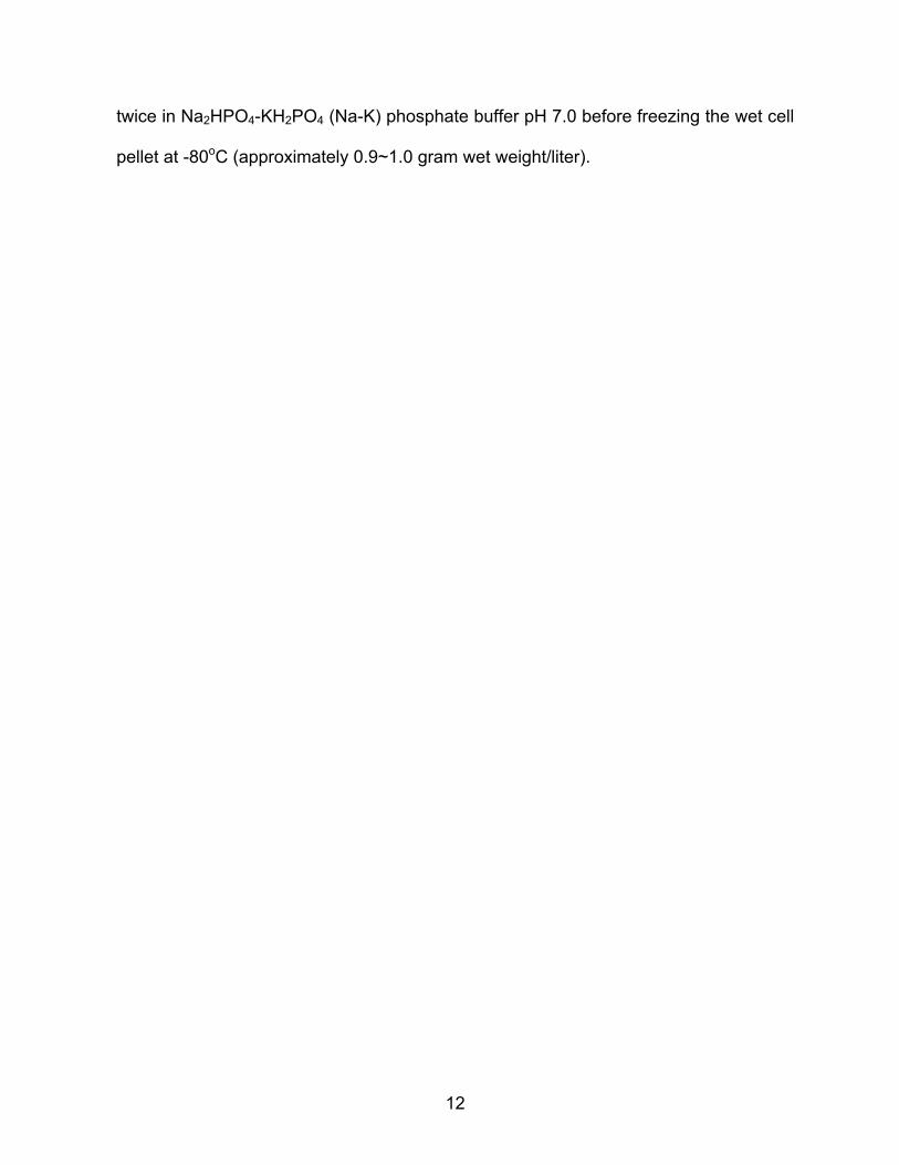
these conditions. Fig 5A shows the protein elution pattern and recovery of Nit activity
following the second round of anion exchange chromatography. Nit activity, as shown,
was concentrated in fractions eluting between 0.3 and 0.4 M Na2SO4. These fractions
were pooled, desalted by passage through 10,000 molecular weight cutoff (mwco)
ultramembrane centrifugation tubes, and the concentrated protein was applied to a 24
ml capacity Superdex-200 column. The pattern of elution from this column
is shown in Fig 5B and shows that activity was recovered in individual fractions
calibrated to contain proteins in the approximate 110 kDa range. At each step of the
purification process the relative Nit activity towards fumaronitrile versus cyanide as the
substrate was measured and proteins recovered in various fractions analyzed by SDS-
PAGE. The three-step protocol to enzyme isolation was performed on three separate
occasions for which the overall results are summarized in Table 3. The results show
that the enrichment in activity for assays conducted with FMN were paralleled by a
simultaneous enrichment in activity towards cyanide suggesting that a single enzyme
was responsible for the activity observed with both substrates. The final purified
sample exhibited 18 times greater activity with FMN than for cyanide. In contrast, the
overall purification achieved for the cyanide degrading activity was almost five times
that achieved when FMN served as the assay substrate. Fig 6 shows the gel profile
pattern for proteins present in a single fraction recovered after gel filtration
chromatography. Visible were four major protein bands of 42, 40, 39, and 38 kDa.
Similar migrating bands was also detected in fractions obtained following the 1st and

27
2nd rounds of anion-exchange chromatography steps, but overall, fewer proteins bands
were visible on gels while progressing from one step to the next as expected. In order
to determine which protein(s) was responsible for the observed FMN and cyanide
degrading activities, individual bands from the gel were excised and subjected to mass
spectrometry. These experiments were conducted in collaboration with Dr. Barney
Venables of the UNT Protein Proteomics Laboratory. Table 5 shows a summary of the
results in which it can be seen that only the 38 kDa species returned a match for a
protein annotated as a nitrilase (Nit). Thus, it was concluded that a single Nit protein
from Pf11764 was responsible for the observed activity towards both FMN and
cyanide as substrates.
28
100 200 300 400
A
10 20 30 40
B
FIG 5. Elution of proteins and Nit activity present in cell extracts of P.
fluorescens NCIMB 11764 following anion-exchange (A) and gel-filtration
chromatography (B). In A and B respectively, 29 and 2.8 mg protein was
applied. Proteins in (B) were eluted with 20 mM Na2HPO4, pH 8.0, at a flow
rate of 0.5 ml/min.
Absorbance
Activity
Salt
Activity
Absorbance

29

30
.
FIG 6. SDS-PAGE analysis of Pf11764 proteins recovered in fractions exhibiting Nit
activity. Lane 1, molecular weight standards: phosphorylase B, 97 kDa; BSA, 66 kDa;
ovalbumin, 45 kDa; carbonic anhydrase, 31kDa (5 µg); lane 2, 1st ion-exchange
column (10 µg); lane 3, 2nd ion-exchange column (10 µg); lane 4, gel filtration column.
(2 µg).
42 kDa 40 kDa
39 kDa
38 kDa

31

32
Comparative Analysis of Recombinant and Native Isolated Pf11764 Nit Enzymes
Pallab Ghosh and Dr. Jung-Huyn Lee of our laboratory succeeded in cloning Nit
using Pf11764 DNA sequence arrived at by gene-walking both up and downstream of
the identified Nit region (unpublished results). The nit content of the plasmid construct
(pE101D::nitPG) was verified by DNA sequencing and the translated amino acid
sequence showed more than 50% identity to other Nit1C-derived Nit proteins providing
strong evidence that the correct enzyme had been cloned (data not shown).
However, in this case the predicted mass of the enzyme was 34.5 kDa which is
somewhat below that determined for the enzyme isolated from the wild-type as
described here. Limited expression studies for the recombinant protein showed that
indeed, a protein of the correct mass was produced (as determined by gel
electrophoresis and confirmed by mass spectrometry) (data not shown). Thus, a
question then arose as to whether or not the cloned (34.5 kDa) recombinant (NitPG)
and native (38 kDa) enzymes (NitCC) were different. To further compare them, the
amino acid sequence for the Nit protein returned from a Mascot MS-ion search of
matching peptides with the Pf11764 enzyme (e.g., gi|186473966 from B. phymatum
STM815) was aligned with the sequence predicted for the recombinant. These data
are shown in Fig 7. Included also in this comparison is the sequence of one
additional Nit enzyme (gi|91784392) from the related bacterium, B. xenovorans LB400.
Aside from the obvious differences between NitPG and NitCC as far as total amino acid
content is concerned, additional differences may be noted. First, with regards to the
highlighted regions (referencing peptides returned from the Mascot search), It may be
noted that for the more carboxyl oriented peptide a 100% match for the two sequences

33
FIG 7. Alignment of amino acid sequences for the recombinant NitPG protein from P. fluorescens
Pf11764 (PfPG), the MASCOT returned peptide match with the Pf11764 NitCC protein derived from
Burkholderia phymatum STM815 (Bup), and Nit from Burkholderia xenovorans LB400 (Bux) using
the ClustalW program available at ExPASy – Tools (www.ebi.ac.uk/Tools/clustalw2/index.html).
Highlighted blocks of amino acids are those returned from a peptide search of amino acids
matching those present in Pf11764 NitCC. Consensus amino acids representing the conserved
catalytic triad are also shown highlighted. Identical residues are noted by asterisk ( * ), similar
ones with a colon ( : ), and less similar by a dot ( . ). Non-conservative charges are left blank
below the alignment.
PfPG Bup Bux
PfPG Bup Bux
PfPG Bup Bux
PfPG Bup Bux
PfPG Bup Bux
PfPG Bup Bux
PfPG Bup Bux
PfPG Bup Bux

34
exists. In contrast, for the more amino-terminal peptide there is a two amino-acid
mismatch (one on each end, see also Table 5). There are additional differences that
can be noted only one of which will attention be drawn to here. This includes the
occurrence of a phenylalanine residue (F) next to the conserved catalytic triad-
cysteine (C) for the Bup protein as opposed to tryptophan (W) in the case of NitPG and
BxLB400. Thus, despite the relative high homology of all three proteins, differences
can be discerned which could be important as far as biochemical function is
concerned. This is important because both NitPG and NitBux originate from known
Nit1C cluster genes while the NitBup enzyme does not as is evident from an
examination of the Bup genome.
To further probe the question whether or not the NitPG recombinant and the
isolated NitCC proteins were different the relative activities of the two towards FMN and
cyanide were compared. As already noted, the native NitCC enzyme exhibited about a
18-fold higher activity with FMN than it did for cyanide. In contrast, studies performed
with the recombinant enzyme by P. Ghosh revealed about a 10,000-fold higher activity
with FMN compared with cyanide (Table 5). Consequently, these results gave further
reason to think that the two enzymes were not the same. The comparative properties
of the two are further summarized in Table 5.

35

36
CHAPTER IV
DISCUSSION
This project was initiated for the purpose of identifying the enzyme from P.
fluorescens NCIMB 11764 shown previously to be capable of degrading cyanide
independently but also capable of joining with several other enzymes to produce a
novel enzyme complex with high cyanide scavenging ability (Fernandez and Kunz,
2005). The latter complex known as CNO has important physiological implications
because of its ability to serve as an efficient cyanide detoxification system (micromolar
range) and also because it is known to be required for utilization of cyanide as the sole
nitrogen source. Previous studies suggested that the enzyme component of CNO
capable of attacking cyanide on its own (but only at much higher concentrations then
when combined with other CNO partners) was a likely cyanide dihydratase (CynD).
These enzymes represent a small subset of enzymes grouped in branch 1 of the large
superfamily of enzymes classified as nitrilases (E.C. 3.5.53) that act on cyanide
specifically, catalyzing its direct hydrolysis to ammonia and formic acid. Besides
CynD enzymes, branch 1 includes many enzymes that catalyze a similar conversion of
higher nitriles referred to in general, as nitrilases (Nit) (Pace and Brenner, 2001).
Extensive efforts in our laboratory to verify that the enzyme in question was indeed
related to other CynD enzymes from bacteria were unsuccessful. Instead, while the
enzyme was shown to fall into the same branch of the superfamily as CynD’s (branch
1) it was not sufficiently homologous to warrant its being considered an ortholog
thereof. Nonetheless, for reasons already discussed there was reason to think that

37
the Pf11764 Nit enzyme could be involved in cyanide metabolism, and therefore,
efforts to isolate and characterize the enzyme were put forward.
In the final analysis, two Nit proteins have now been successfully isolated from
Pf11764. One of these is a recombinant enzyme (NitPG) arrived at by cloning the
corresponding gene and the other is the native enzyme (NitCC) whose purification from
Pf11764 is described here. One possible outcome of the research undertaken is that
the same Nit enzyme as already cloned (NitPG) might have been expected to be
recovered in attempts to isolate Nit from the native organism, however, this turned out
not to be the case. Instead differences between the two in terms of molecular mass,
predicted amino acid sequence and substrate specificity were found suggesting that
the two are different enzymes. This is not completely surprising when one considers
that duplicate genes (paralogs) in organisms encoding different enzyme isoforms are
fairly common among genomes. A question then arises what is the genetic origin of
the NitCC enzyme since it has already been established that NitPG originates from a
Nit1C cluster (based on extensive gene-walking experiments performed surrounding
the nit region)? One possibility is that there exists in Pf11764 a second Nit1C cluster
from which NitCC originates. That two separate Nit1C clusters might exist in one
organism is certainly a possibility and indeed exists for some bacteria as an
examination of their genomes reveals (e.g., Gluconoacetobacter diazotrophicus PA1
5). At a minimum, results showing that cyanide induced Nit enzyme activity and that
this parallels the apparent induction also of a Nit1C-encoded Hyp1 protein (Ghosh,
2009), provides strong evidence of an effect of cyanide on Nit1C expression
(regardless of whether there exist one or two clusters). On the other hand, there is
38
nothing to exclude the possibility that the NitCC product is encoded by a gene
completely separate from that residing among Nit1C genes but whose expression also
responds to cyanide as an environmental signal. If the NitBup match with the
Pf11764NitCC enzyme for peptides detected by MS is any indication, then it might be
deduced that NitCC is not of Nit1C origin because NitBup is not Nit1C encoded.
A question that arises is, why was the native counterpart of NitPG not recovered
during enzyme purification if, as expected, significant levels of the enzyme could also
be assumed to be present? First, it is possible that the purification protocol employed
was not selective for the NitPG enzyme. This seems somewhat unlikely because no
evidence for the recovery of Nit activity in fractions other than those containing
proteins eluting within a narrow range was observed. Second, it is possible that the
NitPG protein was present but somehow was degraded perhaps by protease action.
The detection of smaller polypeptides in fractions subjected to multiple rounds of gel
filtration chromatography which was occasionally observed (data not shown) could be
taken as evidence that protein degradation occurs. Similarly, the identification of
proteins on gels matching those of known chaperones (e.g. groEL) (data not shown)
gives reason to think that the presence of such proteins might be explained on the
basis that they are needed stabilize Nit or prevent it from being degraded. Finally,
another possibility is that despite the expected induction of NitPG by cyanide, it is, in
fact, not induced but instead, it is the NitCC protein that is the major Nit isoform made.
Is there reason to think that the recovered NitCC enzyme is more closely related
to what was earlier proposed to be a CynD-like enzyme? The answer to this
question depends somewhat on being able to combine the purified enzyme with other

39
components necessary for CNO and determine whether the reconstituted system
supports CNO activity. However, this will require that all other components are re-
isolated since quantities needed to perform such a reconstitution experiment are not
currently available. The higher activity exhibited towards cyanide by the NitCC enzyme
compared to NitPG gives reason to believe that of the two, it is the stronger candidate
for a match with what was earlier published as a possible CynD (Fernandez and Kunz,
2005). Indeed, the similarity in specific activities (~ 0.026 U/mg when assayed with
cyanide) of the two recovered at the same stage of purification (e.g., 1st round anion-
exchange) gives further reason to think they are the same.
A curious observation that deserves comment is the rather dramatic increase in
enrichment achieved for cyanide serving as the assay substrate opposed to FMN. As
shown in Table 3, for cyanide the enrichment achieved was approximately 188 fold
compared to 39-fold when FMN was employed as the assay substrate. The reasons
for this are not understood, but repeated observations have shown that as proteins in
Pf11764 are resolved the observed Nit activity appears to increase over what might
otherwise be expected based on the rather low activity present in crude extracts.
Why this should be the case is not known but one possible explanation is that the Nit
protein is unavailable for enzyme action or inactive when there are many other
proteins present (for example, in crude extracts). As already mentioned, it was not
uncommon to detect chaperone proteins co-purifying with NitCC. While this may
stabilize the enzyme it may at the same time restrict its availability to the substrate
(e.g., cyanide). The large oligomeric structures that nitrilases are known to take
(Thuku et al., 2008) also implies that rather complex interactions may occur which

40
could influence enzyme activity. Whether these types of interactions if proven serve
any biological role remains to be determined. However, that they are not outside the
realm of possibilities is supported by findings that the known mammalian Nit1
counterpart (and the fused NitFhit protein in Drosophila melanogaster and
Caenorhabditis elegans) has tumor suppressor capability (Pace et al., 2000). In this
case, one of the proposed Nit1 roles is that of a stabilizing agent for cell growth
regulators (Semba et al., 2006). Another reason to suspect that NitCC has a tendency
to bind other proteins comes from the fact that it was initially discovered to be one of
four components of CNO (Fernandez and Kunz, 2005). This gave rise to a proposed
model for CNO (see Fig. 1, Introduction) of a novel assembly of enzymes responsible
for cyanide conversion. The high affinity for cyanide displayed by the combination of
CNO enzymes compared with the apparent low affinity for putative NitCC (e.g., 5 µM
versus 5 mM) (NitCC being assumed here to be the same as CynD for which the
apparent Km for cyanide was earlier estimated at 5 mM [Fernandez et al., 2004b]
implies that not only does the substrate affinity change when the component proteins
interact, but curiously, so does the mechanism – from hydrolytic to oxygenolytic in
chemistry. To explain how these rather dramatic physical and chemical changes
may be taking place will require that each CNO component be separately
characterized. Towards that objective, this project has provided new insights on the
Nit component.

41
REFERENCES
1. Agency For Toxic Substances and Disease Registry. 1993. Cyanide toxicity. Am. Family Phys. 48:107-114.
2. Bendall, D. S., and W. D. Bonner. 1971. Cyanide-insensitive respiration in plant mitochondria. Plant Physiol. 47(2): 236-245.
3. Castric, P. A. 1981. The metabolism of hydrogen cyanide by bacteria. In:
Vennesland, B. et al. (eds) Cyanide in biology. Academic Press. London & New York, p 233-262.
4. Conn, E. E. 1980. Cyanogenic compounds. Ann. Rev. of Plant Physiol. 31:433-
451.
5. Dolghih, E. 2004. Bacterial cyanide assimilation: pterin cofactor and enzymatic requirements for substrate oxidation. M.S. thesis. University of North Texas, Denton, TX.
6. Fawcett, J. K., and J. E. Scott. 1960. A rapid and precise method for the
determination of urea. J. Clin. Pathol. 13:156-159.
7. Fernandez, R. F., Wessler, H., Benjamin, R., and Kunz, D. 2001. Tn5 mutagenesis of cyanide-assimilatory functions in Pseudomonas fluorescens NCIMB 11764. In Abstracts of the General Meeting of the American Society for Microbiology, K106, p. 466, Orlando, FL.
8. Fernandez, R. F., E. Dolghih, D. A. Kunz. 2004a. Enzymatic assimilation of
cyanide via pterin-dependent oxygenolytic cleavage to ammonia and formate in Pseudomonas fluorescens NCIMB 11764. Appl. Environ. Microbiol. 70(1): 121-128.
9. Fernandez, R. F., E. Dolghih, and D. A. Kunz. 2004b. Purification of cyanide
dihydratase from Pseudomonas fluorescens NCIMB 11764. In Abstracts of the General Meeting of the American Society for Microbiology, K-165, New Orleans, LA.
10. Fernandez, R. F. and D. A. Kunz. 2005. Bactrial cyanide oxygenase is a suite
of enzymes catalyzing the scavenging and adventitious utilization of cyanide as a nitrogenous growth substrate. J. Bacteriol. 187: 6396-6402.
11. Frehner M., M. Scalet, E. E. Conn. 1990. Pattern of the cyanide-potential in
developing fruits: implications for plants accumulating cyanogenic monoglucosides (Phaseolus lunatus) or cyanogenic diglucosides in their seeds (Linum usitatissimum, Prunus amygdalus). Plant Physiol. 94(1): 28-34.
42
12. Ghosh, P. 2009. Linkage of a nitrilase-containing Nit1C gene cluster to cyanide utilization in Pseudomonas fluorescens NCIMB 11764. M.S. thesis. University of North Texas, Denton, TX.
13. Grant, N. G., and M. H. Hommersand. 1974. The respiratory chain of Chlorella protothecoides: I. inhibitor responses and cytochrome components of whole cells. Plant Physiol. 54(1): 50-56.
14. Harris, R. E., and C. J. Knowles. 1983a. Isolation and growth of a
Pseudomonas species that utilizes cyanide as a source of nitrogen. J. Gen. Microbiol. 129(4):1005-11.
15. Harris, R. E., and C. J. Knowles. 1983b. The conversion of cyanide to
ammonia by extracts of a strain of Pseudomonas fluorescens that utilize cyanide as a source of nitrogen for growth. FEMS Microbiol. Lett. 20:337-341.
16. Heinemann, U., D. Engels, S. Bürger, C. Kiziak, R. Mattes, A. Stolz. 2003.
Cloning of a nitrilase gene from the cyanobacterium Synechocystis sp. strain PCC6803 and heterologous expression and characterization of the encoded protein. Appl. Environ. Microbiol. 69(8): 4359-4366.
17. Homan, E. R. 1988. Reactions, processes and materials with potential for
cyanide exposure. In: Ballantyne B. and Marrs T. C. (eds) Clinical and Experimental Toxicology of Cyanides, (pp 1-21). Wright, IOP Publishing Ltd., Bristol U.K.
18. Jenrich, R., I. Trompetter, S. Bak, C. E. Olsen, B. L. Møller, and M.
Piotrowski. 2007. Evolution of heteromeric nitrilase complexes in poaceae with new functions in nitrile metabolism. Proc. Natl. Acad. Sci. U S A. 104(47):18848-18853.
19. Knowles, C. J. 1988. Cyanide utilization and degradation by microorganisms.
In Cyanide Compounds in Biology, CIBA Found. Symp. 140:3-15.
20. Knowles, C. J., A. W. Bunch. 1986. Microbial cyanide metabolism. Adv. Microb. Physiol. 27: 73-111.
21. Kojima, M., N. Iwatsuki, E. S. Data, C. D. Villegas, I. Uritani. 1983. Changes
of cyanide content and linamarase activity in wounded cassava roots. Plant physiol. 72(1): 186-189.
22. Kunz, D. A., O. Nagappan, J. Silva-Avalos, and G. T. Delong. 1992.
Utilization of cyanide as a nitrogenous substrate by Pseudomonas fluorescens NCIMB 11764: evidence for multiple pathways of metabolic conversion. Appl. Environ. Microbiol. 58:2022-2029.

43
23. Kunz, D. A., Wang, C. -S., Chen, J. -L. 1994. Alternative routes of enzymic cyanide metabolism in Pseudomonas fluorescens NCIMB 11764. Microbiology. 140: 1705-12.
24. Kunz, D. A., J. L. Chen, G. Pan. 1998. Accumulation of alpha-keto acids as
essential components in cyanide assimilation by Pseudomonas fluorescens NCIMB 11764. Appl Eviron Microbiol. 64(11): 4452-4459.
25. Kunz, D. A., R. Fernandez, P. Parab. 2001. Evidence that bacterial cyanide
oxygenase is a pterin-dependent hydroxylase. Biochem. Biophys. Res. Commun. 287: 514-518.
26. Lambert, J. L., J. Ramassamy, and J. V. Paukstells. 1975. Stable reagents
for the colorimetric determination of cyanide by modified Konig reaction. Anal. Chem. 47:916-918.
27. Lennox, E. S. 1995. Transduction of linked genetic characters of the host
bacteriophage P1. Virology 1: 190-206.
28. Martínková, L., V. Kren. 2010. Biotransformations with nitrilases. Curr. Opin. Chem. Biol. 14(2):130-7.
29. O’Reilly, C., P. D. Turner. 2003. The nitrilase family of CN hydrolysing
enzymes - a comparative study. J. Appl. Microbio. 95(6):1161-1174.
30. Pace, H. C., S. C. Hodawadekar, A. Draganescu, J. Huang, P. Bieganowski, Y. Pekarsky, C. M. Croce, and C. Brenner. 2000. Crystal structure of the worm NitFhit Rosetta stone protein reveals a Nit tetramer binding tow Fhit dimers. Curr. Biol. 10:907-917.
31. Pace, H. C., C. Brenner. 2001. The nitrilase superfamily: classification,
structure and function. Genome Biol. 2(1): reviews0001.1-0001.9
32. Poulton, J. E. 1988. Localization and catabolism of cyanogenic glycosides. In Cyanide compounds in Biology, CIBA Found Symp. 140: 67-81.
33. Podar, M., J. R. Eads, T. H. Richardson. 2005. Evolution of a microbial
nitrilase gene family: a comparative and environmental genomics study. BMC Evol. Biol. 5: 42.
34. Raybuck, S. 1992. Microbes and microbial enzymes for cyanide degradation.
Biodegradation. 3: 3-18.
35. Rhoads, D. M., A. L. Ambach, C. R. Sweet, A. M. Lennon, G. S. Rauch, and J. N. Siedow. 1998. Regulation of cyanide-resistant alternative oxidase of plant mitochondria. J. Biol. Chem. 273:30750-30756.

44
36. Ryan, R. W., R. C. Tilton. 1977. Isolation of rhodanese from Pseudomonas aeruginosa by affinity chromatography. J. Gen. Microbiol. 103: 197-199.
37. Semba, S., S. Y. Han, H. R. Qin, K. A. McCorkell, D. Iliopoulos , Y.
Pekarsky, T. Druck , F. Trapasso, C. M. Croce, K. Huebner. 2006. Biological functions of mammalian Nit1, the counterpart of the invertebrate NitFhit Rosetta stone protein, a possible tumor suppressor. J Biol Chem. 281(38):28244-28353.
38. Silver, M., and D. P. Kelly. 1976. Rhodanase from Thiobacillus A2: Catalysis
of reactions of thiosulphate with dihydrolipoate and dihydrolipoamide. J. Gen. Microbiol. 97: 277-284.
39. Silva-Avalos, J., M. G. Richmond, O. Nagappan, and D. A. Kunz. 1990.
Degradation of the metal-cyano complex tetracyanonickelate(II) by cyanide-utilizing bacterial isolates. Appl. Environ. Microbiol. 56: 3664-3670.
40. Solomonson, L.P. 1981. Cyanide as a metabolic inhibitor. P11-28. In B.
Vennesland et al. (ed), Cyanide in biology. Academic Press, Inc., New York.
41. Sorbo, B. H. 1953. Crystalline rhodanase I. Purification and physicochemical examination. Acta. Chem. Scand. 7:1129-1136.
42. Thuku, R. N., D. Brady, M. J. Benedik, B. T. Sewell. 2008. Microbial
nitrilases: versatile, spiral forming, industrial enzymes. J. Appl. Microbiol. 106(3):703-727.
43. Vergote, D., E. R. Macagno, M. Salzet, and P. E. Sautière. 2006. Proteome
modifications of the medicinal leech nervous system under bacterial challenge. Proteomics. 6: 4817-4825.
44. Zagrobelny, M., S. Bak, B. L. Moller. 2008. Cyanogenesis in plants and
arthropods. Phytochemistry. 69(7): 1457-68.