Purification and properties of a cell-bound bacteriocin from a ...
5
Vol. 25, No. 2 ANTIMICROBIAL AGENTS AND CHEMOTHERAPY, Feb. 1984, p. 253-257 0066-4804/841020253-05$02.00O0 Copyright © 1984, American Society for Microbiology Purification and Properties of a Cell-Bound Bacteriocin from a Bacteroides fragilis Strain J. A. SOUTHERN,1 W. KATZ,1 AND D. R. WOODS2* State Vaccine Institute, Pinelands, 7405,1 and Department of Microbiology, University of Cape Town, Rondebosch, 7700,2 South Africa Received 24 August 1983/Accepted 21 November 1983 A cell-bound bacteriocin was extracted from a Bacteroidesfragilis BF-11 strain by treating the cells with a low-molarity buffer (0.01 M Tris-hydrochloride, pH 8.0). Sucrose osmotic shock experiments and ultrasonic lysis of whole cells indicated that the majority of the bacteriocin was located at the cell surface. Culture supernatants contained no significant bacteriocin activity. The bacteriocin was purified by DEAE- cellulose and Sephacryl S200 chromatography and had an apparent molecular weight of approximately 7,000. It was relatively heat stable and was inactivated by proteases. There was a delay of approximately 3.5 h before DNA, RNA, and protein synthesis were inhibited by the bacteriocin. Inhibition of macromolecular synthesis coincided with lysis of the susceptible indicator strain. There are numerous reports on the characterization of bacteriocins produced by aerobic bacteria (7, 9, 11, 22), but the bacteriocins produced by strains of the anaerobic genus Bacteroides have not been extensively studied. Booth et al. (1) have investigated the production of a high-molecular- weight bacteriocin (>300,000) by a Bacteroides strain and the role of this strain in the ecology of the colon. Mossie et al. (12) purified a lower-molecular-weight bacteriocin (about 16,000) from a Bacteroides fragilis Bf-1 strain and reported that it inhibited RNA synthesis. Nakamura et al. (15) studied a cell-bound bacteriocin-like protein (melaninocin) which had an apparent molecular weight of about 105,000 and was produced by Bacteroides melaninogenicus. Since the production of more than one bacteriocin by a single bacterial strain is not uncommon (22), we investigated the production of a cell-bound bacteriocin by a derivative of the B. fragilis Bf-1 strain Bf-11 which had lost the ability to produce the cell-free bacteriocin described by Mossie et al. (12). MATERIALS AND METHODS Bacterial strains. A derivative of B. fragilis Bf-1 (designat- ed Bf-11) which had lost the ability to produce the cell-free bacteriocin which was studied by Mossie et al. (12-14) was used for the production of the bacteriocin. The susceptible indicator strain (Bf-2) and the 10 rifampin-resistant Bf-2 mutants have been described previously (13). Media and bacteriocin assay. The bacteriocin was pro- duced in a complex medium which contained, in grams per liter, Difco tryptic soy broth, 24; Difco yeast extract, 10; glucose, 1; and L-cysteine-hydrochloride 0.5. The bacterio- cin was assayed with porcelain beads (18) containing 20-,ul samples which were placed on the surface of a 3-ml seeded soft agar overlay on brain heart infusion agar plates which contained, in grams per liter, Difco brain heart infusion, 37; Difco yeast extract, 5; L-cysteine-hydrochloride, 0.5; and agar, 15. Bacteriocin titers in arbitrary units (AU) were expressed as the reciprocal of the highest doubling dilution that gave a zone of inhibition surrounding the bead. The minimal medium of Varel and Bryant (23) was used for the * Corresponding author. analysis of macromolecular synthesis. Hemin and mena- dione (Sigma Chemical Co.) were added to all the media (5 and 0.5 mg/liter, respectively). Protein concentrations were estimated by the method of Bradford (2). Bacteriocin purification. Overnight cultures (100 ml) of B. fragilis Bf-11 were harvested by centrifugation, and the cells were suspended in 10 ml of extraction buffer (0.01 M Tris- hydrochloride, pH 8.0) for 30 min at 20°C. The bacteriocin was also extracted by the sucrose osmotic shock method of Willis et al. (24). The cells were disrupted by ultrasonication (Heat Systems-Ultrasonics Inc.) at 4°C. All subsequent operations were performed at 4°C. The supernatant obtained after centrifugation was dialyzed in dialysis tubing with a molecular cutoff of 6,000 to 8,000 against distilled water for 16 h. The bacteriocin was adsorbed onto a column (25 by 200 mm) of freshly regenerated DEAE-cellulose (DE-52; What- man, Inc.) equilibrated with 0.01 M Tris-hydrochloride (pH 8.0) and washed with the same buffer containing 0.01 M KCI. The activity was eluted with a linear gradient of 0.01 to 0.2 M KCI in the same buffer, and fractions were collected and assayed for bacteriocin activity. Fractions showing bacterio- cin activity were pooled, dialyzed against distilled water for 6 h, lyophilized, reconstituted in about 1/10 of the original volume, and loaded onto a Sephacryl S200 (Pharmacia) column (25 by 850 mm) equilibrated with 0.1 M Tris- hydrochloride buffer (pH 8.0) containing 0.5 M NaCl. This buffer was used to elute 5-ml fractions from the column at a flow rate of 30 ml/h. Active fractions were pooled, filter sterilized (0.22-ixm Millex-GS; Millipore Corp.), and lyophi- lized. The molecular weight of the bacteriocin was determined by Sephacryl S200 chromatography (two 10-by-400-mm col- umns connected in series) and sodium dodecyl sulfate- polyacrylamide gel electrophoresis (SDS-PAGE). The col- umn was calibrated with cytochrome c (12,400 molecular weight), ovalbumin (45,000 molecular weight), and Transfer- rin (90,000 molecular weight). Discontinuous SDS-PAGE was carried out by the methods described by Laemmli (10) and O'Farrell (17), and 10 to 20% polyacrylamide gradient gels gave the best resolution of the bacteriocin. Bacteriocin stability. The effect of pH on the stability of the purified bacteriocin was determined in 0.1 M citrate-phos- phate-borate-hydrochloride buffer (3). A purified bacteriocin 253
Transcript of Purification and properties of a cell-bound bacteriocin from a ...
Vol. 25, No. 2ANTIMICROBIAL AGENTS AND CHEMOTHERAPY, Feb. 1984, p. 253-2570066-4804/841020253-05$02.00O0Copyright © 1984, American Society for Microbiology
Purification and Properties of a Cell-Bound Bacteriocin from aBacteroides fragilis Strain
J. A. SOUTHERN,1 W. KATZ,1 AND D. R. WOODS2*State Vaccine Institute, Pinelands, 7405,1 and Department of Microbiology, University of Cape Town, Rondebosch, 7700,2
South AfricaReceived 24 August 1983/Accepted 21 November 1983
A cell-bound bacteriocin was extracted from a Bacteroidesfragilis BF-11 strain by treating the cells witha low-molarity buffer (0.01 M Tris-hydrochloride, pH 8.0). Sucrose osmotic shock experiments andultrasonic lysis of whole cells indicated that the majority of the bacteriocin was located at the cell surface.Culture supernatants contained no significant bacteriocin activity. The bacteriocin was purified by DEAE-cellulose and Sephacryl S200 chromatography and had an apparent molecular weight of approximately7,000. It was relatively heat stable and was inactivated by proteases. There was a delay of approximately 3.5h before DNA, RNA, and protein synthesis were inhibited by the bacteriocin. Inhibition of macromolecularsynthesis coincided with lysis of the susceptible indicator strain.
There are numerous reports on the characterization ofbacteriocins produced by aerobic bacteria (7, 9, 11, 22), butthe bacteriocins produced by strains of the anaerobic genusBacteroides have not been extensively studied. Booth et al.(1) have investigated the production of a high-molecular-weight bacteriocin (>300,000) by a Bacteroides strain andthe role of this strain in the ecology of the colon. Mossie etal. (12) purified a lower-molecular-weight bacteriocin (about16,000) from a Bacteroides fragilis Bf-1 strain and reportedthat it inhibited RNA synthesis. Nakamura et al. (15) studieda cell-bound bacteriocin-like protein (melaninocin) whichhad an apparent molecular weight of about 105,000 and wasproduced by Bacteroides melaninogenicus.
Since the production of more than one bacteriocin by asingle bacterial strain is not uncommon (22), we investigatedthe production of a cell-bound bacteriocin by a derivative ofthe B. fragilis Bf-1 strain Bf-11 which had lost the ability toproduce the cell-free bacteriocin described by Mossie et al.(12).
MATERIALS AND METHODSBacterial strains. A derivative of B. fragilis Bf-1 (designat-
ed Bf-11) which had lost the ability to produce the cell-freebacteriocin which was studied by Mossie et al. (12-14) wasused for the production of the bacteriocin. The susceptibleindicator strain (Bf-2) and the 10 rifampin-resistant Bf-2mutants have been described previously (13).Media and bacteriocin assay. The bacteriocin was pro-
duced in a complex medium which contained, in grams perliter, Difco tryptic soy broth, 24; Difco yeast extract, 10;glucose, 1; and L-cysteine-hydrochloride 0.5. The bacterio-cin was assayed with porcelain beads (18) containing 20-,ulsamples which were placed on the surface of a 3-ml seededsoft agar overlay on brain heart infusion agar plates whichcontained, in grams per liter, Difco brain heart infusion, 37;Difco yeast extract, 5; L-cysteine-hydrochloride, 0.5; andagar, 15. Bacteriocin titers in arbitrary units (AU) wereexpressed as the reciprocal of the highest doubling dilutionthat gave a zone of inhibition surrounding the bead. Theminimal medium of Varel and Bryant (23) was used for the
* Corresponding author.
analysis of macromolecular synthesis. Hemin and mena-dione (Sigma Chemical Co.) were added to all the media (5and 0.5 mg/liter, respectively). Protein concentrations wereestimated by the method of Bradford (2).
Bacteriocin purification. Overnight cultures (100 ml) of B.fragilis Bf-11 were harvested by centrifugation, and the cellswere suspended in 10 ml of extraction buffer (0.01 M Tris-hydrochloride, pH 8.0) for 30 min at 20°C. The bacteriocinwas also extracted by the sucrose osmotic shock method ofWillis et al. (24). The cells were disrupted by ultrasonication(Heat Systems-Ultrasonics Inc.) at 4°C. All subsequentoperations were performed at 4°C. The supernatant obtainedafter centrifugation was dialyzed in dialysis tubing with amolecular cutoff of 6,000 to 8,000 against distilled water for16 h.The bacteriocin was adsorbed onto a column (25 by 200
mm) of freshly regenerated DEAE-cellulose (DE-52; What-man, Inc.) equilibrated with 0.01 M Tris-hydrochloride (pH8.0) and washed with the same buffer containing 0.01 M KCI.The activity was eluted with a linear gradient of 0.01 to 0.2 MKCI in the same buffer, and fractions were collected andassayed for bacteriocin activity. Fractions showing bacterio-cin activity were pooled, dialyzed against distilled water for6 h, lyophilized, reconstituted in about 1/10 of the originalvolume, and loaded onto a Sephacryl S200 (Pharmacia)column (25 by 850 mm) equilibrated with 0.1 M Tris-hydrochloride buffer (pH 8.0) containing 0.5 M NaCl. Thisbuffer was used to elute 5-ml fractions from the column at aflow rate of 30 ml/h. Active fractions were pooled, filtersterilized (0.22-ixm Millex-GS; Millipore Corp.), and lyophi-lized.The molecular weight of the bacteriocin was determined
by Sephacryl S200 chromatography (two 10-by-400-mm col-umns connected in series) and sodium dodecyl sulfate-polyacrylamide gel electrophoresis (SDS-PAGE). The col-umn was calibrated with cytochrome c (12,400 molecularweight), ovalbumin (45,000 molecular weight), and Transfer-rin (90,000 molecular weight). Discontinuous SDS-PAGEwas carried out by the methods described by Laemmli (10)and O'Farrell (17), and 10 to 20% polyacrylamide gradientgels gave the best resolution of the bacteriocin.
Bacteriocin stability. The effect ofpH on the stability of thepurified bacteriocin was determined in 0.1 M citrate-phos-phate-borate-hydrochloride buffer (3). A purified bacteriocin
253

254 SOUTHERN, KATZ, AND WOODS
TABLE 1. Purification and specific activities of bacteriocin preparations
Purification step Vol (ml) Protein Total Bacteriocin Total Sp act Purification(,ug/mi) protein (mg) activity (AU/ml) activity (AU) (AU/mg) (fold)
Culture supernatant 1,800 <10 <18 <220%o Sucrose-2 mM EDTA extract 60 <10 <0.6 2Osmotic shock fluid after sucrose 50 1,400 70 32 1,600 22.8Extract of ultrasonicated cells after 50 2,280 114 8 400 3.5
sucrose osmotic shockExtract of ultrasonicated whole cells 50 3,680 184 32 1,600 8.60.01 M Tris extract 50 290 14.5 32 1,600 110.3 1Extract of ultrasonicated cells after 25 3,550 177.5 16 400 2.25 0.02
0.01 M Tris extractionDE.52 activity peak, pooled 38 83 3.154 16 608 192 1.74S200 activity peak, pooled 11 8 0.088 32 352 4,000 36.2
preparation was dialyzed against distilled water, and 10 ,ulwas added to 90 ,ul of the various pH buffers in steps of 1 pHunit from pH 3 to 10. After 4 h at 8 to 10°C, the samples wereassayed for residual activity.The susceptibility of the bacteriocin to proteolytic en-
zymes was tested by incubation of a purified preparation ofbacteriocin with protease from Streptomyces griseus (Sig-ma) at a concentration of 1 mg/ml (pH 7.6) at 37°C for 30 min.Trypsin (Difco, 1:250), DNase and RNase (Miles Labora-tories, Inc.) were also tested under the same conditions.After incubation, the mixtures were heated at 65°C for 20min to reduce any possible interference by the enzymes inthe assay of the bacteriocin.
This heat treatment reduced the activity of the S. griseusprotease by 50% and trypsin by 90%o. The sensitivity of B.fragilis Bf-2 cells to the bacteriocin was not affected bypretreatment with 0.5 mg of the proteases per ml for 10 min,followed by washing in growth medium.
Synthesis of proteins and nucleic acids. Syntheses of DNA,RNA, and protein by the indicator strain (Bf-2) in thepresence of bacteriocin were determined under stringentanaerobic conditions by the incorporation of [2-14C]thymi-dine (5 ,ug/ml, 3 ,uCi/ml), [3H]uracdil (20 jig/ml, 15 ,uCi/ml),and [35S]methionine (25 pg/ml, 20 ,uCi/ml), respectively, intocold trichloroacetic acid-precipitable material. Exponential-phase cells were labeled for 30 min before the addition ofpurified bacteriocin (8 AU/ml).RNA polymerase assay. RNA polymerase was purified
from the B. fragilis Bf-2 indicator strain by the method ofDavison (4). The RNA polymerase assay has been describedpreviously (19).
RESULTSBacteriocin purification. Culture supernatants ofB. fragilis
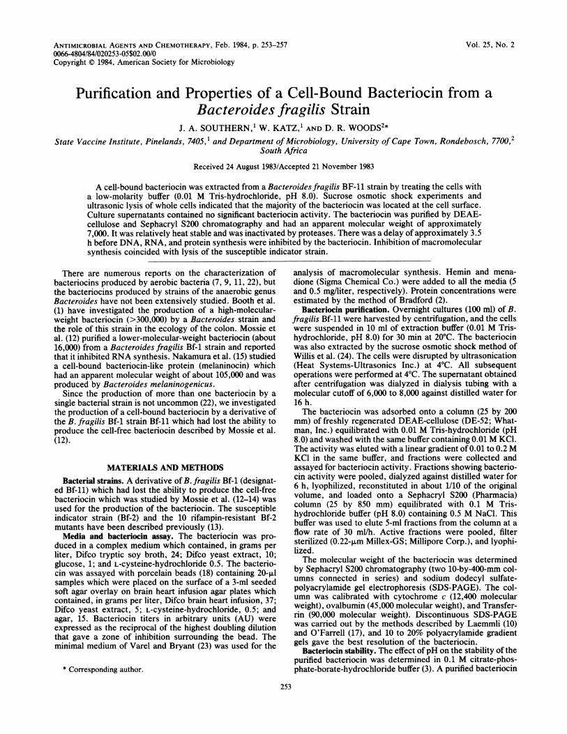
Bf-11 contained very low levels of bacteriocin activity.Relatively high levels of bacteriocin activity were obtainedafter treatment of B. fragilis Bf-11 with the low-molaritybuffer (Table 1). The bacteriocin was eluted from DEAE-cellulose by a 0.01 to 0.2 M KCl gradient and was purifiedfurther by Sephacryl S200 chromatography. SDS-PAGE ofthe purified active preparation revealed a single protein band(Fig. 1 and 2).
Since SDS interfered with the bacteriocin assay, theactivity of the single band after SDS-PAGE could not bedetermined. PAGE of an undenatured sample of the bacte-riocin on a 5 to 15% polyacrylamide gradient (pH 8.25) wascarried out. Under these conditions, the bacteriocin was notresolved as well as on SDS-PAGE, and a broad band whichwas shown to have bacteriocin activity was obtained.A 36-fold purification was achieved after Sephacryl S200
chromatography (Table 1 and Fig. 2). The bacteriocin wastotally retained by dialysis tubing with a molecular cutoff of6,000 to 8,000 and was not detected in the dialysate after 16 hof dialysis. However, it was not retained by dialysis tubingwith a 12,000 to 14,000 molecular weight cutoff and wasestimated to have a molecular weight of 7,200 or 6,400 bySephacryl S200 chromatography and SDS-PAGE, respec-tively. Bacteriocin activity was not obtained in higher-molecular-weight fractions after Sephacryl S200 chromatog-raphy (Fig. 3).
Cellular localization of the bacteriocin. Culture superna-tants of B. fragilis Bf-11 contained very low levels ofbacteriocin (Table 1). The majority of the bacteriocin wasreleased from the cells either by treatment with the low-molarity buffer or by sucrose osmotic shock (Table 1).Treatments of the cells with 20%o sucrose and 2 mM EDTA
FIG. 1. Bacterial extracts analyzed by SDS-polyacrylamide gra-dient (10 to 20%) electrophoresis and stained with Coomassie blueR250. Lane a, Purified and concentrated bacteriocin; lane b, culturesupernatant; lane c, 20% sucrose-2 mM EDTA extract; lane d,osmotic shock fluid after sucrose; lane e, extract of ultrasonicatedwhole cells; lane f, 0.01 M Tris extract; lane g, supernatant fromultrasonic lysate of osmotically shocked (sucrose) cells; lane h,extract of ultrasonicated cells after 0.01 M Tris extraction; lane m,marker proteins ovalbumin, human growth hormone, cytochrome c(bovine), and insulin B chain (molecular weights shown in thou-sands). Equal volumes of the cell extracts described in Table 1 wereapplied to lanes b through h.
ANTIMICROB. AGENTS CHEMOTHER.

CELL-BOUND BACTERIOCIN FROM B. FRAGILIS 255
FIG. 2. SDS-PAGE of extracts before and after purification byDEAE-cellulose and Sephacryl S200 chromatography. Lanes showTris (0.01 M) extract of the B. fragilis Bf-2 indicator strain (a) andthe Bf-11 producer strain (b); fractions showing bacteriocin activityafter DEAE-cellulose (c) and Sephacryl S200 (d) chromatography.MW, Molecular weight (x 103) markers (Pierce Lo-Ranger) aprotinin(6.5), cytochrome c (12.5), and soybean trypsin inhibitor (21.5).Lowest band was associated with fractions which showed bacterio-cin activity. Stained with Biorad silver stain.
did not release the bacteriocin, and the appropriate bacterio-cin band was not present after SDS-PAGE (Fig. 1, lane c).Extraction with the low-molarity buffer resulted in a higherspecific activity of the bacteriocin than extraction by sucroseosmotic shock, which released more contaminating non-bacteriocin proteins (Table 1 and Fig. 1). Examination of thecells by phase-contrast microscopy indicated that they werenot lysed or disintegrated by these extraction procedures.This is also shown by the difference in the protein profilesbetween the extracts and the extracted cells after ultrasoni-cation (Fig. 1, lanes f and h).
Ultrasonicated cell preparations did not result in an in-creased yield of bacteriocin (Table 1). Although ultrasonica-tion disrupted the cells, it did not inactivate the bacteriocin.
Bacteriocin stability. The optimum pH for the stability ofthe purified bacteriocin was between pH 6 and 9. There wasa marked reduction in activity above pH 9 and somereduction in activity below pH 6. The bacteriocin was stableat 50 and 60°C for 5 and 3 h, respectively. At 70°C, there wasan initial 50% loss in activity after 1 h but no further loss inactivity over 4 h. After autoclaving at 121°C for 15 min, 25%of the initial activity was retained.The bacteriocin was inactivated 85% by S. griseus prote-
ase but only 30% by trypsin at a similar concentration.DNase and RNase did not have any affect on activity.Untreated bacteriocin retained its titer after the heat treat-ment used to reduce the activity of the enzymes.
Specificity of the bacteriocin. The bacteriocin susceptibilityof 31 B. fragilis isolates from different clinical samples wasdetermined. These isolates had been characterized as differ-ent from each other on the basis of phage susceptibility,
bacteriocin production, and bacteriocin susceptibility. Ofthese isolates, eight were susceptible to the bacteriocin. B.fragilis ATCC 29771 was resistant to the bacteriocin. Thebacteriocin did not affect isolates of Escherichia coli or ofKlebsiella, Acinetobacter, Pseudomonas, and Staphylococ-cus species.
Since B. fragilis Bf-11 was a derivative of B. fragilis Bf-1,which was shown by Mossie et al. (12-14) to produce anextracellular low-molecular-weight bacteriocin which inhib-ited RNA polymerase activity, it was important to establishthat the cell-bound bacteriocin was a different bacteriocin.Mossie et al. (13) isolated 10 rifampin-resistant B. fragilis Bf-2 mutants from the indicator strain which were shown to beresistant to the bacteriocin described by Mossie et al. (12).These rifampin-resistant mutants were susceptible to thecell-bound bacteriocin, which did not inhibit B. fragilis RNApolymerase in the in vitro RNA polymerase assay.
Nucleic acid and protein synthesis. There was a delay ofapproximately 3.5 h before DNA, RNA, and protein synthe-sis was inhibited at about the same time, and the inhibition ofmacromolecular synthesis coincided with a decrease inturbidity of the cultures (Fig. 4).
DISCUSSIONThe low-molecular-weight bacteriocin we have described
was extracted by suspending the cells in low-molarity bufferand by osmotic shocking. These treatments did not lyse thecells, and it is concluded that the bacteriocin is cell-bound.The inhibitor we have described can be considered to be a
bacteriocin since it is a bactericidal protein with a narrowactivity spectrum and is inactive against the producer strain(9, 22). The B. fragilis bacteriocin differs from most other
z0
IIz
IL0
0
FRACTION NUMBER
FIG. 3. Gel filtration chromatography of bacteriocin prepara-tions on Sephacryl S200. A, purification of a concentrated samplefrom DEAE-cellulose showing percent transmittance at 254 nm (0)and bacteriocin activity (M). B, Molecular weight estimation usingtransferrin (T, 90,000), ovalbumin (0, 45,000), and cytochrome c (C,12,400). The column was eluted with 0.1 M Tris-hydrochloride (pH8.0) containing 0.5 M NaCl at a flow rate of 10 ml/h.
VOL. 25, 1984

256 SOUTHERN, KATZ, AND WOODS
o5
104 i
-J A~~~~~~~I
~0/.
103 '0 1 2 3 4 5 6
TIME hFIG. 4. Effect of bacteriocin on DNA, RNA, and protein synthe-
sis in B. fragilis Bf-2 cultures. Cells were labeled with [14C]thymi-dine with (0) and without (0) bacteriocin; [3H]uracil with (0) andwithout (U) bacteriocin; and [35S]methionine with (A) and without(A) bacteriocin. The bacteriocin was added at 30 min.
bacteriocins reported to date in that it has a very lowmolecular weight (approximately 7,000) (23).The next smallest native Bacteroides bacteriocin reported
is produced by B. fragilis Bf-1 and has a molecular weight ofapproximately 16,000 (12). The 16,000-molecular-weightbacteriocin inhibited RNA polymerase activity. Althoughthe 7,000-molecular-weight bacteriocin is produced by aderivative of the strain which produced the 16,000-molecu-lar-weight bacteriocin, it is a different bacteriocin. The16,000-molecular-weight bacteriocin was detected in culturesupernatants and inhibited RNA synthesis immediately.DNA, RNA, and protein synthesis were inhibited simulta-neously by the 7,000-molecular-weight bacteriocin after a3.5-h delay. Furthermore, this bacteriocin was active againstB. fragilis Bf-2 rifampin-resistant mutants which were isolat-ed from the Bf-2 indicator strain and were resistant to the16,000-molecular-weight bacteriocin. The B. fragilis bacte-riocin described by Booth et al. (1) is completely different inthat it is very large, with a molecular weight of 300,000. The7,000-molecular-weight B. fragilis bacteriocin resembles me-laninocin from B. melaninogenicus in that both bacteriocinswere not detected in culture supernatant and had to beextracted from the cells (15). Other low-molecular-weight
bacteriocins which have been reported are colicin V (4,000molecular weight) and a bacteriocin (5,000 molecular weight)from Staphylococcus aureus (6, 16).Although the mode of action of the 7,000-molecular-
weight bacteriocin has not been identified, the long delaybetween the addition of the bacteriocin to susceptible cellsand the inhibition of macromolecular synthesis indicates thatthe mode of action differs from that of most bacteriocins,which affect some aspect of cell metabolism relatively rapid-ly (within 10 min) (9). Inhibition of macromolecular synthe-sis by the B. fragilis bacteriocin coincides with a decrease inturbidity, and it is suggested that the bacteriocin may be alytic agent. Pesticin acts as a lysozyme and has a protractedeffect on susceptible cells before causing lysis and theinhibition of macromolecular synthesis (5, 8). Colicin M,which also induces lysis of susceptible cells (20), has beenshown to be an inhibitor of murein biosynthesis (21).
LITERATURE CITED1. Booth, S. J., J. L. Johnson, and T. D. Wilkins. 1977. Bacterio-
cin production by strains of Bacteroides isolated from humanfeces and the role of these strains in the bacterial ecology of thecolon. Antimicrob. Agents Chemother. 11:718-724.
2. Bradford, M. 1976. A rapid and sensitive method for thequantitation of microgram quantities of protein utilizing theprincipal of protein-dye binding. Anal. Biochem. 72:248-254.
3. CIBA-GEIGY Corp. 1975. Documenta Geigy, p. 280. In K.Diem and C. Lentner (ed.), Scientific tables, 7th ed. CIBAGEIGY Corp., Basel.
4. Davison, B. L., T. Leighton, and J. C. Rabinowitz. 1979. Purifi-cation of Bacillus subtilis RNA polymerase with Heparin-Agarose. J. Biol. Chem. 254:9220-9226.
5. Ferber, D. M., and R. R. Brubaker. 1979. Mode of action ofpesticin: N-acetylglucosaminidase activity. J. Bacteriol.139:495-501.
6. Frick, K. K., R. L. Quackenbush, and J. Konisky. 1981. Cloningof immunity and structural genes for colicin V. J. Bacteriol.148:498-507.
7. Hardy, K. G. 1975. Colicinogeny and related phenomena. Bac-teriol. Rev. 39:464-515.
8. Hu, P. C., and R. R. Brubaker. 1974. Characterization of pesti-cin: separation of antibacterial activities. J. Biol. Chem.249:4749-4753.
9. Konisky, J. 1982. Colicins and other bacteriocins with estab-lished modes of action. Annu. Rev. Microbiol. 36:125-144.
10. Laemmli, U. K. 1970. Cleavage of structural proteins during theassembly of the head of bacteriophage T4. Nature (London)227:680-685.
11. Mayr-Harting, A., A. J. Hedges, and R. C. W. Berkeley. 1972.Methods for studying bacteriocins. Methods Microbiol. 7A:316-363.
12. Mossie, K. J., D. T. Jones, F. T. Robb, and D. R. Woods. 1979.Characterization and mode of action of a bacteriocin producedby a Bacteroidesfragilis strain. Antimicrob. Agents Chemother.16:724-730.
13. Mossie, K. J., D. T. Jones, F. T. Robb, and D. R. Woods. 1980.Rifampin and bacteriocin resistance in Bacteroides fragilis.Antimicrob. Agents Chemother. 17:838-841.
14. Mossie, K. J., F. T. Robb, D. T. Jones, and D. R. Woods. 1981.Inhibition of ribonucleic acid polymerase by a bacteriocin fromBacteroides fragilis. Antimicrob. Agents Chemother. 20:437-442.
15. Nakamura, T., S. Fujimura, N. Obata, and N. Yamazaki. 1981.Bacteriocin-like substance (melaninocin) from oral Bacteroidesmelaninogenicus. Infect. Immun. 31:28-32.
16. Nakamura, T., N. Yamazaki, H. Taniguchi, and S. Fujimura.1983. Production, purification, and properties of a bacteriocinfrom Staphylococcus aureus isolated from saliva. Infect. Im-mun. 39:609-614.
17. O'Farrell, P. H. 1975. High resolution two-dimensional electro-phoresis of proteins. J. Biol. Chem. 250:4007-4021.
I I I- I v a
aJII
JII
II
I
II
I
I
I
I.. ilI
I.. .AI
I
IJ
0-
ANTIMICROB. AGENTS CHEMOTHER.

CELL-BOUND BACTERIOCIN FROM B. FRAGILIS 257
18. Richardson, H., A. H. Emslie-Smith, and B. W. Senior. 1968.Agar diffusion method for the assay of colicins. Appl. Microbi-ol. 16:1468-1474.
19. Robb, S. M., D. R. Woods, F. T. Robb, and J. K. Struthers.1977. Rifampicin-resistant mutant supporting bacteriophagegrowth on stationary phase Achromobacter cells. J. Gen. Virol.35:117-123.
20. Schalier, K., R. Dreher, and V. Braun. 1981. Structural andfunctional properties of colicin M. J. Bacteriol. 146:54-63.
21. Schaller, K., J.-V. Holtje, and V. Braun. 1982. Colicin M is an
inhibitor of murein biosynthesis. J. Bacteriol. 152:994-1000.22. Tagg, J. R., A. S. Dajani, and L. W. Wannamaker. 1976. Bacte-
riocins of gram-positive bacteria. Bacteriol. Rev. 40:722-756.23. Varel, V. H., and M. P. Bryant. 1974. Nutritional features of
Bacteroides fragilis subsp. fragilis. Appl. Microbiol. 28:251-257.
24. Willis, R. C., R. G. Morris, C. Cirakoglu, G. D. Schelienberg,N. H. Gerber, and C. E. Furlong. 1974. Preparation of theperiplasmic binding proteins from Salmonella typhimurium andEscherichia coli. Arch. Biochem. Biophys. 161:64-75.
VOL. 25, 1984