PUOs and Viral Diagnosis - Virology Research · PDF filePUOs and Viral Diagnosis Alison M...
50
PUOs and Viral Diagnosis Alison M Kesson Virologist, Microbiologist and Paediatric Infectious Diseases Physician Conjoint Assoc Professor of Paediatrics, University of Sydney
Transcript of PUOs and Viral Diagnosis - Virology Research · PDF filePUOs and Viral Diagnosis Alison M...

PUOs and Viral Diagnosis
Alison M Kesson Virologist, Microbiologist and Paediatric
Infectious Diseases PhysicianConjoint Assoc Professor of Paediatrics,
University of Sydney

PUO• “Fever higher than 38.3oC on several
occasions persisting without diagnosis for at least 3 weeks in spite of at least 1 weeks investigation in hospital.”
– Petersdorf and Beeson 1961

PUO• Arbitrary definition but useful.• The exacting definition is historic and not
rigorously applied.• Now there is “Fever Without Source”
(FWS) for a fever of recent onset without explanation determined by history or examination.
• PUO is fever for 8 days with no obvious source after initial investigation.

FWS vs PUO• Overlap exists• Frequent cause of one may not be frequent
cause in the other.• Recent onset of fever usually warrants more
immediate evaluation than PUO.• PUO rarely presents as an emergency but does
require timely, not urgent, diagnosis and treatment.
• Empiric antibiotics often indicated in FWS but not often in PUO.

Fever Without Source• FWS is fever for 1 week with no obvious
source.• 5-10% with up to 20% of children • Peak incidence is 2nd year of life• Many are self limiting infectious diseases• Some in prodromal phase of specific
illness eg sinusitis, hepatitis, mononucleosis.
• May be indicator of Kawasaki disease

PUO• Five main etiological categories
– Infection– Neoplasm– Connective Tissue Disease– Miscellaneous– Undiagnosed

“Subtypes” of PUO• Community acquired• Nosocomial• Immunodeficient• HIV-related

Approach to Diagnosis of PUO• History for Community Acquired PUO
– Duration and pattern of fever– Age of patient– Sexual History– Contact with other ill people– Vaccination history– Travel History– Animal, insect exposure– Previous medical treatment inc blood products

Approach to Diagnosis of PUO• Physical Exam - extras
– Sinus tenderness– Mouth ulceration– Fundi – chorioretinitis– Chest – pneumonitis– Abdomen – hepatic tenderness, splenomegaly– Lymphadenopathy– Arthralgia / Arthritis– Rash

Approach to Diagnosis of PUO• Investigations
– FBC and differential– EUC, LFTs, Amylase, CRP, ESR– Urinalysis + culture– Investigation for infectious causes guided by
history and exam– Consider Rh F, ANA , ANCA- Other investigations and imaging as indicated

Viral Causes in CA-PUO• Mononucleosis – EBV, CMV• Zoster• Enterovirus • Adenovirus• Respiratory viruses – parainfluenza, RSV• Hepatitis – A,B,C,D,E• Alphaviruses – Ross River, Barmah Forest virus.
Sindbis• Flaviviruses - Dengue

EBV rash

Mononucleosis• Prodrome of 2 to 5 days• Acute phase
– Fever, sore throat, malaise, fatigue– Fever may last 4 to 5 weeks– Lymphadenopathy (80%), hepatomegaly
(60%), splenomegaly (50%).– EBV causes 80-95% others usually CMV– Children <5 usually heterophile antibody
negative.

Mononucleosis• CMV induced mononucleosis• Less common
– Sore throat– Lymphadenopathy– Atypical lymphocytes
• Prominent splenomegaly

Adenoviruses
• ds DNA virus• non-enveloped• At least 52 serotypes are
known• classified into 6
subgenera: A to F

Adenovirus syndromes• 1. Pharyngitis 1, 2, 3, 5, 7
2. Pharyngoconjunctival fever 3, 7 3. Acute respiratory disease 4, 7, 14, 21 4. Pneumonia 1, 2, 3, 7 5. Follicular conjunctivitis 3, 4, 11 6. Epidemic keratoconjunctivitis 8, 19, 37 7. Pertussis-like syndrome 5 8. Acute haemorrhagic cystitis 11, 21 9. Acute infantile gastroenteritis 40, 41 10. Intussusception 1, 2, 5 11. Severe disease in AIDS and other immunocompromised patients 5, 34, 35 12. Meningitis 3, 7

Enterovirus Infections• Enter and replicate in the GIT• Prevalent summer and autumn• Cause disease in variety of organs
– Meningitis / encephalitis– Myocarditis– URTI, pneumonia– Hepatitis– Vomiting and diarrhoea – non-specific

Zoster in 2 year old

Zoster• Zoster in children can be difficult to
diagnose if there is no history of chickenpox.
• Maternal chickenpox in pregnancy is assoc with increased risk of zoster in young children.
• Zoster is usually less severe and much less associated pain.

Roseola• Roseola infantum is a common acute illness of
young children characterised by a fever of 3-5 days duration, rapid defervescence and then the appearance of an erythematous macular or maculopapular rash that persists for 1-2 days
• The most important complication are convulsions and other neurological symptoms.

Roseola• It is estimate that HHV-6 - 73.5%, HHV-7 -
10.2% and other - 16.2% of roseola cases.
• HHV6 appears to be the major cause of roseola.

Roseola infantum – HHV-6
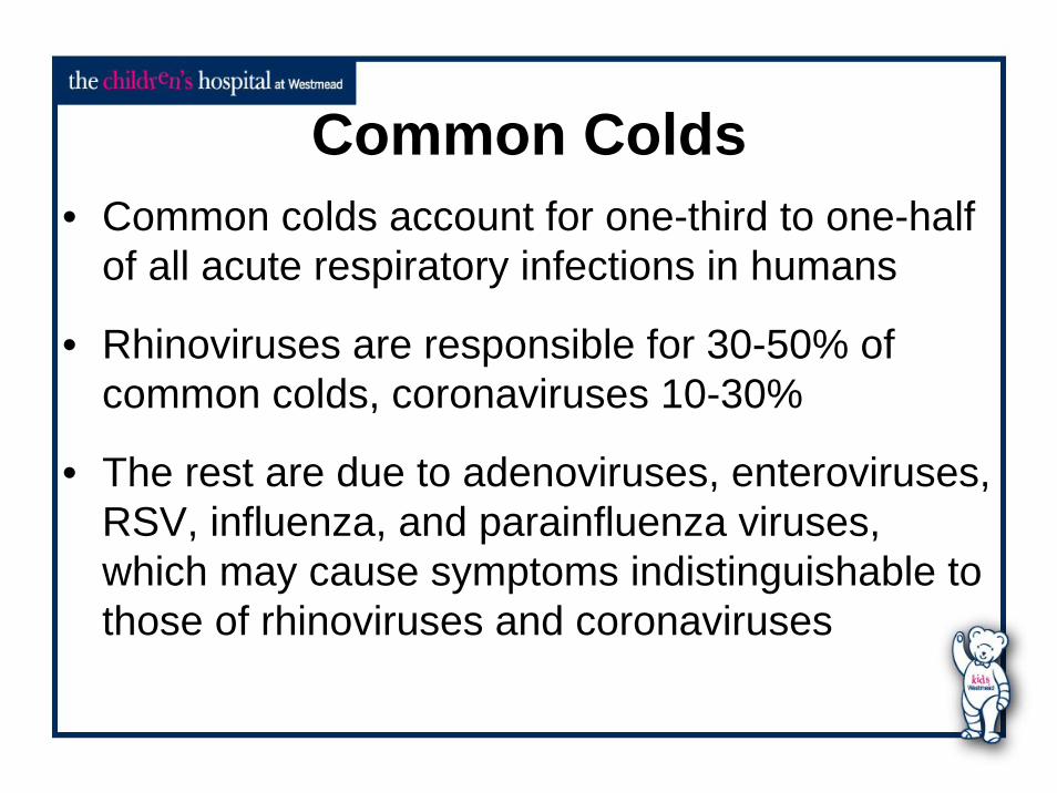
Common Colds• Common colds account for one-third to one-half
of all acute respiratory infections in humans
• Rhinoviruses are responsible for 30-50% of common colds, coronaviruses 10-30%
• The rest are due to adenoviruses, enteroviruses, RSV, influenza, and parainfluenza viruses, which may cause symptoms indistinguishable to those of rhinoviruses and coronaviruses

Hepatitis A• Worldwide endemicity of HAV varies within
and between countries.• Clinical presentation varies with age of
patient and reflects endemicity.• Infection in childhood (pre-adolescence)
often asymptomatic or mild

Hepatitis A• Can be non-specific with little or no
jaundice.• Diagnosis:
– raised LFTs– HAV IgM pos– HAV PCR
• Protection by passive or active vaccination

Hepatitis A virus
Hepatitis B• Non-specific prodrome of fever, malaise and
nausea.• Clinical features non-specific and inc jaundice
variable.• Manifestations and outcome variable by age.• Diagnosis
– Serology– HBV DNA PCR
• Protection by passive or active vaccination

Hepatitis B virus

Hepatitis E Virus• Similar incubation, prodroma and clinical
presentation to hepatitis A• History of travel to endemic area• Diagnosis by serology• Mortality of 10 to 20% in pregnant women

Hepatitis E Virus

Hepatitis E Virus

Nosocomial Acquired PUO• Bacterial causes
– Pneumonia, wound infection, UTI, IV lines etc• Viral causes
– Respiratory viruses– Hepatitis A, parvovirus, CMV from blood
products

PUO in Immunodeficient patients• Neutropenic patients not responding to
antibiotics– CMV, HHV-6– EBV and PTLD– HSV– VZV– Resp viruses – Parainfluenza, Influenza, RSV– Blood products – CMV, Parvovirus, Hep A.

PUO in Immunodeficient patients• The probability of viral infection and type
of viral infection is related to the degree of immunosuppression.
• Disease is a function of both infection and the immune response.
• PUO due to viral infection common in this group as they have difficulty clearing infection and partial immunity may confound the clinical picture.

Timing of most frequent virus infections post-transplantation

Solid Organ Transplants - CMV• Solid organ transplants in children, unlike
in adults, are often CMV mismatched – CMV-P/ CMV+D
• CMV prophylaxis given but seroconversion occurs often with non-specific febrile illness and difficult to diagnose.
• Severity of CMV disease related to transplant with renal<liver<lung<BMT.

Solid Organ Transplants - CMV• CMV infection often associated with
rejection and a careful balance between immunosuppression and CMV therapy must be achieved.
• May progress to pneumonitis with significant morbidity / mortality if unrecognised.
• Diagnosis by serology, culture, p65 antigenaemia and/or CMV PCR.

Solid Organ Transplants - EBV• Post-transplant lymphoproliferative
disorder (PTLD) is due to EBV.• Due to uncontrolled proliferation of EBV
infected B cells where cellular immunity is suppressed.

Solid Organ Transplants - EBV• EBV – PTLP presents as
– infectious mononucleosis like syndrome or – fever and lymphomatous infiltrate into lymph
nodes, spleen, liver, lung, brain, intestine, kidney, bone marrow.

Solid Organ Transplants - EBV• EBV-PTLD more common in transplant
patients with severe T cell immuno-suppression– BMT– Anti-thymocyte globulin– Primary EBV after solid organ transplant.
• Can occur in any patient with high dose immuno-suppressive therapy or inherited T cell deficiency.

Solid Organ Transplants - EBV• Diagnosis
– EBV seroconversion – insensitive– EBV PCR – qualitative or quantitative

Solid Organ Transplants - HSV• If unrecognised and untreated mouth ulceration
may extend to oesophagus and lung.• Asymptomatic shedding with systemic disease
e.g. hepatitis• Extensive visceral involvement of GIT, liver,
bone marrow, adrenals.• Genital infection.• Aciclovir prophylaxis decreases incidence but
resistance can develop.

Solid Organ Transplants – HHV-6
• Most individuals infected with HHV-6 by 2 years.
• HHV-6 detected in immunocompromised individuals often associated with CMV .
• Difficult to assign disease to HHV-6• Evidence of disease in patients with high
HHV-6 viral loads by PCR testing.

Solid Organ Transplants – HHV-7• Biologically and epidemiologically related
to HHV-6 implies they should share disease manifestations.
• Sporadic descriptions of encephalitis, hepatitis and pityriasis rosea.
• May be broad undefined spectrum of disease.
• No strong evidence of disease

Solid Organ Transplants • Blood transfusion risks
– HCV, HBV, HIV, HTLV-1/2 and CMV low risk with screened blood and products.
– Hep A and West Nile virus (USA).– Parvovirus with significant haemopoietic
suppression

PUO in BMT Patients• In children who have MUD BMT or T cell
depleted allografts there is a high risk of disseminated disease from adenovirus infections.
• Present as:– Haemorrhagic cystitis– Persistent diarrhoea– Pneumonitis– Hepatitis– Encephalitis - rarely

Fever in HIV patients• Primary HIV infection – mononucleosis like
syndrome• Opportunistic Infections• Immune reconstitution disease.

Fever in HIV patients• CMV• B cell lymphomas very similar to PTLD.• All CNS lymphomas are EBV pos. Others
are 60-70% EBV pos

Summary• History and Physical Exam• Consider immunological status• Consider non-infectious causes.• Be strategic in approach to optimise
predictive value of tests.

Thank You