Pulmonary thromboembolism due to severe … · · 2013-03-18 115 Cho KH, et al. Pulmonary...
4
LETTER TO THE EDITOR Korean J Intern Med 2013;28:112-115 http://dx.doi.org/10.3904/kjim.2013.28.1.112 Copyright © 2013 The Korean Association of Internal Medicine This is an Open Access article distributed under the terms of the Creative Commons Attribution Non-Commercial License (http://creativecommons.org/licenses/ by-nc/3.0/) which permits unrestricted noncommercial use, distribution, and reproduction in any medium, provided the original work is properly cited. pISSN 1226-3303 eISSN 2005-6648 http://www.kjim.org Heart Research Center, Chonnam National University Hospital, Gwangju, Korea Pulmonary thromboembolism due to severe hyperhomocysteinemia associated with a methyltetrahydrofolate reductase mutation Kyung Hoon Cho, Myung Ho Jeong, Doo Sun Sim, Young Joon Hong, Ju Han Kim, Youngkeun Ahn, and Jung Chaee Kang Received: September 7, 2009 Revised : October 27, 2009 Accepted: November 16, 2009 Correspondence to Myung Ho Jeong, M.D. Heart Research Center, Chonnam National University Hospital, 42 Jebong-ro, Dong-gu, Gwangju 501-757, Korea Tel: +82-62-220-6243 Fax: +82-62-227-3105 E-mail: [email protected] To the Editor, Pulmonary thromboembolism (PTE) ranges from asymptomatic, inciden- tally discovered emboli to massive emboli causing immediate death. Treatment can reduce the risk of death, and appropriate primary pro- phylaxis is usually effective. Certain risk factors increase the likelihood for acute deep venous thrombosis and thus PTE. Knowing the risk factors for a given patient should help the physician choose appropriate diag- nostic and prophylactic strategies. Elevated levels of homocysteine (Hcy) are associated with an increased risk for thrombosis [1,2]. We present a case of PTE due to severe hyperhomo- cysteinemia. A 66-year-old man was admitted to our hospital because of dyspnea on mild exertion for 3 days. He was a smoker and had a 10-year history of hypertension with medication. He had no known history of diabetes, pulmonary tuberculosis, or hepatitis. He had no medication history except for antihypertensive agents. His fam- ily history was non-specific. On ar- rival, his vital signs included temper- ature, 36.3°C; blood pressure, 140/90 mmHg; pulse rate, 88 beats/min; and respiratory rate, 32 beats/min. The patient showed an acutely ill appearance at the time of admission. On physical examination, he had in- creased jugular venous pressure of 10 cm H 2 O. Auscultation in the left up- per sternal area revealed a normal S1 and a loud P 2 with a grade IV systolic ejection murmur in the pulmonic area. No abdominal tenderness or organomegaly was noted. His neu- rological examination was normal. Peripheral edema was not observed. Arterial blood gas analysis revealed pH, 7.429; pCO 2 , 33.6 mmHg; pO 2 , 72.2 mmHg; HCO 3 - , 21.8 mmol/L; and O 2 saturation, 94.5%. His complete blood cell count results were white cells, 11,200/mm 3 ; hemoglobin, 14.5 mg/dL; hematocrit, 40.8%; and plate- lets, 157,000/mm 3 . The coagulation profile included activated partial- thromboplastin time, 39.2 seconds (range, 26.5 to 41); prothrombin time, 11.8 seconds (range, 9.8 to 13); fibrino- gen, 284.9 mg/dL (range, 180 to 350); fibrinogen degradation products, 8.9 µg/mL (range, 0 to 5); and D-dimer, 0.72 mg/L (range, 0 to 0.3). His creati- nine level had increased to 1.9 mg/dL (range, 0.5 to 1.2). The lipid profile re- sults showed low density lipoprotein- cholesterol, 121 mg/dL (range, 0 to 120); triglycerides, 133 mg/dL (range,
Transcript of Pulmonary thromboembolism due to severe … · · 2013-03-18 115 Cho KH, et al. Pulmonary...

LETTER TO THE EDITOR
Korean J Intern Med 2013;28:112-115http://dx.doi.org/10.3904/kjim.2013.28.1.112
Copyright © 2013 The Korean Association of Internal MedicineThis is an Open Access article distributed under the terms of the Creative Commons Attribution Non-Commercial License (http://creativecommons.org/licenses/by-nc/3.0/) which permits unrestricted noncommercial use, distribution, and reproduction in any medium, provided the original work is properly cited.
pISSN 1226-3303eISSN 2005-6648
http://www.kjim.org
Heart Research Center, Chonnam National University Hospital, Gwangju, Korea
Pulmonary thromboembolism due to severe hyperhomocysteinemia associated with a methyltetrahydrofolate reductase mutationKyung Hoon Cho, Myung Ho Jeong, Doo Sun Sim, Young Joon Hong, Ju Han Kim, Youngkeun Ahn, and Jung Chaee Kang
Received: September 7, 2009Revised : October 27, 2009Accepted: November 16, 2009
Correspondence to Myung Ho Jeong, M.D.Heart Research Center, Chonnam National University Hospital, 42 Jebong-ro, Dong-gu, Gwangju 501-757, KoreaTel: +82-62-220-6243 Fax: +82-62-227-3105 E-mail: [email protected]
To the Editor,Pulmonary thromboembolism (PTE) ranges from asymptomatic, inciden-tally discovered emboli to massive emboli causing immediate death. Treatment can reduce the risk of death, and appropriate primary pro-phylaxis is usually effective. Certain risk factors increase the likelihood for acute deep venous thrombosis and thus PTE. Knowing the risk factors for a given patient should help the physician choose appropriate diag-nostic and prophylactic strategies.
Elevated levels of homocysteine (Hcy) are associated with an increased risk for thrombosis [1,2]. We present a case of PTE due to severe hyperhomo-cysteinemia.
A 66-year-old man was admitted to our hospital because of dyspnea on mild exertion for 3 days. He was a smoker and had a 10-year history of hypertension with medication. He had no known history of diabetes, pulmonary tuberculosis, or hepatitis. He had no medication history except for antihypertensive agents. His fam-ily history was non-specif ic. On ar-rival, his vital signs included temper-ature, 36.3°C; blood pressure, 140/90 mmHg; pulse rate, 88 beats/min; and respiratory rate, 32 beats/min.
The patient showed an acutely ill appearance at the time of admission. On physical examination, he had in-creased jugular venous pressure of 10 cm H2O. Auscultation in the left up-per sternal area revealed a normal S1 and a loud P2 with a grade IV systolic ejection murmur in the pulmonic area. No abdominal tenderness or organomegaly was noted. His neu-rological examination was normal. Peripheral edema was not observed. Arterial blood gas analysis revealed pH, 7.429; pCO2, 33.6 mmHg; pO2, 72.2 mmHg; HCO3
-, 21.8 mmol/L; and O2 saturation, 94.5%. His complete blood cell count results were white cells, 11,200/mm3; hemoglobin, 14.5 mg/dL; hematocrit, 40.8%; and plate-lets, 157,000/mm3. The coagulation prof ile included activated partial-thromboplastin time, 39.2 seconds (range, 26.5 to 41); prothrombin time, 11.8 seconds (range, 9.8 to 13); fibrino-gen, 284.9 mg/dL (range, 180 to 350); fibrinogen degradation products, 8.9 µg/mL (range, 0 to 5); and D-dimer, 0.72 mg/L (range, 0 to 0.3). His creati-nine level had increased to 1.9 mg/dL (range, 0.5 to 1.2). The lipid profile re-sults showed low density lipoprotein-cholesterol, 121 mg/dL (range, 0 to 120); triglycerides, 133 mg/dL (range,
113www.kjim.orghttp://dx.doi.org/10.3904/kjim.2013.28.1.112
Cho KH, et al. Pulmonary thromboembolism and hyperhomocysteinemia
50 to 150); and lipoprotein(a), 10 mg/dL (range, 0 to 30). The levels of cardiac biomarkers were elevated: tropo-nin I, 0.35 ng/mL (range, 0 to 0.05); C-reactive protein, 1.6 mg/dL (range, 0.1 to 1); and N-terminal prohor-mone brain natriuretic peptide, 3,106 pg/mL (range, < 262). The estimated creatinine clearance rate using the Cockcroft-Gault formula was 33.92 mL/min/1.73 m2. An electrocardiogram revealed sinus rhythm at a rate of 65 beats per minute and ST-T segment changes over leads III, aVF, and V1–3. Chest radiography revealed no definitive abnormalities such as cardiomegaly, pulmo-nary congestion, or pneumonic infiltration.
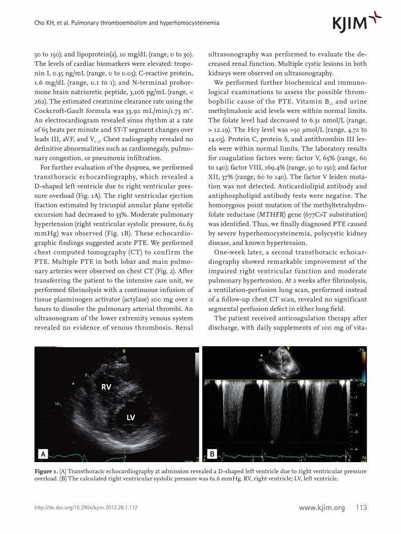
For further evaluation of the dyspnea, we performed transthoracic echocardiography, which revealed a D-shaped left ventricle due to right ventricular pres-sure overload (Fig. 1A). The right ventricular ejection fraction estimated by tricuspid annular plane systolic excursion had decreased to 33%. Moderate pulmonary hypertension (right ventricular systolic pressure, 61.63 mmHg) was observed (Fig. 1B). These echocardio-graphic findings suggested acute PTE. We performed chest computed tomography (CT) to conf irm the PTE. Multiple PTE in both lobar and main pulmo-nary arteries were observed on chest CT (Fig. 2). After transferring the patient to the intensive care unit, we performed fibrinolysis with a continuous infusion of tissue plasminogen activator (actylase) 100 mg over 2 hours to dissolve the pulmonary arterial thrombi. An ultrasonogram of the lower extremity venous system revealed no evidence of venous thrombosis. Renal
ultrasonography was performed to evaluate the de-creased renal function. Multiple cystic lesions in both kidneys were observed on ultrasonography.
We performed further biochemical and immuno-logical examinations to assess the possible throm-bophilic cause of the PTE. Vitamin B12 and urine methylmalonic acid levels were within normal limits. The folate level had decreased to 6.31 nmol/L (range, > 12.19). The Hcy level was >50 µmol/L (range, 4.72 to 14.05). Protein C, protein S, and antithrombin III lev-els were within normal limits. The laboratory results for coagulation factors were: factor V, 65% (range, 60 to 140); factor VIII, 169.4% (range, 50 to 150); and factor XII, 37% (range, 60 to 140). The factor V leiden muta-tion was not detected. Anticardiolipid antibody and antiphospholipid antibody tests were negative. The homozygous point mutation of the methyltetrahydro-folate reductase (MTHFR) gene (677C>T substitution) was identified. Thus, we finally diagnosed PTE caused by severe hyperhomocysteinemia, polycystic kidney disease, and known hypertension.
One-week later, a second transthoracic echocar-diography showed remarkable improvement of the impaired right ventricular function and moderate pulmonary hypertension. At 2 weeks after fibrinolysis, a ventilation-perfusion lung scan, performed instead of a follow-up chest CT scan, revealed no significant segmental perfusion defect in either lung field.
The patient received anticoagulation therapy after discharge, with daily supplements of 100 mg of vita-
Figure 1. (A) Transthoracic echocardiography at admission revealed a D-shaped left ventricle due to right ventricular pressure overload. (B) The calculated right ventricular systolic pressure was 61.6 mmHg. RV, right ventricle; LV, left ventricle.
A B

114 www.kjim.org http://dx.doi.org/10.3904/kjim.2013.28.1.112
The Korean Journal of Internal Medicine Vol. 28, No. 1, January 2013
min B6 and 2 mg of folate to prevent recurrence of the PTE. The serum level of Hcy decreased to 12.05 µmol/L, and PTE had not recurred after 6 months of clinical follow-up.
A recent revolution has occurred in the field of ve-nous thromboembolism (VTE), which encompasses deep venous thrombosis and PTE. VTE-related deaths have been estimated at 300,000 annually in the United States. Although less common in certain regions such as Asia, VTE is a worldwide problem, particularly in people with known risk factors.
Hcy is a sulf hydryl amino acid derived from the metabolic conversion of methionine, which is depen-dent on vitamins (folic acid, B12, and B6) as cofactors or cosubstrates. It is clear that hyperhomocysteinemia promotes atherosclerosis and thrombosis in suscepti-ble animal models, but the pathophysiological mecha-nisms of these effects are less well understood. The overall prevalence of hyperhomocysteinemia in the total population is 5% to 7%. Several clinical studies have reported that elevated levels of Hcy are associated with an increased risk for coronary artery disease and VTE [1,2]. A meta-analysis found that for every increase of 5 µmol/L in plasma Hcy concentration, there was an average increase of 60% (odds ratio, 16; 95% confidence interval, 1.1 to 2.34) in the incidence of VTE [2].
Plasma levels of Hcy increase with age, are lower in fertile women than in men, and increase after meno-pause. Major determinants of plasma Hcy levels in-
clude genetic abnormalities, diet (vitamin B2, B6, B12, and folate intake), renal function, cigarette smoking, and coffee and tea consumption.
The most frequent cause of severe hyperhomocyste-inemia (characterized by fasting plasma levels of Hcy > 100 µmol/L) is a homozygous deficiency of cystathione β-synthase. The prevalence of this deficiency in the general population is approximately one in 335,000, varying between one in 65,000 (Ireland), and one in 900,000 (Japan). Approximately 5% to 10% of severe hyperhomocysteinemia cases are caused by inherited defects in remethylation. The homozygous point mu-tation of MTHFR (677C>T substitution), which cata-lyzes the reduction of methylene-tetrahydrofolate to methyltetrahydrofolate, is the most common inherited defect of the remethylation pathway.
The homozygous MTHFR point mutation leads to a thermolabile enzyme variant with decreased basal enzymatic activity. It has been found in a large pro-portion of both normal subjects (5%) and patients with vascular disease (17%) in Western countries [3]. Moon et al. [4] reported that the proportion of patients with a homozygous MTHFR C677T gene mutation in Korea was 14% to 20%. Homozygous Korean patients with the MTHFR mutation and coronary artery disease had significantly higher fasting Hcy levels than those in the wild-type enzyme (homozygotes, 18.83 ± 6.37 µmol/L; wild type, 12.36 ± 3.21 µmol/L; p < 0.01) [4]. Three me-ta-analyses related to the association between MTHFR
Figure 2. Chest computed tomography at admission revealed multiple pulmonary thromboemboli in the right lobar (A, arrow) and both main (B, arrows) pulmonary arteries.
A B

115www.kjim.orghttp://dx.doi.org/10.3904/kjim.2013.28.1.112
Cho KH, et al. Pulmonary thromboembolism and hyperhomocysteinemia
677TT (homozygous deficiency) and VTE showed that the TT genotype is associated with increased risk for VTE compared with the wild-type homozygous geno-type [2].
It is unclear whether correcting mild to moder-ate hyperhomocysteinemia reduces the risk for VTE. However, there is clear evidence that the very high VTE risk of patients with severe hyperhomocystein-emia is due to cystathione β-synthase deficiency; this deficiency decreases dramatically with vitamin sup-plementation, thereby lowering the very high plasma Hcy levels [5]. No side effects have been reported in tri-als in which folic acid with or without other vitamins were used for various indications.
In our patient, severe hyperhomocysteinemia was the most possible causative factor for the PTE. The severe hyperhomocysteinemia was associated with old age, smoking, folate deficiency, renal insufficiency, and a homozygous MTHFR C677T gene mutation. With the administration of folate and vitamin B6, the Hcy level decreased to 12.05 µmol/L within 3 months, and the patient had no evidence of PTE recurrence during a 6-month clinical follow-up.
This patient with PTE due to severe hyperhomocys-teinemia associated with the MTHFR gene mutation was successfully managed with antithrombotics, fo-late, and vitamin B6. Although it is uncertain whether treatment with vitamins in patients with mild to moderate hyperhomocysteinemia decreases the risk for PTE, we should bear in mind that a patient with a PTE due to severe hyperhomocysteinemia precipitated by a homozygous MTHFR C677T gene mutation can be successfully managed with inexpensive and safe vita-min supplementation.
Keywords: Pulmonary embolism; Hyperhomocystein-emia; Folate
Conflict of interestNo potential conflict of interest relevant to this article is reported.
REFERENCES
1. den Heijer M, Koster T, Blom HJ, et al. Hyperhomocys-teinemia as a risk factor for deep-vein thrombosis. N Engl J Med 1996;334:759-762.
2. Den Heijer M, Lewington S, Clarke R. Homocysteine, MTHFR and risk of venous thrombosis: a meta-analysis of published epidemiological studies. J Thromb Hae-most 2005;3:292-299.
3. Kang SS, Wong PW, Susmano A, Sora J, Norusis M, Rug-gie N. Thermolabile methylenetetrahydrofolate reduc-tase: an inherited risk factor for coronary artery disease. Am J Hum Genet 1991;48:536-545.
4. Moon KW, Chung WS, Youn HJ, et al. The frequency distribution of methylenetetrahydrofolate reductase (MTHFR) polymorphism and association between the genotypes and total homocysteine level in patients with coronary artery disease. Korean Circ J 1999;29:781-787.
5. Yap S, Boers GH, Wilcken B, et al. Vascular outcome in patients with homocystinuria due to cystathionine beta-synthase deficiency treated chronically: a multi-center observational study. Arterioscler Thromb Vasc Biol 2001;21:2080-2085.