Pulmonary Function Testing Function Tests … · • 6 Minute walk test: ... disease progression,...
51
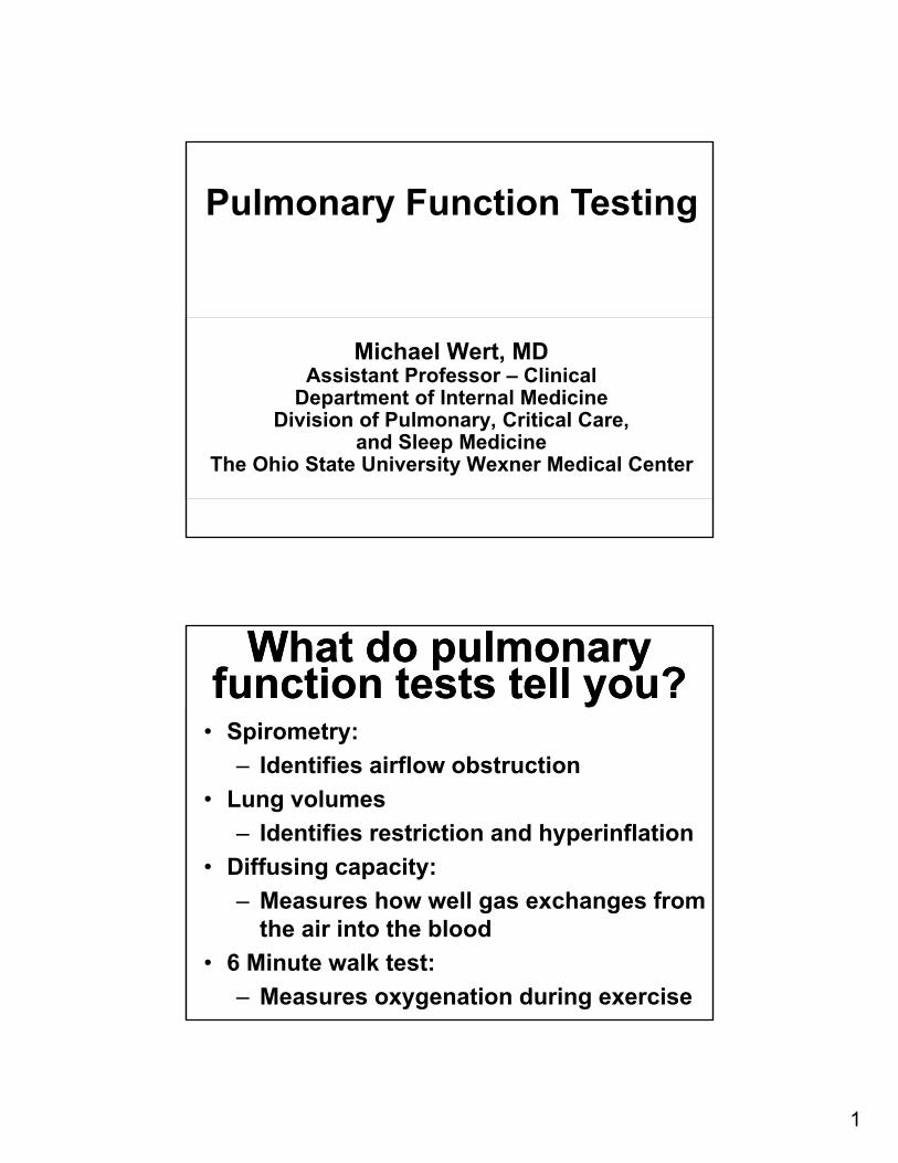
1 Michael Wert, MD Assistant Professor – Clinical Department of Internal Medicine Division of Pulmonary, Critical Care, and Sleep Medicine The Ohio State University Wexner Medical Center Pulmonary Function Testing What do pulmonary function tests tell you? What do pulmonary function tests tell you? • Spirometry: ‒ Identifies airflow obstruction • Lung volumes ‒ Identifies restriction and hyperinflation • Diffusing capacity: ‒ Measures how well gas exchanges from the air into the blood • 6 Minute walk test: ‒ Measures oxygenation during exercise
Transcript of Pulmonary Function Testing Function Tests … · • 6 Minute walk test: ... disease progression,...
1
Michael Wert, MDAssistant Professor – Clinical
Department of Internal MedicineDivision of Pulmonary, Critical Care,
and Sleep MedicineThe Ohio State University Wexner Medical Center
Pulmonary Function Testing
What do pulmonary function tests tell you?
What do pulmonary function tests tell you?
• Spirometry:
‒ Identifies airflow obstruction
• Lung volumes
‒ Identifies restriction and hyperinflation
• Diffusing capacity:
‒ Measures how well gas exchanges from the air into the blood
• 6 Minute walk test:
‒ Measures oxygenation during exercise
2
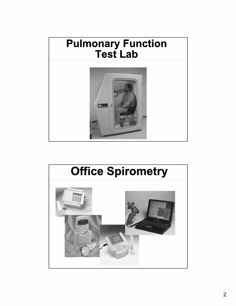
Pulmonary Function Test Lab
Pulmonary Function Test Lab
Office SpirometryOffice Spirometry
3
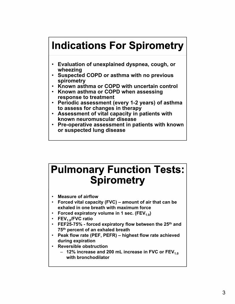
Indications For SpirometryIndications For Spirometry
• Evaluation of unexplained dyspnea, cough, or wheezing
• Suspected COPD or asthma with no previous spirometry
• Known asthma or COPD with uncertain control• Known asthma or COPD when assessing
response to treatment• Periodic assessment (every 1-2 years) of asthma
to assess for changes in therapy• Assessment of vital capacity in patients with
known neuromuscular disease• Pre-operative assessment in patients with known
or suspected lung disease
Pulmonary Function Tests:Spirometry
Pulmonary Function Tests:Spirometry
• Measure of airflow• Forced vital capacity (FVC) – amount of air that can be
exhaled in one breath with maximum force• Forced expiratory volume in 1 sec. (FEV1.0)• FEV1.0/FVC ratio• FEF25-75% - forced expiratory flow between the 25th and
75th percent of an exhaled breath• Peak flow rate (PEF, PEFR) – highest flow rate achieved
during expiration• Reversible obstruction
‒ 12% increase and 200 mL increase in FVC or FEV1.0
with bronchodilator
4
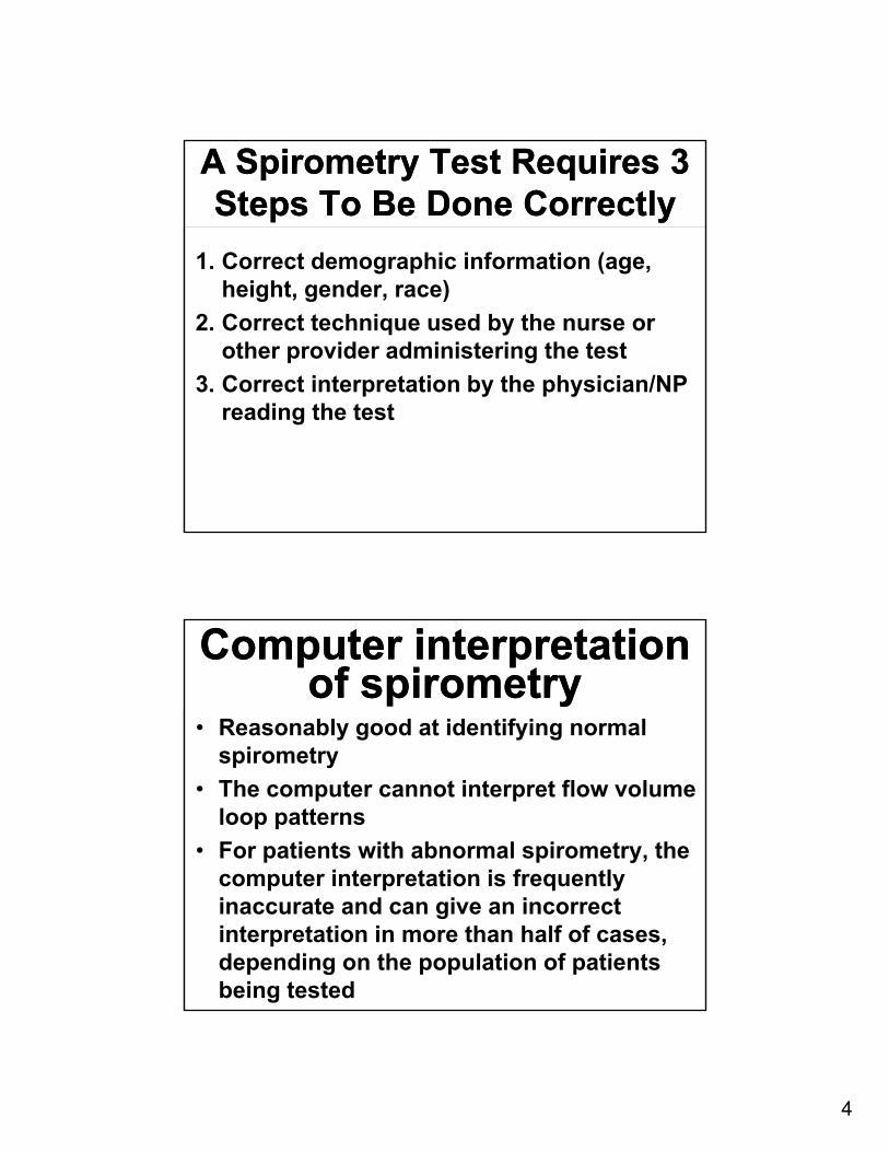
A Spirometry Test Requires 3 Steps To Be Done Correctly
A Spirometry Test Requires 3 Steps To Be Done Correctly
1. Correct demographic information (age, height, gender, race)
2. Correct technique used by the nurse or other provider administering the test
3. Correct interpretation by the physician/NP reading the test
Computer interpretation of spirometry
Computer interpretation of spirometry
• Reasonably good at identifying normal spirometry
• The computer cannot interpret flow volume loop patterns
• For patients with abnormal spirometry, the computer interpretation is frequently inaccurate and can give an incorrect interpretation in more than half of cases, depending on the population of patients being tested
5
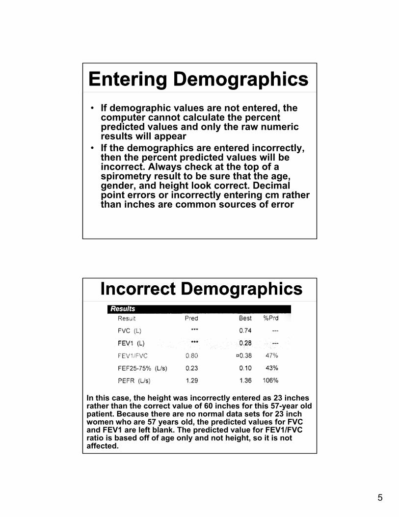
Entering DemographicsEntering Demographics• If demographic values are not entered, the
computer cannot calculate the percent predicted values and only the raw numeric results will appear
• If the demographics are entered incorrectly, then the percent predicted values will be incorrect. Always check at the top of a spirometry result to be sure that the age, gender, and height look correct. Decimal point errors or incorrectly entering cm rather than inches are common sources of error
Incorrect DemographicsIncorrect Demographics
In this case, the height was incorrectly entered as 23 inches rather than the correct value of 60 inches for this 57-year old patient. Because there are no normal data sets for 23 inch women who are 57 years old, the predicted values for FVC and FEV1 are left blank. The predicted value for FEV1/FVC ratio is based off of age only and not height, so it is not affected.
6
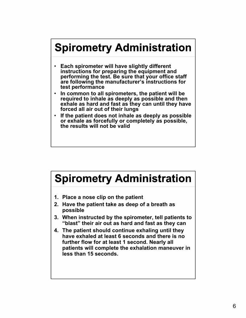
Spirometry AdministrationSpirometry Administration
• Each spirometer will have slightly different instructions for preparing the equipment and performing the test. Be sure that your office staff are following the manufacturer’s instructions for test performance
• In common to all spirometers, the patient will be required to inhale as deeply as possible and then exhale as hard and fast as they can until they have forced all air out of their lungs
• If the patient does not inhale as deeply as possible or exhale as forcefully or completely as possible, the results will not be valid
Spirometry AdministrationSpirometry Administration
1. Place a nose clip on the patient2. Have the patient take as deep of a breath as
possible3. When instructed by the spirometer, tell patients to
“blast” their air out as hard and fast as they can4. The patient should continue exhaling until they
have exhaled at least 6 seconds and there is no further flow for at least 1 second. Nearly all patients will complete the exhalation maneuver in less than 15 seconds.
7
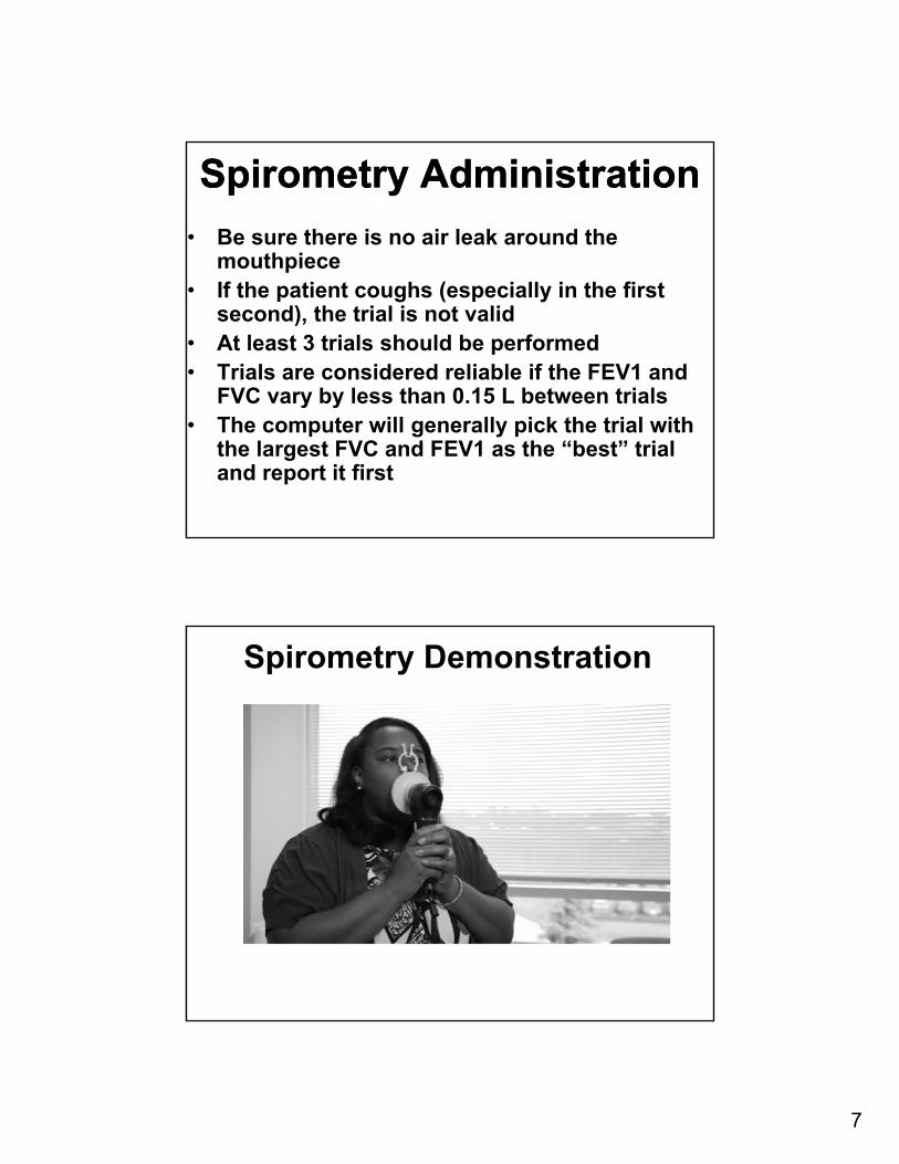
Spirometry AdministrationSpirometry Administration
• Be sure there is no air leak around the mouthpiece
• If the patient coughs (especially in the first second), the trial is not valid
• At least 3 trials should be performed• Trials are considered reliable if the FEV1 and
FVC vary by less than 0.15 L between trials• The computer will generally pick the trial with
the largest FVC and FEV1 as the “best” trial and report it first
Spirometry Demonstration
8
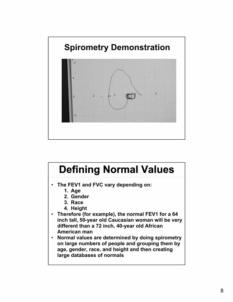
Spirometry Demonstration
Defining Normal ValuesDefining Normal Values• The FEV1 and FVC vary depending on:
1. Age2. Gender3. Race4. Height
• Therefore (for example), the normal FEV1 for a 64 inch tall, 50-year old Caucasian woman will be very different than a 72 inch, 40-year old African American man
• Normal values are determined by doing spirometry on large numbers of people and grouping them by age, gender, race, and height and then creating large databases of normals
9
Defining obstructionDefining obstruction• Obstruction is present if the FEV1/FVC ratio is
reduced• There are several different ways of defining a low
FEV1/FVC ratio. The two most common are:1. American Thoracic Society: defines a low
FEV1/FVC by comparison to large databases of normal subjects. A low FEV1/FVC is then defined as less than the 5th percentile of normal subjects stratified by age
2. Global Initiative for Obstructive Lung Disease (GOLD): uses a fixed number for all people regardless of age and defines a low FEV1/FVC as less than 70% for everyone
The FEV1/FVC Ratio Changes With Age
The FEV1/FVC Ratio Changes With Age
• The FEV1/FVC ratio declines in normal people as they get older‒ An average FEV1/FVC in a 20 year old is 87%‒ An average FEV1/FVC in an 84 year old is 71%
• The lower limit of normal in an 84 year old is 59%!
• The ATS definition of obstruction takes this age variation into account
• The GOLD definition of obstruction does not‒ Some normal older patients may be mis-
classified as being obstructed when using the GOLD criteria
10
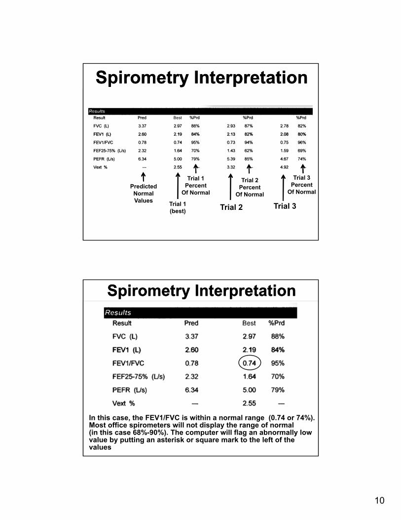
Spirometry InterpretationSpirometry Interpretation
PredictedNormalValues
Trial 1(best) Trial 2 Trial 3
Trial 1Percent
Of Normal
Trial 2Percent
Of Normal
Trial 3Percent
Of Normal
Spirometry InterpretationSpirometry Interpretation
In this case, the FEV1/FVC is within a normal range (0.74 or 74%). Most office spirometers will not display the range of normal(in this case 68%-90%). The computer will flag an abnormally lowvalue by putting an asterisk or square mark to the left of the values
11
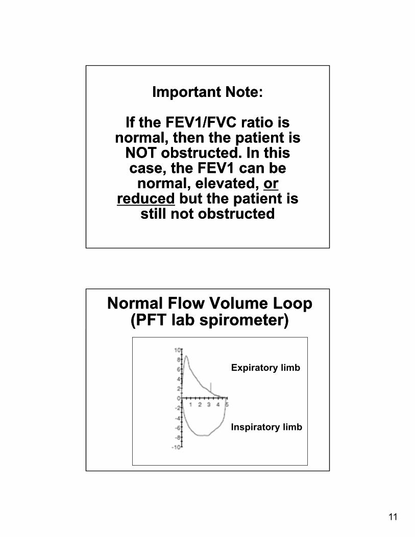
Important Note:
If the FEV1/FVC ratio is normal, then the patient is
NOT obstructed. In this case, the FEV1 can be normal, elevated, or
reduced but the patient is still not obstructed
Important Note:
If the FEV1/FVC ratio is normal, then the patient is
NOT obstructed. In this case, the FEV1 can be normal, elevated, or
reduced but the patient is still not obstructed
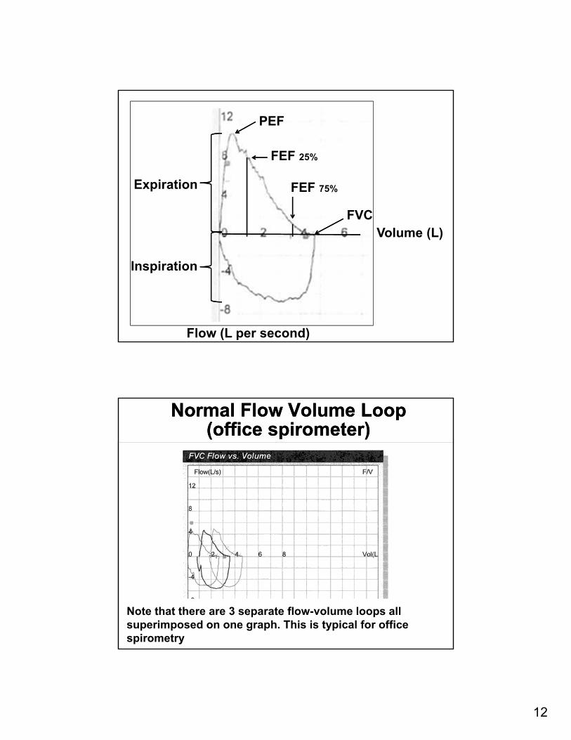
Normal Flow Volume Loop (PFT lab spirometer)
Normal Flow Volume Loop (PFT lab spirometer)
Expiratory limb
Inspiratory limb
12
Expiration
Inspiration
PEF
FEF 25%
FEF 75%
FVCVolume (L)
Flow (L per second)
Normal Flow Volume Loop (office spirometer)
Normal Flow Volume Loop (office spirometer)
Note that there are 3 separate flow-volume loops allsuperimposed on one graph. This is typical for office spirometry
13
TracheostenosisTracheostenosis
1086420
-2-4-6-8-10
1 2 3 4 5 6 8
Vocal Cord DysfunctionVocal Cord Dysfunction
1086420
-2-4-6-8-10
1 2 3 4 5 6 8
14
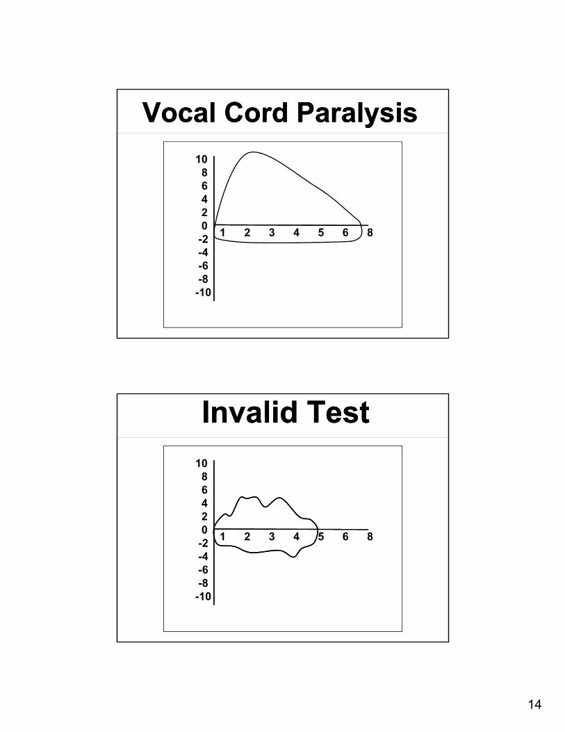
Vocal Cord ParalysisVocal Cord Paralysis
1086420
-2-4-6-8-10
1 2 3 4 5 6 8
Invalid TestInvalid Test
1086420
-2-4-6-8-10
1 2 3 4 5 6 8
15
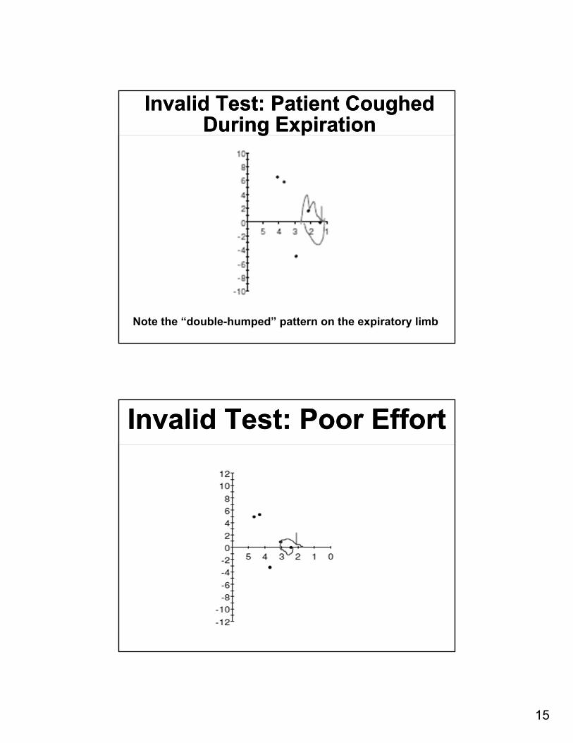
Invalid Test: Patient Coughed During Expiration
Invalid Test: Patient Coughed During Expiration
Note the “double-humped” pattern on the expiratory limb
Invalid Test: Poor EffortInvalid Test: Poor Effort
16
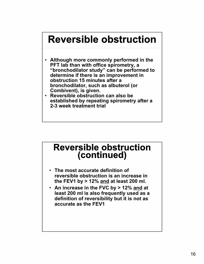
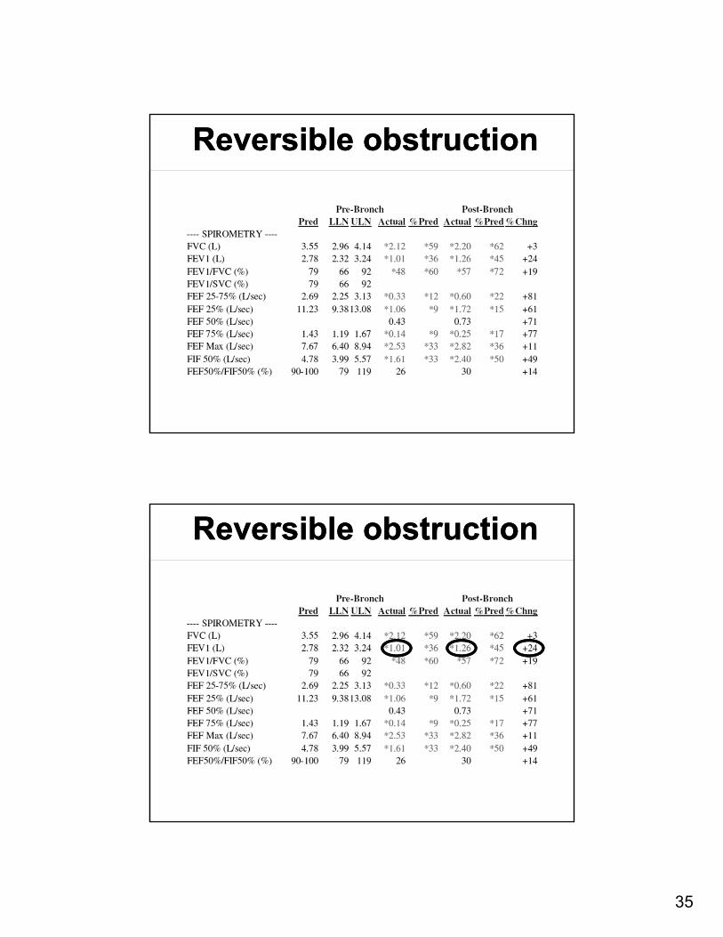
Reversible obstructionReversible obstruction
• Although more commonly performed in the PFT lab than with office spirometry, a “bronchodilator study” can be performed to determine if there is an improvement in obstruction 15 minutes after a bronchodilator, such as albuterol (or Combivent), is given.
• Reversible obstruction can also be established by repeating spirometry after a 2-3 week treatment trial
Reversible obstruction (continued)
Reversible obstruction (continued)
• The most accurate definition of reversible obstruction is an increase in the FEV1 by > 12% and at least 200 ml.
• An increase in the FVC by > 12% and at least 200 ml is also frequently used as a definition of reversibility but it is not as accurate as the FEV1
17
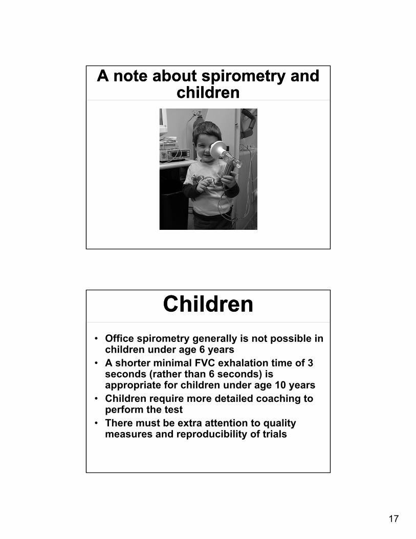
A note about spirometry and children
A note about spirometry and children
ChildrenChildren• Office spirometry generally is not possible in
children under age 6 years• A shorter minimal FVC exhalation time of 3
seconds (rather than 6 seconds) is appropriate for children under age 10 years
• Children require more detailed coaching to perform the test
• There must be extra attention to quality measures and reproducibility of trials
18
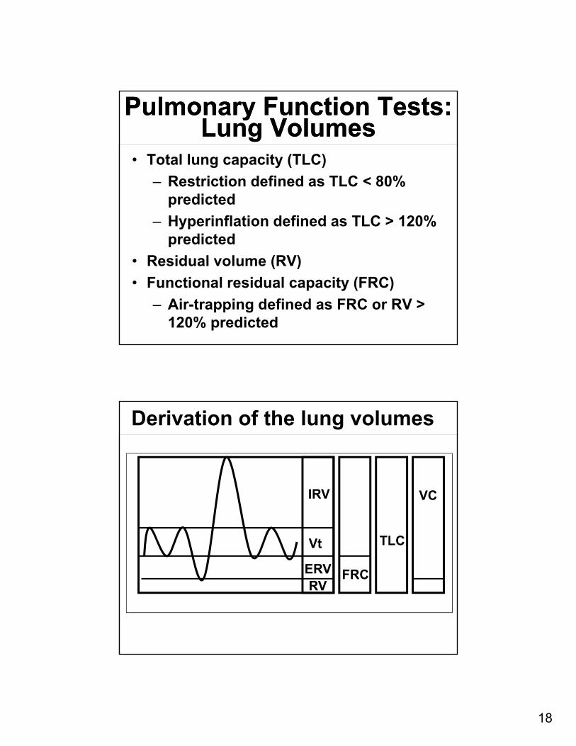
Pulmonary Function Tests:Lung Volumes
Pulmonary Function Tests:Lung Volumes
• Total lung capacity (TLC)
‒ Restriction defined as TLC < 80% predicted
‒ Hyperinflation defined as TLC > 120% predicted
• Residual volume (RV)
• Functional residual capacity (FRC)
‒ Air-trapping defined as FRC or RV > 120% predicted
IRV
Vt
ERVRV
FRC
TLC
VC
Derivation of the lung volumes
19
Causes of RestrictionCauses of Restriction
• Interstitial lung disease
• Alveolar filling processes
• Chest wall impairment
• Respiratory muscle weakness
Diagnosing Restriction Based On Spirometry
Diagnosing Restriction Based On Spirometry
• The only confident way to diagnose restriction is by full lung volume measurements with measurement of the total lung capacity (TLC).
• You can suspect restriction if the FVC is low on spirometry but this can be fraught with error.‒ Many patients with COPD will have a low FVC‒ The FVC is often low even when the TLC is normal
• If the FVC is low and you suspect restriction, you should order lung volumes in the PFT lab to confirm restriction
20
Diagnosing Restriction Based On Spirometry (continued)
Diagnosing Restriction Based On Spirometry (continued)
• However, in some diseases, following the FVC serially can be a good marker of lung capacity and respiratory muscle strength‒ Patients with interstitial lung disease‒ Patients with neuromuscular weakness
• When using the FVC to follow these patients for disease progression, it is important that the test be done with consistent technique, preferably by the same individual(s). Often, this is best accomplished in the PFT lab or in clinics that regularly care for neuromuscular patients.
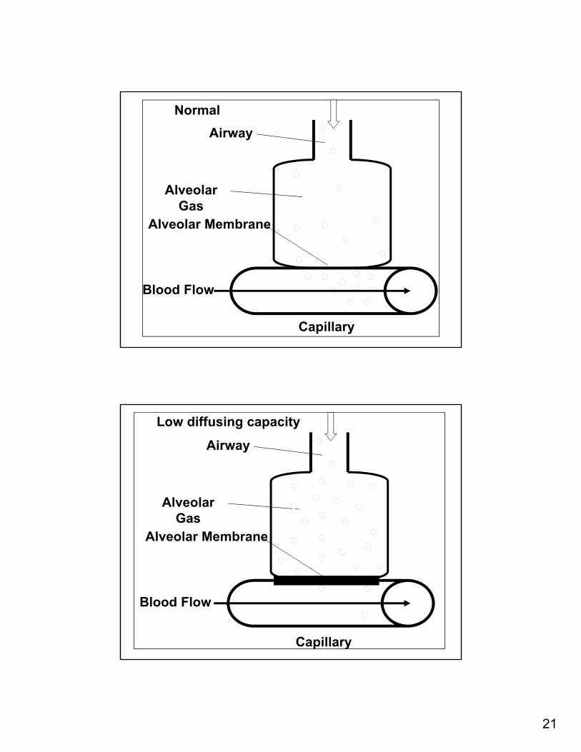
Pulmonary Function Tests:Diffusing Capacity
Pulmonary Function Tests:Diffusing Capacity
• Measure of gas exchange across the alveolar/capillary membrane
• Dependent on surface area, gas solubility, membrane thickness, and transit time
• Affected by age, body size, gender, hemoglobin, and lung volume
• Measured by carbon monoxide uptake
21
Airway
Alveolar Gas
Alveolar Membrane
Capillary
Blood Flow
Normal
Airway
Alveolar Gas
Alveolar Membrane
Capillary
Blood Flow
Low diffusing capacity
22
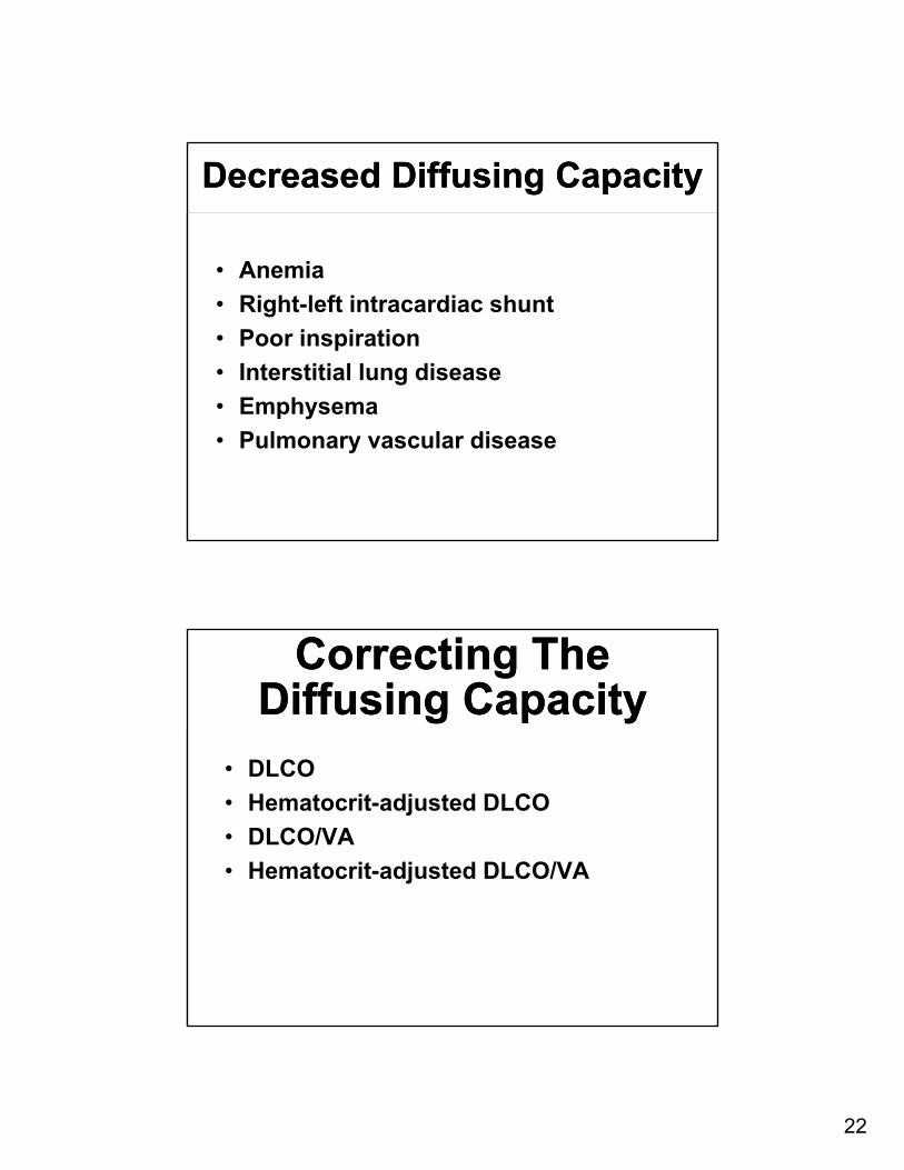
Decreased Diffusing CapacityDecreased Diffusing Capacity
• Anemia
• Right-left intracardiac shunt
• Poor inspiration
• Interstitial lung disease
• Emphysema
• Pulmonary vascular disease
Correcting The Diffusing Capacity
Correcting The Diffusing Capacity
• DLCO
• Hematocrit-adjusted DLCO
• DLCO/VA
• Hematocrit-adjusted DLCO/VA
23
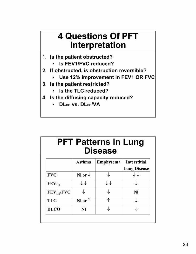
4 Questions Of PFT Interpretation
4 Questions Of PFT Interpretation
1. Is the patient obstructed?• Is FEV1/FVC reduced?
2. If obstructed, is obstruction reversible?• Use 12% improvement in FEV1 OR FVC
3. Is the patient restricted?• Is the TLC reduced?
4. Is the diffusing capacity reduced?• DLCO vs. DLCO/VA
Asthma Emphysema Interstitial
Lung Disease
FVC Nl or
FEV1.0
FEV1.0/FVC Nl
TLC Nl or
DLCO Nl
PFT Patterns in Lung Disease
24
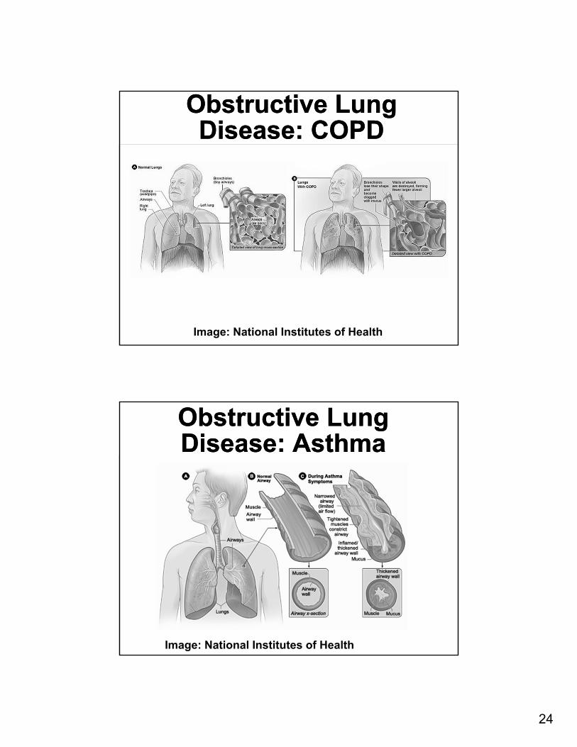
Obstructive Lung Disease: COPD
Obstructive Lung Disease: COPD
Image: National Institutes of Health
Obstructive Lung Disease: AsthmaObstructive Lung Disease: Asthma
Image: National Institutes of Health
25
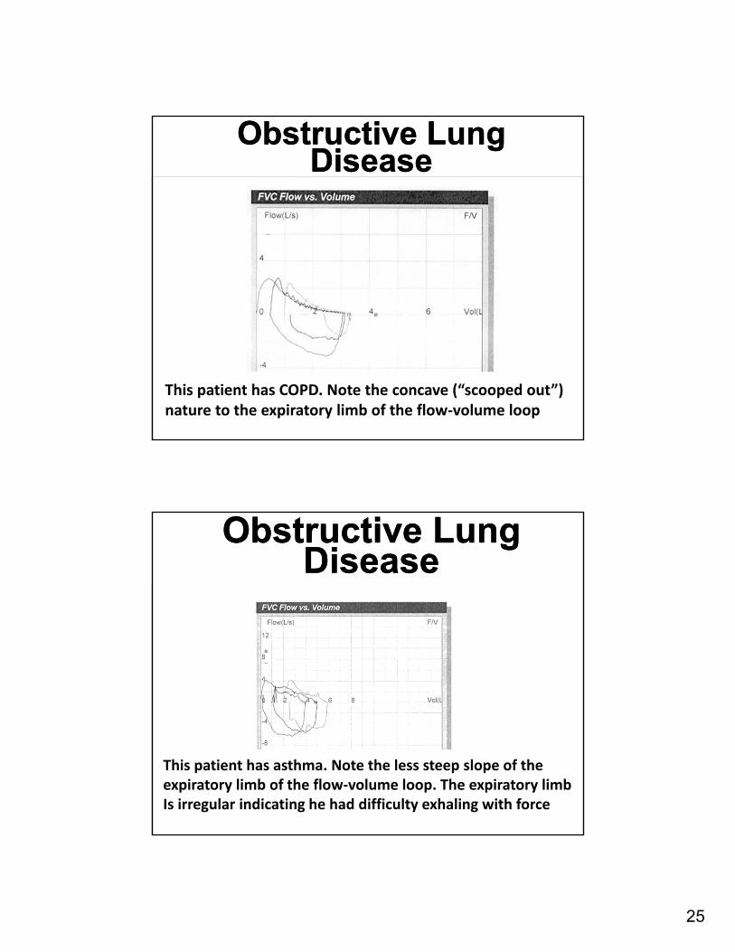
Obstructive Lung Disease
Obstructive Lung Disease
This patient has COPD. Note the concave (“scooped out”) nature to the expiratory limb of the flow‐volume loop
Obstructive Lung Disease
Obstructive Lung Disease
This patient has asthma. Note the less steep slope of theexpiratory limb of the flow‐volume loop. The expiratory limbIs irregular indicating he had difficulty exhaling with force
26
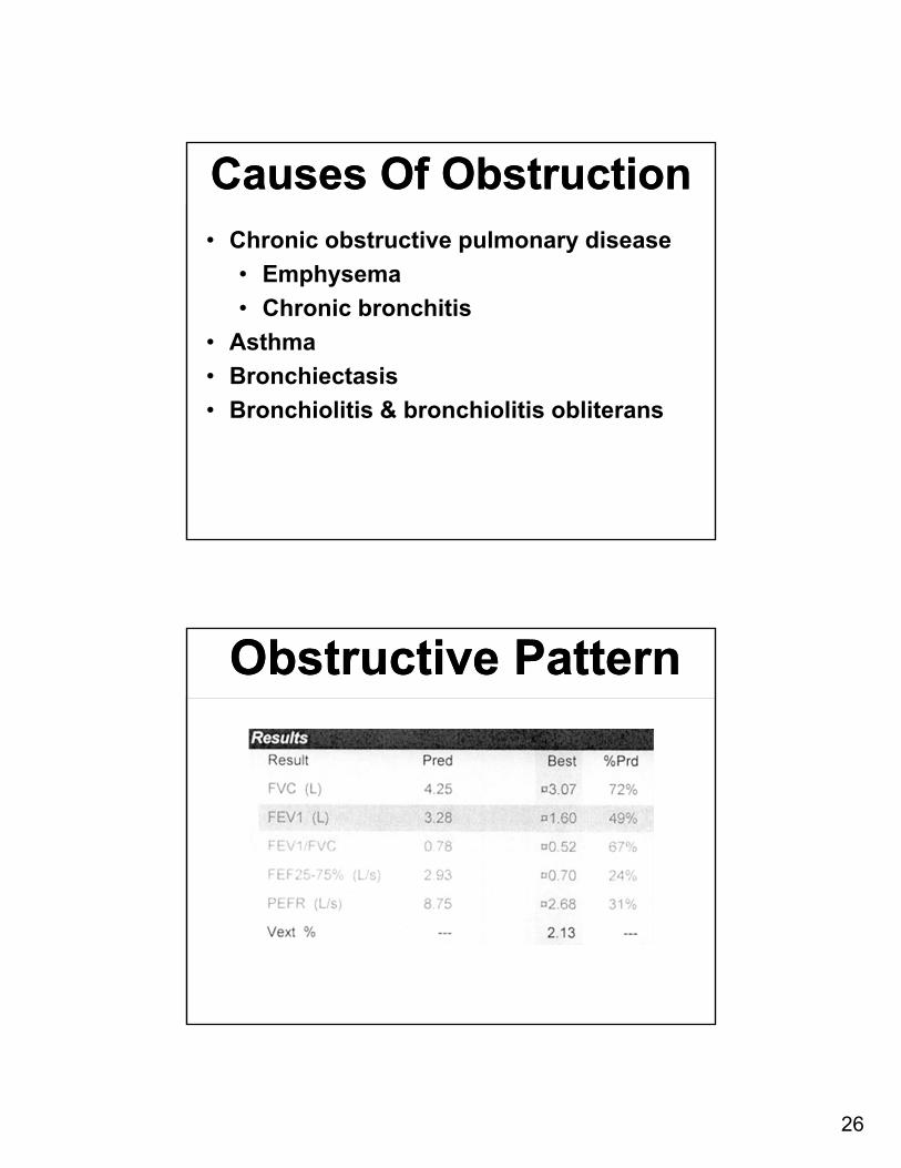
Causes Of ObstructionCauses Of Obstruction• Chronic obstructive pulmonary disease
• Emphysema
• Chronic bronchitis
• Asthma
• Bronchiectasis
• Bronchiolitis & bronchiolitis obliterans
Obstructive PatternObstructive Pattern
27
Obstructive PatternObstructive Pattern
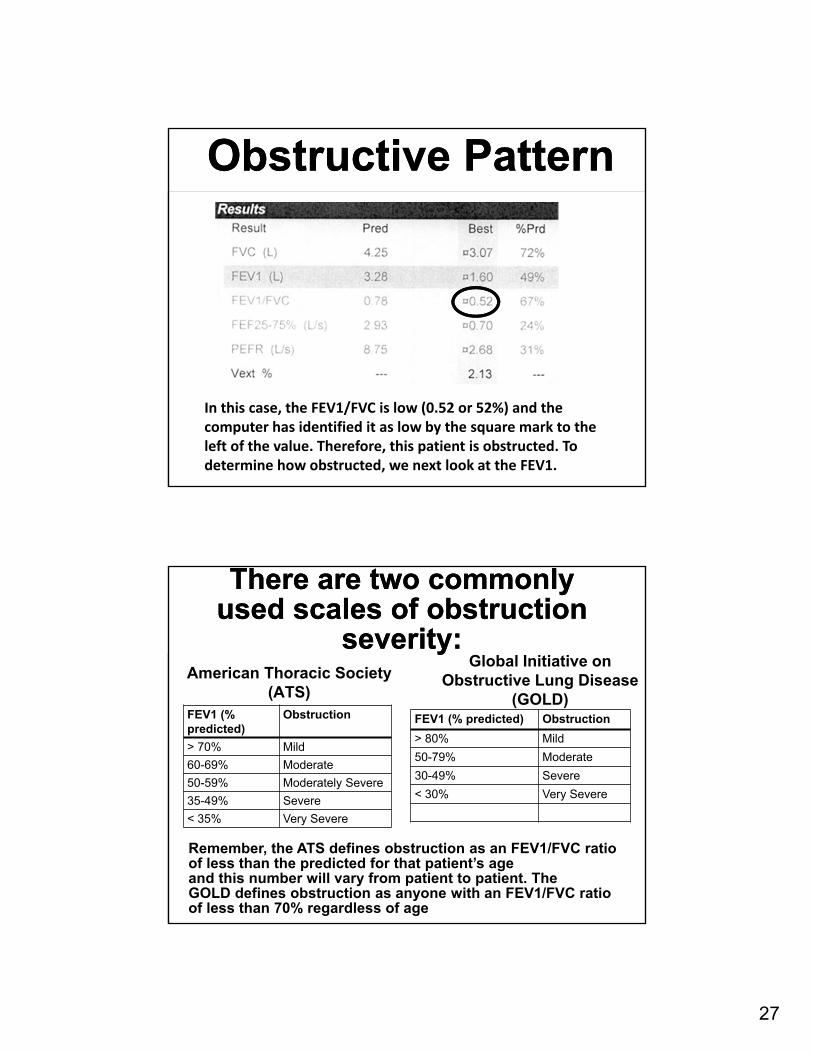
In this case, the FEV1/FVC is low (0.52 or 52%) and thecomputer has identified it as low by the square mark to the left of the value. Therefore, this patient is obstructed. To determine how obstructed, we next look at the FEV1.
There are two commonly used scales of obstruction
severity:
There are two commonly used scales of obstruction
severity:American Thoracic Society
(ATS)FEV1 (% predicted)
Obstruction
> 70% Mild
60-69% Moderate
50-59% Moderately Severe
35-49% Severe
< 35% Very Severe
Global Initiative on Obstructive Lung Disease
(GOLD)FEV1 (% predicted) Obstruction
> 80% Mild
50-79% Moderate
30-49% Severe
< 30% Very Severe
Remember, the ATS defines obstruction as an FEV1/FVC ratioof less than the predicted for that patient’s ageand this number will vary from patient to patient. TheGOLD defines obstruction as anyone with an FEV1/FVC ratio of less than 70% regardless of age
28
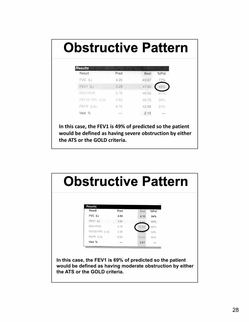
Obstructive PatternObstructive Pattern
In this case, the FEV1 is 49% of predicted so the patient would be defined as having severe obstruction by eitherthe ATS or the GOLD criteria.
Obstructive PatternObstructive Pattern
In this case, the FEV1 is 69% of predicted so the patient would be defined as having moderate obstruction by eitherthe ATS or the GOLD criteria.
29
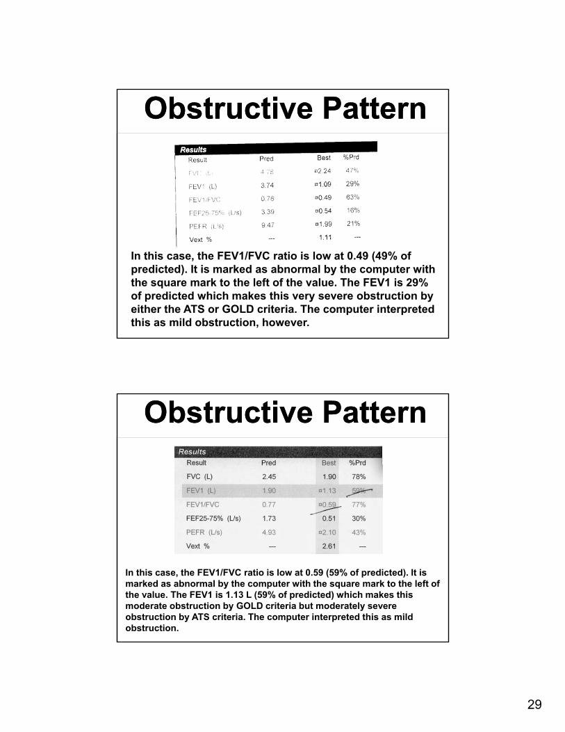
Obstructive PatternObstructive Pattern
In this case, the FEV1/FVC ratio is low at 0.49 (49% of predicted). It is marked as abnormal by the computer with the square mark to the left of the value. The FEV1 is 29% of predicted which makes this very severe obstruction by either the ATS or GOLD criteria. The computer interpreted this as mild obstruction, however.
Obstructive PatternObstructive Pattern
In this case, the FEV1/FVC ratio is low at 0.59 (59% of predicted). It is marked as abnormal by the computer with the square mark to the left of the value. The FEV1 is 1.13 L (59% of predicted) which makes this moderate obstruction by GOLD criteria but moderately severe obstruction by ATS criteria. The computer interpreted this as mild obstruction.
30
Obstructive PatternObstructive Pattern
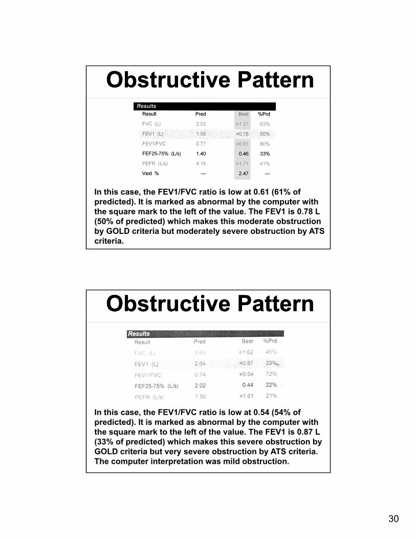
In this case, the FEV1/FVC ratio is low at 0.61 (61% of predicted). It is marked as abnormal by the computer with the square mark to the left of the value. The FEV1 is 0.78 L (50% of predicted) which makes this moderate obstruction by GOLD criteria but moderately severe obstruction by ATS criteria.
Obstructive PatternObstructive Pattern
In this case, the FEV1/FVC ratio is low at 0.54 (54% of predicted). It is marked as abnormal by the computer with the square mark to the left of the value. The FEV1 is 0.87 L (33% of predicted) which makes this severe obstruction by GOLD criteria but very severe obstruction by ATS criteria. The computer interpretation was mild obstruction.
31
TracheostenosisTracheostenosis
TracheostenosisTracheostenosis
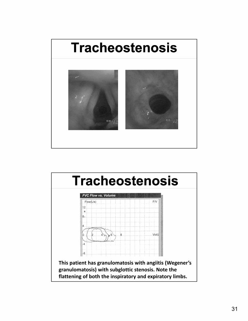
This patient has granulomatosis with angiitis (Wegener’sgranulomatosis) with subglottic stenosis. Note theflattening of both the inspiratory and expiratory limbs.
32
TracheostenosisTracheostenosis
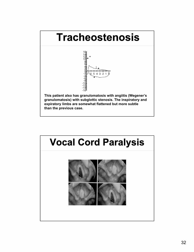
This patient also has granulomatosis with angiitis (Wegener’sgranulomatosis) with subglottic stenosis. The inspiratory and expiratory limbs are somewhat flattened but more subtlethan the previous case.
Vocal Cord ParalysisVocal Cord Paralysis
33
Paralyzed Vocal CordsParalyzed Vocal Cords
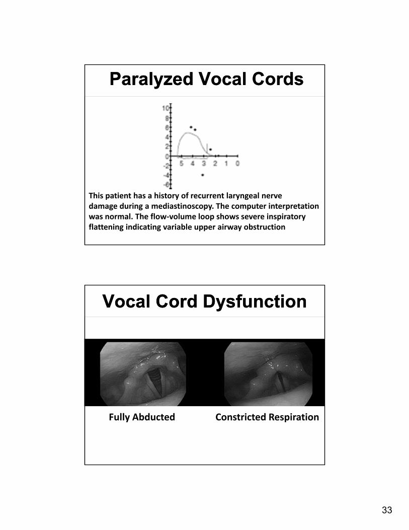
This patient has a history of recurrent laryngeal nervedamage during a mediastinoscopy. The computer interpretation was normal. The flow‐volume loop shows severe inspiratory flattening indicating variable upper airway obstruction
Vocal Cord DysfunctionVocal Cord Dysfunction
Fully Abducted Constricted Respiration
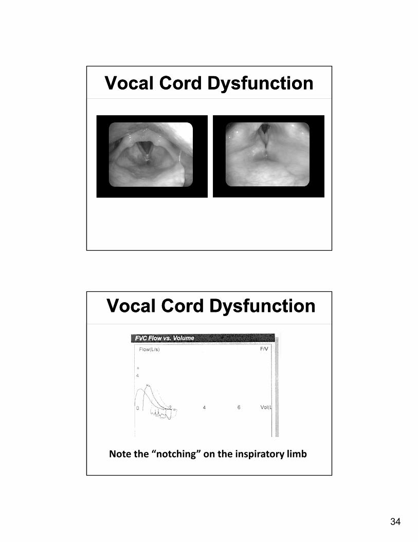
34
Vocal Cord DysfunctionVocal Cord Dysfunction
Vocal Cord DysfunctionVocal Cord Dysfunction
Note the “notching” on the inspiratory limb
35
Reversible obstructionReversible obstruction
Reversible obstructionReversible obstruction
36
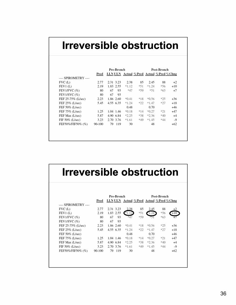
Irreversible obstructionIrreversible obstruction
Irreversible obstructionIrreversible obstruction
37
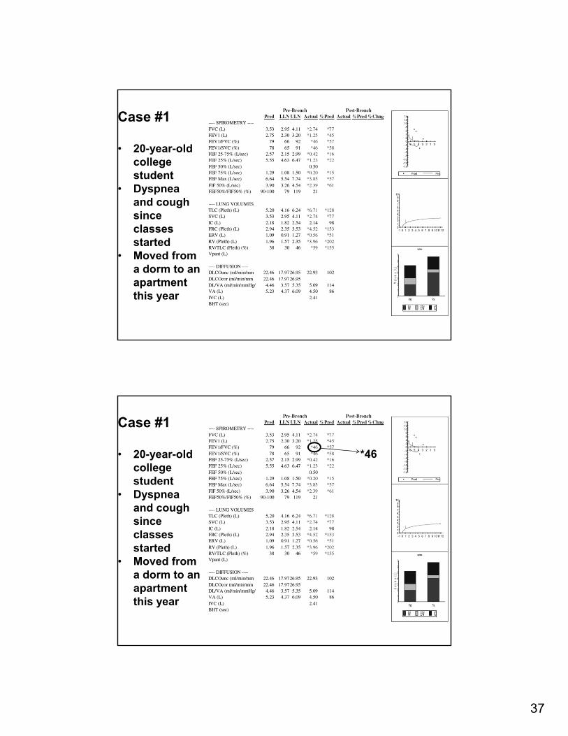
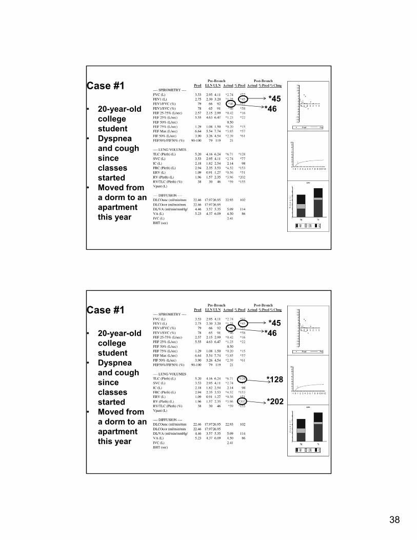
Case #1
• 20-year-old college student
• Dyspnea and cough since classes started
• Moved from a dorm to an apartment this year
Case #1
• 20-year-old college student
• Dyspnea and cough since classes started
• Moved from a dorm to an apartment this year
*46
38
Case #1
• 20-year-old college student
• Dyspnea and cough since classes started
• Moved from a dorm to an apartment this year
*46*45
Case #1
• 20-year-old college student
• Dyspnea and cough since classes started
• Moved from a dorm to an apartment this year
*46*45
*128
*202
39
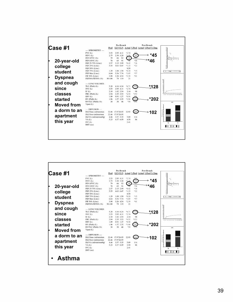
Case #1
• 20-year-old college student
• Dyspnea and cough since classes started
• Moved from a dorm to an apartment this year
*46*45
*128
*202
102
Case #1
• 20-year-old college student
• Dyspnea and cough since classes started
• Moved from a dorm to an apartment this year
*46*45
*128
*202
102
• Asthma
40
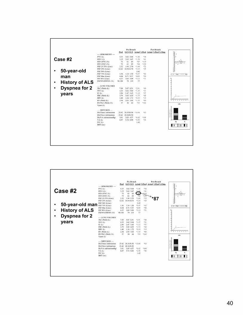
Case #2
• 50-year-old man
• History of ALS• Dyspnea for 2
years
Case #2
• 50-year-old man• History of ALS• Dyspnea for 2
years
*87
41
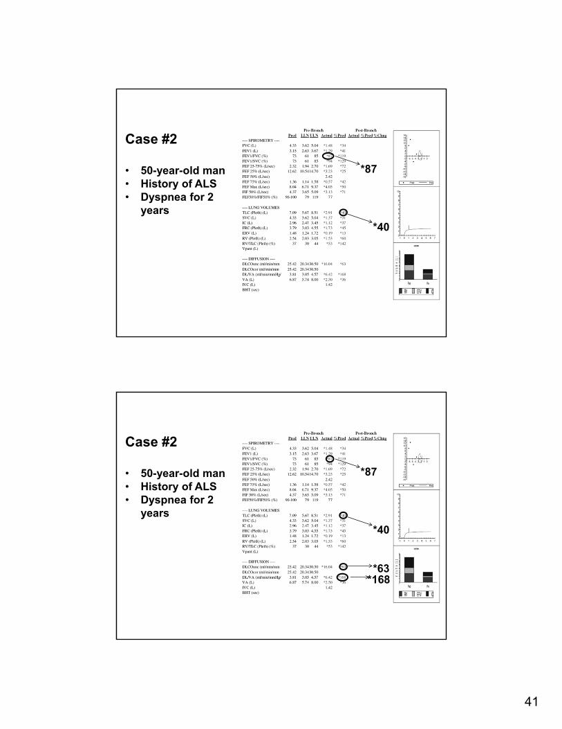
Case #2
• 50-year-old man• History of ALS• Dyspnea for 2
years
*87
*40
Case #2
• 50-year-old man• History of ALS• Dyspnea for 2
years
*87
*40
*63*168
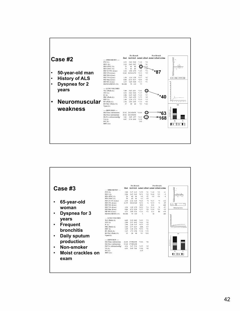
42
Case #2
• 50-year-old man• History of ALS• Dyspnea for 2
years
• Neuromuscular weakness
*87
*40
*63*168
Case #3
• 65-year-old woman
• Dyspnea for 3 years
• Frequent bronchitis
• Daily sputum production
• Non-smoker• Moist crackles on
exam
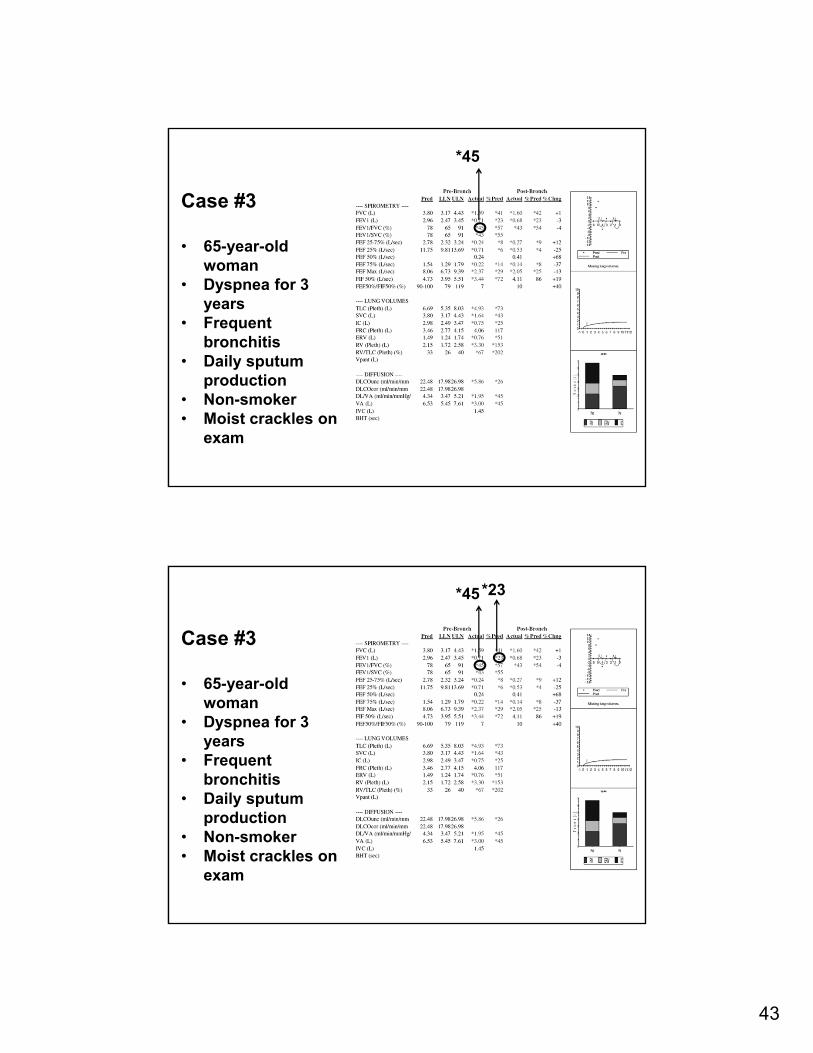
43
Case #3
• 65-year-old woman
• Dyspnea for 3 years
• Frequent bronchitis
• Daily sputum production
• Non-smoker• Moist crackles on
exam
*45
Case #3
• 65-year-old woman
• Dyspnea for 3 years
• Frequent bronchitis
• Daily sputum production
• Non-smoker• Moist crackles on
exam
*45*23
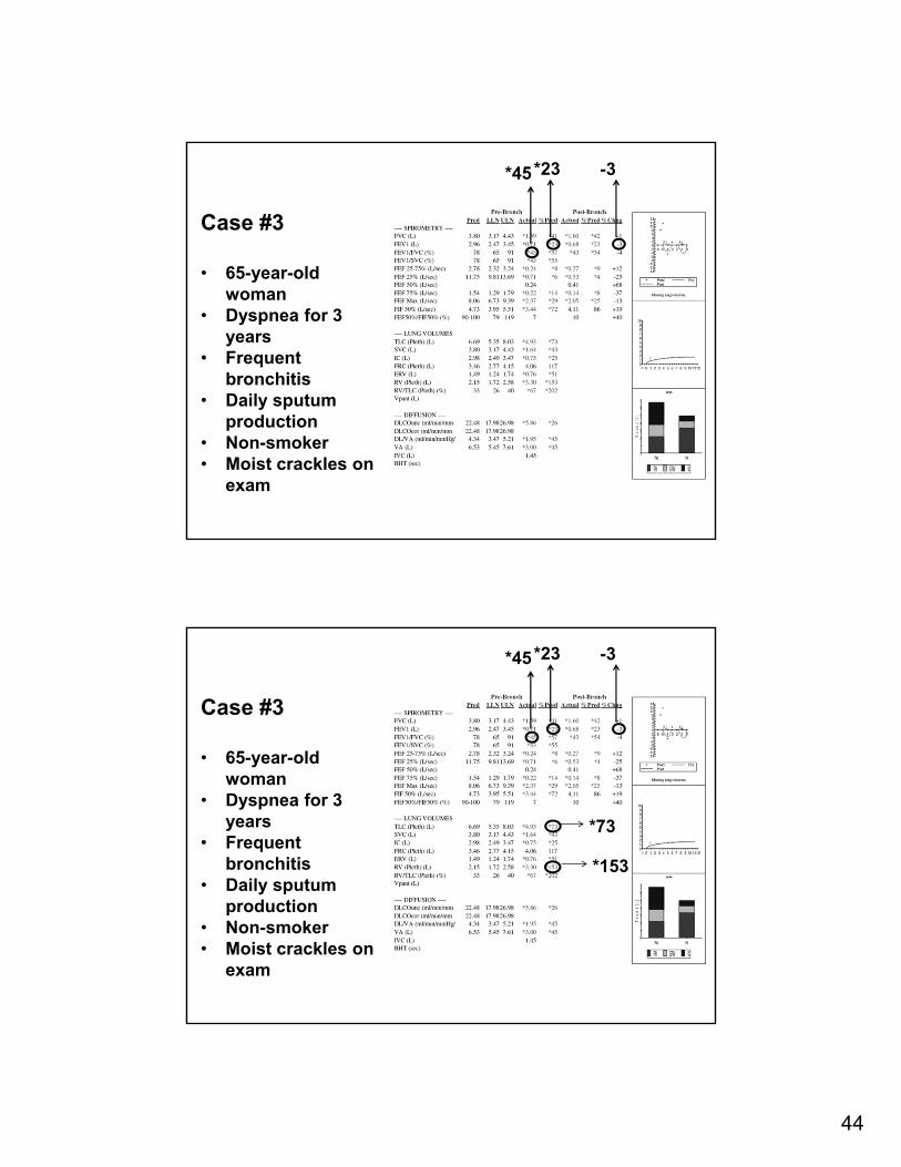
44
Case #3
• 65-year-old woman
• Dyspnea for 3 years
• Frequent bronchitis
• Daily sputum production
• Non-smoker• Moist crackles on
exam
*45*23 -3
Case #3
• 65-year-old woman
• Dyspnea for 3 years
• Frequent bronchitis
• Daily sputum production
• Non-smoker• Moist crackles on
exam
*45
*73
*153
*23 -3
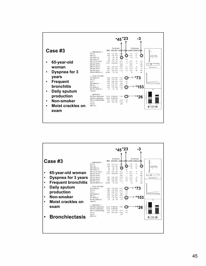
45
Case #3
• 65-year-old woman
• Dyspnea for 3 years
• Frequent bronchitis
• Daily sputum production
• Non-smoker• Moist crackles on
exam
*45
*73
*26
*153
*23 -3
Case #3
• 65-year-old woman• Dyspnea for 3 years• Frequent bronchitis• Daily sputum
production• Non-smoker• Moist crackles on
exam
• Bronchiectasis
*45
*73
*26
*153
*23 -3
46
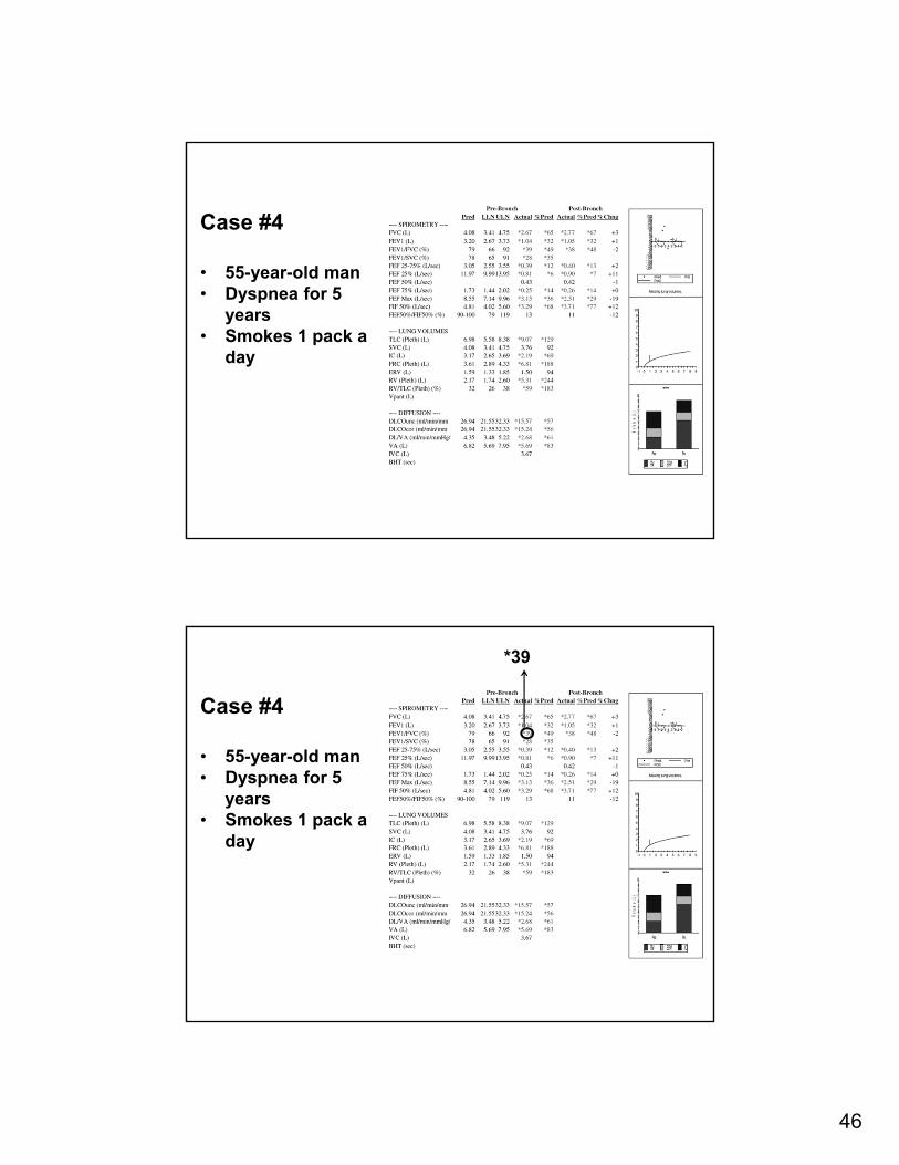
Case #4
• 55-year-old man• Dyspnea for 5
years• Smokes 1 pack a
day
Case #4
• 55-year-old man• Dyspnea for 5
years• Smokes 1 pack a
day
*39
47
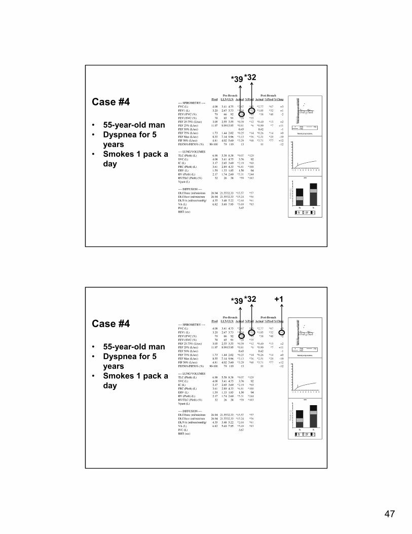
Case #4
• 55-year-old man• Dyspnea for 5
years• Smokes 1 pack a
day
*39*32
Case #4
• 55-year-old man• Dyspnea for 5
years• Smokes 1 pack a
day
*39*32 +1
48
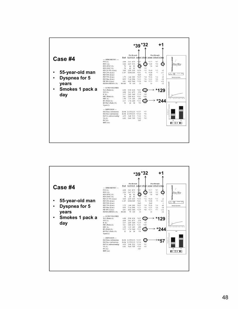
Case #4
• 55-year-old man• Dyspnea for 5
years• Smokes 1 pack a
day
*39
*129
*244
*32 +1
Case #4
• 55-year-old man• Dyspnea for 5
years• Smokes 1 pack a
day
*39
*129
*57
*244
*32 +1
49
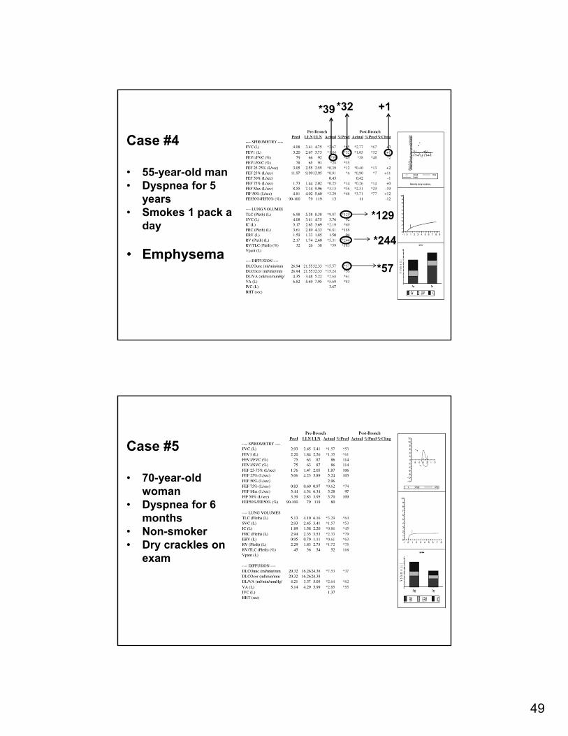
Case #4
• 55-year-old man• Dyspnea for 5
years• Smokes 1 pack a
day
• Emphysema
*39
*129
*57
*244
*32 +1
Case #5
• 70-year-old woman
• Dyspnea for 6 months
• Non-smoker• Dry crackles on
exam
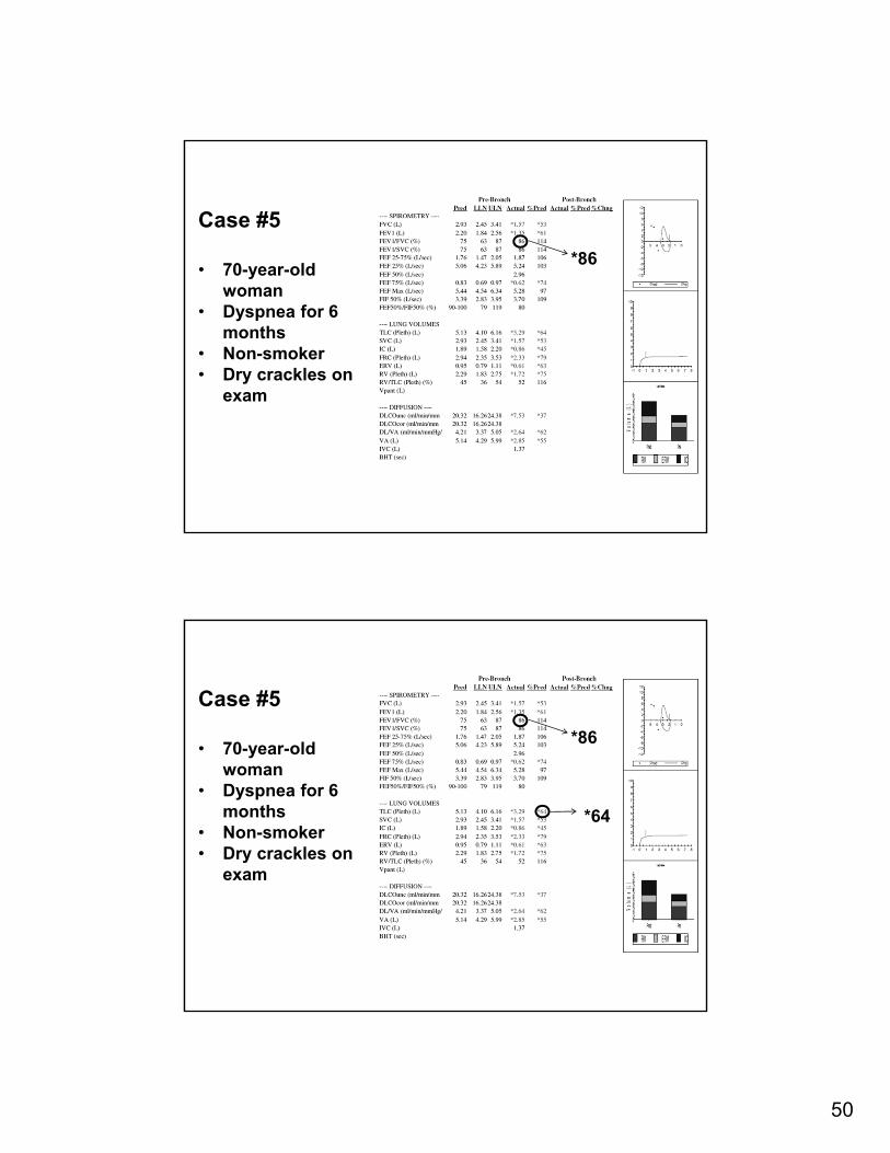
50
Case #5
• 70-year-old woman
• Dyspnea for 6 months
• Non-smoker• Dry crackles on
exam
*86
Case #5
• 70-year-old woman
• Dyspnea for 6 months
• Non-smoker• Dry crackles on
exam
*86
*64
51
Case #5
• 70-year-old woman
• Dyspnea for 6 months
• Non-smoker• Dry crackles on
exam
*86
*64
*37
Case #5
• 70-year-old woman
• Dyspnea for 6 months
• Non-smoker• Dry crackles on
exam
• Idiopathic pulmonary fibrosis
*86
*64
*37