Pulmonary Embolism Aortic Aneurysm Aortic Dissection Nursing 313, Fall 2011.
43
Pulmonary Embolism Aortic Aneurysm Aortic Dissection Nursing 313, Fall 2011
-
Upload
magnus-garrett -
Category
Documents
-
view
221 -
download
2
Transcript of Pulmonary Embolism Aortic Aneurysm Aortic Dissection Nursing 313, Fall 2011.

Pulmonary EmbolismAortic AneurysmAortic Dissection
Nursing 313, Fall 2011

Incidence >650,000 cases diagnosed per year in US Third most common cause of death in
hospitalized patients Greatest risk those who have a DVT Recent trends: men > women Risk doubles every ten years after 60
2

Risk Factors
http://www.cholesterolcholestrol.com/virchows-triad.jpg3
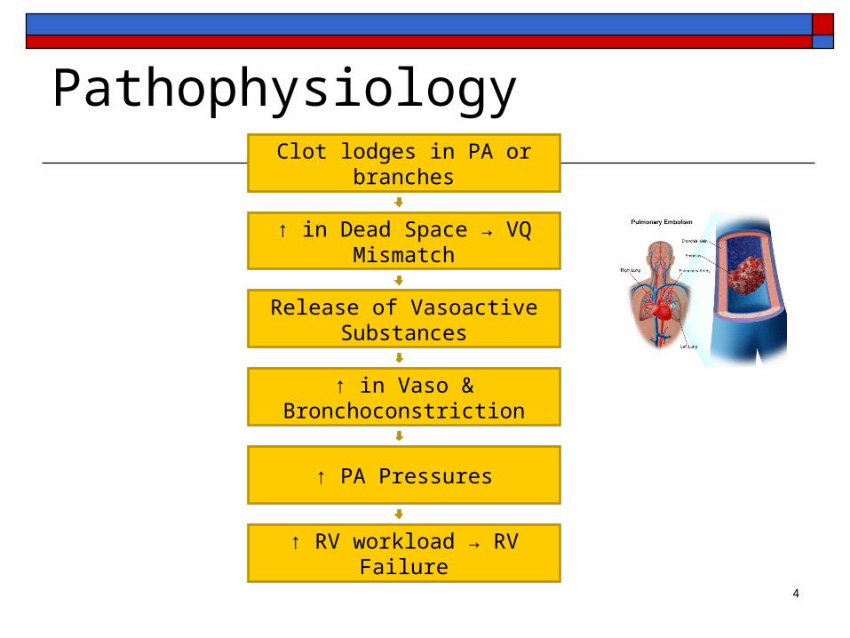
PathophysiologyClot lodges in PA or branches
↑ in Dead Space → VQ Mismatch
↑ PA Pressures
↑ in Vaso & Bronchoconstriction
↑ RV workload → RV Failure
Release of Vasoactive Substances
4

Respiratory System
A balancing act between ventilation (V) and perfusion (Q)
5

Perfusion / ventilation mismatchV/Q mismatch
Obstructed area has absent or diminished blood flow
Alveoli ventilated but not perfused which causes increased dead space
Severity depends on size of embolism and degree of vascular obstruction
Possibility of severe hypotension and shock
6

Pulmonary Embolism
7

Signs and Symptoms
Sudden onset of dyspnea Sudden onset of pleuritic chest pain General signs of hypoxemia Feeling of impending doom ↑ Anxiety Cough, possible hemoptysis Mild fever Diaphoresis
8
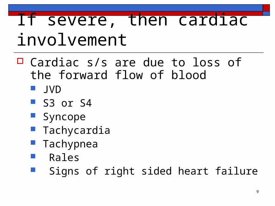
If severe, then cardiac involvement Cardiac s/s are due to loss of the forward flow
of blood JVD S3 or S4 Syncope Tachycardia Tachypnea Rales Signs of right sided heart failure
9

Diagnosis D-Dimer assay – possible method of ruling
out a PE Spiral CT scan Pulmonary Angiography (gold standard) V/Q scan CXR, ECG, ABGs
10

Emergency Management Stabilize the cardiopulmonary system
Oxygen Insertions of IV lines Treat hypotension – vasoactive drugs if ↓BP Diagnostic testing: scans, ABGs, labs Indwelling urinary catheter Sedatives or pain relief as needed Prevent further emboli from forming
11

Pharmacological ManagementHeparin Heparin- initial and preferred treatment Does not affect the existing clot PTT/ therapeutic range 1.5 to 2 times normal Antidote: protamine sulfate
12

Warfarin Warfarin (Coumadin) Interferes with the synthesis of the vitamin K 3-4 days for therapeutic benefit INR between 2.0 – 2.5 for those with PE Antidote – Vitamin K
13

Thrombolytic Therapy tPA, Alteplase, Reteplase Converts plasminogen to plasmin Specific contraindications:
Recent CVA; active bleeding; surgery in past 10 days; trauma; recent labor and delivery, severe hypertension
Major complication: bleeding
14
Nursing Management of Patients on Anticoagulation Therapy
Frequent vital signs Hematest stools Handle patient gently Avoid IM injections & veni-punctures Use electric shaver & soft toothbrushes Nothing per rectum Avoid foods containing vitamin K Monitor labs
15

Surgical Management
Thrombectomy or Embolectomy Vena Cava Interruption (Filters)
Indicated in patients who may not tolerate anticoagulation
16

Greenfield Filter
17

Nursing Management
Identify patients at risk Manage pain! Psychosocial support for patient and family Utilize nursing interventions to minimize risk
Early ambulation Watch labs, VS, targeted assessments for
cardiopulmonary system Patient & family teaching on importance of lifestyle
changes18

19

Aortic Aneurysms
20

Flash from the past….
21

Factoids Atherosclerosis damages the lining of the aorta Majority below the renal arteries Exact cause of aneurysm unknown More common in Caucasians More common in men than women
Higher risk of rupture and death in women compared with men with same size aneurysms
Rare in young females, but related to pregnancy
22

Incidence & Risk Factors 13th leading cause of death in men aged 65-75 Up to 13% of individuals with AA have multiple aneurysms Causes about 9,000 deaths / year Risk factors:
Atherosclerosis Patients born with bicuspid aortic valves (new) Hypertension Age Smoking, cholesterol, ↑lipids Genetic CT disorders (Marfan’s) History of crack use in pregnancy Positive family history
Often found incidentally23

Classification: Morphology
24

Abdominal More common (75%) Usually infra-renal
Thoracic Less common (25%) > chance of rupture &
dissection
Classification: Location
25

Clinical Manifestations
Abdominal Aortic Aneurysms Asymptomatic (early) Symptoms caused by expansion (later)
Chest or abdominal pain, flank pain, scrotal pain Pulsatile mass palpated (80%) , bruit + Feels like heart beating in abdomen
Ruptured aneurysm is a medical emergency
26
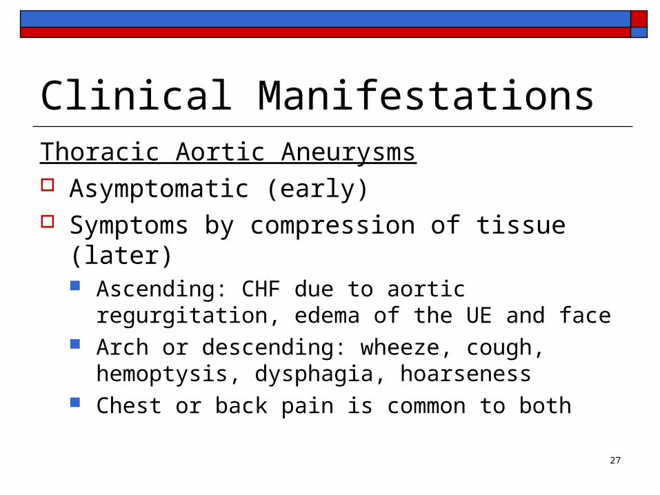
Thoracic Aortic Aneurysms Asymptomatic (early) Symptoms by compression of tissue (later)
Ascending: CHF due to aortic regurgitation, edema of the UE and face
Arch or descending: wheeze, cough, hemoptysis, dysphagia, hoarseness
Chest or back pain is common to both
Clinical Manifestations
27
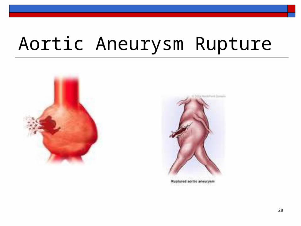
Aortic Aneurysm Rupture
28

Aortic Dissection Occurs when there is a tear in the intimal
lining (or adventitia) and blood gets diverted into the channel → ↓ intravascular volume
http://www.youtube.com/watch?v=ZtanUq95pTk
29
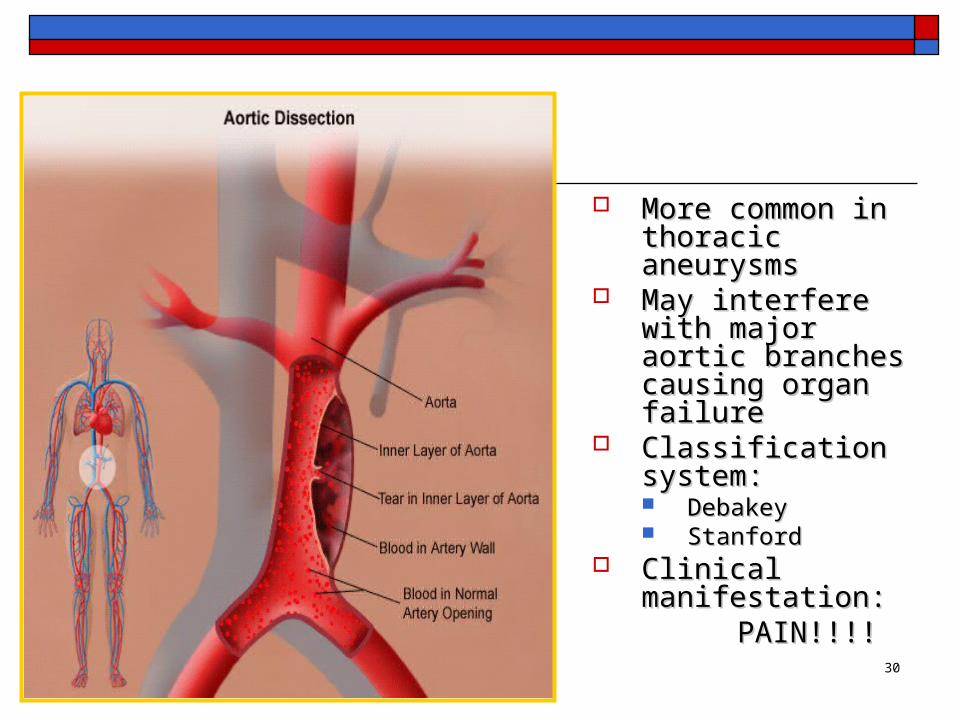
More common in More common in thoracic aneurysmsthoracic aneurysms
May interfere with May interfere with major aortic branches major aortic branches causing organ failurecausing organ failure
Classification system:Classification system: Debakey Debakey StanfordStanford
Clinical manifestation:Clinical manifestation:PAIN!!!!PAIN!!!!
30
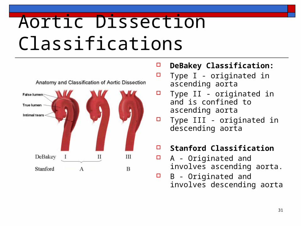
Aortic Dissection Classifications DeBakey Classification: Type I - originated in ascending
aorta Type II - originated in and is
confined to ascending aorta Type III - originated in
descending aorta
Stanford Classification A - Originated and involves
ascending aorta. B - Originated and involves
descending aorta
31

Dx tests for Aortic Dissections Trans thoracic echocardiogram (TTE) Trans esophageal echocardiogram (TEE) Computed Tomography Angiography (CTA) Magnetic Resonance Angiography Ultrasonography Aortography
32
Medical Management ofAortic Aneurysm
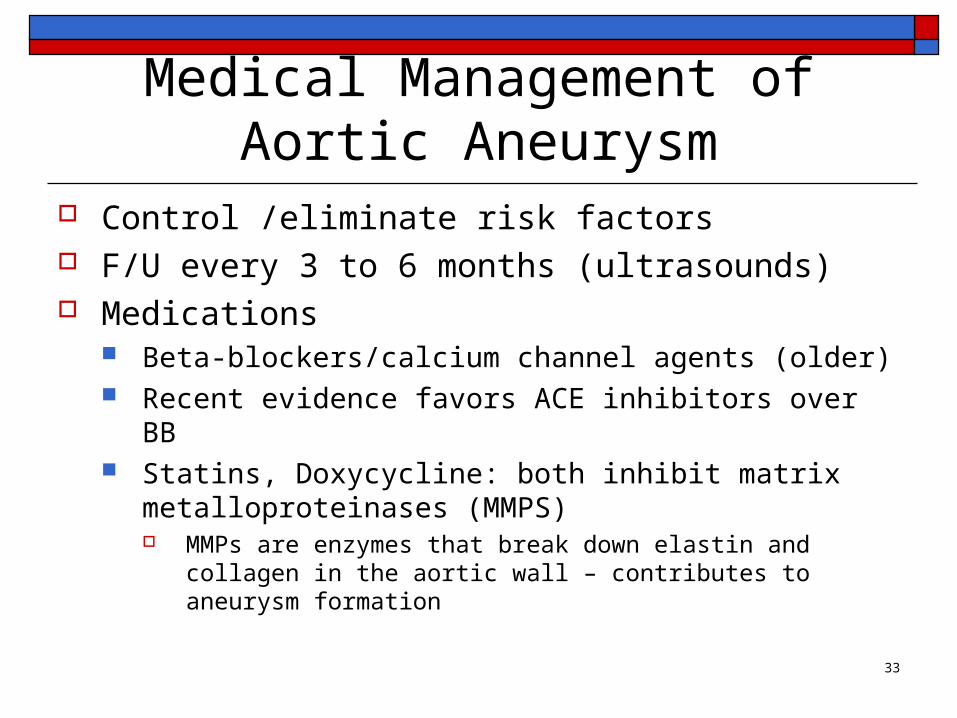
Control /eliminate risk factors F/U every 3 to 6 months (ultrasounds) Medications
Beta-blockers/calcium channel agents (older) Recent evidence favors ACE inhibitors over BB Statins, Doxycycline: both inhibit matrix
metalloproteinases (MMPS) MMPs are enzymes that break down elastin and collagen in the
aortic wall – contributes to aneurysm formation
33

General Indications for Surgical Repair of Aortic Aneurysms Diameter ≥5.5 cm (men) For women, 4.5-5.0 cm (due to greater incidence of
rupture) Ascending Thoracic Diameter ≥5.5 cm (5 cm in patients with Marfan
syndrome) Symptoms suggesting expansion or compression of
surrounding structures Rapidly expanding aneurysms (growth rate >0.5 cm
over a 6-month period) Symptomatic aneurysm
34

http://www.cvtsa.com/ListofConditions/A-444-62.html 35

Open AneurysmectomyOpen Aneurysmectomy
36
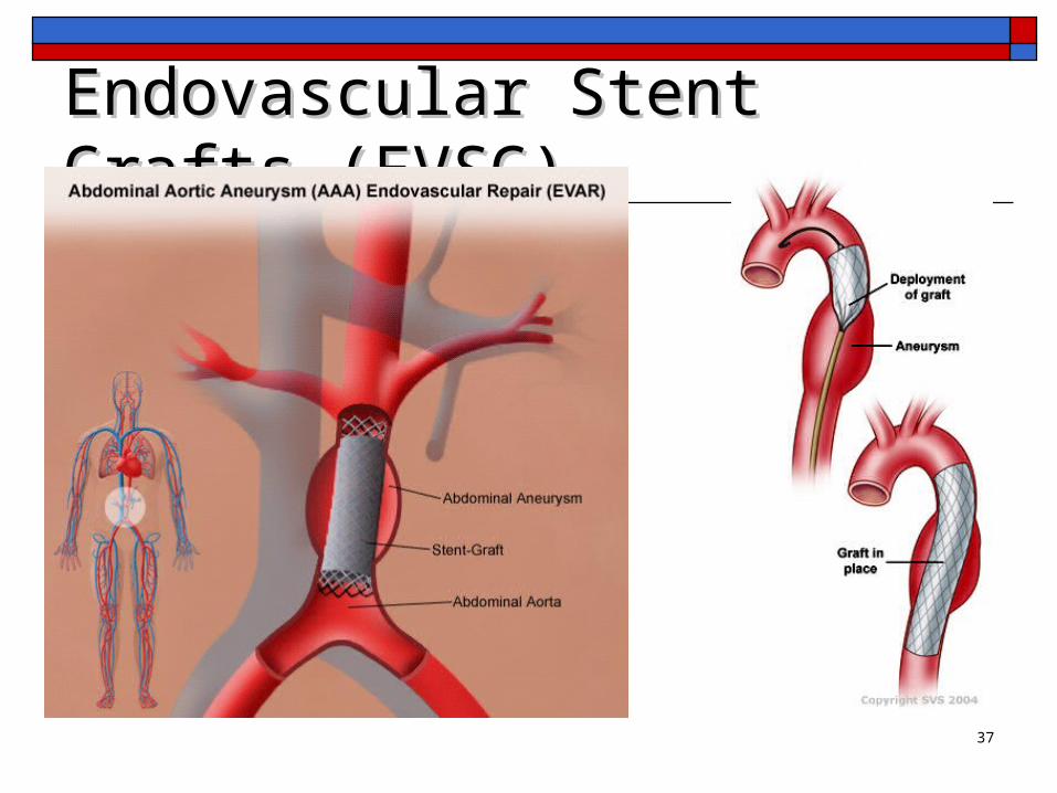
Endovascular Stent Grafts (EVSG)Endovascular Stent Grafts (EVSG)
37
EVAR 60% of aneurysm repairs in the US Fewer immediate complications than
conventional surgery More interventions are needed after 2 years
with EVAR (graft leaks, graft migration or infection, bowel perforation, etc)
Similar survival rates at 6 years (EVAR vs. open)
38

Post op care of surgical repair of AAA Post op complications
MI Cerebral infarct or ischemia to spinal cord Hypovolemia Respiratory distress Paralytic ileus Renal Failure
39

Postoperative nursing management Cardiovascular
Monitor for dysrhythmias Control of BP Monitor labs
Renal Hourly urines Monitor renal indicators
40

Postoperative nursing management Respiratory
Prevention of complications Meticulous pulmonary hygiene Control pain
Gastrointestinal Assess motility
NGT for suctioning (ileus) Monitor girth Provide nutrition Administer antibiotics
41

Postoperative nursing management Neurological
Neurovascular checks Graft Occlusion or Rupture
Assess for changes in pulses; temperature & color of extremities; severe pain
Abdominal distention Decreased urine output
Post endovascular stent procedure complications
42

43