
Pulmonary embolism
67
-
Upload
hosam-atef -
Category
Health & Medicine
-
view
60 -
download
0
Transcript of Pulmonary embolism


PULMONARY EMBOLISM
Dr. HOSAM ATEF
LECTURER OF ANESTHESIOLOGY&ICU


Pulmonary Embolism, Infarction
• Embolism : Impaction of a thrombus or foreign matter in the pulmonary vascular bed.
• Infarction : The pathological changes which develop in the lung as a result of pulmonary embolism.

Pulmonary Thrombo-embolism
• Thrombosis of peripheral veins , embolization of pulmonary arteries , and pulmonary infarction.
• Primary thrombosis in pulmonary arteries and veins

Pulmonary Embolism, Prevalence
• PE : The cause of, or a major contributory factor to, death in 7-9% of necropsy cases
• PM Pul. Angiographic technique : 14-18%
• Considering smaller thrombi : 60%
• PE is a major contributory factor to death in 50 000-200 000 patients per year in USA

EMBOLUS
• Thrombotic
• Non-thrombotic : Fat, Air, Tumour , Amniotic fluid, IV Drug abusers.

Pathogenesis of Vascular Thrombosis
• Decrease in blood flow below a certain critical level.
• Increase in coagulability of blood.
• Damage of the vessel wall.

RISK FACTORS
• Bed rest• Post-operative• After severe
blood loss and trauma
• CHF• Varicose veins• Advancing age
• Obesity• Post-partum• Malignancy• DM• Pneumonia• Debilitating
diseases• 1ry polycythemia• Race, Diet

PE, Clinical Features
• Size of the embolus and blood vessel occluded.
• State of the lung.
• Associated disease(s).
PE , Clinical Features
• Massive Pulmonary Embolism ( MPE )
• Pulmonary Infarction ( PI )
• Obliterative Pulmonary Hypertension

Massive Pulmonary Embolism MPE
• CLINICAL SETTING• ELDERLY,POSSIBLY OBESE• AROUND THE 10th DAY POST-OP.• CALLING FOR BED-PAN• EXPIRING SUDDENLY OR WHILE
IN THE ACT OF DEFECATION • IMMEDIATELY FATAL,2/3 DIE IN
THE FIRST TWO HOURS


Massive Pulmonary Embolism MPE
• SHOCK• DYSPNEA• APPREHENSION• TACHYCARDIA• SWEATING
• CHEST PAIN• FAINTNESS• CYANOSIS• AF• COLLAPSE

MPE, Differential Diagnosis
• Myocardial Infarction.
• Dissecting Aortic Aneurysm.
• Peumothorax.
• Major Pulmonary Collapse.
• Shock.
• Perforating Peptic Ulcer.
• Acute Pancreatitis.


CARDIOGENIC PULMONARY EDEMA
• SUDDEN ONSET OF DYSPNEA• SOMETIMES SEVERE CHEST PAIN• PINK FROTHY SPUTUM• EXTREME ANXIETY AND
ORTHOPNEA • DIAPHORESIS AND CYANOSIS• TACHYPNEA AND AIR HUNGER• WHEEZING• DIFFUSE MOIST RALES,GALLOP

ACUTE PULMONARY EDEMA IN COPD
• HISTORY OF PREVIOUS HEART DIS.
• SUDDEN,NOT ACUTE OR INSIDIOUS, ONSET OF DYSPNEA
• PINK FROTHY SPUTUM• DIFFUSE MOIST RALES,PULSUS
ALTERNANS,GALLOP,MURMURS• CXR,ECG,ABG


PNEUMOTHORAX
• SHARP UNILATERAL CHEST PAIN• DYSPNEA;EXTREME IN TENSION
PNX• PRIMARY, SECONDARY,
TRAUMATIC, BAROTRAUMA• TACHYPNOEIC• RAPID LOW VOLUME PULSE• HYPOTENSION• SURGICAL EMPHYSEMA
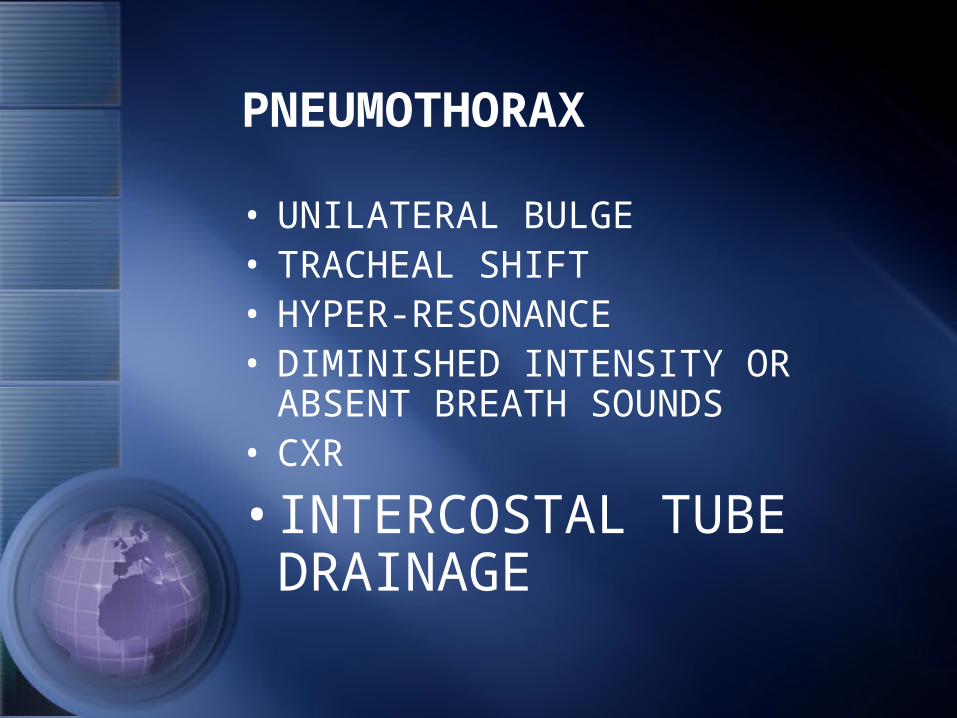
PNEUMOTHORAX
• UNILATERAL BULGE• TRACHEAL SHIFT• HYPER-RESONANCE• DIMINISHED INTENSITY OR
ABSENT BREATH SOUNDS• CXR
• INTERCOSTAL TUBE DRAINAGE


MASSIVE PULMONARY COLLAPSE
• CLINICAL SETTING
• TRACHEAL SHIFT
• UNILATERAL DULLNESS
• DIMINISHED OR ABSENT BREATH SOUNDS
• CXR
• BRONCHOSCOPY


COMPREHENSIVE ASSESSMENT
• HISTORY : PT., PT.’S RELATIVES, WITNESS
• PHYSICAL EXAMINATION: GENERAL RESPIRATORY CARDIOVASCULAR

COMPREHENSIVE ASSESSMENT
• INVESTIGATIVE STUDIES
• ECG
• ABG
• CXR
• ELECTROLYTES
• ENZYMES

Pulmonary Infarction, Pathology
• Blood Vessels: Engorgement, Hemorrhage from distended necrotic capillaries, Granulation tissue repair , Fibrous scar
• Bronchioles: usually survive, may turn bronchiactatic

Pulmonary Infarction, Pathology
• Bacterial Infection : Abscess Source : embolus, blood-borne, bronchi .
• Pleural Complications : Pleurisy, Pleural effusion, Empyema.

Pulmonary Infarction, Clinical Picture
• Pleuritic chest pain, Pleural rub, Pleural effusion
• Hemoptysis: in only 50% of cases• Finding the source of
embolization: in only 60% of cases• Tachcardia( more than 100/ min )
Tachypnoea • Jaundice, Cyanosis

Pulmonary Infarction, Clinical Picture
• Locally: No Physical Findings, Consolidation, Diminished Intensity of Breath Sounds, Crepitus, Wheezing Chest
• Pleural Rub• Signs of Pleural Effusion

Pulmonary Infarction, Clinical Picture
• With Infection: Worsening of the Clinical Status: Abscess or Empyema
• Persistent Fever, Malaise, Sweating
• Increasing Pulse Rate• Leucocytosis more than 20 000• Chest X-Ray

Clinical Features of PTE
Silent Asymptomatic Probably more frequent than we realize
Without Infarction Breathlessness, Tachycardia, Anxiety, Restlessness
Usually Transient

Clinical Features of PTE
With Infarction Dyspnea, Hemoptysis, Pleutitic Pain, Friction Rub, Fever, Brochospasm.
If you wait for these features, you will miss perhaps 60% of patients with embolism

Clinical Features of PTEWith Hemodynamic Impairment
Angina, Tachycardia, P++, Gallop, JVP++, Hypotension, Cyanosis, Syncope
This means obstruction of 30-50% of pulmonary vascular bed

Value of Diagnostic Tests in PTE
Chest X-Ray Elevated Diaphragm, Wedge-shaped opacity, Atelectasis, Pleural Effusion
It may be Normal after acute PE
ECG Sinus Tachycardia, S1, Q3, T3, Rt. Axis P-Pulmonale, Incomplete RBBB , Arrhythmias
Chest X-Ray and ECG should be Routine

Value of Diagnostic Tests in PTE
Isoenzyme Pattern
Normal Only helpful in distinguishing PE from MI
Leucocytic Count
Under 15 000 If over 15 000, consider Bacterial Sepsis

Value of Diagnostic Tests in PTE
Arterial Blood Gases ( ABG )
Hypoxemia, Hypocapnia
Non-specific
Alveolar-Arterial Oxygen Tension Difference
Increased Difference
More Sensitive but Non-specific .

Value of Diagnostic Tests in PTE
Radioactive Scanning
Abnormal Lung Perfusion with Normal Ventilation . High, Intermediate and Low Probability .
Non-specific, Too many False-positive . A Normal Perfusion Scan with a Normal CXR rules out PE .

Value of Diagnostic Tests in PTE
Pulmonary Angiogram
Intravacular Filling Defect or Vessel Cut-off
The Most Reliable but Invasive
D-Dimers A Good Negative Predictive Test
Elevated in DIC, Pregnancy, Severe Infection, Trauma, Malignancy, Surgery, Liver Disease

Value of Diagnostic Tests in PTE
Spiral Computed Tomography
•Comparable to Angiography.•Cases with Ventilation-Perfusion scan of Intermediate Probability
Poor Quality may be obtained as a result of motion artifacts.
Clinical Assessment
Clinical Scoring Plus ECG, Radiographic Findings, Perfusion Scan.
This may restrict the need for Angiography to a minority.

Clinical Probability of PE
• High Probability ( 90% ): Presence of at least one of three symptoms ( Sudden onset Dyspnea, Chest Pain, or Fainting ) not explained otherwise and associated with : (1) Any two of the following abnormalities: ECG signs of RV overload, Radiographic signs of Oligemia, Amputation of hilar artety, or Pulmonary consolidations compatible with infarction; (2) Any one of the above three radiographic abnormalities.

Clinical Probability of PE
• Intermediate Probability (50%): Presence of one of the above symptoms, not explained otherwise, but not associated with the above ECG and Radiographic abnormalities, or associated with ECG signs of RV overload only.

Clinical Probability of PE
• Low Probability (10%): Absence of the above three symptoms, or identification of an alternative diagnosis that may account for their presence (e.g.,exacerbation of COPD, Pneumonia, Lung Edema, Pneumothorax, Myocardial Infarction, and others).

•A Normal Ventilation-Perfusion Scan Excludes Pulmonary Embolism

•The Combination of A High-Probability Ventilation-Perfusion Scan Plus A High Clinical Suspicion is Diagnostic for Pulmonary Embolism.

•A Low-Probability or Normal Lung Scan with a Low Clinical Suspicion makes the diagnosis of Pulmonary Embolism Unlikely

•Spiral CT is A Primary Diagnostic Modality in Suspected Pulmonary Embolism.
.It is the Diagnostic Test of Choice when
Ventilation-Perfusion Scans are judged to be Intermediate.

Massive Pulmonary Embolism MPE
• 2/3 die in the first 2 hours.• A Medical Emergency.• Resuscitate : Succeed : Pulmonary
Angiography.• Less than 75% decrease in peripheral
perfusion : Streptokinase.• 75% or more decrease in peripheral
perfusion : Embolectomy.

Massive Pulmonary Embolism MPE
Recombinent-tissue type plasminogen activator ; t-PA
Altepase : 100mg over 2 hours.

Pulmonary Embolism, Prevention
• Patients at risk: Early Ambulation.
• Risk Factors.
• During Operations.
• Prophylactic Heparin.
• Vein Ligation.
• Filters.

Pulmonary Embolism, Active Treatment
• Embolectomy.
• Recombinant tissue-plasminogen activator, t-PA.
• Streptokinase and Urokinase.

Pulmonary Embolism, Active Treatment
• Heparin, Low-Molecular-Weight Heparin.
• Oral Anticoagulants.
• Filters, Vein Ligation.
• Adjuvants: Oxygen, Antibiotics, Rest in bed.

Streptokinase
• 600 000 U in 1/2 h,
• Then 100 000 U/h for 72h.
• Thrombin clotting time.
• EACA: Local and Systemic, Fresh Blood, and Fresh Frozen Plasma.

Heparin
• 15 000 - 25 000 U iv Bolus,• Then 40 000 - 60 000 U/ 24 h, or 20
U/Kg/h• Partial Thromboplastin Time ( PTT ).• Infusion Pump.• Absolute and Relative Cotraindications.• Protamine Sulphate.

Low-Molecular-Weight-Heparin
• Greater Bioavailability.
• Can be given Subcutaneously.
• Longer duration of Anticoagulant effect.
• A fixed dose can be used, PTT monitoring is not necessary.
• Enoxaparin: 1 mg/kg every 12h.

Oral Anticoagulants
• For How Long ?
• Prothrombin Time.
• Drug-Drug Interaction.
• Vitamin K.

THANK YOU


