Pulmonary Diseases of the Newborn
40
PULMONARY DISEASES OF THE NEWBORN
-
Upload
allyna-samantha-lucas -
Category
Documents
-
view
11 -
download
0
description
Medicine
Transcript of Pulmonary Diseases of the Newborn
PULMONARY DISEASES OF THE
NEWBORN
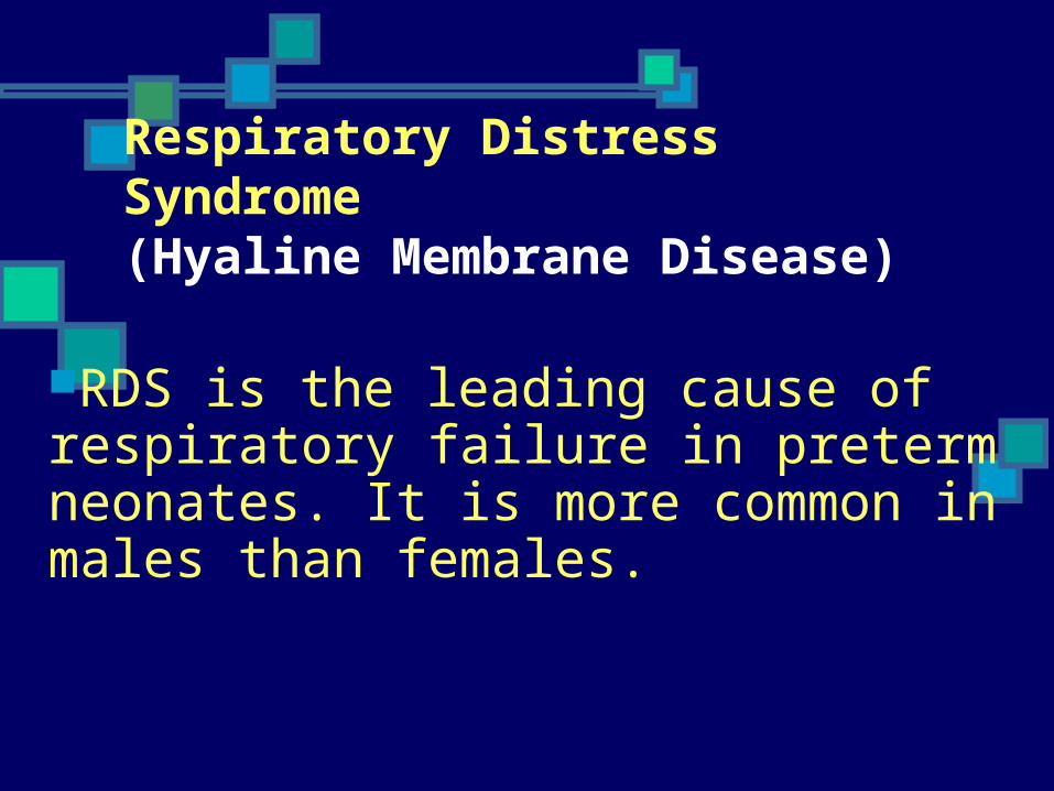
Respiratory Distress Syndrome (Hyaline Membrane Disease)
RDS is the leading cause of respiratory failure in preterm neonates. It is more common in males than females.
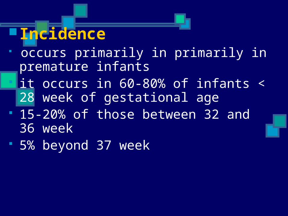
Incidence occurs primarily in primarily in
premature infants it occurs in 60-80% of infants < 28
week of gestational age 15-20% of those between 32 and 36
week 5% beyond 37 week
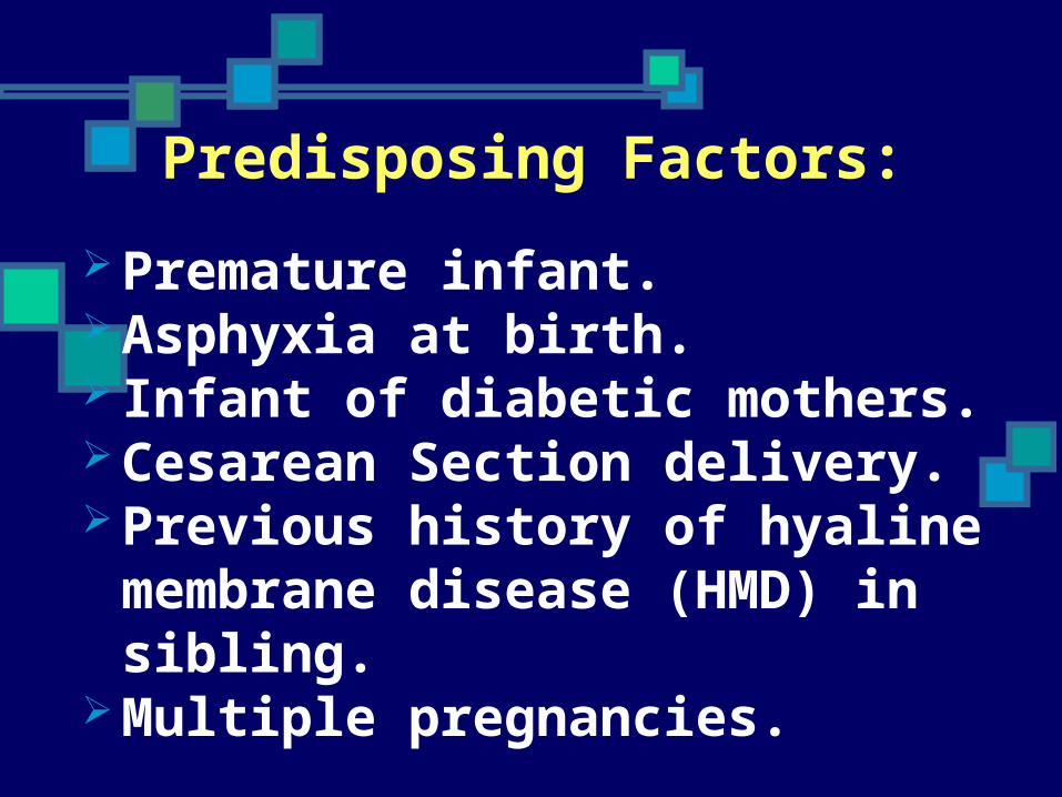
Predisposing Factors:
Premature infant. Asphyxia at birth. Infant of diabetic mothers. Cesarean Section delivery. Previous history of hyaline
membrane disease (HMD) in sibling. Multiple pregnancies.
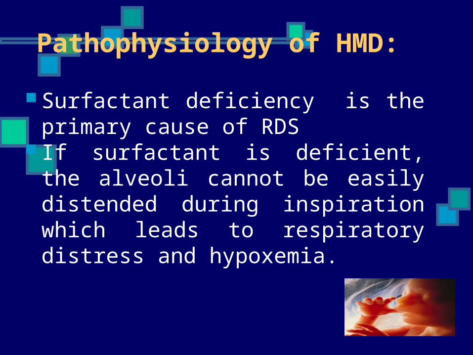
Pathophysiology of HMD:
Surfactant deficiency is the primary cause of RDS
If surfactant is deficient, the alveoli cannot be easily distended during inspiration which leads to respiratory distress and hypoxemia.
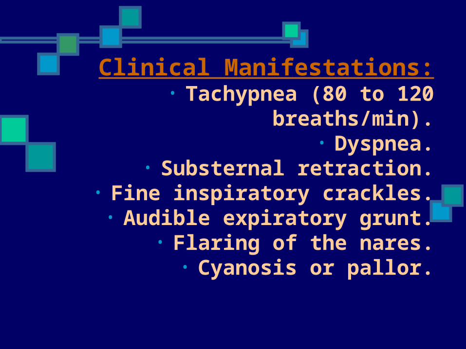
Clinical Manifestations:• Tachypnea (80 to 120 breaths/min).
• Dyspnea.• Substernal retraction.
• Fine inspiratory crackles.• Audible expiratory grunt.
• Flaring of the nares.• Cyanosis or pallor.
Diagnostic Tests:• Chest x-ray shows congested lung
field with a ground- glass appearance that represents alveolar
atelectasis, and dark streaks.
• Respiratory and metabolic acidosis is determined by blood gas
analysis.
Therapeutic Management
• *Maintain adequate ventilation and oxygenation.
*Oxygen should be warmed and humidified
*Maintain a neutral thermal environment
Therapeutic Management
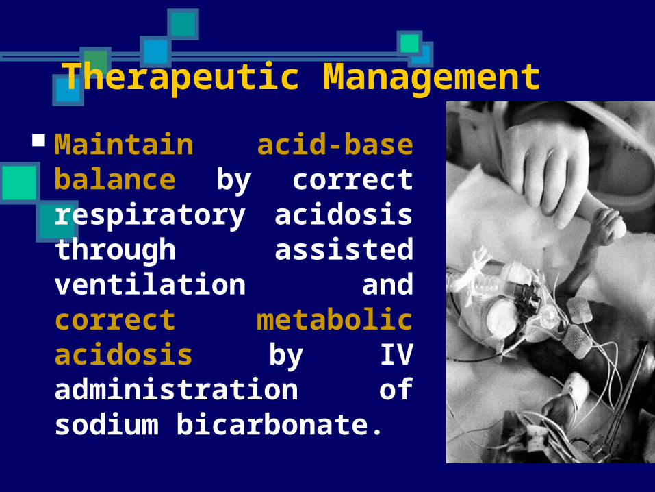
Maintain acid-base balance by correct respiratory acidosis through assisted ventilation and correct metabolic acidosis by IV administration of sodium bicarbonate.

Maintain adequate hydration and
electrolytes level. Nutrition is provided by parenteral
therapy during the acute stage. Surfactant therapy installed in
trachea.
• Nipple and gavage feeding are contraindicated in any situation
that creates a marked increase in respiratory rate because of the greater hazards of aspiration.
Prevention of HMD prevention of premature delivery.
Administration of corticosteroids to the mother (24 hours to 7 days before delivery).
Prophylactic administration of artificial surfactant into trachea of premature neonate.
Prognosis
• RDS is a self- limiting disease if mild, and following a period of
deterioration (approximately 48 hrs) and in the absence of complications,
affected neonates begin to improve by 72 hours.
Prognosis• Neonates who survive the first
96 hours have a reasonable chance of recovery. Surfactant
therapy decreased the use of long term ventilation and
decreased period of stay in hospital. It also improves the
outcome.

Prognosis• Neonates who survive the first
96 hours have a reasonable chance of recovery. Surfactant
therapy decreased the use of long term ventilation and
decreased period of stay in hospital. It also improves the
outcome.

Transient Tachypnea of the Newborn
Transient Tachypnea of the Newborn
usually follows uneventual normal preterm or term vaginal delivery or CS delivery
Is encountered by all physicians who take care of newborn infants
Clinical Manifestations
early onset of tachypnea Retractions respiratory grunting Occasionally cyanosis that is relieved by
minimal oxygen(<40%) Patient recovered rapidly within 3 days No rales or rhonchi X rays shows prominent vascular markings,
fluid in the intralobar fissures, ovearation, flat diaphragms
TTN vs RDS
TTN sudden recovery of infants
X ray findings of RDS ( hypoaeration, diffuse riticulogranules pattern, air bronchogram
Etiology
secondary to slow absorption of flat lung fluid resulting in decrease pulmonary compliance and tidal volume, increased dead space
Treatment and Management
supportive Observe for the development of respiratory
fatigue and signs of clinical deterioration that may suggest some other diagnosis
NEONATAL PNEUMONIA
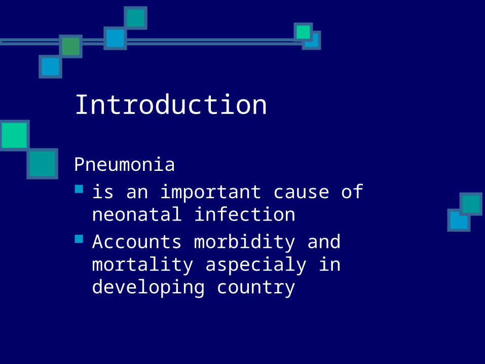
Introduction
Pneumonia is an important cause of neonatal
infection Accounts morbidity and mortality
aspecialy in developing country

Pathogenesis
Routes of acquisition: Varies in part with
the time of onset of pneumonia
Early – onset pneumonia Late - onset pneumonia
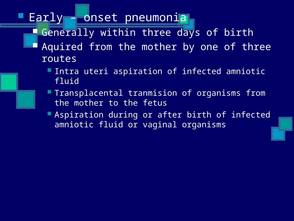
Early – onset pneumonia Generally within three days of birth Aquired from the mother by one of three routes
Intra uteri aspiration of infected amniotic fluid Transplacental tranmision of organisms from the
mother to the fetus Aspiration during or after birth of infected amniotic
fluid or vaginal organisms
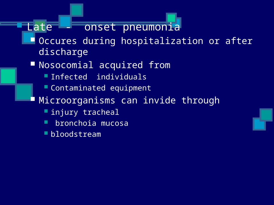
Late - onset pneumonia Occures during hospitalization or after
discharge Nosocomial acquired from
Infected individuals Contaminated equipment
Microorganisms can invide through injury tracheal bronchoia mucosa bloodstream
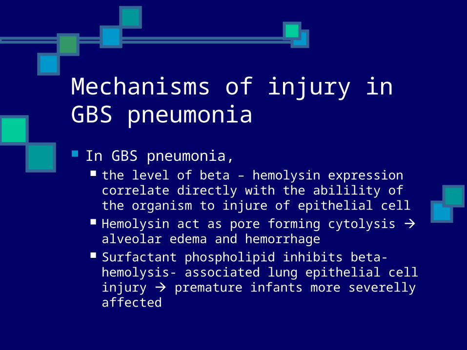
Mechanisms of injury in GBS pneumonia
In GBS pneumonia, the level of beta – hemolysin expression
correlate directly with the abilility of the organism to injure of epithelial cell
Hemolysin act as pore forming cytolysis alveolar edema and hemorrhage
Surfactant phospholipid inhibits beta- hemolysis- associated lung epithelial cell injury premature infants more severelly affected
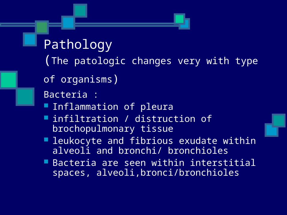
Pathology (The patologic changes very with type of
organisms) Bacteria : Inflammation of pleura infiltration / distruction of
brochopulmonary tissue leukocyte and fibrious exudate within
alveoli and bronchi/ bronchioles Bacteria are seen within interstitial
spaces, alveoli,bronci/bronchioles
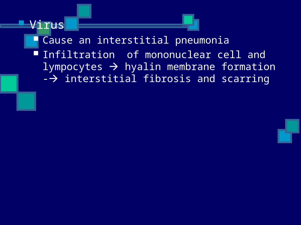
Virus Cause an interstitial pneumonia Infiltration of mononuclear cell and
lympocytes hyalin membrane formation - interstitial fibrosis and scarring

Microbiology
Cause : Bacterial Viral Spirochetal Protozoan Fungal pathogens
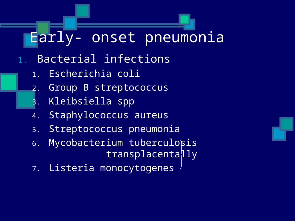
Early- onset pneumonia1. Bacterial infections
1. Escherichia coli2. Group B streptococcus3. Kleibsiella spp4. Staphylococcus aureus5. Streptococcus pneumonia6. Mycobacterium tuberculosis
transplacentally7. Listeria monocytogenes
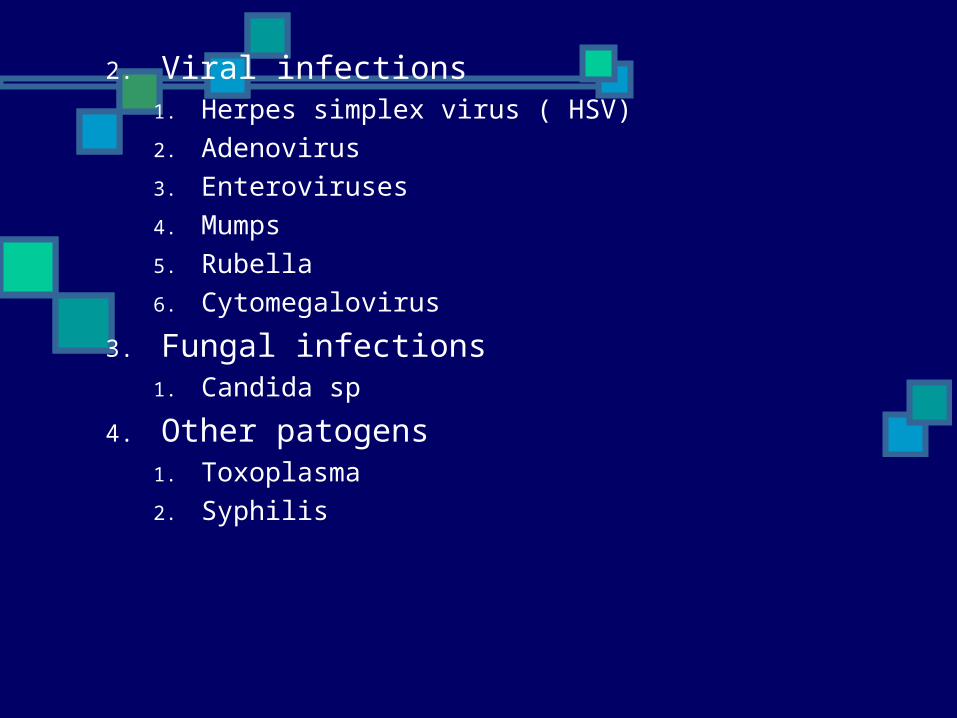
2. Viral infections1. Herpes simplex virus ( HSV) 2. Adenovirus3. Enteroviruses4. Mumps 5. Rubella6. Cytomegalovirus
3. Fungal infections1. Candida sp
4. Other patogens 1. Toxoplasma2. Syphilis
Late – onset pneumonia
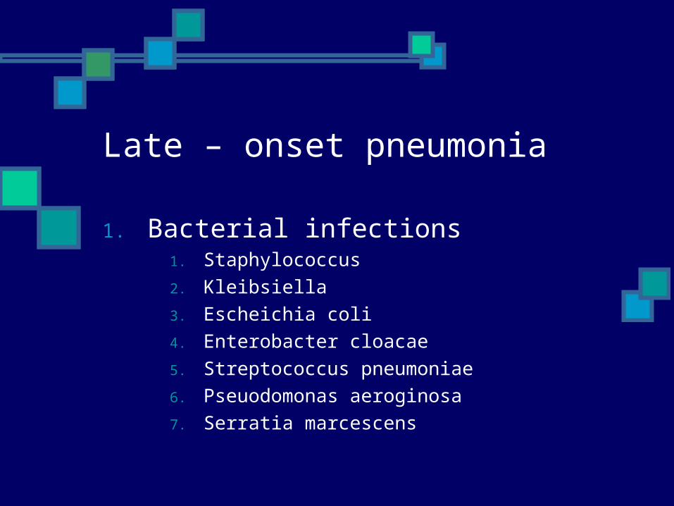
1. Bacterial infections1. Staphylococcus2. Kleibsiella3. Escheichia coli4. Enterobacter cloacae5. Streptococcus pneumoniae6. Pseuodomonas aeroginosa7. Serratia marcescens
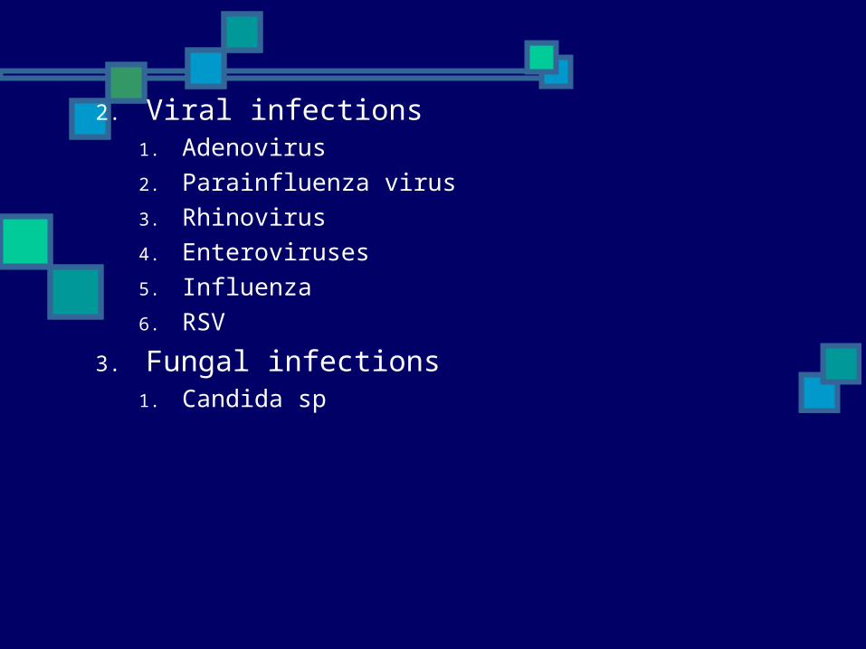
2. Viral infections1. Adenovirus 2. Parainfluenza virus3. Rhinovirus 4. Enteroviruses5. Influenza 6. RSV
3. Fungal infections1. Candida sp
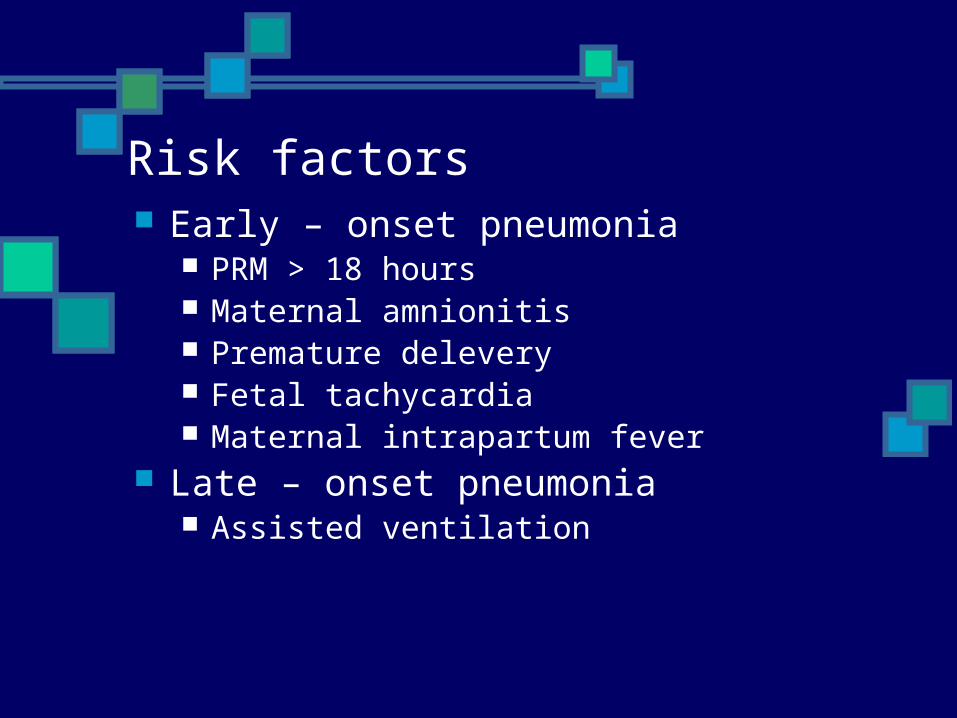
Risk factors Early – onset pneumonia
PRM > 18 hours Maternal amnionitis Premature delevery Fetal tachycardia Maternal intrapartum fever
Late – onset pneumonia Assisted ventilation
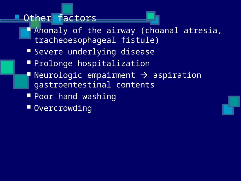
Other factors Anomaly of the airway (choanal atresia,
tracheoesophageal fistule) Severe underlying disease Prolonge hospitalization Neurologic empairment aspiration
gastroentestinal contents Poor hand washing Overcrowding
Clinical manifestation
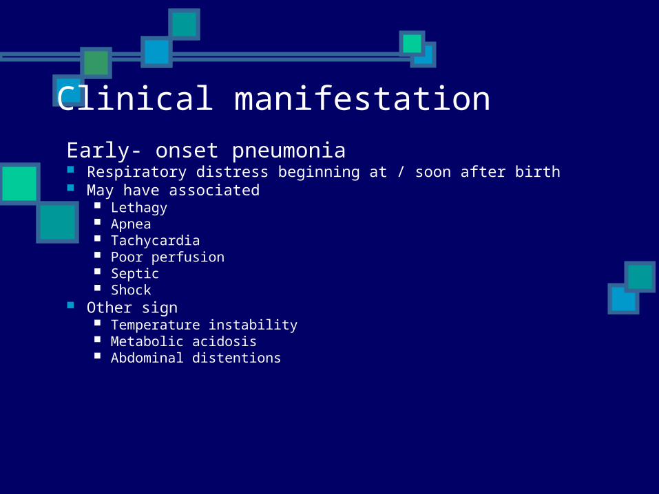
Early- onset pneumonia Respiratory distress beginning at / soon after birth May have associated
Lethagy Apnea Tachycardia Poor perfusion Septic Shock
Other sign Temperature instability Metabolic acidosis Abdominal distentions
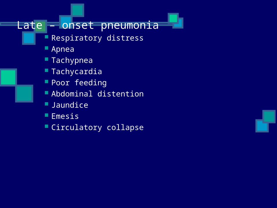
Late – onset pneumonia Respiratory distress Apnea Tachypnea Tachycardia Poor feeding Abdominal distention Jaundice Emesis Circulatory collapse
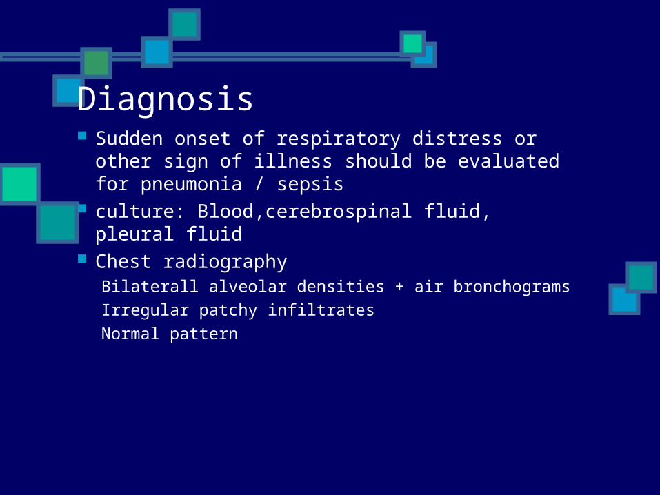
Diagnosis Sudden onset of respiratory distress or other
sign of illness should be evaluated for pneumonia / sepsis
culture: Blood,cerebrospinal fluid, pleural fluid
Chest radiographyBilaterall alveolar densities + air bronchogramsIrregular patchy infiltratesNormal pattern
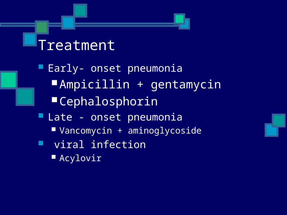
Treatment Early- onset pneumonia
Ampicillin + gentamycinCephalosphorin
Late - onset pneumonia Vancomycin + aminoglycoside
viral infection Acylovir