Pulmonary Actinomycosis: CT Findings...(b) CT scan at the level of subcarina shows mass-like...
6
&}J 0. iæ pp. 90 1- 906. 1989 Journal of Kor ea n Radiological Society. 25(6) 901-906. 1989 Pulmonary Actinomycosis: CT Findings Chun Hwan Han , M.D. , Jung-Gi 1m, M.D. , Kil Sun Park , M.D. , Man Chung Han , M.D. Department of Radi ol ogy, Coll ege of Medi cine, Seo ul National University conso li dation ) , 1 CT 1- 2 cm Index Words: Lung, mycos is Lung , CT Ac tinomycos is Pulmonary actinomycosis is believed to be a rare disease. The radiological manifestations of the disease are for the most part nonspecific but the chest wall lesion is strongly suggestive of acti- nomycosis 1 ). As this classical presentation with chest wall involvement is uncommon by early administration of antibiotics , the diagnosis of pu- lmonary actinomycosis is easly missed. The a u- Re cei ved Septem ber 1, Accepted Septem ber 12. 1989 thors performed a retrospective analysis of five proved cases of actinomycosis and described the chest radiograph and CT findings Case Reports Case 1. A man had a six months' duration of chest pain and developed mild fever and cough with blood-tinged sputum. Antituberculosis the- ra py was not effective. Sputum smear and culture for tubercle bacilli was negative. WBC count on - 901 -
Transcript of Pulmonary Actinomycosis: CT Findings...(b) CT scan at the level of subcarina shows mass-like...
大 韓 放 껴 t 1없 찮 &}J 0. iæ t:í~25찬 第6i>>,~ pp. 90 1- 906. 1989 Journal of Korean Radio log ical Society. 25(6) 901-906. 1989
Pulmonary Actinomycosis: CT Findings
Chun Hwan Han, M.D. , Jung-Gi 1m, M.D. , Kil Sun Park, M.D. ,
Man Chung Han, M.D.
Department of Radiology, College of Medicine, Seo ul Nation al University
〈국문초록〉
흉부 방선상균병 (Actinomycosis)의 전산화단층촬영 소견
서울대학교 의파대학 방사선과학교실
한춘환·임정기·박길선·한만청
흉부의 방선상균뱅 ( Actinom ycosis )은 근래 항생제의 보편화된 사용으로 방사선학석 소견을 나
타내는 상태에서 발견되는 경우가 애우 회귀하으로 방사선학적 강벨 잔안에 포함되지 않는 경우
가대부분이마. 저자둥은최근폐 앙선상균영으로진단된 5예의 단순흉부 X선 및 CT 소견을정
리보고한마.
환자의 임상 증상은 기칭 및 혈담 그러고 흉부 동통 둥이었고 발열은 심하지 않았다. 단순 흉부
X선에서는 4예에서 만성적인 경계가 불분영한 기공경결 (ai r- s pace conso lidation ), 흑은 종괴양
의 소견을 보였다 CT상 챔염부위는 좌우폐 , 흑은 특정 폐엽의 호말 경향을 보이지 않았으며 , 1
내지 2개의 대엽을 청엄하였마. CT 양상은 4예에서 종괴양의 펴1 경결을 보였으며 3예에서 는 공동
파 내부에 괴사를 시사하는 처음영도 부위를 포함하고 있었마. 흉막바후가 전례에서 있었A며 그
중 3예에서 늑막삼출이 판찰되었다. 3예 에서 종격동 럼프절이 직경 1-2 cm 정도로 매대해 있
었마. 폐엽간열을 통파하는 영변의 파급은 1예에서 판창되었다.
흉부방선상균뱅은만성의 페 경결 및 공동이 주된소견으로 폐암또는 폐 결핵 둥과강벨을요하
며 상기의 진단이 배제 될 경우 이 질환의 가능성을 고려하여야 할 것£로 사료된다.
Index Words: Lung, mycos is
Lung , CT
Actinomycos is
Pulmonary actinomycosis is believed to be a
rare disease. The radiological manifestations of the
disease are for the most part nonspecific but the
chest wall lesion is strongly suggestive of acti
nomycosis1). As this classical presentation with
chest wall involvement is uncommon by early
administration of antibiotics , the diagnosis of pu
lmonary actinomycosis is easly missed. The au-
이 논문은 1 989년 9월 1 일 접수히여 1989년 9월 1 2일에 채 택되었음 Received September 1, Accepted September 12. 1989
thors performed a retrospective analysis of five
proved cases of actinomycosis and described the
chest radiograph and CT findings
Case Reports
Case 1.
A 65-year-이d man had a six months ' duration
of chest pain and developed mild fever and cough
with blood-tinged sputum. Antituberculosis the
rapy was not effective. Sputum smear and culture
for tubercle bacilli was negative . WBC count on
- 901 -
- 大韓放射線옆學會註 · 第25卷 第 6 號 1989 -
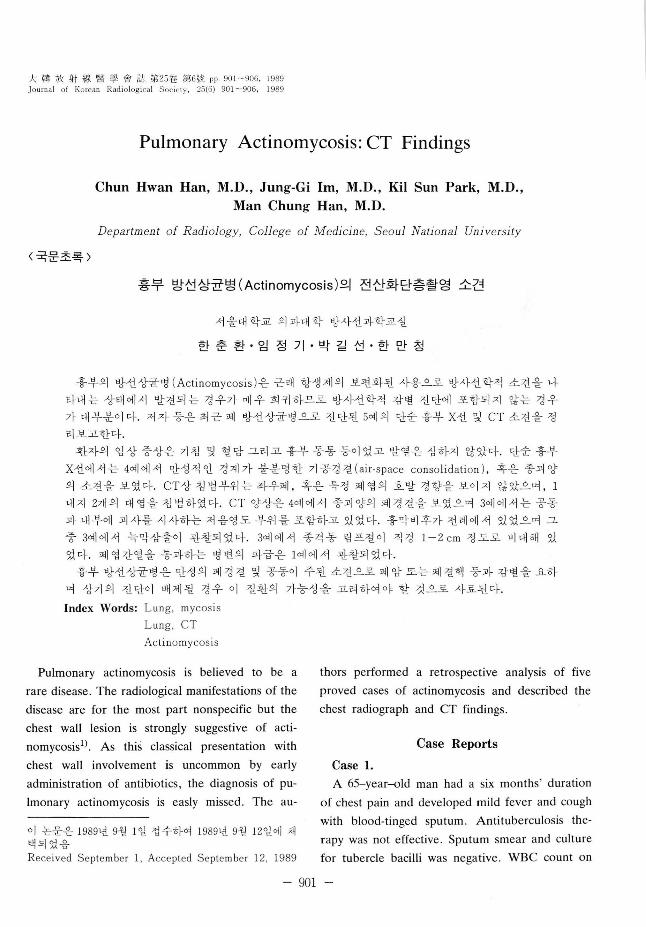
admission was 13,000. A chest radiograph showed
streaky hazy density in the upper division of the
left upper lobe (Fig. la) and later became a mass
like consolidation(Fig. lb). CT scans showed a
nodular marginated mass-like lesion and a cavity
with air-fluid level (Fig. lc) . Transpleural exte
nsion of consolidation from the left upper lobe to
the left lower lobe was demonstrated (Fig. ld).
c d
Pleural thickening was visible along the peripheral
margin of the mass-like lesion. Mediastinal lymph
nodes were aggregated and enlarged about 1 cm in
diameter. Bronchoscopy revealed mild luminal
narrowing of the posterior segment of the left up
per lobe without a endotracheal lesion. Percutane
ous fine needle aspiration biopsy confirmed acti
nomycosls .
‘
Fig. 1. Serial chest radiographs(a , b) and CT scans (c , d) in a 65-year old male patient (case 1) (a) Ill-defined hazy air-space consolidation associated with linear densities is seen in the left upper peripheral lung field (b) Chest radiograph taken seven months after (a) shows peripheral consolidation- like lesion which has bee n increased in area and density (c) CT scan at the leve l of the tracheal carina obtained ten days before (b) shows peripherally located air-space consolidation in the anterior segment of the lelt upper lobe with re latively sharp transition 01 the normal lung and lobulated border , especially posteriorly , mimicking a solid mass (arrowheads). Note also cavity with air fluid level (arrow) with in the consolidated lung and enlarged , aggregated lym ph nodes in A - P window area (d) Lung window setting of (c) show irregular marginated posterior border of the lung lesion extending beyond the expected position 01 the major fi ssure (arrowheads)
- 902
- Chun Hwan Han‘ et al:Pulmonary Actinomycosis -
Case 2.
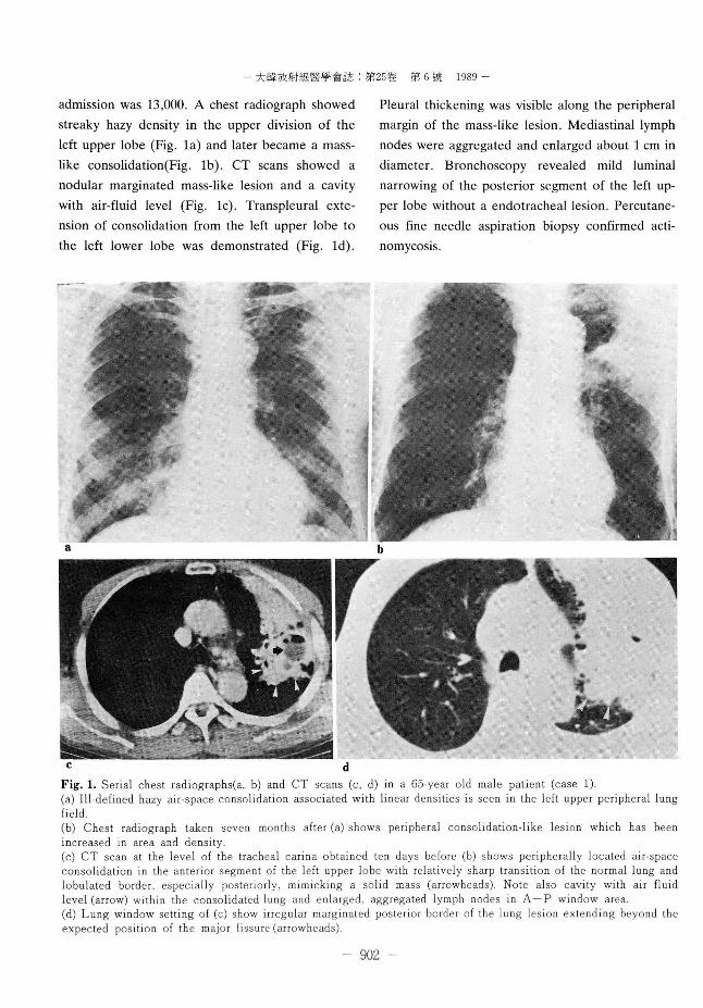
A 65-year-old male patient presented about six
months' history of cough and occasional hemopty
sis. Mild fever and dyspnea occurred. WBC count
was within normal range _ A chest radiograph sho
wed a mass-Iike lesion in the right middle lobe .
CT scans showed a mass-Iike consolidation in the
right middle lobe , the right lower lobe and the left
lower lobe (Fig . 2b). Pleural thickening and bi
lateral effusion were identified. Right tracheobr
onchial and subcarinal lymph nodes were enlarged
about 1- 2 cm in diameter and a calcified node was
detected (Fig . 2a) . Bronchoscopy showed a bronc
hial narrowing of the right middle lobe , but no en
dobronchial mass was found . Percutaneous aspir
ation biopsy confirmed the globoid colonies of
organisms , highly suggesting actinomycosis . The
patient was treated with penicillin for 6 months
and made a recovery.
Case 3.
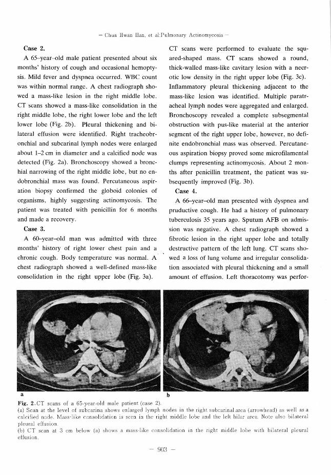
A 60-year-이d man was admitted with three
months' history of right lower chest pain and a
chronic cough. Body temperature was normal. A
chest radiograph showed a well-defined mass-Iike
consolidation in the right upper lobe (Fig. 3a) .
CT scans were performed to evaluate the squ
ared-shaped mass . CT scans showed a round , thick-walled mass-Iike cavitary lesion with a necr
otic low density in the right upper lobe (Fig . 3c) .
Inflammatory pleural thickening adjacent to the
mass-Iike lesion was identified. Multiple paratr
acheal lymph nodes were aggregated and enlarged.
Bronchoscopy revealed a complete subsegmental
obstruction with pus-Iike material at the anterior
segment of the right upper lobe , however , no defi
nite endobronchial mass was observed . Percutane
ous aspiration biopsy proved some microfilamental
clumps representing actinomycosis. About 2 mon
ths after penicillin treatment , the patient was su
bsequently improved (Fig. 3b)
Case 4.
A 66-year-이d man presented with dyspnea and
pruductive cough . He had a history of pulmonary
tuberculosis 35 years ago . Sputum AFB on admis
sion was negative. A chest radiograph showed a
fibrotic lesion in the right upper lobe and totally
destructive pattern of the left lung. CT scans sho
wed a loss of lung volume and irregular consolida
tion associated with pleural thickening and a small
amount of effusion. Left thoracotomy was perfor-
a b
Fig. 2. CT scans 01 a 65-year-o ld mal e patient (case 2) (a) Scan at the leve l of subca ri na shows enlarged lymph nodes in the right subcarinal area (a rrow head) as we ll as a calcified node. Mass' like conso lidation is seen in th e right mi dd le lobe and the left hi lar area. No te also bi lateral pleural effusion (b) CT scan at 3 cm be low (a) shows a mass- like consolidat ion in the right midd le lobe with bil ate ral pleura l effusion
m m
a
c
大韓放射線짧볕會誌 : 第25卷 第 6 號 1989 -
med. The pleural cavity was obliterated with thick
adhesion from the apex to the diaphragm. Left
lung was destroyed with a caseation material. On
pathological examination a small lesion of acti
nomycosis was found within the destroyed lung by
tuberculosis.
Case 5.
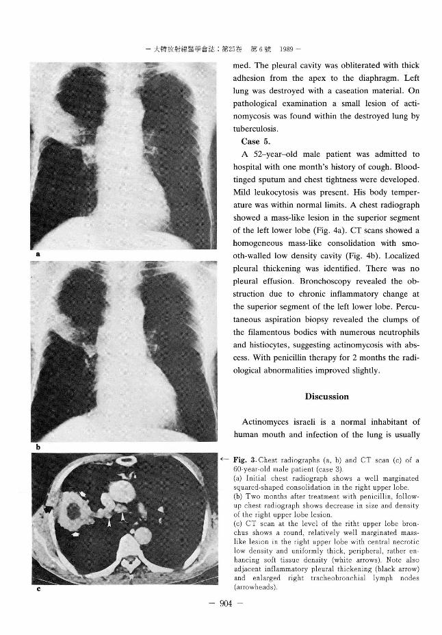
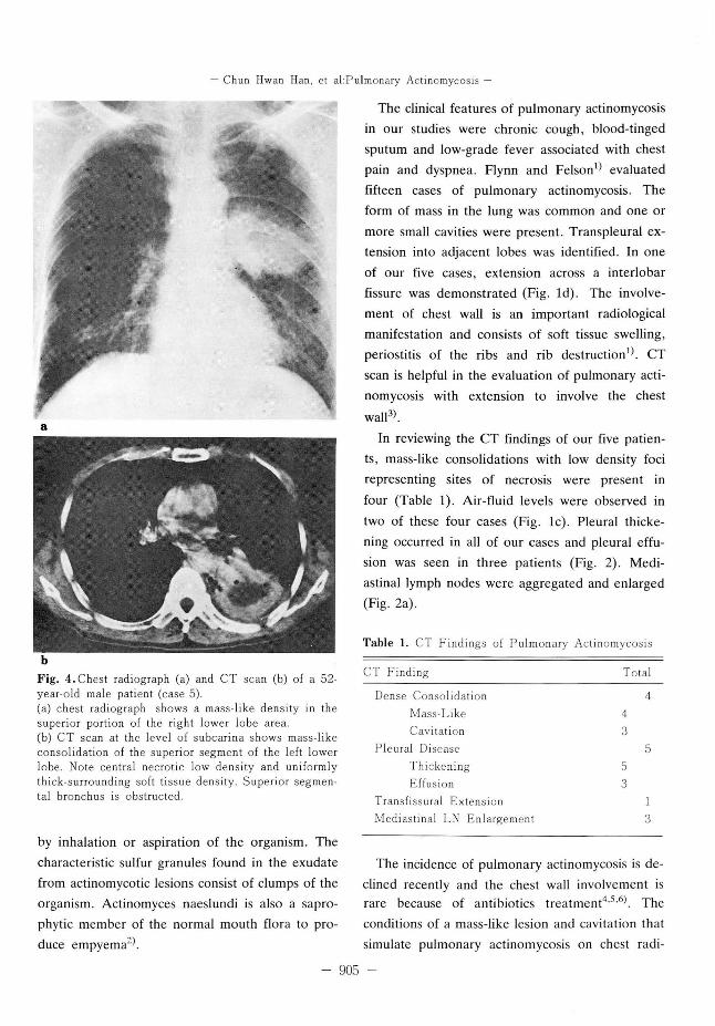
A 52-year-old male patient was admitted to
hospital with one month’s history of cough. Blood
tinged sputum and chest tightness were developed .
Mild leukocytosis was present. His body temper
ature was within normal limits . A chest radiograph
showed a mass-like lesion in the superior segment
of the left lower lobe (Fig. 4a) . CT scans showed a
homogeneous mass-like consolidation with smo
oth-walled low density cavity (Fig. 4b). Localized
pleural thickening was identified . There was no
pleural effusion. Bronchoscopy revealed the ob
struction due to chronic inflammatory change at
the superior segment of the left lower lobe . Percu
taneous aspiration biopsy revealed the clumps of
the filamentous bodies with numerous neutrophils
and histiocytes, suggesting actinomycosis with abs
cess. With penicillin therapy for 2 months the radi
ological abnormalities improved slightly.
Discussion
Actinomyces israeli is a normal inhabitant of
human mouth and infection of the lung is usually
• Fig. 3. Chest radiographs (a, b) and CT scan (c) of a 60-year-old male patient (case 3)
- 904
(a) lnitial chest radiograph shows a well marginated squared-shaped consolidation in the right upper lobe (b) Two months after treatment with penicillin , followup chest radiograph shows decrease in size and density of the right upper lobe lesion (c) CT scan at the level of the rith t upper lobe bronchus shows a round , relatively well marginated mass like lesion in the right upper lobe with central necrotic low density and unifo rmly thick, peripheral, rather enhancing soft tiss ue density (white arrows). Note al s。
adjacent inflammatory pleural thickening (black arrow) and enlarged right tracheo bronchial lymph nodes (arrowheads)
a
- Chun Hwan Han, et al:P ulmonary Act inomycos is
b Fig. 4. Chest radiograph (a) and CT scan (b) of a 52-year-o ld male patient (case 5) (a) chest radi ograph shows a mass- li ke density in the superior portion of the right lower lobe area (b) CT scan at the level of subcarina shows mass -like conso lidation of the superior segment of the left lower lobe. Note ce ntral necrotic low density and uniformly thick-surrounding soft tissue density. Superior segmental bronchus is obstructed
The c1inical features of pulmonary actinomycosis
in our studies were chronic cough , blood-tinged
sputum and low-grade fever associated with chest
pain and dyspnea. Flynn and Felson l) evaluated
fifteen cases of p비monary actinomycosis . The
form of mass in the lung was common and one or
more small cavities were presen t. Transpleural ex
tension into adjacent lobes was identified. In one
of our five cases , extension across a interlobar
fissure was demonstrated (Fig. 1d). The involve
ment of chest wall is an important radiological
manifestation and consists of soft tissue swelling , periostitis of the ribs and rib destruction 1). CT
scan is helpful in the evaluation of p비monary acti
nomycosis with extension to involve the chest
wall3)
In reviewing the CT findings of our five patien
ts , mass-like consolidations with low density foci
representing sites of necrosis were present in
four (Table 1). Air-fluid levels were observed in
two of these four cases (Fig . 1c) . Pleural thicke
ning occurred in all of our cases and pleural effu
sion was seen in three patients (Fig . 2). Medi
astinal Iymph nodes were aggregated and enlarged
(Fig . 2a).
Table 1. CT Fi nd ings 01 P ulmonary Act in omycosis
CT Finding Total
4
4
3
5 5
3
3
Dense Consolidation Mass.Like Cavitation
P leural Disease Thickening Effu sion
Translissural Exte nsion Mediasti nal LN Enl argement
by inhalation or aspiration of the organism. The
characteristic sulfur granules found in the exudate The incidence of pulmonary actinomycosis is de-
from actinomycotic lesions consist of c1umps of the c1ined recently and the chest wa ll involvement is
organism . Actinomyces naeslundi is also a sapro- rare because of antibiotics treatment4 ,5 ,6) The
phytic member of the normal mouth flora to pro- conditions of a mass-like lesion and cavitation that
duce empyema2). simulate pulmonary actinomycosis on chest radi-
%
- 大韓放射線훌훌學會註 : 第25卷 第 6 號 1989 -
ograph are lung cancer and pulmonary tubercu
losis7l . Indeed, lung cancer and pulmonary tuber
culosis are more common than actinomycosis and
tend to involve the chest wall frequently , so that
pulmonary actinomycosis is hardly considered to
be included in the differential diagnosis of mass
like and cavitary lung lesions.
REFERENCES
1. Flynn ME , Felson B. The roentgen manifestations
of thoracic actinomycosis. A]R 110:707-716, 1970
2. Karetzky MS , Garvey ]W. Empyema due to Acti.
nomycosis naeslundi. Chest 65:229-230, 1974
3. Webb WR , Sagel SS. Actinomycosis involving the
chest wall: CT findings. A]R 139:1007-1009, 1982
4. Frank P , Strickland B. Pulmonary actinomycosis.
Br ] Radiol 47:373-378, 1974
5. Slade PR , Slesser BV, Southgate J Thoracic acti
nomycosis. Thorax 28:73-85, 1973
6. Allen III HA , Scatarige ]C , Kim MH. A ctinomy
COSlS ‘ CT findings in six patients. A]R
149:1255-1258, 1987
7. Balikian ]P , Cheng TH , Costello P , Herman PG
Pulmonary actinomycosis. Radiology 128:613-616,
1978
- 906 -