Ptosis and Blepharoplasty Surgery - Larrabee Center for ... · edema from trauma or allergies,...
4
Ptosis and Blepharoplasty Surgery Donna J. Millay, MD, Wayne F. Larrabee, Jr, MD \s=b\ Acquired senile ptosis of the upper eyelid is a common condition in the elder- ly population and is often accompanied by varying amounts of dermatochalasis, which may mask its presence. Blepharo- plasty of the ptotic lid, without repair of the ptosis, may result in exaggeration of the drooping eyelid postoperatively. Acquired senile ptosis is produced by dehiscence or disinsertion of the levator aponeurosis and is characterized by a high or absent eyelid crease, thinning of the upper eyelid tissue, and normal leva- tor function. Frequent causes of acquired ptosis include dermatochalasis, eyelid edema from trauma or allergies, previous ocular surgery, or use of contact lenses. When discovered, repair of the ptosis should be performed at the time of bleph- aroplasty to produce the best functional and cosmetic result. It is important for every surgeon who performs blepharo- plasties to be aware of the presentation and management of acquired senile pto- sis for consistent surgical results. (Arch Otolaryngol Head Neck Surg 1989;115:198-201) Acquired upper lid ptosis is a com- -i\. mon condition in the elderly population and is often accompanied by varying amounts of dermatochala¬ sis. Blepharoplasty of the ptotic lid may result in exaggeration of the drooping eyelid postoperatively, with a poor cosmetic result. It is important for every surgeon who performs blepharoplasties to be able to recog¬ nize this problem and manage it appropriately. To that end, important aspects of acquired senile ptosis, including etiology, pathophysiology, physical diagnosis, and surgical repair, will be discussed. Relevant anatomy of the eyelid will also be presented. ANATOMY AND PHYSIOLOGY The anatomy of the upper eyelid is the real key to understanding ptosis and ptosis surgery (Fig l).1·2 The upper eyelid consists of several layers of tissue that are all distinct structures. The outermost layer of the eyelid is the skin, which is unique in that it is the thinnest skin in the body, with essentially no subcutaneous fat. The orbicularis oculi muscle lies just below the skin and is divided into a pretarsal and preseptal portion. The lower third of the pretarsal segment is firmly adherent to the lower third of the tarsus, while the remainder is attached to the levator aponeurosis. The orbital septum, found just be¬ neath the preseptal portion of the orbicularis oculi muscle, is a fibrous layer of tissue that originates from the periorbital periosteum and ex¬ tends downward to fuse with the leva- tor aponeurosis, about 2 to 3 mm above the upper border of the tarsus. The next structure identified is the preaponeurotic fat pad of the upper eyelid, which is found in a pocket beneath the orbital septum and lying atop the levator aponeurosis. This fat pad is a key anatomic structure in distinguishing the orbital septum from the levator aponeurosis, two structures that may have a similar appearance at surgery. The levator palpebrae muscle and its aponeurosis are identified with upward retraction of the pad. The muscle originates from the lesser wing of the sphenoid at the orbital apex and extends for¬ ward for 40 mm, with its aponeurosis continuing for another 15 mm. At approximately 10 mm above the tar¬ sus, the muscle divides into an anteri¬ or lamellae, which becomes the fibrous levator aponeurosis, and a posterior lamellae. The aponeurosis inserts on the anterior inferior two thirds of the tarsus as well as the pretarsal orbicularis oculi muscle and skin. The posterior lamellae contains nonstriated muscle fibers, Müller's muscle, and inserts on the superior tarsal border. The innermost layer of the eyelid is conjunctiva, which is closely adherent to Müller's muscle. The last major structure of the upper eyelid is the tarsus, a plate of dense fibrous and elastic tissue, which pro¬ vides support for the upper eyelid. It measures 10 mm in height and pro¬ vides attachments for the levator apo¬ neurosis, Müller's muscle, and con¬ junctiva. Innervation of the eyelid includes the seventh nerve, which supplies the orbicularis oculi muscle, and the third nerve, which supplies the levator mus¬ cle. Müller's muscle is under sympa¬ thetic control and has a primary func¬ tion of maintaining eyelid tone. ETIOLOGY There are many etiologies of acquired ptosis, but the list is greatly reduced when considering the other¬ wise healthy elderly population. In patients with good levator function, the usual cause of acquired ptosis is thinning and stretching or disruption of the levator aponeurosis, which occurs when the minimal attachment of the aponeurosis to the tarsal plate is insufficient to withstand any exces¬ sive pull. There are many common Accepted for publication Dec 2, 1988. From the Department of Reconstructive and Plastic Surgery, Virginia Mason Clinic, Seattle. Read before the American Academy of Facial Plastic and Reconstructive Surgery, Palm Beach, Fla, April 29, 1988. Reprint requests to Virginia Mason Clinic, 33501 First Way S, Federal Way, WA 98003 (Dr Millay). DownloadedFrom:http://jamanetwork.com/byWayneLarrabeeon11/08/2016
Transcript of Ptosis and Blepharoplasty Surgery - Larrabee Center for ... · edema from trauma or allergies,...
Ptosis and Blepharoplasty SurgeryDonna J. Millay, MD, Wayne F. Larrabee, Jr, MD
\s=b\Acquired senile ptosis of the uppereyelid is a common condition in the elder-ly population and is often accompaniedby varying amounts of dermatochalasis,which may mask its presence. Blepharo-plasty of the ptotic lid, without repair ofthe ptosis, may result in exaggeration ofthe drooping eyelid postoperatively.Acquired senile ptosis is produced bydehiscence or disinsertion of the levatoraponeurosis and is characterized by a
high or absent eyelid crease, thinning ofthe upper eyelid tissue, and normal leva-tor function. Frequent causes of acquiredptosis include dermatochalasis, eyelidedema from trauma or allergies, previousocular surgery, or use of contact lenses.When discovered, repair of the ptosisshould be performed at the time of bleph-aroplasty to produce the best functionaland cosmetic result. It is important forevery surgeon who performs blepharo-plasties to be aware of the presentationand management of acquired senile pto-sis for consistent surgical results.
(Arch Otolaryngol Head Neck Surg1989;115:198-201)
Acquired upper lid ptosis is a com--i\. mon condition in the elderlypopulation and is often accompaniedby varying amounts of dermatochala¬sis. Blepharoplasty of the ptotic lidmay result in exaggeration of thedrooping eyelid postoperatively, witha poor cosmetic result. It is importantfor every surgeon who performsblepharoplasties to be able to recog¬nize this problem and manage itappropriately. To that end, important
aspects of acquired senile ptosis,including etiology, pathophysiology,physical diagnosis, and surgicalrepair, will be discussed. Relevantanatomy of the eyelid will also bepresented.
ANATOMY AND PHYSIOLOGYThe anatomy of the upper eyelid is
the real key to understanding ptosisand ptosis surgery (Fig l).1·2 The uppereyelid consists of several layers oftissue that are all distinct structures.The outermost layer of the eyelid isthe skin, which is unique in that it isthe thinnest skin in the body, withessentially no subcutaneous fat. Theorbicularis oculi muscle lies justbelow the skin and is divided into a
pretarsal and preseptal portion. Thelower third of the pretarsal segmentis firmly adherent to the lower thirdof the tarsus, while the remainder isattached to the levator aponeurosis.The orbital septum, found just be¬neath the preseptal portion of theorbicularis oculi muscle, is a fibrouslayer of tissue that originates fromthe periorbital periosteum and ex¬tends downward to fuse with the leva-tor aponeurosis, about 2 to 3 mmabove the upper border of the tarsus.The next structure identified is thepreaponeurotic fat pad of the uppereyelid, which is found in a pocketbeneath the orbital septum and lyingatop the levator aponeurosis. This fatpad is a key anatomic structure indistinguishing the orbital septumfrom the levator aponeurosis, twostructures that may have a similarappearance at surgery. The levatorpalpebrae muscle and its aponeurosisare identified with upward retractionof the pad. The muscle originatesfrom the lesser wing of the sphenoid
at the orbital apex and extends for¬ward for 40 mm, with its aponeurosiscontinuing for another 15 mm. Atapproximately 10 mm above the tar¬sus, the muscle divides into an anteri¬or lamellae, which becomes thefibrous levator aponeurosis, and a
posterior lamellae. The aponeurosisinserts on the anterior inferior twothirds of the tarsus as well as thepretarsal orbicularis oculi muscle andskin. The posterior lamellae containsnonstriated muscle fibers, Müller'smuscle, and inserts on the superiortarsal border. The innermost layer ofthe eyelid is conjunctiva, which isclosely adherent to Müller's muscle.The last major structure of the uppereyelid is the tarsus, a plate of densefibrous and elastic tissue, which pro¬vides support for the upper eyelid. Itmeasures 10 mm in height and pro¬vides attachments for the levator apo¬neurosis, Müller's muscle, and con¬
junctiva.Innervation of the eyelid includes
the seventh nerve, which supplies theorbicularis oculi muscle, and the thirdnerve, which supplies the levator mus¬cle. Müller's muscle is under sympa¬thetic control and has a primary func¬tion of maintaining eyelid tone.
ETIOLOGYThere are many etiologies of
acquired ptosis, but the list is greatlyreduced when considering the other¬wise healthy elderly population. Inpatients with good levator function,the usual cause of acquired ptosis isthinning and stretching or disruptionof the levator aponeurosis, whichoccurs when the minimal attachmentof the aponeurosis to the tarsal plateis insufficient to withstand any exces¬
sive pull. There are many common
Accepted for publication Dec 2, 1988.From the Department of Reconstructive and
Plastic Surgery, Virginia Mason Clinic, Seattle.Read before the American Academy of Facial
Plastic and Reconstructive Surgery, Palm Beach,Fla, April 29, 1988.Reprint requests to Virginia Mason Clinic,
33501 First Way S, Federal Way, WA 98003 (DrMillay).
Downloaded From: http://jamanetwork.com/ by Wayne Larrabee on 11/08/2016

Fig 1.—Depiction of major structures of upper eyelid.
conditions that may cause a levatordehiscence or disinsertion. The addedweight of excessive skin in dermato¬chalasis may produce a levator dehis¬cence and is probably the most com¬mon cause of acquired senile ptosis.3Marked eyelid edema from trauma,surgery, or allergies may also result ina levator disinsertion. Mechanicaltrauma to the eyelids from the use ofcontact lenses may produce a levatordisinsertion at any age.4 Myastheniagravis should always be considered inthe differential diagnosis of acquiredptosis, particularly in the patient withsevere ptosis bilaterally. Regardlessof the etiology of levator dehiscence ordisinsertion, surgery can usuallyproduce good, reliable results.
EXAMINATION OF THE PATIENTWITH PTOSIS
A careful history should beobtained and a physical examinationshould be performed when a ptotic lid
is discovered.5 The history shouldinclude the time of onset, as a congen¬ital ptosis usually involves a dysfunc¬tional levator muscle and is treateddifferently from an acquired ptosis. Ahistory of other coexistent symptoms,particularly neurologic or muscular,should be obtained to rule out moreserious disease processes, such as
myasthenia gravis, tumors, or othercentral nervous system disorders. Ahistory of eyelid edema, surgery, orthe use of contact lenses is suggestiveof levator aponeurosis disinsertion.Once a history of benign acquired
ptosis is established the physicalexamination should take place. Thereare several characteristics that definean acquired senile ptosis on physicalexamination. There should be a highor absent eyelid crease and thinningof the upper eyelid tissue to the extentthat the iris may be visible throughthe closed eyelid. It is necessary thatthe levator muscle function normally
to diagnose a case as one of acquiredsenile ptosis. Eyes should be examinedin upward and downward gaze, as wellas in the primary position. In acquiredsenile ptosis, the amount of ptosisremains the same in relation to thenormal eyelid in all levels of gaze. Theamount of ptosis should be docu¬mented either by direct measurementor by estimation. For example, thenormal eyelid covers the superior lim-bus by 1 to 2 mm, and the distancefrom the superior limbus to the mid-pupillary point is 5.5 mm, so an eyelidat the midpupillary line would repre¬sent a ptosis of 3.5 to 4.5 mm. A ptosisof 1 to 2 mm is considered mild; 3 mm,moderate; and 4 mm or greater,severe. Levator muscle functionshould be measured in all patientswith lid ptosis. To evaluate function ofthe levator muscle, pressure is firstplaced over the brow with the thumbor finger to prevent participation ofthe frontalis muscle in lifting the eye-
Downloaded From: http://jamanetwork.com/ by Wayne Larrabee on 11/08/2016
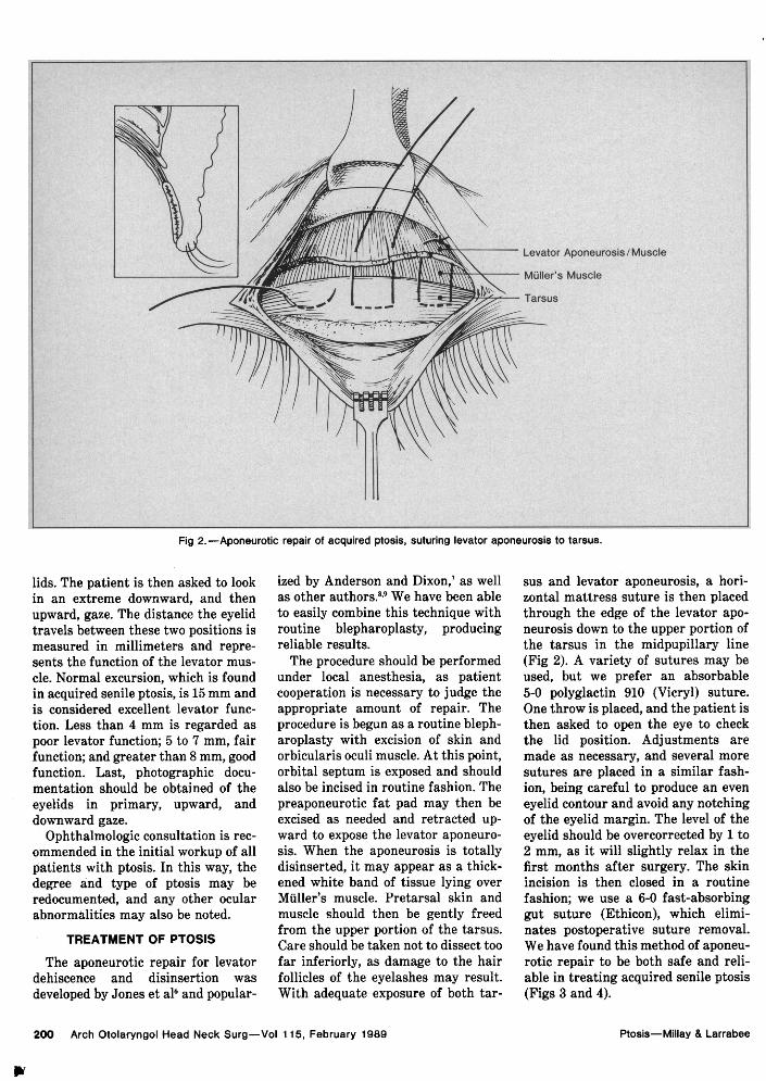
Fig 2.—Aponeurotic repair of acquired ptosis, suturing levator aponeurosis to tarsus.
lids. The patient is then asked to lookin an extreme downward, and thenupward, gaze. The distance the eyelidtravels between these two positions ismeasured in millimeters and repre¬sents the function of the levator mus¬cle. Normal excursion, which is foundin acquired senile ptosis, is 15 mm andis considered excellent levator func¬tion. Less than 4 mm is regarded as
poor levator function; 5 to 7 mm, fairfunction; and greater than 8 mm, goodfunction. Last, photographic docu¬mentation should be obtained of theeyelids in primary, upward, anddownward gaze.Ophthalmologic consultation is rec¬
ommended in the initial workup of allpatients with ptosis. In this way, thedegree and type of ptosis may beredocumented, and any other ocularabnormalities may also be noted.
TREATMENT OF PTOSIS
The aponeurotic repair for levatordehiscence and disinsertion was
developed by Jones et al6 and popular-
ized by Anderson and Dixon,7 as wellas other authors.8-9 We have been ableto easily combine this technique withroutine blepharoplasty, producingreliable results.The procedure should be performed
under local anesthesia, as patientcooperation is necessary to judge theappropriate amount of repair. Theprocedure is begun as a routine bleph¬aroplasty with excision of skin andorbicularis oculi muscle. At this point,orbital septum is exposed and shouldalso be incised in routine fashion. Thepreaponeurotic fat pad may then beexcised as needed and retracted up¬ward to expose the levator aponeuro¬sis. When the aponeurosis is totallydisinserted, it may appear as a thick¬ened white band of tissue lying over
Müller's muscle. Pretarsal skin andmuscle should then be gently freedfrom the upper portion of the tarsus.Care should be taken not to dissect toofar inferiorly, as damage to the hairfollicles of the eyelashes may result.With adequate exposure of both tar-
sus and levator aponeurosis, a hori¬zontal mattress suture is then placedthrough the edge of the levator apo¬neurosis down to the upper portion ofthe tarsus in the midpupillary line(Fig 2). A variety of sutures may beused, but we prefer an absorbable5-0 polyglactin 910 (Vicryl) suture.One throw is placed, and the patient isthen asked to open the eye to checkthe lid position. Adjustments aremade as necessary, and several moresutures are placed in a similar fash¬ion, being careful to produce an even
eyelid contour and avoid any notchingof the eyelid margin. The level of theeyelid should be overcorrected by 1 to2 mm, as it will slightly relax in thefirst months after surgery. The skinincision is then closed in a routinefashion; we use a 6-0 fast-absorbinggut suture (Ethicon), which elimi¬nates postoperative suture removal.We have found this method of aponeu¬rotic repair to be both safe and reli¬able in treating acquired senile ptosis(Figs 3 and 4).
Downloaded From: http://jamanetwork.com/ by Wayne Larrabee on 11/08/2016
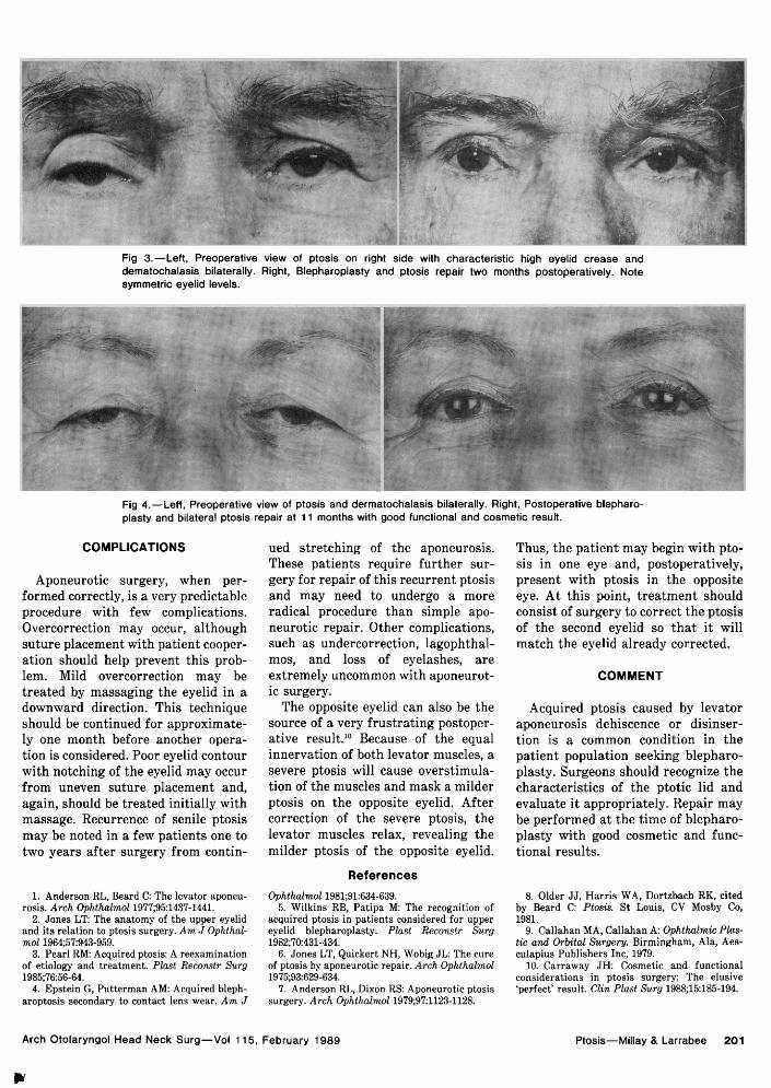
Fig 3.—Left, Preoperative view of ptosis on right side with characteristic high eyelid crease anddematochalasis bilaterally. Right, Blepharoplasty and ptosis repair two months postoperatively. Notesymmetric eyelid levels.
Fig 4.—Left, Preoperative view of ptosis and dermatochalasis bilaterally. Right, Postoperative blepharo¬plasty and bilateral ptosis repair at 11 months with good functional and cosmetic result.
COMPLICATIONS
Aponeurotic surgery, when per¬formed correctly, is a very predictableprocedure with few complications.Overcorrection may occur, althoughsuture placement with patient cooper¬ation should help prevent this prob¬lem. Mild overcorrection may betreated by massaging the eyelid in adownward direction. This techniqueshould be continued for approximate¬ly one month before another opera¬tion is considered. Poor eyelid contourwith notching of the eyelid may occurfrom uneven suture placement and,again, should be treated initially withmassage. Recurrence of senile ptosismay be noted in a few patients one totwo years after surgery from contin-
1. Anderson RL, Beard C: The levator aponeu¬rosis. Arch Ophthalmol 1977;95:1437-1441.
2. Jones LT: The anatomy of the upper eyelidand its relation to ptosis surgery. Am J Ophthal¬mol 1964;57:943-959.
3. Pearl RM: Acquired ptosis: A reexaminationof etiology and treatment. Plast Reconstr Surg1985;76:56-64.
4. Epstein G, Putterman AM: Acquired bleph¬aroptosis secondary to contact lens wear. Am J
ued stretching of the aponeurosis.These patients require further sur¬
gery for repair of this recurrent ptosisand may need to undergo a moreradical procedure than simple apo¬neurotic repair. Other complications,such as undercorrection, lagophthal¬mos, and loss of eyelashes, are
extremely uncommon with aponeurot¬ic surgery.The opposite eyelid can also be the
source of a very frustrating postoper¬ative result.10 Because of the equalinnervation of both levator muscles, asevere ptosis will cause overstimula-tion of the muscles and mask a milderptosis on the opposite eyelid. Aftercorrection of the severe ptosis, thelevator muscles relax, revealing themilder ptosis of the opposite eyelid.
References
Thus, the patient may begin with pto¬sis in one eye and, postoperatively,present with ptosis in the oppositeeye. At this point, treatment shouldconsist of surgery to correct the ptosisof the second eyelid so that it willmatch the eyelid already corrected.
COMMENT
Acquired ptosis caused by levatoraponeurosis dehiscence or disinser¬tion is a common condition in thepatient population seeking blepharo¬plasty. Surgeons should recognize thecharacteristics of the ptotic lid andevaluate it appropriately. Repair maybe performed at the time of blepharo¬plasty with good cosmetic and func¬tional results.
Ophthalmol 1981;91:634-639.5. Wilkins RB, Patipa M: The recognition of
acquired ptosis in patients considered for uppereyelid blepharoplasty. Plast Reconstr Surg1982;70:431-434.
6. Jones LT, Quickert NH, Wobig JL: The cureof ptosis by aponeurotic repair. Arch Ophthalmol1975;93:629-634.
7. Anderson RL, Dixon RS: Aponeurotic ptosissurgery. Arch Ophthalmol 1979;97:1123-1128.
8. Older JJ, Harris WA, Dortzbach RK, citedby Beard C: Ptosis. St Louis, CV Mosby Co,1981.
9. Callahan MA, Callahan A: Ophthalmic Plas-tic and Orbital Surgery. Birmingham, Ala, Aes-culapius Publishers Inc, 1979.
10. Carraway JH: Cosmetic and functionalconsiderations in ptosis surgery: The elusive'perfect' result. Clin Plast Surg 1988;15:185-194.
Downloaded From: http://jamanetwork.com/ by Wayne Larrabee on 11/08/2016