Psychosurgery and the limbic system DESMOND KELLY
9
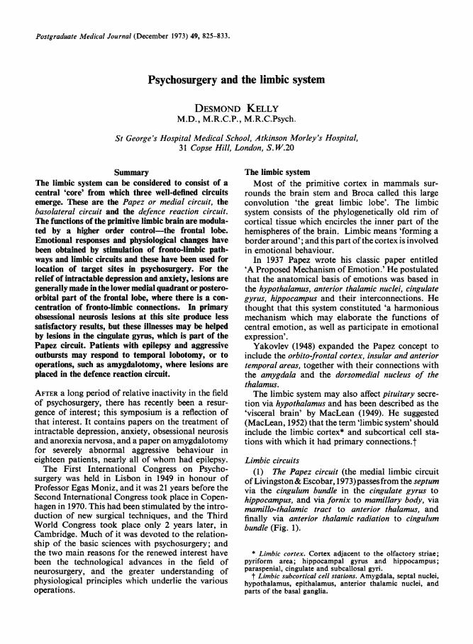
Postgraduate Medical Journal (December 1973) 49, 825-833. Psychosurgery and the limbic system DESMOND KELLY M.D., M.R.C.P., M.R.C.Psych. St George's Hospital Medical School, Atkinson Morley's Hospital, 31 Copse Hill, London, S. W.20 Summary The limbic system can be considered to consist of a central 'core' from which three well-defined circuits emerge. These are the Papez or medial circuit, the basolateral circuit and the defence reaction circuit. The functions of the primitive limbic brain are modula- ted by a higher order control-the frontal lobe. Emotional responses and physiological changes have been obtained by stimulation of fronto-limbic path- ways and limbic circuits and these have been used for location of target sites in psychosurgery. For the relief of intractable depression and anxiety, lesions are generally made in the lower medial quadrant or postero- orbital part of the frontal lobe, where there is a con- centration of fronto-limbic connections. In primary obsessional neurosis lesions at this site produce less satisfactory results, but these illnesses may be helped by lesions in the cingulate gyrus, which is part of the Papez circuit. Patients with epilepsy and aggressive outbursts may respond to temporal lobotomy, or to operations, such as amygdalotomy, where lesions are placed in the defence reaction circuit. AFTER a long period of relative inactivity in the field of psychosurgery, there has recently been a resur- gence of interest; this symposium is a reflection of that interest. It contains papers on the treatment of intractable depression, anxiety, obsessional neurosis and anorexia nervosa, and a paper on amygdalotomy for severely abnormal aggressive behaviour in eighteen patients, nearly all of whom had epilepsy. The First International Congress on Psycho- surgery was held in Lisbon in 1949 in honour of Professor Egas Moniz, and it was 21 years before the Second International Congress took place in Copen- hagen in 1970. This had been stimulated by the intro- duction of new surgical techniques, and the Third World Congress took place only 2 years later, in Cambridge. Much of it was devoted to the relation- ship of the basic sciences with psychosurgery; and the two main reasons for the renewed interest have been the technological advances in the field of neurosurgery, and the greater understanding of physiological principles which underlie the various operations. The limbic system Most of the primitive cortex in mammals sur- rounds the brain stem and Broca called this large convolution 'the great limbic lobe'. The limbic system consists of the phylogenetically old rim of cortical tissue which encircles the inner part of the hemispheres of the brain. Limbic means 'forming a border around'; and this part of the cortex is involved in emotional behaviour. In 1937 Papez wrote his classic paper entitled 'A Proposed Mechanism of Emotion.' He postulated that the anatomical basis of emotions was based in the hypothalamus, anterior thalamic nuclei, cingulate gyrus, hippocampus and their interconnections. He thought that this system constituted 'a harmonious mechanism which may elaborate the functions of central emotion, as well as participate in emotional expression'. Yakovlev (1948) expanded the Papez concept to include the orbito-frontal cortex, insular and anterior temporal areas, together with their connections with the amygdala and the dorsomedial nucleus of the thalamus. The limbic system may also affect pituitary secre- tion via hypothalamus and has been described as the 'visceral brain' by MacLean (1949). He suggested (MacLean, 1952) that the term 'limbic system' should include the limbic cortex* and subcortical cell sta- tions with which it had primary connections.t Limbic circuits (1) The Papez circuit (the medial limbic circuit of Livingston& Escobar, 1973)passes from the septum via the cingulum bundle in the cingulate gyrus to hippocampus, and via fornix to mamillary body, via mamillo-thalamic tract to anterior thalamus, and finally via anterior thalamic radiation to cingulum bundle (Fig. 1). * Limbic cortex. Cortex adjacent to the olfactory striae; pyriform area; hippocampal gyrus and hippocampus; paraspenial, cingulate and subcallosal gyri. t Limbic subcortical cell stations. Amygdala, septal nuclei, hypothalamus, epithalamus, anterior thalamic nuclei, and parts of the basal ganglia.
Transcript of Psychosurgery and the limbic system DESMOND KELLY
Postgraduate Medical Journal (December 1973) 49, 825-833.
Psychosurgery and the limbic system
DESMOND KELLYM.D., M.R.C.P., M.R.C.Psych.
St George's Hospital Medical School, Atkinson Morley's Hospital,31 Copse Hill, London, S. W.20
SummaryThe limbic system can be considered to consist of acentral 'core' from which three well-defined circuitsemerge. These are the Papez or medial circuit, thebasolateral circuit and the defence reaction circuit.The functions of the primitive limbic brain are modula-ted by a higher order control-the frontal lobe.Emotional responses and physiological changes havebeen obtained by stimulation of fronto-limbic path-ways and limbic circuits and these have been used forlocation of target sites in psychosurgery. For therelief of intractable depression and anxiety, lesions aregenerally made in the lower medial quadrant or postero-orbital part of the frontal lobe, where there is a con-centration of fronto-limbic connections. In primaryobsessional neurosis lesions at this site produce lesssatisfactory results, but these illnesses may be helpedby lesions in the cingulate gyrus, which is part of thePapez circuit. Patients with epilepsy and aggressiveoutbursts may respond to temporal lobotomy, or tooperations, such as amygdalotomy, where lesions areplaced in the defence reaction circuit.
AFTER a long period of relative inactivity in the fieldof psychosurgery, there has recently been a resur-gence of interest; this symposium is a reflection ofthat interest. It contains papers on the treatment ofintractable depression, anxiety, obsessional neurosisand anorexia nervosa, and a paper on amygdalotomyfor severely abnormal aggressive behaviour ineighteen patients, nearly all of whom had epilepsy.The First International Congress on Psycho-
surgery was held in Lisbon in 1949 in honour ofProfessor Egas Moniz, and it was 21 years before theSecond International Congress took place in Copen-hagen in 1970. This had been stimulated by the intro-duction of new surgical techniques, and the ThirdWorld Congress took place only 2 years later, inCambridge. Much of it was devoted to the relation-ship of the basic sciences with psychosurgery; andthe two main reasons for the renewed interest havebeen the technological advances in the field ofneurosurgery, and the greater understanding ofphysiological principles which underlie the variousoperations.
The limbic systemMost of the primitive cortex in mammals sur-
rounds the brain stem and Broca called this largeconvolution 'the great limbic lobe'. The limbicsystem consists of the phylogenetically old rim ofcortical tissue which encircles the inner part of thehemispheres of the brain. Limbic means 'forming aborder around'; and this part ofthe cortex is involvedin emotional behaviour.
In 1937 Papez wrote his classic paper entitled'A Proposed Mechanism of Emotion.' He postulatedthat the anatomical basis of emotions was based inthe hypothalamus, anterior thalamic nuclei, cingulategyrus, hippocampus and their interconnections. Hethought that this system constituted 'a harmoniousmechanism which may elaborate the functions ofcentral emotion, as well as participate in emotionalexpression'.Yakovlev (1948) expanded the Papez concept to
include the orbito-frontal cortex, insular and anteriortemporal areas, together with their connections withthe amygdala and the dorsomedial nucleus of thethalamus.The limbic system may also affect pituitary secre-
tion via hypothalamus and has been described as the'visceral brain' by MacLean (1949). He suggested(MacLean, 1952) that the term 'limbic system' shouldinclude the limbic cortex* and subcortical cell sta-tions with which it had primary connections.t
Limbic circuits(1) The Papez circuit (the medial limbic circuit
of Livingston& Escobar, 1973)passesfrom the septumvia the cingulum bundle in the cingulate gyrus tohippocampus, and via fornix to mamillary body, viamamillo-thalamic tract to anterior thalamus, andfinally via anterior thalamic radiation to cingulumbundle (Fig. 1).
* Limbic cortex. Cortex adjacent to the olfactory striae;pyriform area; hippocampal gyrus and hippocampus;paraspenial, cingulate and subcallosal gyri.
t Limbic subcortical cell stations. Amygdala, septal nuclei,hypothalamus, epithalamus, anterior thalamic nuclei, andparts of the basal ganglia.
826 Desmond Kelly
Giglate gyrus
c
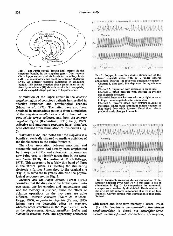
FIG. 1. The Papez circuit (broken line): passes via thecingulum bundle, in the cingulate gyrus, from septum(S) to hippocampus, and via fornix to mamillary body(M), via mamillothalamic tract to anterior thalamus(AT), via anterior thalamic radiations to cingulumbundle. The defence reaction circuit (solid line): passesfrom hypothalamus (H) via stria terminalis to amygdala,and via amygdalo-fugal pathway to hypothalamus.
Stimulation of the Papez circuit in the anteriorcingulate region of conscious patients has resulted inaffective responses and physiological changes(Meyer et al., 1973). The latter have also beenobtained in unconscious patients from stimulationof the cingulum bundle below and in front of thegenu of the corpus callosum, and from the anteriorcingulate region (Richardson, 1973; Kelly, 1972).Affective and autonomic responses have, therefore,been obtained from stimulation of this circuit (Fig.2).Yakovlev (1965) had noted that the cingulum is a
bundle strategically situated to mediate activities ofthe limbic cortex to the entire forebrain.The close association between emotional and
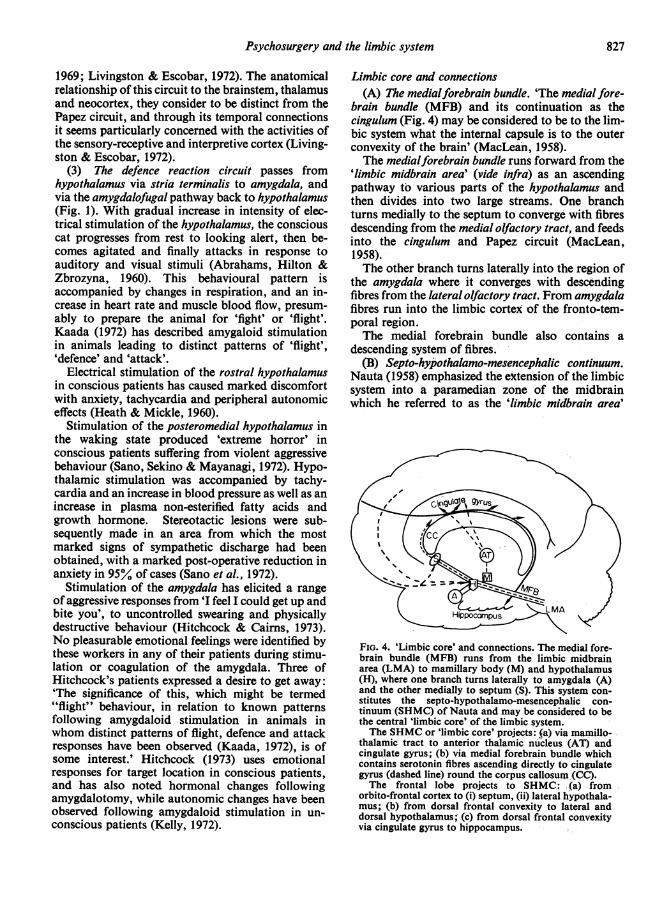
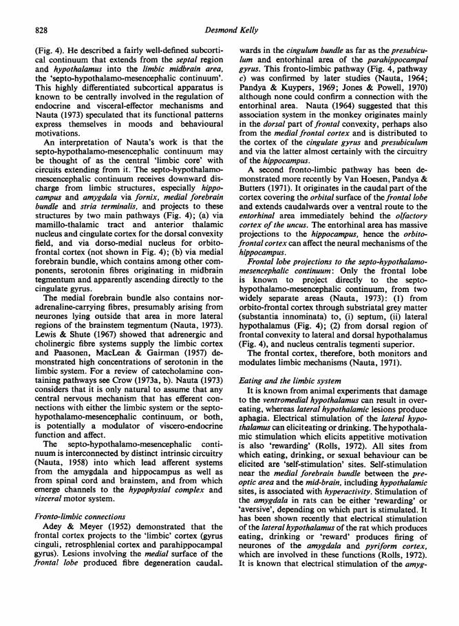
autonomic pathways had already been emphasizedby Livingston (1953), and autonomic responses arenow being used to identify target sites in the cingu-lum bundle (Kelly, Richardson & Mitchegl-Heggs,1973). This appears to be a fairly thin band of fibresin the mertical plane, as inserting the stimulatingelectrode a further 3 mm deep to the original site(Fig. 3) is sufficient to greatly diminish the physio-logical responses seen in Fig. 2.Memory and the Papez circuit. Turner (1973)
considers that the division of the limbic system intotwo parts, onesfor emotion and temperament andone for memory is justified, since he effects ofablation operations on the two parts are quitedifferent. Anterior cingulate (Kelly & Mitchell-Heggs, 1973), or posterior cingulate (Turner, 1973)lesions have no detectable effect on memory,whereas other structures in the Papez circuit, suchas the hippocampus, fornix, mamillary bodies andmammillo-thalamic tract, are apparently concerned
(3))[9 ,e H. ..
(4)
(5 1-7 3'-3'/
FIG. 2. Polygraph recording during stimulation of theanterior cingulate gyrus with 10 V under generalanaesthesia showing the following autonomic changes:
Channel 1, time (sec), line depressed during stimula-tion.Channel 2, respiration with decrease in amplitude.Channel 3, blood pressure with increase in systolicand diastolic pressures.Channel 4, heart rate increase with very slight increasein finger pulse amplitude after stimulation.Channel 5, forearm blood flow (ml/100 ml/min) isincreased. Finger pulse amplitude reflects changes inskin blood flow while forearm blood flow reflectspredominantly changes in muscle.
(2) .
(35
(47 6lllEEl D l E i E
(5)/1 1XS-31 131'I~~~~~~~~~~~p
-.-.-.-.. -. -. ............
FIG. 3. Polygraph recording during stimulation of theanterior cingulate gyrus with 10 V 3 mm deep to site ofstimulation in Fig. 2. By comparison the autonomicchanges are considerably diminished. Restimulation ofthe original site restored autonomic changes in all fourchannels. Current spread from stimulation is thus fairlylimited.
with recent and long-term memory (Turner, 1973).(2) The basolateral circuit-orbital frontal-tem-
poral-amygdalar-is closed via amygdalar-dorso-medial thalamic-frontal connections (tivingston,
Psychosurgery and the limbic system
1969; Livingston & Escobar, 1972). The anatomicalrelationship of this circuit to the brainstem, thalamusand neocortex, they consider to be distinct from thePapez circuit, and through its temporal connectionsit seems particularly concerned with the activities ofthe sensory-receptive and interpretive cortex (Living-ston & Escobar, 1972).
(3) The defence reaction circuit passes fromhypothalamus via stria terminalis to amygdala, andvia the amygdalofugal pathway back to hypothalamus(Fig. 1). With gradual increase in intensity of elec-trical stimulation of the hypothalamus, the consciouscat progresses from rest to looking alert, then be-comes agitated and finally attacks in response toauditory and visual stimuli (Abrahams, Hilton &Zbrozyna, 1960). This behavioural pattern isaccompanied by changes in respiration, and an in-crease in heart rate and muscle blood flow, presum-ably to prepare the animal for 'fight' or 'flight'.Kaada (1972) has described amygaloid stimulationin animals leading to distinct patterns of 'flight','defence' and 'attack'.
Electrical stimulation of the rostral hypothalamusin conscious patients has caused marked discomfortwith anxiety, tachycardia and peripheral autonomiceffects (Heath & Mickle, 1960).
Stimulation of the posteromedial hypothalamus inthe waking state produced 'extreme horror' inconscious patients suffering from violent aggressivebehaviour (Sano, Sekino & Mayanagi, 1972). Hypo-thalamic stimulation was accompanied by tachy-cardia and an increase in blood pressure as well as anincrease in plasma non-esterified fatty acids andgrowth hormone. Stereotactic lesions were sub-sequently made in an area from which the mostmarked signs of sympathetic discharge had beenobtained, with a marked post-operative reduction inanxiety in 95°/ of cases (Sano et al., 1972).
Stimulation of the amygdala has elicited a rangeof aggressive responses from 'I feel I could get up andbite you', to uncontrolled swearing and physicallydestructive behaviour (Hitchcock & Cairns, 1973).No pleasurable emotional feelings were identified bythese workers in any of their patients during stimu-lation or coagulation of the amygdala. Three ofHitchcock's patients expressed a desire to get away:'The significance of this, which might be termed"flight" behaviour, in relation to known patternsfollowing amygdaloid stimulation in animals inwhom distinct patterns of flight, defence and attackresponses have been observed (Kaada, 1972), is ofsome interest.' Hitchcock (1973) uses emotionalresponses for target location in conscious patients,and has also noted hormonal changes followingamygdalotomy, while autonomic changes have beenobserved following amygdaloid stimulation in un-conscious patients (Kelly, 1972).
Limbic core and connections(A) The medialforebrain bundle. 'The medial fore-
brain bundle (MFB) and its continuation as thecingulum (Fig. 4) may be considered to be to the lim-bic system what the internal capsule is to the outerconvexity of the brain' (MacLean, 1958).The medialforebrain bundle runs forward from the
'limbic midbrain area' (vide infra) as an ascendingpathway to various parts of the hypothalamus andthen divides into two large streams. One branchturns medially to the septum to converge with fibresdescending from the medial olfactory tract, and feedsinto the cingulum and Papez circuit (MacLean,1958).The other branch turns laterally into the region of
the amygdala where it converges with descendingfibres from the lateral olfactory tract. From amygdalafibres run into the limbic cortex of the fronto-tem-poral region.The medial forebrain bundle also contains a
descending system of fibres.(B) Septo-hypothalamo-mesencephalic continuum.
Nauta (1958) emphasized the extension of the limbicsystem into a paramedian zone of the midbrainwhich he referred to as the 'limbic midbrain area'
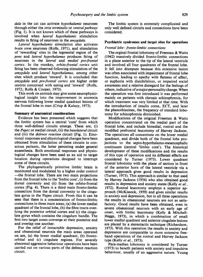
FIG. 4. 'Limbic core' and connections. The medial fore-brain bundle (MFB) runs from the limbic midbrainarea (LMA) to mamillary body (M) and hypothalamus(H), where one branch turns laterally to amygdala (A)and the other medially to septum (S). This system con-stitutes the septo-hypothalamo-mesencephalic con-tinuum (SHMC) of Nauta and may be considered to bethe central 'limbic core' of the limbic system.The SHMC or 'limbic core' projects: fa) via mamillo-
thalamic tract to anterior thalamic nucleus (AT) andcingulate gyrus; (b) via medial forebrain bundle whichcontains serotonin fibres ascending directly to cingulategyrus (dashed line) round the corpus callosum (CC).The frontal lobe projects to SHMC: (a) from
orbito-frontal cortex to (i) septum, (ii) lateral hypothala-mus; (b) from dorsal frontal convexity to lateral anddorsal hypothalamus; (c) from dorsal frontal convexityvia cingulate gyrus to hippocampus.
827
Desmond Kelly
(Fig. 4). He described a fairly well-defined subcorti-cal continuum that extends from the septal regionand hypothalamus into the limbic midbrain area,the 'septo-hypothalamo-mesencephalic continuum'.This highly differentiated subcortical apparatus isknown to be centrally involved in the regulation ofendocrine and visceral-effector mechanisms andNauta (1973) speculated that its functional patternsexpress themselves in moods and behaviouralmotivations.An interpretation of Nauta's work is that the
septo-hypothalamo-mesencephalic continuum maybe thought of as the central 'limbic core' withcircuits extending from it. The septo-hypothalamo-mescencephalic continuum receives downward dis-charge from limbic structures, especially hippo-campus and amygdala via fornix, medial forebrainbundle and stria terminalis, and projects to thesestructures by two main pathways (Fig. 4); (a) viamamillo-thalamic tract and anterior thalamicnucleus and cingulate cortex for the dorsal convexityfield, and via dorso-medial nucleus for orbito-frontal cortex (not shown in Fig. 4); (b) via medialforebrain bundle, which contains among other com-ponents, serotonin fibres originating in midbraintegmentum and apparently ascending directly to thecingulate gyrus.The medial forebrain bundle also contains nor-
adrenaline-carrying fibres, presumably arising fromneurones lying outside that area in more lateralregions of the brainstem tegmentum (Nauta, 1973).Lewis & Shute (1967) showed that adrenergic andcholinergic fibre systems supply the limbic cortexand Paasonen, MacLean & Gairman (1957) de-monstrated high concentrations of serotonin in thelimbic system. For a review of catecholamine con-taining pathways see Crow (1973a, b). Nauta (1973)considers that it is only natural to assume that anycentral nervous mechanism that has efferent con-nections with either the limbic system or the septo-hypothalamo-mesencephalic continuum, or both,is potentially a modulator of viscero-endocrinefunction and affect.The septo-hypothalamo-mesencephalic conti-
nuum is interconnected by distinct intrinsic circuitry(Nauta, 1958) into which lead afferent systemsfrom the amygdala and hippocampus as well asfrom spinal cord and brainstem, and from whichemerge channels to the hypophysial complex andvisceral motor system.
Fronto-limbic connectionsAdey & Meyer (1952) demonstrated that the
frontal cortex projects to the 'limbic' cortex (gyruscinguli, retrosphlenial cortex and parahippocampalgyrus). Lesions involving the medial surface of thefrontal lobe produced fibre degeneration caudal-
wards in the cingulum bundle as far as the presubicu-lum and entorhinal area of the parahippocampalgyrus. This fronto-limbic pathway (Fig. 4, pathwayc) was confirmed by later studies (Nauta, 1964;Pandya & Kuypers, 1969; Jones & Powell, 1970)although none could confirm a connection with theentorhinal area. Nauta (1964) suggested that thisassociation system in the monkey originates mainlyin the dorsal part of frontal convexity, perhaps alsofrom the medial frontal cortex and is distributed tothe cortex of the cingulate gyrus and presubiculumand via the latter almost certainly with the circuitryof the hippocampus.A second fronto-limbic pathway has been de-
monstrated more recently by Van Hoesen, Pandya &Butters (1971). It originates in the caudal part ofthecortex covering the orbital surface of the frontal lobeand extends caudalwards over a ventral route to theentorhinal area immediately behind the olfactorycortex of the uncus. The entorhinal area has massiveprojections to the hippocampus, hence the orbito-frontal cortex can affect the neural mechanisms of thehippocampus.
Frontal lobe projections to the septo-hypothalamo-mesencephalic continuum: Only the frontal lobeis known to project directly to the septo-hypothalamo-mesencephalic continuum, from twowidely separate areas (Nauta, 1973): (1) fromorbito-frontal cortex through substriatal grey matter(substantia innominata) to, (i) septum, (ii) lateralhypothalamus (Fig. 4); (2) from dorsal region offrontal convexity to lateral and dorsal hypothalamus(Fig. 4), and nucleus centralis tegmenti superior.The frontal cortex, therefore, both monitors and
modulates limbic mechanisms (Nauta, 1971).
Eating and the limbic systemIt is known from animal experiments that damage
to the ventromedial hypothalamus can result in over-eating, whereas lateral hypothalamic lesions produceaphagia. Electrical stimulation of the lateral hypo-thalamus can eliciteating or drinking. Thehypothala-mic stimulation which elicits appetitive motivationis also 'rewarding' (Rolls, 1972). All sites fromwhich eating, drinking, or sexual behaviour can beelicited are 'self-stimulation' sites. Self-stimulationnear the medial forebrain bundle between the pre-optic area and the mid-brain, including hypothalamicsites, is associated with hyperactivity. Stimulation ofthe amygdala in rats can be either 'rewarding' or'aversive', depending on which part is stimulated. Ithas been shown recently that electrical stimulationof the lateral hypothalamus of the rat which produceseating, drinking or 'reward' produces firing ofneurones of the amygdala and pyriform cortex,which are involved in these functions (Rolls, 1972).It is known that electrical stimulation of the amyg-
828
Psychosurgery and the limbic system
dala in the cat can activate hypothalamic neuronesthrough either the stria terminalis or ventralpathway(Fig. 1). It is not known which of these pathways isinvolved when lateral hypothalamic stimulationresults in firing of neurones in the amygdala.
Lateral hypothalamic stimulation also activatesbrain stem neurones (Rolls, 1971), and stimulationof 'rewarding' sites in the tegmental region of thepons and lateral hypothalamus produces firing ofneurones in the lateral and medial pre-frontalcortex. In the monkey, orbito-frontal cortex unitfiring has been observed following stimulation of theamygdala and lateral hypothalamus, among othersites which produce 'reward'. It is concluded thatamygdala and pre-frontal cortex are higher ordercentres concerned with eating and 'reward' (Rolls,1972; Rolls & Cooper, 1973).This work on animals may give some neurophysio-
logical insight into the improvement in anorexianervosa following lower medial quadrant lesions ofthe frontal lobe in man (Crisp & Kalucy, 1973).
Summary of anatomical considerationsEvidence has been presented which suggests that
the limbic system has a central 'core' from whichthree well-defined circuits emerge. These are, (i)the Papez or medial circuit, (ii) the basolateral circuitand (iii) the defence reaction circuit (Fig. 1). Emo-tional responses and physiological changes have beenobtained from stimulation of these circuits in con-scious patients, the latter persisting under generalanaesthesia. Both emotional responses and physio-logical changes have been used as an aid to targetlocation during operations designed to interruptsome of these circuits.The phylogenetically primitive limbic brain is
monitored and modulated by a higher order control-the frontal lobe. There are two main projectionsfrom the frontal lobe to the 'limbic core', (i) from thedorsal convexity and (ii) from the orbito-frontalcortex (Fig. 4). There is a third main fronto-limbicconnection from the dorsal convexity to the cingu-late gyrus in the Papez circuit. From this it can beseen that there is a concentration of fronto-limbicconnections in three main areas; (a) the lower medialquadrant of the frontal lobe, (b) the fibre tracts over-lying the posterior orbital cortex and (c) the cingu-late gyrus which contains the cingulum bundle. Thefirst two target zones converge at their posterior endand overlap one another.For the relief of intractable depression, anxiety
and obsessional neurosis the main areas operatedon are, (a) the lower medial quadrant, (b) fronto-orbital and (c) cingulate regions. For severelyabnormal aggressive behaviour operations have beencarried out on various parts of the defence reactioncircuit.
The limbic system is extremely complicated andonly well defined circuits and connections have beenconsidered.
Psychiatric syndromes and target sites for operationsFrontal lobe: fronto-limbic connectionsThe orginal frontal lobotomy of Freeman & Watts
(1942) massively divided fronto-limbic connectionsin a plane anterior to the tip of the lateral ventricleand involved all four quadrants of the frontal lobe.It fell into disrepute because this extensive lesionwas often associated with impairment of frontal lobefunction, leading to apathy with flatness of affect,or euphoria with disinhibition, or impaired socialawareness and a relative disregard for the feelings ofothers, indicative ofamajorpersonalitychange. Whenthe operation was first introduced it was performedmainly on patients with chronic schizophrenia, forwhich treatment was very limited at that time. Withthe introduction of insulin coma, ECT, and laterthe phenothiazines, the frequency of standard lobo-tomy for schizophrenia diminished.
Modifications of the original Freeman & Wattsoperation concentrated on the medial part of thefrontal lobe, and include bimedial, rostral and themodified prefrontal leucotomy of Harvey Jackson.The operations all concentrate on the lower medialquadrant, and divide both of the frontal lobe pro-jections to the septo-hypothalamo-mesencephaliccontinuum (central 'limbic core'). The historicaldevelopment of these modifications and the resultsof this type of operation and its different effects areconsidered by Turner (1973). Lower quadrantfrontal lobotomy with the plane of section in frontof the anterior horn of the lateral ventricle via alateral approach gives good results in depression(Turner, 1973). This approach is similar to that usedby Harvey Jackson (1954) who also obtained goodresults in depression and anxiety states (Kelly et al.,1972). Rostral leucotomy employs a superior ap-proach (McKissock, 1959) and gives similar resultsin anxiety and depression, but in all three operationsthe results in obsessional neurosis are not as satis-factory. Good results have been obtained, even insevere obsessional neurosis with an early age ofonset, with limbic leucotomy (Kelly & Mitchell-Heggs, 1973), in which a combination of smalllower medial quadrant and anterior cingulate lesionsare placed by a stereotactic technique (Richardson,1973). With this operation the results in anxiety anddepression are comparable to more extensive free-hand operations of the rostral and Harvey Jacksontype (Kelly et al., 1973).
Para-median lobotomy is considered by Turner(1973) to benefit patients with anxiety and impulsivebehaviour, usually of an aggressive nature. Young
829
830 Desmond Kelly
patients with obsessional neurosis do not usuallyrespond to this operation, however.The other main target site in the frontal lobe is the
posterior orbito-frontal region. Le Gros Clark &Meyer (1950) demonstrated fibres which pass fromArea 13 in the orbital cortex to the hypothalamus.The orbital undercut of Scoville was modified byKnight (1964), who subsequently developed astereotactic operation restricted to the last 2 cm of a6 cm restricted orbital undercutting incision. Auto-nomic responses can be obtained from Area 13, andSmith (1945) and Livingston et al. (1948a) obtainedrespiratory and vascular responses from a continuouszone of cortex extending from the anterior cingulategyrus to the posterior orbital surface in the monkey.Lesions in these areas produced loss of fear, di-minished aggression and increased tameness (Ward,1948).In patients, pre-leucotomy stimulation of the
orbital part of the frontal lobe also resulted inchanges in respiration and blood pressure and wassometimes accompanied by intense emotion (Living-ston et al., 1948b). Knight (1973) describes his ex-tensive experience with stereotactic tractotomy inthis symposium, with very good results in depressionand anxiety but less satisfactory results in obsessionalneurosis. 'There does seem, however, to be a rela-tively small group of cases characterized by pureobsessional symptoms, without depression, of earlyage of onset, which has a bad prognosis.' The targetsite of stereotactic tractotomy overlaps the lowermedial quadrant target site of limbic leucotomy-although the approaches are from different angles-the plane of the lesion is almost horizontal in theformer (Knight, 1973; Fig. 3), and vertical in thelatter (Richardson, 1973; Fig. 2).
In the lower medial quadrant, and the posterior-orbital part, of the frontal lobe there is a concentra-tion of fronto-limbic connections (Fig. 4) and it istempting to speculate that it is the division of thesethat causes the beneficial effects of stereotactictractotomy and lower medial quadrant lesions.
It can be seen from Figs. 1 and 4 that limbic con-nections are concentrated in two main areas: in thelower medial quadrant and posterior-orbito-frontallobe and in the anterior cingulate gyrus. It is re-markable how these two widely separated targetzones yield very similar autonomic responses, andthis suggests that fibres may be stimulated whichmediate similar functional pathways (Figs. 2 and 5).
It is noteworthy that moving from these targetsites by as little as 3-4mm may be sufficient to almostcompletely abolish the responses. The symmetryof autonomic response pattern from either side of thebrain in the lower medial quadrant of the frontallobe and anterior cingulate gyrus is also impressive.Recently, increase in cortisol secretion has been
(2)N
..R:H:...:.:........:..: ..
.. ............... ................................... .. ..
0
(4) 9
105A;''96 '- 13S
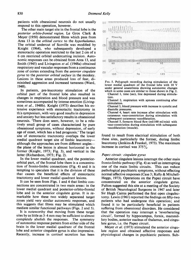
FIG. 5. Polygraph recording during stimulation of thelower medial quadrant of the frontal lobe with 10 Vunder general anaesthesia showing autonomic changeswhich in some cases are similar to those shown in Fig. 2.Channel 1, time (sec), line depressed during stimula-tion.Channel 2, respiration with apnoea continuing afterstimulation.Channel 3, blood pressure with increase in systolic anddiastolic pressures.Channel 4, heart rate increase after stimulation withcutaneous vaso-constriction during stimulation withsubsequent cutaneous vasodilatation.Channel 5, forearm blood flow (ml/100 ml/min) withvaso-constriction during stimulation with subsequentvasodilatation (muscle).
found to result from electrical stimulation of boththese sites, particularly the former, during limbicleucotomy (Jenkins & Frankel, 1973). The maximumincrease in cortisol was 375 %.
Papez circuit: cingulate gyrusAnterior cingulate lesions interrupt the other main
fronto-limbic pathway (Fig. 4) as well as interruptingone of the main limbic circuits. This can reducepathological psychiatric symptoms, without effectingnormal affective responses (Case 3; Kelly & Mitchell-'Heggs, 1973). Operations on the Papez circuit haveconcentrated on the anterior cingulate gyrus.Fulton suggested this site at a meeting of the Societyof British Neurological Surgeons in 1947 and laterSir Hugh Cairns performed the first open cingulec-tomy. Lewin (1961) reported the findings on fifty-twopatients who had undergone this operation; andfound it to be particularly beneficial in patientssuffering from obsessional disorders. He consideredthat the operation may interrupt a 'reverberatingcircuit', formed by hippocampus, fornix, mammil-lary bodies, anterior nucleus of thalamus, and cingu-late gyrus, i.e. the Papez circuit.Meyer et al. (1973) stimulated the anterior cingu-
late region and obtained affective responses andphysiological changes in psychiatric patients. Sub-
Psychosurgery and the limbic system 831
sequently, anterior cingulotomy resulted in signi-ficant overall improvement in a variety of psychiatricdisorders. Ballantine et al. (1967) and Brown &Lighthill (1968) also obtained good results withlesions placed by stereotaxis in the anterior cin-gulate gyrus.
The baso-lateral and defence reaction circuitsPersonality disorders associated with epilepsy, and
aggressive outbursts or 'uncontrollable rage, usuallyof a particular kind ushered in by experiences ofsudden fear, panic or stark terror' respond to tem-poral lobotomy (Turner, 1973). The site of operationis at, or just behind the uncus of the hippocampusand in, or just behind the amygdaloid nucleus. Fearis abolished if ictal, but chronic anxiety is generallynot helped. Hitchcock & Cairns (1973) and Bala-sumramaniam & Ramamurthi (1968) have alsofound stereotactic amygdalotomy beneficial inmodifying aggressive behaviour associated withepilepsy. In both these operations lesions are beingplaced in the defence reaction circuit. In addition tooperations on the amygdala, other workers haveobtained clinical improvement from other sites inthe defence reaction circuit. In patients sufferingfrom severely disturbed destructive behaviour,Sano et al. (1972) operated on the postero-medialhypothalamus, and Burzaco (1973) on the striaterminalis.
Psychosurgery for anorexia nervosaCrisp & Kalucy (1973) describe in this symposium
the effects of leucotomy on four patients withprimary anorexia nervosa, and conclude that in thissyndrome leucotomy has and will continue to havea limited but very definite place in treatment. Thedetailed reports illustrating the relationship be-tween weight gain and psychological change are ofconsiderable interest, and the application of therepertory grid technique to the evaluation of changesin attitude following leucotomy adds another di-mension to the methods now available for post-operative assessment. The inability of one of theirpatients to tolerate the psychological consequences ofleucotomy has important implications, and under-lines the difficulty in predicting the effect of the opera-tion in very neurotic vulnerable personalities. Thecomplex relationship between eating and 'reward'and the pathways involved-are only now beginningto be explored in depth.
ConclusionThere still appears to be a place for psychosur-
gery, 'the challenge being to precisely locate theindications within the total field of psychiatry'(Bridges & Bartlett, 1973). It must be rememberedthat affective disorders carry a 15%4 risk of death
from suicide, and psychosurgery can relieve intoler-able suffering and improve the quality of life whenall other treatments have failed. The main indica-tions are intractable depression, anxiety and obses-sional neurosis. Some patients with schizophrenia,and very rarely patients with anorexia nervosa, canalso be helped. Operations for seriously aggressivebehaviour, such as posterior cingulectomy andparamedian frontal lobotomy (Turner, 1973),amygdalotomy and postero-medial hypothalamo-tomy are more controversial, and experience in thiscountry is, so far, limited.The various target sites chosen by different neuro-
surgeons can be understood in terms of lesions placedin different parts of well defined fronto-limbicconnections and limbic circuits.
Technical advances in neurosurgery are likely tolead to a decrease in the number of 'free hand'operations and an increase in the use of stereotactictechniques. Far more sophisticated methods ofpsychiatric, psychological, biochemical and psycho-physiological assessment are now available, and willbe increasingly used.
Progress is likely to come from close multi-disciplinary co-operation, as indicated in this sym-posium, and with increasing sophistication in thisfield, it would probably be wise for this highlyspecialized type of treatment to be concentrated in arelatively few centres as suggested by Bridges &Bartlett (1973). It is hoped that there will come a timewhen psychosurgery is no longer required for thetiny minority of psychiatric patients for whom, atpresent, no other type of treatment is effective.Meanwhile, the major question is, if all else fails,which patients are suitable for psycho-surgery, andwhich operation should they have?
ReferencesABRAHAMS, V.C., HILTON, S.M. & ZBROZYNA, A. (1960)
Active muscle vasodilatation produced by stimulation ofthe brain stem: its significance in the defence reaction.Journal of Physiology, 154, 491.
ADEY, W.R. & MEYER, M. (1952) Hippocampal and hypo-thalamic connections of the temporal lobe in the monkey.Brain, 75, 358.
BALASUMRAMANIAM, V. & RAMAMURTHI, B. (1968) Stereo-taxic amygdalotomy. Proceedings of the AustralianAssociation of Neurologists, 5, 277.
BALLANTINE, H.T., CASSIDY, W.L., FLANAGAN, N.B. &MARINO, R. (1967) Stereotactic anterior cingulotomy forneuropsychiatric illness and intractable pain. Journal ofNeurosurgery, 26, 488.
BRIDGES, P.K. & BARTLETT, J.R. (1973) The work of apsychosurgical unit. Postgraduate Medical Journal, 49, 855.
BROWN, H.M. & LIGHTHILL, J.A. (1968) Selective anteriorcingulotomy: a psychosurgical evaluation. Journal ofNeurosurgery, 29, 513.
BURZACO, J.A. (1973) Fundus striae terminalis, an optimaltarget in sedative stereotactic surgery. In: Surgical Ap-proaches in Psychiatry (Ed by Laitinen and Livingston),p. 135. Medical and Technical Publishing Co. Ltd.Lancaster.
832 Desmond Kelly
CLARK, W.E. Le GROS & MEYER, M. (1950) Anatomicalrelationships between the cerebral cortex and hypo-thalamus. British Medical Bulletin, 6, 341.
CRISP, A.H. & KALUCY, R.L. (1973) The effect of leucotomyin intractable adolescent weight phobia (primary anorexianervosa). Postgraduate Medical Journal, 49, 883.
CROW, T.J. (1973a) Catecholamine-containing neurones andelectrical self-stimulation: a review of some data. Psycho-logical Medicine, 2, 414.
CROW, T.J. (1973b) Catecholamine-containing neurones andelectrical self-stimulation: a theoretical interpretation andsome psychiatric implications. Psychological Medicine, 3,66.
FREEMAN, W. & WATrS, J. (1942) Psychosurgery. Charles C.Thomas, Springfield. Illinois.
HEATH, R.G. & MICKLE, W.A. (1960) Evaluation of sevenyears experience with depth electrode studies in humanpatients. In: Electrical Studies on the Unanesthetized Brain(Ed. by Ramey and O'Doherty). New York.
HITCHCOCK, E. & CAIRNS, V. (1973) Amygdalotomy. Post-graduate Medical Journal, 49, 894.
JACKSON, H. (1954) Leucotomy-a recent development.Journal of Mental Science, 100, 62.
JENKINS, J. & FRANKEL, R. (1973) Personal communication.JONES, E.G. & POWELL, T.P.S. (1970) An anatomical study
of converging sensory pathways within the cerebral cortexof the monkey. Brain, 93, 793.
KAADA, B. (1972) Stimulation and regional ablation of theamygdaloid complex with reference to functional repre-sentations. In: The Neurobiology of the Amygdala (Ed. byB. E. Eleftheriou), p. 205. Plenum Press, New York.
KELLY, D. (1972) Physiological changes during operations onthe limbic system in man. Conditional Reflex, 7, 127.
KELLY, D. & MITCHELL-HEGGS, N. (1973) Stereotactic limbicleucotomy: a follow-up study of thirty patients. Post-graduate Medical Journal, 49, 865.
KELLY, D., RICHARDSON, A. & MITCHELL-HEGGS, N. (1973)Stereotactic limbic leucotomy: neurophysiological aspectsand operative technique. British Journal of Psychiatry, 123,133.
KELLY, D., RICHARDSON, A., MITCHELL-HEGGS, N., GREEN-uP, J., CHEN, C. & HAFNER, R.J. (1973) Stereotactic limbicleucotomy: a preliminary report on forty patients. BritishJournal of Psychiatry, 123, 141.
KELLY, D., WALTER, C.J.S., MITCHELL-HEGGS, N. & SAR-GANT, W. (1972) Modified leucotomy assessed clinically,physiologically and psychologically at six weeks andeighteen months. British Journal of Psychiatry, 120, 19.
KNIGHT, G. (1964) The orbital cortex as an objective in thesurgical treatment of mental illness. British Journal ofSurgery, 51, 114.
KNIGHT, G. (1973) Further observations from an experienceof 660 cases of stereotactic tractotomy. PostgraduateMedical Journal, 49, 845.
LEWIN, W. (1961) Observations on selective leucotomy.Journal of Neurology, Neurosurgery and Psychiatry, 24, 37.
LEWIS, P.R. & SHUTE, C.C.D. (1967) The cholinergic limbicsystem: projections to hippocampal formation, medialcortex, nuclei of the ascending cholinergic reticular system,and the subfornical organ and supra-optic crest. Brain,90, 521.
LIVINGSTON, K.E. (1953) Cingulate cortex isolation for thetreatment of psychoses and psychoneuroses. ResearchPublications of the Association for Research in Nervous andMental Disease, 31, 374.
LIVINGSTON, K.E. (1969) The frontal lobes revisited: thecase for a second look. Archives of Neurology, 20, 90.
LiVINGSTON, K.E. & EsCOBAR, A. (1972) The continuingevolution of the limbic system concept. In: Psychosurgery(Ed. by Hitchcock, Laitinen and Vaernet), p. 25. Charles CThomas, Springfield, Illinois.
LIVINGSTON, R.B., CHAPMAN, W.P., LIVINGSTON, K.E. &KRAINTZ, L. (1948a) Stimulation of the orbital surface ofman prior to frontal lobotomy. Research Publications ofthe Association for Research in Nervous and MentalDisease, 27, 421.
LIVINGSTON, R.B., FULTON, J.F., DELGADO, J.M.R., SACHS,F. JR, BRENDLER, S.J. & DAVIS, G.D. (1948b) Stimulationand regional ablation of orbital surface of frontal lobe.Research Publications of the Association for Research inNervous and Mental Disease, 27, 405.
MAcLEAN, P.D. (1949) Psychosomatic disease and the'visceral brain'. Recent developments bearing on thePapez theory of emotion. Psychosomatic Medicine, 11, 338.
MACLEAN, P.D. (1952) Some psychiatric implications ofphysiological studies on frontotemporal portion of thelimbic system (visceral brain). Electroencephalography andClinical Neurophysiology, 4, 407.
MAcLEAN, P.D. (1958) Contrasting functions of limbic andneocortical systems of the brain and their relevance topsychophysiological aspects of medicine. American Journalof Medicine, 25, 61 1.
MEYER, G., McELHANY, M., MARTIN, W. & McGRAw, C.P.(1973) Stereotactic cingulotomy. In: Surgical Approachesin Psychiatry (Ed. by Laitinen and Livingston), p. 39.Medical and Technical Publishing Co. Ltd, Lancaster.
McKISSOCK, W. (1959) Discussion on psychosurgery. Pro-ceedings of the Royal Society of Medicine, 52, 206.
NAUTA, W.J.H. (1958) Hippocampal projections and re-lated neural pathways to the midbrain in the cat. Brain, 81,319.
NAUTA, W.J.H. (1964) Some efferent connections of the pre-frontal cortex in the monkey. In: The Frontal GranularCortexand Behaviour (Ed. by J. M. Warren and K. Akert),p. 397. McGraw-Hill, New York.
NAUTA, W.J.H. (1971) The problem of the frontal lobe:a reinterpretation. Journal of Psychiatric Research, 8, 167.
NAUTA, W.J.H. (1973) Connections of the frontal lobe withthe limbic system. In: Surgical Approaches in Psychiatry(Ed. by Laitinen and Livingston), p. 303. Medical andTechnical Publishing Co. Ltd, Lancaster.
PAASONEN, M.K., MACLEAN, P.D., & GAIRMAN, N.J. (1957)5-Hydroxytryptamine (serotonin, enteramine) content ofstructures of the limbic system. Journal of Neurochemistry,1, 326.
PANDYA, D.N. & KUYPERS, H.G.J.M. (1969) Cortico-corticalconnections in the rhesus monkey. Brain Research, 13, 13.
PAPEZ, J.W. (1937) A proposed mechanism of emotion.Archives of Neurology and Psychiatry, 38, 725.
RICHARDSON, A. (1973) Stereotactic limbic leucotomy:surgical technique. Postgraduate Medical Journal, 49, 860.
ROLLS, E.T. (1971) Involvement of brainstem units in medialforebrain bundle self-stimulation. Physiology and Be-havior, 7, 297.
ROLLS, E.T. (1972) Activation of amygdaloid neurones inreward, eating and drinking elicited by electrical stimula-tion of the brain. Brain Research, 45, 365.
ROLLS, E.T. & COOPER, S.J. (1973) Activation of neuronesin the prefrontal cortex by brain-stimulation reward in therat. Brain Research (in press).
SANO, K., SEKINO, H. & MAYANAGI, Y. (1972) Results ofstimulation and destruction of the posterior hypothalamusin cases with violent, aggressive or restless behaviors. In:Psychosurgery (Ed. by Hitchcock Laitinen and Vaernet),p. 57. Charles C. Thomas, Springfield, Illinois.
SMITH, W.K. (1945) The functional significance of the rostralcingular cortex as revealed by its responses to electricalexcitation. Journal of Neurophysiology, 8, 241.
TURNER, E. (1973) Custom psychosurgery. PostgraduateMedical Journal, 49, 834.
Psychosurgery and the limbic system 833
VAN HOESEN, G.W., PANDYA, D.N. & BUTTERS, N. (1971)Cortical efferents to the entorhinal cortex of the rhesusmonkey. Science, 175, 1471.
WARD, A.A. JR (1948) The anterior cingular gyrus andpersonality. Research Publications of the Association ofNervous and Mental Disease, 27, 438.
YAKOVLEV, P.I. (1948) Motility, behavior and the brain:stereodynamic organization and neural co-ordinates ofbehavior. Journal ofNervous and MentalDiseases, 107, 313-
YAKOVLEV, P.1. (1965) Personal communication to Ballan.tine et al., 1967.