Psoriatic arthritis
51
PSORIATIC ARTHRITIS Abdelrahman Amer Mousa Resident of Rheumatology & Physical medicine Mansoura University Hospital
-
Upload
mansoura-university-hospitals -
Category
Education
-
view
48 -
download
0
Transcript of Psoriatic arthritis

PSORIATIC ARTHRITIS
Abdelrahman Amer Mousa
Resident of Rheumatology & Physical medicine
Mansoura University Hospital

How prevalent is psoriasis and psoriatic arthritis
in the general population???
•Epidemiologic studies suggest that the prevalence of psoriasis is
approximately 2% to 3%. Whites are affected
• (two times) more often than other ethnic groups. The estimates of
inflammatory arthritis accompanying
•psoriasis range from 7% to 42% (average 26%).

prevalence of arthritis is relatively equal between the sexes.
However, However, in patients with spinal involvement, the male to female
ratio is almost 3:1
. Men also tend to have a higher prevalence of DIP-only involvement,
whereas women tend to have a higher prevalence of symmetric
polyarthritis.
Most patients present between the ages of 35 and 50 years. However,
juvenile psoriatic arthritis is also well
recognized and usually presents between ages 9 and 12 years

Is there a relationship between the onset of
psoriasis and the onset of arthritis?
Psoriasis precedes arthritis by an average of 8 to 10 years in 67%
of patients.
Arthritis precedes psoriasis or occurs simultaneously in 33% of
patients, particularly in childhood and in older patients (>age 50
years).

What are the current classification criteria for psoriatic
arthritis?The CASPAR (Classification of Psoriatic Arthritis) criteria are:
1. Evidence of psoriasis (current, past, family): two points if current
history of psoriasis, one point others.
2. Psoriatic nail dystrophy: one point
3. Negative rheumatoid factor: one point.
4. Dactylitis (current, past history): one point.
5. Radiographic evidence of juxtaarticular new bone formation: one
point.
Three or more points have 99% specificity and 92% sensitivity for
diagnosis of psoriatic arthritis
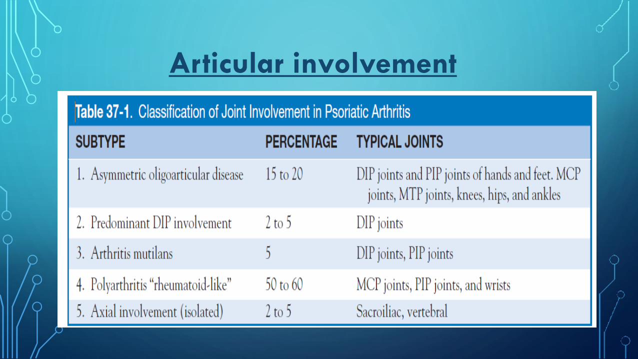
Articular involvement

The majority have symmetric polyarthritis or asymmetric
oligoarthritis of the hands and feet. Often the DIP joints
become stiff, swollen, and tender in an asymmetric fashion.
When present, involvement of the DIP joints helps distinguish
psoriatic arthritis from rheumatoid arthritis, but sometimes
results in confusion with osteoarthritis or gout. Other
joints that are affected by psoriatic arthritis include the knees,
hips, and sternoclavicular joints.
Regardless of the number of symptomatic joints at disease
onset, most patients progress to additional joint involvement
in the absence of effective treatment.
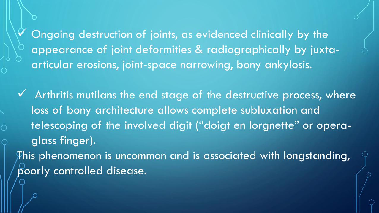
Ongoing destruction of joints, as evidenced clinically by the
appearance of joint deformities & radiographically by juxta-
articular erosions, joint-space narrowing, bony ankylosis.
Arthritis mutilans the end stage of the destructive process, where
loss of bony architecture allows complete subluxation and
telescoping of the involved digit (“doigt en lorgnette” or opera-
glass finger).
This phenomenon is uncommon and is associated with longstanding,
poorly controlled disease.


Arthritis Mutilans
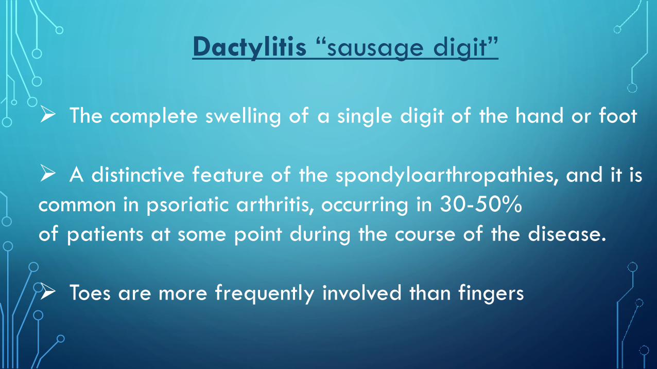
Dactylitis “sausage digit”
The complete swelling of a single digit of the hand or foot
A distinctive feature of the spondyloarthropathies, and it is
common in psoriatic arthritis, occurring in 30-50%
of patients at some point during the course of the disease.
Toes are more frequently involved than fingers


Enthesitis Inflammatory process occurring at the site of insertion of tendons
into bone
up to 40% of psoriatic arthritis patients.
On examination, there is a soft tissue swelling &tenderness to
palpation & may overlying erythema and warmth
Common sites for enthesitis:Achilles tendon, plantar fascia, and
pelvic bones.
Entheseal inflammation may evolve to destruction of the
adjacent bone and joints.

Skin changes All forms of psoriasis are associated with arthritis although classic psoriasis vulgaris is seen
most frequently.
Typical psoriatic lesions are erythematous plaques that produce scaling with scratching
Interestingly, many patients with psoriatic arthritis have only mild to moderate
skin disease, and there has been no consistent correlation between the degree of psoriasis and
the extent of joint involvement.
The psoriasis may be subtle so careful examination of the entire skin surface must be
performed when psoriatic arthritis is suspected
, with particular attention to the hairline, scalp, external auditory canal, periumbilical
area, and gluteal cleft As with uncomplicated psoriasis
,


Nail involvement
Common
Psoriatic nail changes include ridging, pitting,
onycholysis, and hyperkeratosis
May represent the manifestation of psoriasis before
the presence of more characteristic skin lesions.
Nail changes on the affected finger virtually always
occur when psoriaticarthritis affects a DIP joint
.


Spondyloarthropathy
Symptomatic involvement of the sacroiliac joints and
axial skeleton is less common than peripheral joints.
Sacroiliitis usually unilateral & presents with pain and
stiffness in the lower back or buttock.
Tenderness can sometimes be elicited by direct
compression teston SIJ or Gaenslen test

common site of skeletal involvement is the cervical spine.
extensive inflammation and erosion may lead to atlantoaxial
(C1–C2) instability, which can produce cervical myelopathy
as the odontoid process erodes. This process is often clinically
silent and painless. Involvement of other levels of the
spine is also seen in psoriatic arthritis with syndesmophytes,
which often arise from the midpoint of a vertebral body,
bridge adjacent vertebrae, and restrict motion of the spine.
In contrast to the continuous ascending spinal involvement
in ankylosing spondylitis, psoriatic spinal involvement is frequently
discontinuous, affecting noncontiguous vertebrae or
areas

Extraarticular manifestations
Eye disease includes conjunctivitis
in 20% and acute iritis in over 7% of cases. Iritis can be
bilateral and is more commonly associated with
axial involvement.
Other less common features include oral ulcers, urethritis,
nonspecific colitis, and rarely
dilatation of base of aortic arch causing aortic insufficiency.

Can laboratory tests help in diagnosing psoriatic
arthritis? By definition, psoriatic arthritis is classified as a “seronegative”
arthritis, meaning that the rheumatoid factor is typically negative
However, low titer rheumatoid factor can be detected in 5% to 9%
and Anti-CCP in 5% of psoriatic arthritis patients. This can make it
difficult to separate from coexistent rheumatoid arthritis
. However, the presence of DIP involvement, enthesitis, and dactylitis
supports a diagnosis of psoriatic arthritis regardless of the
serologies.

ANA are reported in 10% to 15%.
ESR , CRP and anemia may vary with disease activity
• Patients with an elevated ESR and CRP are more likely to have
polyarticular disease and a worse prognosis
Hyperuricemia is seen in 20% and related to the increased
incidence of the metabolic syndrome seen in patients with psoriatic
disease.
Analysis of synovial fluid reveals inflammatory fluid with a
neutrophilic predominance.Synovial fluid analysis reveals
inflammatory fluid, with
white blood cell counts usually in the 5000–50,000/mcL
range.

What radiographic features help to differentiate psoriatic arthritis
from other inflammatory
diseases?Overall, 45% to 50% of patients will develop erosions within the first 2 years of their
disease and eventually 67% will develop radiographic changes:
• Asymmetric involvement.
• Relative absence of juxtaarticular osteopenia.
• Involvement of DIP joints.
• Erosion of the terminal tufts (acroosteolysis).
• Whittling of the phalanges.
• Cupping of the proximal portion of the phalanges (pencil-in-cup deformity).
• Bony ankylosis distal to metacarpophalangeal (MCP) joints.
• Osteolysis of bones (arthritis mutilans).
• Polyarticular unidigit—MCP,(PIP), and DIP of same finger involved.
• Sacroiliac and spondylitic changes (usually asymmetric).

Arthritis mutilans. Severe destructive changes with multiple erosions
and “pencil-in-cup” deformities

Third left distal interphalangeal joint monoarthritis
with prominent new bone formation.

Periostitis. Radiograph of the thumb shows a marginal erosion of the
base of the distal phalanx, with extensive
cloaking of the surface of this phalanx with irregular periosteal new
bone.
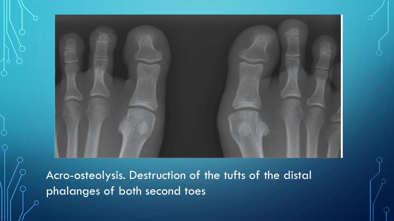
Acro-osteolysis. Destruction of the tufts of the distal
phalanges of both second toes

Asymmetric right-sided sacroiliitis

MSUS is more sensitive than clinical examination in detecting subclinical
synovitis in early psoriatic arthritis
• MSUS features at the enthesis include entheseal thickening,
hypoechoic change, increased vascularity as shown
on power Doppler, tenosynovitis, and bony erosions or
enthesophyte formation
• MSUS guidance for small joint or
entheseal aspiration or injection may have particular application
in patients with psoriatic arthritis.

Right
Right plantar fascia thickening compared with the left.
Left PL fascia long

Transverse section
through the metacarpophalangeal joint showing right
tenosynovitis
Right

Extensor tenosynovitis. Longitudinal ultrasound image of
the dorsum of the wrist shows lobulated, hypoechoic thickening of the extensor tendon sheath (arrowheads)
surrounding an extensor digitorum tendon (T).
Power Doppler has been used and shows hyperaemia (red and orange pixels) in the thickened tendon sheath

Retrocalcaneal bursitis
and enthesitis. Longitudinal ultrasound image with Power Doppler shows hypoechoic thickening of the retrocalcaneal
bursa (arrows), deep to a
severely thickened distal Achilles tendon. Abnormal color flow is demonstrated in the tendon, indicating
neovascularization. There is also irregularity
of the cortex of the calcaneus at the tendon insertion caused by erosions.

MRI
• Showing enthesitis and bone marrow edema at
insertion sites of enthuses
• Sacroiliitis before X-ray changes.

Treatment
• NSAIDS
• Local steroid injection
• DMARDs “Methotrexate, sulfasalazine, leflunomide, and
cyclosporine “
• Apremilast is a tsDMARD acting as a PDE4-inhibitor and has
been demonstrated to be efficacious in PsA

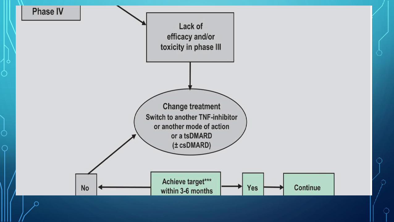
Anti-TNF
”Etanercept,
Infliximab, Adalimumab, Golimumab, & Certolizumab Pegol”
1st choice as well established efficacy/safety balance in PsA
Demonstrated efficacy in PsA, for skin and joint involvement, as
well as in preventing radiographic damage

Ustekinumab is a fully human IgG1κ monoclonal antibody
that binds to the common p40 subunit shared by IL-12 and23.
Secukinumab is a fully human, high affinity,
anti-IL-17A monoclonal antibody that binds to
and neutralizes IL-17A
Both may be useful but are recommended here only as
alternatives, especially if TNFis fail or cannot be applied

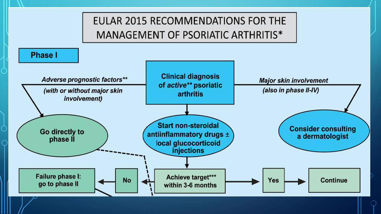
EULAR
recommendations for the management
of psoriatic
arthritis with pharmacological therapies:
2015 update

Active disease: 1 or more tender and inflamed joints; tender enthesis point, dactylitic digit, and/or inflammatory
back pain;
Adverse prognostic factors; 5 active joints; radiographic damage; elevated acute phase reactants; extra-
articular manifestations, especially dactylitis.
The treatment target is clinical remission or, if remission is unlikely to be achievable, at least low disease activity;
clinical remission is the absence of signs and symptoms.
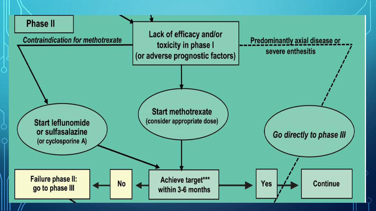
For patients with peripheral arthritis and an inadequate response to at least one csDMARD, in whom TNF
inhibitors are not appropriate. With predominant spinal involvement, active enthesitis and/or dactylitis no
csDMARD needed – use a bDMARD with preferencefor a TNFi.
bDMARD, biological DMARD
csDMARDs, conventional synthetic DMARD
tsDMARD, targeted synthetic DMARD eg such as a PDE4-inhibitor Apremilast