Pseudospread of the Atlas - American Journal of … · · 2014-03-27between the " neural" growth...
4
Richard A. SUSs 1 . 2 Robert D. Zimmerman 1 Norman E. Leeds 1 This article appears in the March/ April 1983 issue of AJNR and the June 1983 issue of AJR. Received May 25, 1982 ; accepted after revi- sion September 30, 1982. Presented at th e ann ual meet ing of th e Am eri- ca n Roent gen Ray Society, San Francisco, March 1981. r Departm enl of Radiology, Montefi ore Hospital an d Medical Cent er, Bronx, NY 10467 . 2 Prese nt address: Departm ent of Radiology , Johns Hopk ins Hospital, 600 N. Wolfe St., Balti- more, MD 21205. Address reprint r eq uests to R. A. Suss. AJNR 4: 183-186, March/ April 1983 0195-6108 / 83 / 0402-018 3 $00 .00 © Ameri can Roent ge n Ray Society Pseudospread of the Atlas: False Sign of Jefferson Fracture in Young Children 1 83 Jefferson fractures are rare prior to teen-age. Three young children examined after trauma exhibited the characteristic spread appearance of the atlas, but fractures were excluded radiographically and clinically . A retrospective study demonstrated a similar appearance , termed " pseudospread ," in most children aged 3 months to 4 years , including over 90% during the second year. Pseudospread results from a discrepancy between the " neural " growth pattern of the atlas and the " somatic " pattern of the axis . An " atlas spread index " is defined and a normal range presented. When an atlas fracture is suggested by apparent lateral spread of the lateral atlas masses , computed tomography is useful to demonstrate an intact atlas ring. The Jefferson fr acture is characterized by a bnormal sepa ration of the later al masses of the atlas (C 1) with fr ac tures of its a nt e ri or a nd post e rior arches. The wedge-shaped lat eral masses are driven apart by a vertica l co mpr essive for ce a pplied to the top of the head [1]. As seen in the ante ropo ste ri or (AP) ope n- mouth view the lateral mas ses ex tend b eyo nd the lateral margin s of the artic ular surface of the axis vertebra (C2) . A similar finding was observed in thr ee chi ldr en after head tr auma, leading to hospit a li zation for further workup of s uspec ted Jeff er so n fr ac tur es. Radiogr ap hi c and clini ca l assessment demon s tr ated no ske letal injury . A retro sp ec tive study of pediatric films was the refore und ertaken in order to determine th e pr evalence , characteristics , and cause of false spread or " p se ud osp read" of the a tl as. Case Reports Case 1 A 5-ye ar-old boy fell down stairs, catching his head in th e banister. He co mplained of head ac he and neck pain. Skull and ce rvica l spine films were initiall y inte rpr eted as nor mal, but upon review a Jefferson fr ac tur e was suggested (fig. 1 A) and the child was reca ll ed for admission. Co mput ed tomography (CT) showe d an int ac t atlas ring (fig. 18). Case 2 A 2' I2- year-old boy clim bed out of his crib and landed " squarely on top of his head ." Decreased motion of his nec k was observed , and ce rvica l spine films showed " displacement of the lateral masses of C1 latera ll y" (fig. 2A). A CT scan showed no fr ac tur e (fig. 2 8) . Case 3 A 3-year-old boy fell out of bed and comp lained of pain and stiffness. Films showed " displ ace d lateral masses of C1 suggest ing a Jefferson fract ur e" (fig. 3A), but CT was normal (fig. 3 8) . In eac h case, pla in films s howed a normal atlas-odontoid distance and no prev ertebr al
Transcript of Pseudospread of the Atlas - American Journal of … · · 2014-03-27between the " neural" growth...

Richard A. SUSs1. 2
Robert D. Zimmerman 1
Norman E. Leeds 1
This article appears in the March/ April 1983 issue of AJNR and the June 1983 issue of AJR.
Received May 25, 1982; accepted after revision September 30,1982.
Presented at the annual meet ing of the American Roentgen Ray Society, San Francisco, March 198 1.
r Departmenl of Radiology, Montefiore Hospital and Medical Center, Bronx, NY 10467.
2 Present address: Department of Radiology , Johns Hopkins Hospital, 600 N. Wolfe St., Baltimore, MD 21205. Address reprint requests to R. A. Suss.
AJNR 4 :183-186, March/ April 1983 0195-6108/ 83 / 0402-0183 $00.00 © American Roentgen Ray Society
Pseudospread of the Atlas: False Sign of Jefferson Fracture in Young Children
183
Jefferson fractures are rare prior to teen-age. Three young children examined after trauma exhibited the characteristic spread appearance of the atlas, but fractures were excluded radiographically and clinically. A retrospective study demonstrated a similar appearance, termed " pseudospread ," in most children aged 3 months to 4 years, including over 90% during the second year. Pseudospread results from a discrepancy
between the " neural " growth pattern of the atlas and the " somatic " pattern of the axis. An " atlas spread index" is defined and a normal range presented. When an atlas fracture is suggested by apparent lateral spread of the lateral atlas masses , computed tomography is useful to demonstrate an intact atlas ring.
The Jefferson fracture is characterized by abnormal separation of the lateral masses of the atlas (C 1) with fractures of its anteri or and posterior arc hes. The wedge-shaped lateral masses are driven apart by a vertical compress ive fo rce applied to the top of the head [1]. As seen in the anteroposteri o r (AP) openmouth view the lateral masses extend beyond the lateral margin s of the arti cular surface of the axis vertebra (C2).
A similar finding was observed in three c hildren after head trauma, lead ing to hospitalization for further workup of suspected Jefferson fractures. Rad iog raphic and c linical assessment demonstrated no ske letal injury. A retrospecti ve study of ped iatric film s was therefore undertaken in order to determine the preva lence , characteristics , and cause of false spread or " pseudospread " of the atl as .
Case Reports
Case 1
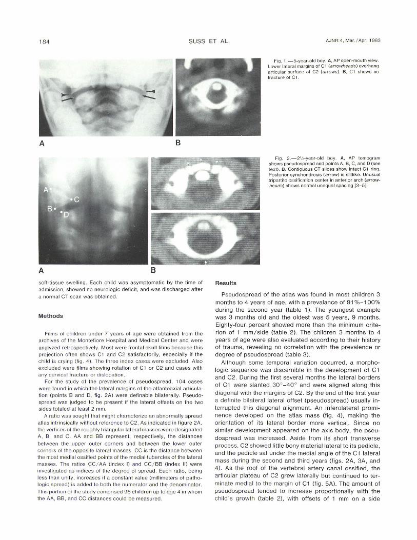
A 5-year-old boy fell down stairs, catc hing his head in th e bani ster. He complained of headache and neck pain . Skull and cervica l spine films were initially interpreted as normal, but upon review a Jefferson fracture was suggested (fig. 1 A) and the child was recall ed for admission. Computed tomography (CT) showed an intact atlas ring (fig. 18).
Case 2
A 2'I2-year-old boy c limbed out of hi s c rib and landed " squarely on top of his head ." Decreased motion of his neck was observed , and cervica l spine films showed " d ispl acement of the lateral masses of C1 laterally " (fig . 2A) . A CT scan showed no fracture (fig . 28) .
Case 3
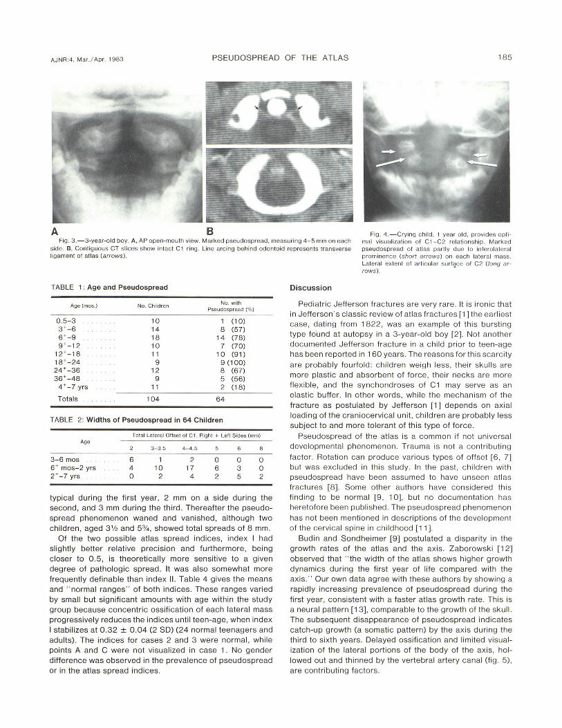
A 3-year-old boy fell out of bed and complained of neci~ pain and stiffn ess. Films showed " displ aced lateral masses of C1 suggesting a Jefferson fracture" (fig . 3 A), but CT was normal (fig. 38) .
In each case, p lain films showed a normal atlas-odontoid distance and no prevertebral
184 SUSS ET AL. AJNR:4 , Mar. l Apr. 1983
A B
A B
soft- tissue swell ing . Each child was asymptomatic by the time of admission, showed no neurolog ic defic it, and was discharged after
a normal CT scan was obtained.
Methods
Fi lms of ch ildren under 7 years of age were obtained from the archives of the Montefiore Hospital and Medical Center and were
analyzed retrospec tively. Most were frontal skull films because this projec tion often shows C1 and C2 sat isfac tori ly, espec ially if the child is c rying (fig. 4). The three index cases were excluded. Also excluded were films showing rotation of C1 or C2 and cases with any cervical frac ture or di slocation.
For the study of the prevalence of pseudospread, 104 cases were found in wh ich the lateral marg ins of the atlantoaxial art iculation (points B and D, fig. 2A) were def inable bi laterally. Pseudospread was judged to be present if th e lateral offse ts on the two sides totaled at least 2 mm .
A ratio was sought that might characteri ze an abn ormally spread atlas intrinsica lly w ithout reference to C2. As ind icated in fi gure 2A, the vertices of th e roughly triangular lateral masses were designated A, B, and C. AA and BB represent , respective ly, the distances
between the upper outer corners and between the lower outer corners of the opposite lateral masses. CC is the d istance between the most medial ossified points of the med ial tuberc les of the lateral masses. The ratios CC I AA (index I) and CC / BB (index II) were investigated as indices of the degree of spread. Each ratio , being less than un ity, increases if a constant value (mi llimeters of patholog ic spread) is added to both the numerator and the denominator.
This portion of the study comprised 96 child ren up to age 4 in whom the AA, BB, and CC distances could be measured.
Results
Fig. 1 .- 5-year-o ld boy. A, AP open-mouth view. Lower lateral marg ins o f C1 (arrowheads ) overhang art icular surface of C2 (arrows ). B, CT shows no frac ture of C1 .
Fig. 2 .- 2 'h -year-old boy. A, AP tomogram shows pseudospread and points A, B , C, and 0 (see tex t) . B , Contiguous CT slices show intact C1 ring. Posterior synchondrosis (arrow) is sli tli ke. Unusual tripartite ossification center in anteri or arch (arro wheads) shows normal unequal spac ing [3 - 5].
Pseudospread of the atl as was found in most children 3 months to 4 years of age, wi th a prevalance of 9 1 %-1 00% during the second year (table 1). The youngest example was 3 months old and the oldest was 5 years, 9 months. Eighty-four percent showed more than the minimum cri terion of 1 mm / side (table 2). The children 3 months to 4 years of age were also evaluated according to their history of trauma, revealing no correlation with the prevalence or degree of pseudospread (tab le 3).
Althoug h some temporal variation occurred , a morpholog ic seq uence was d iscernibl e in the development of C1 and C2 . During the f irst several months the lateral borders of C1 were slanted 30 ° -40° and were aligned along thi s diagonal with the margins of C2 . By the end of the first year a definite bilateral late ral offset (pseudospread) usual ly interrupted this d iagonal alignment. An inferolateral prominence developed on the atlas mass (fi g . 4) , making the orientation of its lateral border more vert ical. Since no sim ilar deve lopment appeared on the axis body, the pseudospread was increased. As ide from its short transverse process, C2 showed li ttl e bony material lateral to its pedic le, and the pedic le sat under the med ial angle of the C1 lateral mass du ring the second and th ird years (fi gs. 2A, 3A, and 4) . As the roof of the vertebral artery canal ossif ied , the art icular plateau of C2 grew lateral ly but continued to terminate medial to the marg in of C1 (f ig. 5A). The amount of pseudospread tended to increase proportional ly with the child 's growth (table 2) , with offsets of 1 mm on a side
AJNR:4. Mar./Apr. 1983 PSEUDOSPREAD OF THE ATLAS 185
A B Fig . 3.-3-year-old boy. A. AP open-mouth view. Marked pseudospread. measuring 4-5 mm on each
side. B. Contiguous CT slices show intact C1 ring. Line arc ing behind odontoid represents transverse ligament of atl as (arrows ).
Fig . 4. - Crying child. 1 year old. provides optimal visualiza ti on o f C1-C2 relati onship. Mark ed pseudospread of atlas partl y due to inferolateral prominence (short arrows) on each lateral mass. Lateral ex tent o f articular surface o f C2 (long ar-
TABLE 1: Age and Pseudospread
Age (mos.) No. Children No. with Pseudospread (% )
0.5-3 10 1 (10) 3+-6 14 8 (57) 6+-9 18 14 (78) 9+-12 10 7 (70)
12+- 18 11 10 (91) 18+-24 9 9 (100) 24+-36 12 8 (67) 36+-48 9 5 (56)
4 + -7 yrs 11 2 (18)
Totals 104 64
TABLE 2: Widths of Pseudospread in 64 Children
Total Lateral Ollset 01 C1 . Righ t + Left Sides (mm) Age
2 3- 3.5 4-4 .5 5 6 8
3-6 mos 6 1 2 0 0 0 6+ mos- 2 yrs 4 10 17 6 3 0 2+-7 yrs 0 2 4 2 5 2
typical during the first year, 2 mm on a side during the second, and 3 mm during the th ird. Thereafter the pseudospread phenomenon waned and vanished, although two children, aged 3% and 5%, showed total spreads of 8 mm.
Of the two possib le atlas spread indices, index I had slightly better relative precision and furthermore, being c loser to 0.5, is theoreticall y more sensitive to a g iven degree of pathologic spread. It was also somewhat more frequently definable than index II . Table 4 gives the means and " normal ranges " of both indices. These ranges varied by small but significant amounts with age within the study group because concentric ossification of each lateral mass progressively reduces the ind ices until teen-age, when index I stabilizes at 0 .32 ± 0.04 (2 SO) (24 normal teenagers and adults). The indices for cases 2 and 3 were normal, while points A and C were not visualized in case 1. No gender difference was observed in the prevalence of pseudospread or in the atlas spread indices .
rows ). •
Discussion
Pediatric Jefferson fractures are very rare. It is ironic that in Jefferson 's c lassic review of atlas fractures [1] the earli est case, dating from 1822, was an example of thi s bursting type fOund at autopsy in a 3-year-old boy [2]. Not another documented Jefferson fracture in a child prior to teen-age has been reported in 160 years. The reasons for thi s scarc ity are probably fourfold: ch ildren weigh less, their skull s are more plastic and absorbent of force , their necks are more f lexible , and the synchondroses of C1 may serve as an elastic buffer. In other words, while the mechanism of the fracture as postulated by Jefferson [1] depends on axial loading of the craniocervical unit, ch ildren are probably less subject to and more tolerant of this type of force.
Pseudospread of the atlas is a common if not universal developmental phenomenon. Trauma is not a contributing factor. Rotation can produce various types of offset [6 , 7] but was exc luded in thi s study. In the past, children with pseudospread have been assumed to have unseen atl as fractures [8]. Some other authors have considered thi s finding to be normal [9 , 10], but no documentation has heretofore been published. The pseudospread phenomenon has not been mentioned in description s of the development of the cervical spine in ch ildhood [11].
Budin and Sondheimer [9] postulated a disparity in the growth rates of the atlas and the axis . Zaborowski [12] observed that · 'the width of the atlas shows higher growth dynamics during the first year of life compared with the ax is. " Our own data agree with these authors by showing a rapidly increasing prevalence of pseudospread during the first year, consistent with a faster atlas growth rate. This is a neural pattern [13] , comparable to the growth of the skull . The subsequent disappearance of pseudospread indicates catch-up growth (a somatic pattern) by the axis during the third to sixth years. Delayed ossification and limited visualization of the lateral portions of the body of the axis, hollowed out and thinned by the vertebral artery canal (fig . 5), are contributing factors .

186 SUSS ET AL. AJ NR:4, Mar. j Apr. 1983
TABLE 3: Trauma and Pseudospread , Ages 3 .5 Months to 4 Years
History
No trauma Possible trauma Trauma, no frac ture Skull fracture
No. with Pseudospread (% )
17 / 23 (74) 9 / 12 (7 5)
25 / 36 (69) 10/ 12 (83)
Mean Spread (mm) ± 2 SO in Positive Cases
4.0 ± 1.8 3.7 ± 1 .3 4 .3 ± 1 .1 3.5 ± 0 .7
Atlas Spread Index CC I AA
0.50 ± 0.03 (n = 29) 0.50 ± 0.04 (n = 13) 0.51 ± 0.03 (n = 34) 0.50 ± 0 .04 (n = 13)
Nole. -AA is the distance between the upper outer corners of the opposite lateral masses: CC is the distance between the most medial ossified points at the medial tuberc les of the lateral masses.
A 8
TABLE 4: Normal Ranges of Atlas Spread Indices
Age
0.5-6 mos t test (2-tailed) 6+ mos-2 yrs t test (2-tailed) 2+-4 yrs
Mean Spread Index ± 2 SO'
I (CC / AA)
0.52 ± 0 .06 p < 0.01
0.50 ± 0.06 p < 0.01
0.48 ± 0.05
II (CC/ SS)
0.63 ± 0.08 p < 0.001
0 .58 ± 0.07 p< 0.01
0 .55 ± 0.07
Note.-AA and SS are the distances between the upper outer and lower outer corners of the opposite lateral masses, respecti ve ly; CC is the distance between the most medial ossified points of each medial tubercle of the lateral masses .
• 95% confidence interval.
Since bilateral lateral offset of C1 with respect to C2 in children is not an indicator of a spread ing process of C1 ,
other means of recognizing a Jefferson fracture without the misleading compari son with C2 would be desirable. CT demonstrates the atl as ring well, but thi s examination is limited by cost, add itional time and radiation, and availability. The atlas spread index is an intrinsic estimator of C1 morphology, completely independent from the phenomenon of
pseudospread. It may therefore be used as supporting evidence to exc lude fracture in a child when a " funny look ing atlas" is actuall y well with in normal limits.
ACKNOWLEDGMENTS
We thank Elnor J . Brown , Marge Eddy, and Carole Wald for assistance in manuscript preparation .
REFERENCES
Fig. 5.-Vertebral artery canal through C2. A, 3 'h-year-old child . Vertebral artery canals (arrows) clearly seen immediately beneath articu lar surfaces of C2. Canal roof becomes thinner laterally (arrowheads) and may continue as cartil age for unknown distance. B, Vertebral arteriog ram from 5-year-old ch ild . As vertebral artery passes upward through C2 it tu rns sharpl y to run laterally directly beneath articular surface of C2. Bony roof of this canal terminates (arrowhead) we ll medial to lateral margin of C1 lateral mass (highlighted by dashed line).
1. Jefferson G. Fracture o f the atl as vertebra. Br J Surg 1920;7: 407 -422
2. Cooper AP. A trea tise on disloca tions and on fractures of the joints. London: Longman, Hurst, Rees, Orme and Brown , 1822 :549- 550
3 . Meckel JF. Ueber die Entwicklung der Centraltheile des Nervensystems bei den Saugthieren. Dtsch Arch Physiol. 1815;1 :648
4. Geipel P: Zur Kenntnis der Spaltbildung des Atl as und Epistropheus. Zentralbl Allg Patho/1955 ;94 : 19-84
5. Silverman FN , Kattan KR. " Trauma " and " no-trauma " of the cervica l spine in pediatric pat ients. In : Kattan KR , ed. " Trauma" and " no-trauma " of the cervical spine. Springfield , IL: Thomas, 1975 : 206- 24 1
6. Paul LW, Moir WW. Non-patholog ic variations in re lat ionship of the upper cervical vertebrae. AJR 1949;62: 519-524
7. Braakman R, Penning L. Injuries of the cervica l spine. Am sterdam: Excerpta Medica, 1971 : 43-45
8. Jacobson G, Adler DC., Examination of the atlanto-axial joint following injury. AJR 1956;76 : 1 08 1-1 094
9. Budin E, Sondheimer F. Lateral spread of the atlas without fracture. Radiology 1966;87: 1 095-1 098
10. Gehweiler JA Jr, Osborne RL Jr, Becker RF. The radiology of vertebral trauma. Philadelphia: Saunders, 1980 : 156-158
11. Bailey OK . The normal cerv ical spine in infants and ch ildren. Radiology 1952 ;59: 712-719
12. Zaborowski Z . Extrafetal development of the axis on the basis of roentgenoanthropometric measurements. Folia Morphol (Warsz) 1978;37: 167 -177
13. Tulsi RS . Growth of the human vertebral co lumn: an osteological study . Acta Anat (Basel) 1971 ;79: 570-580