Pseudomelanocytic nests mimicking atypical melanocytic ... · Pseudomelanocytic nests mimicking...
8
Pseudomelanocytic nests mimicking atypical melanocytic proliferations: first reported cases in the oral cavity Audrey L. Boros, MSc, DDS, a Janice P. Handlers, DDS, b and Raymond J. Melrose, DDS b,c Oral Pathology Associates Inc, Los Angeles, CA, USA; University of Southern California, Los Angeles, CA, USA The concept of pseudomelanocytic nests has been recently described in the dermatology literature. To our knowledge, this entity has yet to be published in the oral pathology literature. We report 2 cases with features of pseudomelanocytic nests. In both instances, nests of cells suspicious for melanocytes were observed. Interpretation of melan-A was negative. Both cases showed strong and diffuse immunoreactivity of the nested cells to CD68. This immunohistochemical staining pattern is most consistent with a melanophage identity. Pseudomelanocytic nests are a recently described entity that represents a potential diagnostic pitfall. Distinguishing pseudomelanocytic nests from an authentic atypical melanocytic proliferation can be challenging and is important for appropriate patient management. Clinicopathologic correlation with cautious interpretation of immunohistochemistry may be necessary to arrive at the correct diagnosis. These cases represent the first reports of pseudomelanocytic nests in the oral pathology literature. (Oral Surg Oral Med Oral Pathol Oral Radiol 2014; 118:461-468) Oral mucosal melanoma accounts for less than 1% of melanomas, affecting approximately 1 in 2 million people annually in the United States. 1,2 Oral melanomas are more common in individuals over 40 years of age, and a slight male predilection is observed. Risk factors for oral melanoma are less well characterized than those for cutaneous melanoma. In general, fair-skinned individuals are at increased risk for developing cutaneous melanoma. Although an increased frequency of oral melanomas has been reported in Japanese and Ugandan Africans, this may be relative to the lower incidence of cutaneous melanomas in these populations. 1,2 High-risk sites for oral mucosal melanomas are the hard palate and maxillary gingiva. 1,2 In a literature review by Rapini et al., 2 75% of cases occurred at the afore- mentioned locations. The majority of oral mucosal mel- anomas are most similar to the acral lentiginous subtype of cutaneous melanoma. 1 Between 30% and 37% of cases are associated with a preexisting pigmented lesion. 2 This likely represents the radial growth phase of the neoplasm and less likely represents a melanoma arising from a discrete preexisting benign melanocytic lesion. The majority of oral melanomas exhibit early invasive char- acteristics. 3 Pigmented lesions of the oral cavity that cannot be definitively diagnosed clinically and radio- graphically should be biopsied for definitive diagnosis. Melanocytic hyperplasia is defined as increased numbers of melanocytes within the basal cell region of the epithelium in the absence of significant confluence, nesting, or pagetoid spread. 4 A ratio of 1 melanocyte per 10 basal keratinocytes is generally accepted as normal, although this ratio is dependent on anatomic location. 4 Atypical melanocytic proliferations may represent the proliferative phase of a melanocytic neoplasm before malignant transformation occurs, making early diagnosis critical for survival. 3,5 As defined by the WESTOP (Western Society of Teachers of Oral Pathology) Banff Workshop on oral mela- nomas, atypical melanocytic proliferations represent melanocytic lesions in which the microscopic archi- tecture or cytology is equivocal. 3 Cellular features of melanocytic atypia include angular and hyperchromatic nuclei and cells with very infrequent mitotic activity. 3 Although sometimes used synonymously with atypical melanocytic proliferation, melanoma in situ should be reserved for morphologically malignant lesions that are incapable of metastasis. 6 The concept of pseudonests, or more precisely pseu- domelanocytic nests, has been reported in the derma- tology literature. 7 This entity is significant because it represents a potential diagnostic pitfall, given that the histologic differential diagnoses include atypical mela- nocytic proliferations, melanoma in situ, and lentigo maligna melanoma. 7-9 Not only is the routine histopa- thology difficult to interpret, but a potential for error exists in the interpretation of immunohistochemical analysis of these lesions. 7-9 The cells that constitute the pseudome- lanocytic nests have not been fully characterized. Based on immunohistochemical analysis, macrophages appear to be a predominant cell type. Occasional lymphocytes, melanocytes, degenerated keratinocytes, or a combina- tion of those may or may not be present in the nests. Pseudomelanocytic nests have been described in the setting of fixed drug eruptions, lichen planus pigmento- sus, and lichenoid lesions including lichenoid keratosis a Associate, Oral Pathology Associates Inc. b Co-director, Oral Pathology Associates Inc. c Emeritus Professor, Herman Ostrow School of Dentistry, University of Southern California. Received for publication Jan 21, 2014; returned for revision Apr 21, 2014; accepted for publication May 14, 2014. Ó 2014 Elsevier Inc. 2212-4403 http://dx.doi.org/10.1016/j.oooo.2014.05.013 461 Vol. 118 No. 4 October 2014 Open access under CC BY-NC-ND license.
Transcript of Pseudomelanocytic nests mimicking atypical melanocytic ... · Pseudomelanocytic nests mimicking...

Vol. 118 No. 4 October 2014
Pseudomelanocytic nests mimicking atypical melanocyticproliferations: first reported cases in the oral cavityAudrey L. Boros, MSc, DDS,a Janice P. Handlers, DDS,b and Raymond J. Melrose, DDSb,c
Oral Pathology Associates Inc, Los Angeles, CA, USA; University of Southern California, Los Angeles, CA, USA
The concept of pseudomelanocytic nests has been recently described in the dermatology literature. To our
knowledge, this entity has yet to be published in the oral pathology literature. We report 2 cases with features of
pseudomelanocytic nests. In both instances, nests of cells suspicious for melanocytes were observed. Interpretation of melan-A
was negative. Both cases showed strong and diffuse immunoreactivity of the nested cells to CD68. This immunohistochemical
staining pattern is most consistent with a melanophage identity. Pseudomelanocytic nests are a recently described entity that
represents a potential diagnostic pitfall. Distinguishing pseudomelanocytic nests from an authentic atypical melanocytic
proliferation can be challenging and is important for appropriate patient management. Clinicopathologic correlation with
cautious interpretation of immunohistochemistry may be necessary to arrive at the correct diagnosis. These cases represent the
first reports of pseudomelanocytic nests in the oral pathology literature. (Oral Surg Oral Med Oral Pathol Oral Radiol 2014;
118:461-468)
Oral mucosal melanoma accounts for less than 1% ofmelanomas, affecting approximately 1 in 2 millionpeople annually in the United States.1,2 Oral melanomasaremore common in individuals over 40 years of age, anda slight male predilection is observed. Risk factors fororal melanoma are less well characterized than those forcutaneousmelanoma. In general, fair-skinned individualsare at increased risk for developing cutaneous melanoma.Although an increased frequency of oral melanomas hasbeen reported in Japanese and Ugandan Africans, thismay be relative to the lower incidence of cutaneousmelanomas in these populations.1,2
High-risk sites for oral mucosal melanomas are thehard palate and maxillary gingiva.1,2 In a literature reviewby Rapini et al.,2 75% of cases occurred at the afore-mentioned locations. The majority of oral mucosal mel-anomas are most similar to the acral lentiginous subtypeof cutaneousmelanoma.1 Between 30%and 37% of casesare associated with a preexisting pigmented lesion.2 Thislikely represents the radial growth phase of the neoplasmand less likely represents a melanoma arising from adiscrete preexisting benign melanocytic lesion. Themajority of oral melanomas exhibit early invasive char-acteristics.3 Pigmented lesions of the oral cavity thatcannot be definitively diagnosed clinically and radio-graphically should be biopsied for definitive diagnosis.
Melanocytic hyperplasia is defined as increasednumbers of melanocytes within the basal cell region of
aAssociate, Oral Pathology Associates Inc.bCo-director, Oral Pathology Associates Inc.cEmeritus Professor, Herman Ostrow School of Dentistry, Universityof Southern California.Received for publication Jan 21, 2014; returned for revision Apr 21,2014; accepted for publication May 14, 2014.� 2014 Elsevier Inc.2212-4403http://dx.doi.org/10.1016/j.oooo.2014.05.013
Open access under CC BY-NC-ND license.
the epithelium in the absence of significant confluence,nesting, or pagetoid spread.4 A ratio of 1 melanocyteper 10 basal keratinocytes is generally accepted asnormal, although this ratio is dependent on anatomiclocation.4 Atypical melanocytic proliferations mayrepresent the proliferative phase of a melanocyticneoplasm before malignant transformation occurs,making early diagnosis critical for survival.3,5 Asdefined by the WESTOP (Western Society of Teachersof Oral Pathology) Banff Workshop on oral mela-nomas, atypical melanocytic proliferations representmelanocytic lesions in which the microscopic archi-tecture or cytology is equivocal.3 Cellular features ofmelanocytic atypia include angular and hyperchromaticnuclei and cells with very infrequent mitotic activity.3
Although sometimes used synonymously with atypicalmelanocytic proliferation, melanoma in situ should bereserved for morphologically malignant lesions that areincapable of metastasis.6
The concept of pseudonests, or more precisely pseu-domelanocytic nests, has been reported in the derma-tology literature.7 This entity is significant because itrepresents a potential diagnostic pitfall, given that thehistologic differential diagnoses include atypical mela-nocytic proliferations, melanoma in situ, and lentigomaligna melanoma.7-9 Not only is the routine histopa-thology difficult to interpret, but a potential for error existsin the interpretation of immunohistochemical analysis ofthese lesions.7-9 The cells that constitute the pseudome-lanocytic nests have not been fully characterized. Basedon immunohistochemical analysis, macrophages appearto be a predominant cell type. Occasional lymphocytes,melanocytes, degenerated keratinocytes, or a combina-tion of those may or may not be present in the nests.
Pseudomelanocytic nests have been described in thesetting of fixed drug eruptions, lichen planus pigmento-sus, and lichenoid lesions including lichenoid keratosis
461

Fig. 1. Clinical photograph depicting a slightly elevated,pigmented lesion of the facial interdental papilla between themandibular central incisors (case 1, lesion 1).
Fig. 2. Low-power photomicrograph of the pigmented lesionin Figure 1. The section shows a somewhat nodular lesioncovered by nondysplastic stratified squamous epithelium.Aggregates of nested cells are present. Scattered foci ofchronic inflammation, some of which is perivascular in nature,are observed in the lamina propria and superficial connectivetissue stroma (case 1, lesion 1; hematoxylin-eosin, originalmagnification � 4). (A high-resolution version of the image isavailable as eSlide: VM00243.)
Fig. 3. Medium-power photomicrograph of the pigmentedlesion in Figure 1. Some of the nested cells have enlarged andhyperchromatic nuclei for melanocytes (case 1, lesion 1;hematoxylin-eosin, original magnification � 10). (A high-reso-lution version of the image is available as eSlide: VM00243.)
Fig. 4. High-power photomicrograph of the pigmented lesion.Some of the nested cells have enlarged, irregular nuclei (case1, lesion 1; hematoxylin-eosin, original magnification � 40).(A high-resolution version of the image is available as eSlide:VM00243.)
ORAL AND MAXILLOFACIAL PATHOLOGY OOOO
462 Boros, Handlers and Melrose October 2014
and lichenoid phototoxic reaction.7-9 The treatment andprognosis for these reactive lesions are differentcompared with those for atypical melanocytic prolifera-tion, melanoma in situ, and lentigo maligna melanoma;thus, distinction between these lesions is essential. To ourknowledge, the present cases are the first 2 reported casesof pseudomelanocytic nests in the oral pathologyliterature.
CASE REPORTCase 1A 66-year-old man presented to a private periodontist’s officefor evaluation of a slightly elevated pigmented lesion of thefacial interdental papilla between the mandibular central in-cisors (Figure 1). A sudden onset of the lesion was reported.The remainder of the oral examination was within normal
limits. No clinical evidence of lichen planus or lichenoidmucositis was observed. The patient’s medical history wasnoncontributory. No medications, prescribed or otherwise,were reported. The social history was negative for smoking. Abiopsy of the lesion was performed.
The initial interpretation of this lesion was that of anatypical melanocytic proliferation (Figures 2 to 4). Theatypical prefix was in reference to the perceived abnormalnesting pattern in combination with the nuclear features ofthese cells. Occasional nuclei in these nested cells wereinterpreted as enlarged with irregular nuclear contours for

Fig. 5. A, Melan-A staining in nested cells was comparablewith the negative control (slide not available). Focal weakcytoplasmic staining was present which was interpreted asnon-specific. A positive internal control highlighting melan-Aimmunoreactivity in the nuclei and cytoplasmic extensionsof melanocytes in the basal cell layer is present (case 1, lesion1; original magnification � 10). (A high-resolution version ofthe image is available as eSlide: VM00244.) B, Medium-power photomicrograph of the same area as seen in panelA. The nested cells show strong, diffuse immunoreactivityto CD68 (case 1, lesion 1; original magnification � 10). (Ahigh-resolution version of the image is available as eSlide:VM00246.)
Fig. 6. Clinical photograph depicting a nonuniformly pig-mented lesion with irregular borders of the buccal gingivaassociated with the left maxillary second molar. (case 1,lesion 2).
OOOO CASE REPORT
Volume 118, Number 4 Boros, Handlers and Melrose 463
typical melanocytes. No mitotic activity was identified withinthese cells. A mild chronic inflammatory infiltrate was pre-sent in the lamina propria and superficial connective tissue. Aprovisional diagnosis of atypical melanocytic proliferationwas rendered. The slides were sent for consultation to anoutside oral and maxillofacial pathology group. Blind to thecontributor’s diagnosis, 2 oral and maxillofacial pathologistsindependently diagnosed the lesion as an atypical melano-cytic proliferation.
In an attempt to better examine the extent of the melanocyticcomponent, melan-A/MART-1 (melanoma antigen recognizedby T cells 1) immunohistochemistry was performed withappropriate positive and negative controls. This stain high-lighted individual melanocytes along the length of the basal celllayer. Melan-A in nested cells (Figure 5, A) was comparablewith the negative control (slide not available). Focal weakcytoplasmic staining was present which was interpreted as non-specific. Based on the hematoxylin-eosin interpretation, thediagnosis remained unchanged. A comment was providedsuggesting that the patient be placed on close and continuedfollow-up, with a recommendation to perform another biopsyfor any new or recurrent pigmented lesions. At this time, a CD68stain was not performed.
Approximately 2 months later, the patient returned to thesame office with a second pigmented lesion. The secondlesion was located on the buccal gingiva associated with themaxillary left second molar (Figure 6). Another biopsy was
performed. A similar microscopic pattern was observed and,once again, a diagnosis of atypical melanocytic proliferationwas rendered (Figure 7). The slide was sent for consultation.All oral and maxillofacial pathologists were in agreement withthe initial diagnosis. Immunohistochemistry for melan-Afound a staining pattern similar to the previous biopsy(Figure 8, A), and the diagnosis remained unchanged. At thistime, CD68 analysis was not performed. Ten days later, pig-mented lesions of the right hard palate adjacent to themaxillary second molar and several pigmented lesions of thelower lip were also noted.
At the request of the patient, material from the biopsies wassent for a consultative opinion at an outside institution whereinterpretation was performed in conjunction with the Depart-ment of Dermatopathology. At this time, immunohistochem-istry for CD68 was performed on both biopsies (Figures 5, B,and 8, B). The nested cells stained strongly positive for CD68.Immunohistochemical stains for melan-A, MiTF (micro-pthalmia-associated transcription factor), and SOX10 (sex-determining region Y box 10) were interpreted as negative(immunohistochemistry not available for MiTF and SOX10).Although the diagnosis of atypical melanocytic proliferationwas in the differential diagnosis, with this additional informa-tion, both lesions were interpreted as “chronic mucositis withpseudomelanocytic nests.”
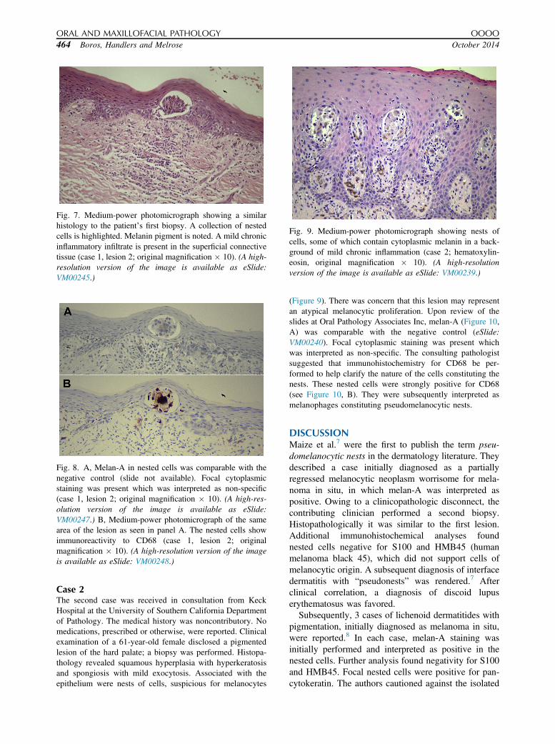
Fig. 8. A, Melan-A in nested cells was comparable with thenegative control (slide not available). Focal cytoplasmicstaining was present which was interpreted as non-specific(case 1, lesion 2; original magnification � 10). (A high-res-olution version of the image is available as eSlide:VM00247.) B, Medium-power photomicrograph of the samearea of the lesion as seen in panel A. The nested cells showimmunoreactivity to CD68 (case 1, lesion 2; originalmagnification � 10). (A high-resolution version of the imageis available as eSlide: VM00248.)
Fig. 9. Medium-power photomicrograph showing nests ofcells, some of which contain cytoplasmic melanin in a back-ground of mild chronic inflammation (case 2; hematoxylin-eosin, original magnification � 10). (A high-resolutionversion of the image is available as eSlide: VM00239.)
Fig. 7. Medium-power photomicrograph showing a similarhistology to the patient’s first biopsy. A collection of nestedcells is highlighted. Melanin pigment is noted. A mild chronicinflammatory infiltrate is present in the superficial connectivetissue (case 1, lesion 2; original magnification � 10). (A high-resolution version of the image is available as eSlide:VM00245.)
ORAL AND MAXILLOFACIAL PATHOLOGY OOOO
464 Boros, Handlers and Melrose October 2014
Case 2The second case was received in consultation from KeckHospital at the University of Southern California Departmentof Pathology. The medical history was noncontributory. Nomedications, prescribed or otherwise, were reported. Clinicalexamination of a 61-year-old female disclosed a pigmentedlesion of the hard palate; a biopsy was performed. Histopa-thology revealed squamous hyperplasia with hyperkeratosisand spongiosis with mild exocytosis. Associated with theepithelium were nests of cells, suspicious for melanocytes
(Figure 9). There was concern that this lesion may representan atypical melanocytic proliferation. Upon review of theslides at Oral Pathology Associates Inc, melan-A (Figure 10,A) was comparable with the negative control (eSlide:VM00240). Focal cytoplasmic staining was present whichwas interpreted as non-specific. The consulting pathologistsuggested that immunohistochemistry for CD68 be per-formed to help clarify the nature of the cells constituting thenests. These nested cells were strongly positive for CD68(see Figure 10, B). They were subsequently interpreted asmelanophages constituting pseudomelanocytic nests.
DISCUSSIONMaize et al.7 were the first to publish the term pseu-domelanocytic nests in the dermatology literature. Theydescribed a case initially diagnosed as a partiallyregressed melanocytic neoplasm worrisome for mela-noma in situ, in which melan-A was interpreted aspositive. Owing to a clinicopathologic disconnect, thecontributing clinician performed a second biopsy.Histopathologically it was similar to the first lesion.Additional immunohistochemical analyses foundnested cells negative for S100 and HMB45 (humanmelanoma black 45), which did not support cells ofmelanocytic origin. A subsequent diagnosis of interfacedermatitis with “pseudonests” was rendered.7 Afterclinical correlation, a diagnosis of discoid lupuserythematosus was favored.
Subsequently, 3 cases of lichenoid dermatitides withpigmentation, initially diagnosed as melanoma in situ,were reported.8 In each case, melan-A staining wasinitially performed and interpreted as positive in thenested cells. Further analysis found negativity for S100and HMB45. Focal nested cells were positive for pan-cytokeratin. The authors cautioned against the isolated

Fig. 10. A, Immunohistochemical stain for melan-A wassimilar to the negative control. Focal cytoplasmic staining waspresent which was interpreted as non-specific. A positive in-ternal control highlighting melan-A immunoreactivity in thenuclei and cytoplasmic extensions of melanocytes in the basalcell layer is present (case 2; original magnification � 10). (Ahigh-resolution version of the image is available as eSlide:VM00240.) B, Medium-power photomicrograph of the samearea of the lesion as seen in panel A. The nested cells showstrong, diffuse immunoreactivity to CD68 (case 2; originalmagnification � 10). (A high-resolution version of the image isavailable as eSlide: VM00241.) (A high-resolution image of anegative control for this case is available as eSlide: VM00242.)
OOOO CASE REPORT
Volume 118, Number 4 Boros, Handlers and Melrose 465
use of melan-A in the evaluation of potential melano-cytic lesions.
Shortly thereafter, Nicholson et al.9 contributed 2similar cases, which were originally diagnosed as len-tigo maligna melanoma but were reclassified as fixeddrug eruptions after clinicopathologic correlation.Again, initial immunohistochemical analysis limited tomelan-A/MART-1 found cytoplasmic staining in nestedcells, which in combination with the histopathologyprompted the diagnosis of lentigo maligna melanoma.Subsequent immunohistochemical tests found S100,HMB45, and MiTF negativity. Further characterizationfor nested cells found focal positivity for CD3 andAE1/AE3. In addition, 1 case highlighted focal CD68immunoreactivity of nested cells.
The 6 aforementioned cases involved lesions of theskin affecting the head and neck, a location which is ofsignificance to the oral health care professional. A po-tential relationship to chronic sun exposure has beensuggested,8 but this is likely not applicable to oral le-sions. Including the present cases, men and womenhave been affected equally. Patients range in age from35 to 76 years (average, 56 years). Pigmented lesions
have been both solitary and multiple. In all cases, astutecorrelation of the histopathology, immunohistochem-istry, and clinical presentation was necessary to arriveat the correct diagnosis. Table I provides a summary ofpublished cases of pseudomelanocytic lesions withinitial interpretations ranging from atypical melanocyticneoplasms to lentigo maligna melanoma.
The histopathologic characteristics of the presentcases included nests of cells in a background of a var-iable chronic inflammatory infiltrate. Pigment was pre-sent within nested cells and within cells in the laminapropria. Based on histopathology alone, it may bedifficult to differentiate atypical melanocytic pro-liferations from pseudomelanocytic nests. Immunohis-tochemical analysis plays an important role in theaccurate diagnosis of these lesions.
CD68 (KP1) is located on lysosomes and therefore isnot entirely cell lineage specific. Despite this, itis generally regarded as a marker for cells of themacrophage lineage. Positivity has been reported inother cell types, including fibroblasts. A wide range ofneoplasms, both histiocyte-related and non-histiocyte-related, have reported immunoreactivity with CD68.Pernick et al.10 published a series of melanoma casesthat exhibited weak staining for CD68. The intensity ofstaining (strong or weak) is generally accepted to be animportant indicator of cell type, with cells of themacrophage lineage staining strongly for CD68.
Melan-A/MART-1, commonly used as a marker formelanocytes, is not as specific as once believed. Bothmelan-A/MART-1 and HMB45 target components ofthe melanosome. These components may be transferredto nonmelanocytic cells, including cells of the macro-phage lineage, resulting in nonspecific cytoplasmicstaining.7-9 Cytoplasmic melan-A and HMB45 stainingis not necessarily synonymous with a melanocyticidentity, highlighting the hazard of relying primarily onthese immunohistochemical stains for diagnosis, espe-cially when performed in isolation.
MiTF is a more specific melanocytic marker thattargets a nuclear transcription factor within melano-cytes,11 resulting in a nuclear staining pattern. However,recent reports also indicate that MiTF is not entirelyspecific for melanocytes and may also react with com-ponents of macrophages and mast cells.12 A case ofpseudomelanocytic nests with rare positivity for MiTFand SOX10 in the setting of lichenoid inflammation hasbeen reported.12 In this case, clinicopathologic correla-tion prevented initial misdiagnosis. To explain theseresults, Silva et al.12 entertained the possibility of eithera new melanocytic entity or a distinct pattern of benignmelanocytic reorganization in the setting of lichenoiddermatitides. Additional studies are needed for furtherclarification. In these instances, CD68 also may helpcharacterize the nested cells.
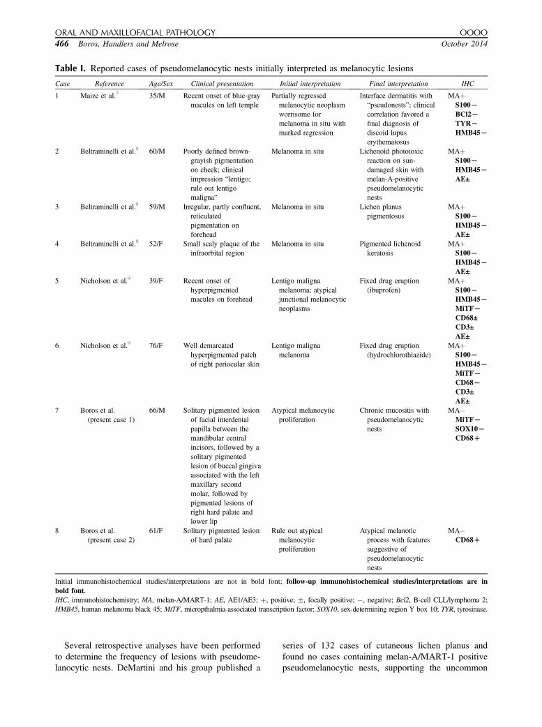
Table I. Reported cases of pseudomelanocytic nests initially interpreted as melanocytic lesions
Case Reference Age/Sex Clinical presentation Initial interpretation Final interpretation IHC
1 Maize et al.7 35/M Recent onset of blue-graymacules on left temple
Partially regressedmelanocytic neoplasmworrisome formelanoma in situ withmarked regression
Interface dermatitis with“pseudonests”; clinicalcorrelation favored afinal diagnosis ofdiscoid lupuserythematosus
MAþS100LBCl2LTYRLHMB45L
2 Beltraminelli et al.8 60/M Poorly defined brown-grayish pigmentationon cheek; clinicalimpression “lentigo;rule out lentigomaligna”
Melanoma in situ Lichenoid phototoxicreaction on sun-damaged skin withmelan-A-positivepseudomelanocyticnests
MAþS100LHMB45LAE±
3 Beltraminelli et al.8 59/M Irregular, partly confluent,reticulatedpigmentation onforehead
Melanoma in situ Lichen planuspigmentosus
MAþS100LHMB45LAE±
4 Beltraminelli et al.8 52/F Small scaly plaque of theinfraorbital region
Melanoma in situ Pigmented lichenoidkeratosis
MAþS100LHMB45LAE±
5 Nicholson et al.9 39/F Recent onset ofhyperpigmentedmacules on forehead
Lentigo malignamelanoma; atypicaljunctional melanocyticneoplasms
Fixed drug eruption(ibuprofen)
MAþS100LHMB45LMiTFLCD68±CD3±AE±
6 Nicholson et al.9 76/F Well demarcatedhyperpigmented patchof right periocular skin
Lentigo malignamelanoma
Fixed drug eruption(hydrochlorothiazide)
MAþS100LHMB45LMiTFLCD68LCD3±AE±
7 Boros et al.(present case 1)
66/M Solitary pigmented lesionof facial interdentalpapilla between themandibular centralincisors, followed by asolitary pigmentedlesion of buccal gingivaassociated with the leftmaxillary secondmolar, followed bypigmented lesions ofright hard palate andlower lip
Atypical melanocyticproliferation
Chronic mucositis withpseudomelanocyticnests
MA�MiTFLSOX10LCD68D
8 Boros et al.(present case 2)
61/F Solitary pigmented lesionof hard palate
Rule out atypicalmelanocyticproliferation
Atypical melanoticprocess with featuressuggestive ofpseudomelanocyticnests
MA�CD68D
Initial immunohistochemical studies/interpretations are not in bold font; follow-up immunohistochemical studies/interpretations are inbold font.IHC, immunohistochemistry; MA, melan-A/MART-1; AE, AE1/AE3; þ, positive; �, focally positive; �, negative; Bcl2, B-cell CLL/lymphoma 2;HMB45, human melanoma black 45; MiTF, micropthalmia-associated transcription factor; SOX10, sex-determining region Y box 10; TYR, tyrosinase.
ORAL AND MAXILLOFACIAL PATHOLOGY OOOO
466 Boros, Handlers and Melrose October 2014
Several retrospective analyses have been performedto determine the frequency of lesions with pseudome-lanocytic nests. DeMartini and his group published a
series of 132 cases of cutaneous lichen planus andfound no cases containing melan-A/MART-1 positivepseudomelanocytic nests, supporting the uncommon
OOOO CASE REPORT
Volume 118, Number 4 Boros, Handlers and Melrose 467
occurrence of pseudomelanocytic nests in lichenoidlesions.13 Analysis with additional immunohistochem-ical markers including CD68 may have also beeninsightful. Abuzeid et al.14 analyzed 53 cases of cuta-neous lupus erythematosus. Among those, 1 of 6 caseswith areas suspicious for pseudomelanocytic nestsshowed focal immunoreactivity for MART-1 and MiTFand lacked immunoreactivity for S100.
In the first case presented here, the nested cellsstained strongly positive for CD68. Melan-A innested cells was comparable with the negative control.Focal cytoplasmic staining was present which wasinterpreted as non-specific. Subsequent immunohisto-chemical evaluation of MiTF and SOX10 was inter-preted as negative; unfortunately, these slides were notavailable for review. Based on the given information,this is more consistent with a staining pattern of mel-anophages, as opposed to a pattern of melanocytestaining. The second case presented also stainedstrongly positive for CD68. Immunohistochemicalstaining for melan-A was similar to the negative con-trol (eSlide: VM00242). Again, focal cytoplasmicstaining was present which was interpreted as non-specific. MiTF and SOX10 immunostains would alsohave been useful but were not used in the evaluation ofthe second case because nested cells were diffuselypositive for CD68.
Clinically, the first case initially presented as a soli-tary lesion of the mandibular gingiva, reported to be ofrapid onset. Approximately 2 months later, a secondlesion, this time of the left posterior maxillary buccalgingiva, was described. In hindsight, this clinical pre-sentation is more consistent with a reactive process thana neoplastic process. The patient continues to be fol-lowed up at regular intervals by the same periodontist.A follow-up time of more than 3 years has been un-eventful, with no new or recurrent lesions reported. Theetiology of the lesions remains unknown. Contributingfactors described in the dermatology literature includelichenoid drug reactions, lichen planus pigmentosus,discoid lupus erythematosus, and sun damage, the latternot being applicable to the majority of the oral cavity.Although no medications were prescribed by theclinician or reported in the history, it is possible that thepatient was taking medications without disclosure.Other possibilities of lichenoid inflammation include areaction to strong flavoring agents, which was not re-ported and with which the histology was not consistent.Lichen planus pigmentosus was also considered in thedifferential diagnosis; however, this was not supportedclinically, because the findings from the remainder ofthe intraoral examination were within normal limits.The social history was negative for smoking, whichhas also been reported to increase pigmentation in theoral cavity.
The second case presented as a solitary pigmentedlesion of the palate, which could represent either areactive or a neoplastic process. In this case, clinicalcorrelation was not as helpful as in the first case. Thispatient was also lost to follow-up.
CONCLUSIONThe cases presented here represent the first 2 cases ofpseudomelanocytic nests reported in the oral pathologyliterature. Histologically, pseudomelanocytic nests maymimic atypical melanocytic proliferations. Clinico-pathologic correlation has proved helpful to achieve anaccurate diagnosis and, in turn, prevent misdiagnosesand potential management complications.
The authors thank Dr Jeffrey Waterman, DDS, for contrib-uting the clinical images.
REFERENCES1. Neville BW, Damm DD, Allen CM, Bouquot JE. Oral and
Maxillofacial Pathology. St Louis, MO: Saunders; 2009.2. Rapini RP, Golitz LE, Greer RO, Krekorian EA, Poulson T.
Primary malignant melanoma of the oral cavity: a review of 177cases. Cancer. 1985;55:1543-1551.
3. Barker BF, Carpenter WM, Daniels TE, et al. Oral mucosalmelanomas: the WESTOP Banff workshop proceedings. WesternSociety of Teachers of Oral Pathology. Oral Surg Oral Med OralPathol Oral Radiol Endod. 1997;83:672-679.
4. Velazquez EF, Murphy GF. Histology of the skin. In: Elder DE,Elenitsas R, Johnson BL Jr, Murphy GF, Xu X, eds. Lever’sHistopathology of the Skin. 10th ed. Philadelphia, PA: LippincottWilliams & Wilkins; 2009.
5. Hicks MJ, Flaitz CM. Oral mucosal melanoma: epidemiology andpathobiology. Oral Oncol. 2000;36:152-169.
6. Buchner A, Merrell PW, Hansen LS, Leider AS. Melanocytichyperplasia of the oral mucosa. Oral Surg Oral Med Oral Pathol.1991;71:58-62.
7. Maize JC, Resneck JS, Shapiro PE, McCalmont TH, LeBoit P.Ducking stray “magic bullets”: a melan-A alert. Am J Dermato-pathol. 2003;25:162-165.
8. Beltraminelli H, Shabrawi-Caelen L, Kerl H, Cerroni L.Melan-A positive “pseudomelanocytic nests”: a pitfall in thehistopathologic and immunohistochemical diagnosis of pig-mented lesions on sun-damaged skin. Am J Dermatopathol.2009;31:305-308.
9. Nicholson KM, Gerami P. An immunohistochemical analysis ofpseudomelanocytic nests mimicking melanoma in situ: report of 2cases. Am J Dermatopathol. 2010;32:633-636.
10. Pernick NL, DaSilva M, Gangi MD, Crissman J, Adsay V.“Histiocytic markers” in melanoma. Mod Pathol. 1999;12:1072-1077.
11. Hemesath TJ, Steingrímsson E, McGill G, et al. Micro-pthalmia, a critical factor in melanocyte development, defines adiscrete transcription factor family. Genes Dev. 1994;8:2770-2780.
12. Silva EY, Goldberg LJ, Mahalingam M, Bhawan J,Wolpowitz D. Nests with numerous SOX10 and MiTF-positivecells in lichenoid inflammation: pseudomelanocytic nests orauthentic melanocytic proliferation? J Cutan Pathol. 2011;38:797-800.

ORAL AND MAXILLOFACIAL PATHOLOGY OOOO
468 Boros, Handlers and Melrose October 2014
13. DeMartini SD, Dalton SR, Ferringer T, Elston DM. Melan-A/MART-1 positive “pseudonests” in lichenoid inflammatorylesions: an uncommon phenomenon. Am J Dermatopathol.2005;27:370-371.
14. Abuzeid M, Dalton SR, Ferringer T, Bernert R, Elston DM.Micropthalmia-associated transcription factor-positive pseu-donests in cutaneous lupus erythematosus. Am J Dermatopathol.2011;33:752-754.
Reprint requests:
Audrey L. Boros, MSc, DDSOral Pathology Associates Inc11500 W Olympic Blvd Suite 390Los Angeles, CA [email protected]