Pseudoaneurysm associated haemosuccus pancreaticus - a ...
4
CASE REPORT Open Access Pseudoaneurysm associated haemosuccus pancreaticus - a rare and dangerous disease Julia Wagenpfeil 1* , Daniel Kütting 1 , Christian P. Strassburg 2 and Carsten Meyer 1 Abstract Background: In patients with upper gastrointestinal bleeding and hemorrhage originating from the major duodenal papilla pseudoaneurysm associated haemosuccus pancreaticus (HP) is a rare differential diagnosis which should be considered. Diagnosis may be challenging, as clinical presentation is often unspecific with only intermittent hemorrhage. Treatment of the causal pseudoaneurysm is mandatory and endovascular coil embolization is the suitable first-line management strategy. Until now there are only a very few studies about this clinical picture and its therapeutic options, especially data regarding whether additional fluid embolization is beneficial/necessary in HP is currently lacking. Case presentation: We report a case of a 59-year-old male patient with chronic pancreatitis and haemosuccus pancreaticus caused by a pancreatico-arterial fistula with an associated inflammatory pseudoaneurysm of the splenic artery. Initially we sought to embolize the pseudoaneurysm with microcoils. As only one coil could be safely deployed in the pseudoaneurysm we additionally employed tissue adhesive embolization in order to achieve complete occlusion of the pseudoaneurysm as well as the pancretico-arterial fistula. In the presented case inflammatory levels decreased following embolization, possibly linked to a decline in pathologic excretion of elastase and autodigestion. As not only the pseudoaneurysm but also the underlying fistula were occluded, the risk of recurrence may conceivably be reduced. Conclusions: Diagnosis of HP is difficult and treatment of the causal pseudoaneurysm is mandatory. Endovascular embolization is the suitable first-line management strategy, complete occlusion of the fistula should be considered when possible. Keywords: Haemosuccus pancreaticus, Upper gastrointestinal bleeding, Chronic pancreatitis, Coil and tissue adhesive (Histoacryl/ Lipiodol) embolization Introduction Haemosuccus pancreaticus (HP), a rare cause of upper gastrointestinal bleeding (1:1500 cases of gastrointestinal hemorrhage) (Subasinghe et al. 2012), is defined as hemorrhage originating from the pancreas, pancreatic duct, or (less frequently) from peri-pancreatic vessels (e. g. splenic artery) into the pancreatic duct. This disease is typically found in patients with chronic pancreatitis (Vimalraj et al. 2009). Increased excretion of elastase, induced by chronic local inflammation, leads to autodiges- tion of peripancreatic vessels or erosion of concomitant pseudocysts into adjacent vessels (Stanley et al. 1976). Diagnosis is challenging and frequently delayed, as hemorrhage is usually only intermittent and not severe enough to cause hemodynamic instability (Mandaliya et al. 2014). Clinical symptoms are similar to those typic- ally found in other forms of gastrointestinal bleeding and include non-specific epigastric pain, hematemesis, melena and hyperamylasemia. Haemosuccus pancreaticus is asso- ciated with a high mortality rate, therapeutic options con- sist of selective radiological embolization or surgery (Subasinghe et al. 2012). © The Author(s). 2020 Open Access This article is licensed under a Creative Commons Attribution 4.0 International License, which permits use, sharing, adaptation, distribution and reproduction in any medium or format, as long as you give appropriate credit to the original author(s) and the source, provide a link to the Creative Commons licence, and indicate if changes were made. The images or other third party material in this article are included in the article's Creative Commons licence, unless indicated otherwise in a credit line to the material. If material is not included in the article's Creative Commons licence and your intended use is not permitted by statutory regulation or exceeds the permitted use, you will need to obtain permission directly from the copyright holder. To view a copy of this licence, visit http://creativecommons.org/licenses/by/4.0/. * Correspondence: [email protected] 1 Department of Radiology, University Hospital Bonn, Venusberg-Campus 1, 53127 Bonn, Germany Full list of author information is available at the end of the article CVIR Endovascular Wagenpfeil et al. CVIR Endovascular (2020) 3:82 https://doi.org/10.1186/s42155-020-00178-3
Transcript of Pseudoaneurysm associated haemosuccus pancreaticus - a ...
CASE REPORT Open Access
Pseudoaneurysm associated haemosuccuspancreaticus - a rare and dangerousdiseaseJulia Wagenpfeil1* , Daniel Kütting1, Christian P. Strassburg2 and Carsten Meyer1
Abstract
Background: In patients with upper gastrointestinal bleeding and hemorrhage originating from the major duodenal papillapseudoaneurysm associated haemosuccus pancreaticus (HP) is a rare differential diagnosis which should be considered.Diagnosis may be challenging, as clinical presentation is often unspecific with only intermittent hemorrhage. Treatment ofthe causal pseudoaneurysm is mandatory and endovascular coil embolization is the suitable first-line management strategy.Until now there are only a very few studies about this clinical picture and its therapeutic options, especially data regardingwhether additional fluid embolization is beneficial/necessary in HP is currently lacking.
Case presentation:We report a case of a 59-year-old male patient with chronic pancreatitis and haemosuccus pancreaticuscaused by a pancreatico-arterial fistula with an associated inflammatory pseudoaneurysm of the splenic artery. Initially wesought to embolize the pseudoaneurysm with microcoils. As only one coil could be safely deployed in the pseudoaneurysmwe additionally employed tissue adhesive embolization in order to achieve complete occlusion of the pseudoaneurysm aswell as the pancretico-arterial fistula. In the presented case inflammatory levels decreased following embolization, possiblylinked to a decline in pathologic excretion of elastase and autodigestion. As not only the pseudoaneurysm but also theunderlying fistula were occluded, the risk of recurrence may conceivably be reduced.
Conclusions: Diagnosis of HP is difficult and treatment of the causal pseudoaneurysm is mandatory. Endovascularembolization is the suitable first-line management strategy, complete occlusion of the fistula should be consideredwhen possible.
Keywords: Haemosuccus pancreaticus, Upper gastrointestinal bleeding, Chronic pancreatitis, Coil and tissue adhesive(Histoacryl/ Lipiodol) embolization
IntroductionHaemosuccus pancreaticus (HP), a rare cause of uppergastrointestinal bleeding (1:1500 cases of gastrointestinalhemorrhage) (Subasinghe et al. 2012), is defined ashemorrhage originating from the pancreas, pancreaticduct, or (less frequently) from peri-pancreatic vessels (e. g.splenic artery) into the pancreatic duct. This disease istypically found in patients with chronic pancreatitis(Vimalraj et al. 2009). Increased excretion of elastase,
induced by chronic local inflammation, leads to autodiges-tion of peripancreatic vessels or erosion of concomitantpseudocysts into adjacent vessels (Stanley et al. 1976).Diagnosis is challenging and frequently delayed, ashemorrhage is usually only intermittent and not severeenough to cause hemodynamic instability (Mandaliyaet al. 2014). Clinical symptoms are similar to those typic-ally found in other forms of gastrointestinal bleeding andinclude non-specific epigastric pain, hematemesis, melenaand hyperamylasemia. Haemosuccus pancreaticus is asso-ciated with a high mortality rate, therapeutic options con-sist of selective radiological embolization or surgery(Subasinghe et al. 2012).
© The Author(s). 2020 Open Access This article is licensed under a Creative Commons Attribution 4.0 International License,which permits use, sharing, adaptation, distribution and reproduction in any medium or format, as long as you giveappropriate credit to the original author(s) and the source, provide a link to the Creative Commons licence, and indicate ifchanges were made. The images or other third party material in this article are included in the article's Creative Commonslicence, unless indicated otherwise in a credit line to the material. If material is not included in the article's Creative Commonslicence and your intended use is not permitted by statutory regulation or exceeds the permitted use, you will need to obtainpermission directly from the copyright holder. To view a copy of this licence, visit http://creativecommons.org/licenses/by/4.0/.
* Correspondence: [email protected] of Radiology, University Hospital Bonn, Venusberg-Campus 1,53127 Bonn, GermanyFull list of author information is available at the end of the article
CVIR EndovascularWagenpfeil et al. CVIR Endovascular (2020) 3:82 https://doi.org/10.1186/s42155-020-00178-3
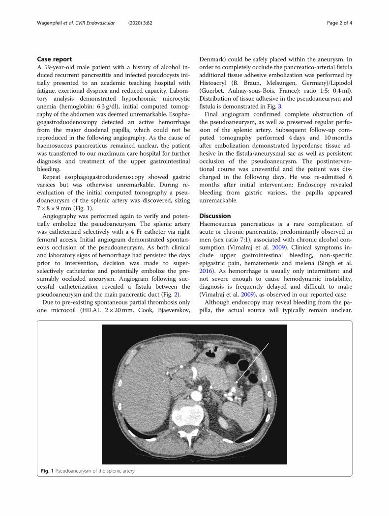
Case reportA 59-year-old male patient with a history of alcohol in-duced recurrent pancreatitis and infected pseudocysts ini-tially presented to an academic teaching hospital withfatigue, exertional dyspnea and reduced capacity. Labora-tory analysis demonstrated hypochromic microcyticanemia (hemoglobin: 6.3 g/dl), initial computed tomog-raphy of the abdomen was deemed unremarkable. Esopha-gogastroduodenoscopy detected an active hemorrhagefrom the major duodenal papilla, which could not bereproduced in the following angiography. As the cause ofhaemosuccus pancreaticus remained unclear, the patientwas transferred to our maximum care hospital for furtherdiagnosis and treatment of the upper gastrointestinalbleeding.Repeat esophagogastroduodenoscopy showed gastric
varices but was otherwise unremarkable. During re-evaluation of the initial computed tomography a pseu-doaneurysm of the splenic artery was discovered, sizing7 × 8 × 9mm (Fig. 1).Angiography was performed again to verify and poten-
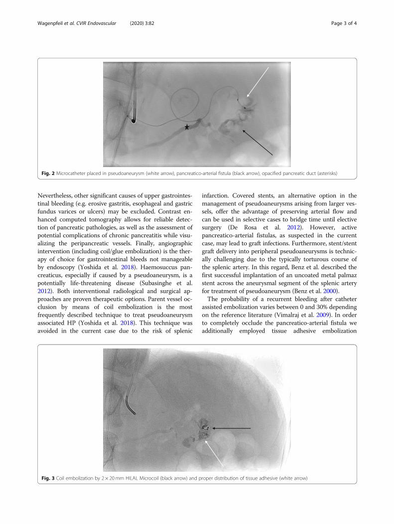
tially embolize the pseudoaneurysm. The splenic arterywas catheterized selectively with a 4 Fr catheter via rightfemoral access. Initial angiogram demonstrated spontan-eous occlusion of the pseudoaneurysm. As both clinicaland laboratory signs of hemorrhage had persisted the daysprior to intervention, decision was made to super-selectively catheterize and potentially embolize the pre-sumably occluded aneurysm. Angiogram following suc-cessful catheterization revealed a fistula between thepseudoaneurysm and the main pancreatic duct (Fig. 2).Due to pre-existing spontaneous partial thrombosis only
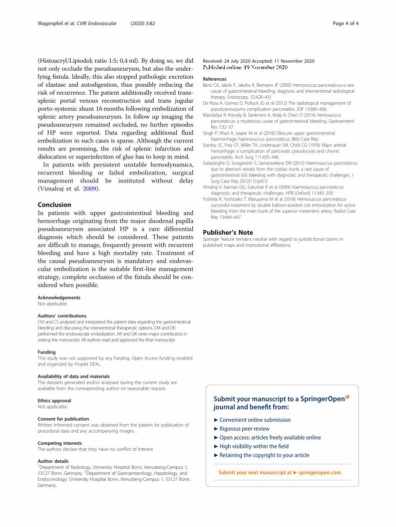
one microcoil (HILAL 2 × 20mm, Cook, Bjaeverskov,
Denmark) could be safely placed within the aneurysm. Inorder to completely occlude the pancreatico-arterial fistulaadditional tissue adhesive embolization was performed byHistoacryl (B. Braun, Melsungen, Germany)/Lipiodol(Guerbet, Aulnay-sous-Bois, France); ratio 1:5; 0,4 ml).Distribution of tissue adhesive in the pseudoaneurysm andfistula is demonstrated in Fig. 3.Final angiogram confirmed complete obstruction of
the pseudoaneurysm, as well as preserved regular perfu-sion of the splenic artery. Subsequent follow-up com-puted tomography performed 4 days and 10monthsafter embolization demonstrated hyperdense tissue ad-hesive in the fistula/aneurysmal sac as well as persistentocclusion of the pseudoaneurysm. The postinterven-tional course was uneventful and the patient was dis-charged in the following days. He was re-admitted 6months after initial intervention: Endoscopy revealedbleeding from gastric varices, the papilla appearedunremarkable.
DiscussionHaemosuccus pancreaticus is a rare complication ofacute or chronic pancreatitis, predominantly observed inmen (sex ratio 7:1), associated with chronic alcohol con-sumption (Vimalraj et al. 2009). Clinical symptoms in-clude upper gastrointestinal bleeding, non-specificepigastric pain, hematemesis and melena (Singh et al.2016). As hemorrhage is usually only intermittent andnot severe enough to cause hemodynamic instability,diagnosis is frequently delayed and difficult to make(Vimalraj et al. 2009), as observed in our reported case.Although endoscopy may reveal bleeding from the pa-
pilla, the actual source will typically remain unclear.
Fig. 1 Pseudoaneurysm of the splenic artery
Wagenpfeil et al. CVIR Endovascular (2020) 3:82 Page 2 of 4
Nevertheless, other significant causes of upper gastrointes-tinal bleeding (e.g. erosive gastritis, esophageal and gastricfundus varices or ulcers) may be excluded. Contrast en-hanced computed tomography allows for reliable detec-tion of pancreatic pathologies, as well as the assessment ofpotential complications of chronic pancreatitis while visu-alizing the peripancreatic vessels. Finally, angiographicintervention (including coil/glue embolization) is the ther-apy of choice for gastrointestinal bleeds not manageableby endoscopy (Yoshida et al. 2018). Haemosuccus pan-creaticus, especially if caused by a pseudoaneurysm, is apotentially life-threatening disease (Subasinghe et al.2012). Both interventional radiological and surgical ap-proaches are proven therapeutic options. Parent vessel oc-clusion by means of coil embolization is the mostfrequently described technique to treat pseudoaneurysmassociated HP (Yoshida et al. 2018). This technique wasavoided in the current case due to the risk of splenic
infarction. Covered stents, an alternative option in themanagement of pseudoaneurysms arising from larger ves-sels, offer the advantage of preserving arterial flow andcan be used in selective cases to bridge time until electivesurgery (De Rosa et al. 2012). However, activepancreatico-arterial fistulas, as suspected in the currentcase, may lead to graft infections. Furthermore, stent/stentgraft delivery into peripheral pseudoaneurysms is technic-ally challenging due to the typically torturous course ofthe splenic artery. In this regard, Benz et al. described thefirst successful implantation of an uncoated metal palmazstent across the aneurysmal segment of the splenic arteryfor treatment of pseudoaneurysm (Benz et al. 2000).The probability of a recurrent bleeding after catheter
assisted embolization varies between 0 and 30% dependingon the reference literature (Vimalraj et al. 2009). In orderto completely occlude the pancreatico-arterial fistula weadditionally employed tissue adhesive embolization
Fig. 2 Microcatheter placed in pseudoaneurysm (white arrow), pancreatico-arterial fistula (black arrow), opacified pancreatic duct (asterisks)
Fig. 3 Coil embolization by 2 × 20mm HILAL Microcoil (black arrow) and proper distribution of tissue adhesive (white arrow)
Wagenpfeil et al. CVIR Endovascular (2020) 3:82 Page 3 of 4
(Histoacryl/Lipiodol; ratio 1:5; 0,4 ml). By doing so, we didnot only occlude the pseudoaneurysm, but also the under-lying fistula. Ideally, this also stopped pathologic excretionof elastase and autodigestion, thus possibly reducing therisk of recurrence. The patient additionally received trans-splenic portal venous reconstruction and trans jugularporto-systemic shunt 16months following embolization ofsplenic artery pseudoaneurysm. In follow up imaging thepseudoaneurysm remained occluded, no further episodesof HP were reported. Data regarding additional fluidembolization in such cases is sparse. Although the currentresults are promising, the risk of splenic infarction anddislocation or superinfection of glue has to keep in mind.In patients with persistent unstable hemodynamics,
recurrent bleeding or failed embolization, surgicalmanagement should be instituted without delay(Vimalraj et al. 2009).
ConclusionIn patients with upper gastrointestinal bleeding andhemorrhage originating from the major duodenal papillapseudoaneurysm associated HP is a rare differentialdiagnosis which should be considered. These patientsare difficult to manage, frequently present with recurrentbleeding and have a high mortality rate. Treatment ofthe causal pseudoaneurysm is mandatory and endovas-cular embolization is the suitable first-line managementstrategy, complete occlusion of the fistula should be con-sidered when possible.
AcknowledgementsNot applicable.
Authors’ contributionsCM and CS analysed and interpreted the patient data regarding the gastrointestinalbleeding and discussing the interventional therapeutic options. CM and DKperformed the endovascular embolization. JW and DK were major contributors inwriting the manuscript. All authors read and approved the final manuscript.
FundingThis study was not supported by any funding. Open Access funding enabledand organized by Projekt DEAL.
Availability of data and materialsThe datasets generated and/or analysed during the current study areavailable from the corresponding author on reasonable request.
Ethics approvalNot applicable.
Consent for publicationWritten informed consent was obtained from the patient for publication ofprocedural data and any accompanying images. .
Competing interestsThe authors declare that they have no conflict of interest.
Author details1Department of Radiology, University Hospital Bonn, Venusberg-Campus 1,53127 Bonn, Germany. 2Department of Gastroenterology, Hepatology, andEndocrinology, University Hospital Bonn, Venusberg-Campus 1, 53127 Bonn,Germany.
Received: 24 July 2020 Accepted: 11 November 2020
ReferencesBenz CA, Jakob P, Jakobs R, Riemann JF (2000) Hemosuccus pancreaticus-a rare
cause of gastrointestinal bleeding: diagnosis and interventional radiologicaltherapy. Endoscopy. 32:428–431
De Rosa A, Gomez D, Pollock JG et al (2012) The radiological management ofpseudoaneurysms complication pancreatitis. JOP 13:660–666
Mandaliya R, Krevsky B, Sankineni A, Walp K, Chen O (2014) Hemosuccuspancreaticus: a mysterious cause of gastrointestinal bleeding. GastroenterolRes 7:32–37
Singh P, Khan A, Jasper M et al (2016) Obscure upper gastrointestinalhaemorrhage: haemosuccus pancreaticus. BMJ Case Rep
Stanley JC, Frey CF, Miller TA, Lindenauer SM, Child CG (1976) Major arterialhemorrhage: a complication of pancreatic pseudocysts and chronicpancreatitis. Arch Surg 111:435–440
Subasinghe D, Sivaganesh S, Samarasekera DN (2012) Haemosuccus pancreaticusdue to aberrant vessels from the coeliac trunk: a rare cause ofgastrointestinal (GI) bleeding with diagnostic and therapeutic challenges. JSurg Case Rep 2012(11):rjs013
Vimalraj V, Kannan DG, Sukumar R et al (2009) Haemosuccus pancreaticus:diagnostic and therapeutic challenges. HPB (Oxford) 11:345–350
Yoshida R, Yoshizako T, Maruyama M et al (2018) Hemosuccus pancreaticussuccessful treatment by double balloon-assisted coil embolization for activebleeding from the main trunk of the superior mesenteric artery. Radiol CaseRep 13:644–647
Publisher’s NoteSpringer Nature remains neutral with regard to jurisdictional claims inpublished maps and institutional affiliations.
Wagenpfeil et al. CVIR Endovascular (2020) 3:82 Page 4 of 4