PSBC: Standards for Obstetrical Ultrasound Assessments
38
Perinatal Services BC Standards for Obstetrical Ultrasound Assessments Table of Contents INTRODUCTION � � � � � � � � � � � � � � � � � � � � � � � � � � � � � � � � � � � � 2 1�0 DEFINITIONS, LICENSURE & CERTIFICATION REQUIREMENTS, SAFETY CONSIDERATION � � � � � � � � � � � � � � � � � � � � � � � � � � � � � � � � 3 Definitions � � � � � � � � � � � � � � � � � � � � � � � � � � � � � � � � � � � � 3 Licensure & Certification Requirements � � � � � � � � � � � � � � � � � � � � 3 Safety Consideration � � � � � � � � � � � � � � � � � � � � � � � � � � � � � � 3 2�0 ABNORMAL OR UNEXPECTED FINDINGS � � � � � � � � � � � � � � � � � � � 3 3�0 STANDARDIZED CHARTS AND FORMULAS� � � � � � � � � � � � � � � � � � � 4 4�0 MINIMUM REQUIRED CONTENT � � � � � � � � � � � � � � � � � � � � � � � � 6 4�1 ALL OB Ultrasound Reports � � � � � � � � � � � � � � � � � � � � � � � � 6 4�2 1st Trimester Ultrasound Report (up to and including 14wks 0d) � � � � � 7 4�3 2nd Trimester Ultrasound Report (14wks 1d – 26wks 6d) � � � � � � � � � 8 4�4 3rd Trimester Ultrasound Report (27wks 0d – term) � � � � � � � � � � � 10 5�0 REFERENCES � � � � � � � � � � � � � � � � � � � � � � � � � � � � � � � � � 11 6�0 DEVELOPMENT COMMITTEE MEMBERS � � � � � � � � � � � � � � � � � � � 12 APPENDIX 1 – SAMPLE SONOGRAPHER WORKSHEET � � � � � � � � � � � � � 13 APPENDIX 2 – REQUIRED IMAGE DOCUMENTATION � � � � � � � � � � � � � � � 14 APPENDIX 3 – PW DOPPLER USE IN OBSTETRIC ULTRASOUND � � � � � � � � 17 APPENDIX 4 – CONTACT INFORMATION AND FDS REFERRAL CRITERIA � � � � 19 APPENDIX 5 – PGSP MINIMUM REPORTING STANDARDS FOR NUCHAL TRANSLUCENCY ULTRASOUND MEASUREMENTS � � � � � � � � � � � � 20 APPENDIX 6 – CROWN RUMP LENGTH (CRL) CHART � � � � � � � � � � � � � � 22 APPENDIX 7 – INTERGROWTH 21ST FETAL GROWTH STANDARDS REVIEW � � 23 APPENDIX 8 – AMNIOTIC FLUID VOLUME � � � � � � � � � � � � � � � � � � � � � 24 APPENDIX 9 – SOGC – 1ST TRIMESTER DATING ULTRASOUND � � � � � � � � � 27 APPENDIX 10 – FETAL SEX DETERMINATION POLICY � � � � � � � � � � � � � � 28 APPENDIX 11 – FETAL SOFT MARKERS � � � � � � � � � � � � � � � � � � � � � � 29 APPENDIX 12 – SOGC – FETAL HEALTH SURVEILLANCE: ANTEPARTUM AND INTRAPARTUM CONSENSUS GUIDELINE � � � � � � � � � � � � � � � � � � 31 APPENDIX 13 – ISUOG PRACTICE GUIDELIES: USE OF DOPPLER ULTRASONOGRAPHY IN OBSTETRICS � � � � � � � � � � � � � � � � � � � 32 APPENDIX 14 – CAR STANDARD FOR PERFORMING DIAGNOSTIC OBSTETRIC ULTRASOUND EXAMINATIONS � � � � � � � � � � � � � � � � � � � � � � � 33 APPENDIX 15 – CAR STANDARD FOR COMMUNICATION OF DIAGNOSTIC IMAGING FINDINGS � � � � � � � � � � � � � � � � � � � � � � � � � � � � � 34 APPENDIX 16 – SOGC – CONTENT OF A COMPLETE ROUTINE SECOND TRIMESTER OBSTETRICAL ULTRASOUND EXAMINATION AND REPORT � 35 APPENDIX 17 – SOGC – FETAL SOFT MARKERS IN OBSTETRIC ULTRASOUND 36 While every attempt has been made to ensure that the information contained herein is clinically accurate and current, Perinatal Services BC acknowledges that many issues remain controversial, and therefore may be subject to practice interpretation� © Perinatal Services BC, 2014 Perinatal Services BC West Tower, Suite 350 555 West 12th Avenue Vancouver, BC Canada V5Z 3X7 Tel: 604-877-2121 www.perinatalservicesbc.ca Effective December 1, 2015
Transcript of PSBC: Standards for Obstetrical Ultrasound Assessments

Perinatal Services BCStandards for Obstetrical Ultrasound Assessments
Table of ContentsINTRODUCTION � � � � � � � � � � � � � � � � � � � � � � � � � � � � � � � � � � � � 2
1�0 DEFINITIONS, LICENSURE & CERTIFICATION REQUIREMENTS, SAFETY CONSIDERATION � � � � � � � � � � � � � � � � � � � � � � � � � � � � � � � � 3Definitions � � � � � � � � � � � � � � � � � � � � � � � � � � � � � � � � � � � � 3Licensure & Certification Requirements � � � � � � � � � � � � � � � � � � � � 3Safety Consideration � � � � � � � � � � � � � � � � � � � � � � � � � � � � � � 3
2�0 ABNORMAL OR UNEXPECTED FINDINGS � � � � � � � � � � � � � � � � � � � 3
3�0 STANDARDIZED CHARTS AND FORMULAS � � � � � � � � � � � � � � � � � � � 4
4�0 MINIMUM REQUIRED CONTENT � � � � � � � � � � � � � � � � � � � � � � � � 64�1 ALL OB Ultrasound Reports � � � � � � � � � � � � � � � � � � � � � � � � 64�2 1st Trimester Ultrasound Report (up to and including 14wks 0d) � � � � � 74�3 2nd Trimester Ultrasound Report (14wks 1d – 26wks 6d) � � � � � � � � � 84�4 3rd Trimester Ultrasound Report (27wks 0d – term) � � � � � � � � � � � 10
5�0 REFERENCES � � � � � � � � � � � � � � � � � � � � � � � � � � � � � � � � � 11
6�0 DEVELOPMENT COMMITTEE MEMBERS � � � � � � � � � � � � � � � � � � � 12
APPENDIX 1 – SAMPLE SONOGRAPHER WORKSHEET � � � � � � � � � � � � � 13
APPENDIX 2 – REQUIRED IMAGE DOCUMENTATION � � � � � � � � � � � � � � � 14
APPENDIX 3 – PW DOPPLER USE IN OBSTETRIC ULTRASOUND � � � � � � � � 17
APPENDIX 4 – CONTACT INFORMATION AND FDS REFERRAL CRITERIA � � � � 19
APPENDIX 5 – PGSP MINIMUM REPORTING STANDARDS FOR NUCHAL TRANSLUCENCY ULTRASOUND MEASUREMENTS � � � � � � � � � � � � 20
APPENDIX 6 – CROWN RUMP LENGTH (CRL) CHART � � � � � � � � � � � � � � 22
APPENDIX 7 – INTERGROWTH 21ST FETAL GROWTH STANDARDS REVIEW � � 23
APPENDIX 8 – AMNIOTIC FLUID VOLUME � � � � � � � � � � � � � � � � � � � � � 24
APPENDIX 9 – SOGC – 1ST TRIMESTER DATING ULTRASOUND � � � � � � � � � 27
APPENDIX 10 – FETAL SEX DETERMINATION POLICY � � � � � � � � � � � � � � 28
APPENDIX 11 – FETAL SOFT MARKERS � � � � � � � � � � � � � � � � � � � � � � 29
APPENDIX 12 – SOGC – FETAL HEALTH SURVEILLANCE: ANTEPARTUM AND INTRAPARTUM CONSENSUS GUIDELINE � � � � � � � � � � � � � � � � � � 31
APPENDIX 13 – ISUOG PRACTICE GUIDELIES: USE OF DOPPLER ULTRASONOGRAPHY IN OBSTETRICS � � � � � � � � � � � � � � � � � � � 32
APPENDIX 14 – CAR STANDARD FOR PERFORMING DIAGNOSTIC OBSTETRIC ULTRASOUND EXAMINATIONS � � � � � � � � � � � � � � � � � � � � � � � 33
APPENDIX 15 – CAR STANDARD FOR COMMUNICATION OF DIAGNOSTIC IMAGING FINDINGS � � � � � � � � � � � � � � � � � � � � � � � � � � � � � 34
APPENDIX 16 – SOGC – CONTENT OF A COMPLETE ROUTINE SECOND TRIMESTER OBSTETRICAL ULTRASOUND EXAMINATION AND REPORT � 35
APPENDIX 17 – SOGC – FETAL SOFT MARKERS IN OBSTETRIC ULTRASOUND 36
While every attempt has been made to ensure that the information contained herein is clinically accurate and current, Perinatal Services BC acknowledges that many issues remain controversial, and therefore may be subject to practice interpretation�© Perinatal Services BC, 2014
Perinatal Services BC West Tower, Suite 350 555 West 12th Avenue Vancouver, BC Canada V5Z 3X7 Tel: 604-877-2121
www.perinatalservicesbc.ca
Effective December 1, 2015

2 Perinatal Services BC
In September 2011, the BC Patient Safety and Quality Council produced a report entitled Investigation into Medical Imaging, Credentialing and Quality Assurance� The report reviewed the existing structure for the licensing and credentialing of physicians in BC’s health authorities, including the processes for quality assurance and peer review� Recommendation #32 in the report designated Perinatal Services BC (PSBC) to develop or adopt and promulgate standards for obstetrical ultrasound assessments in the first, second and third trimesters that are performed in community and tertiary facilities�
In December 2011, PSBC brought together a multidisciplinary committee to develop standards for the required minimum content of an obstetric ultrasound assessment� The committee included radiologists and sonographers from rural and urban areas of practice and both the public and private sector as well as perinatologists� Additional clinical consultation was provided by obstetricians, family physicians, and midwives� Representatives from the BC Radiology Society (BCRS), BC Ultrasonographers’ Society (BCUS), 2Lower Mainland Medical Imaging Integration and the Diagnostic Accreditation Program also provided information and consultation� Standards and clinical practice guidelines from the Canadian Association of Radiologists, the Society of Obstetricians and Gynaecologists of Canada and the International Society of Ultrasound in Obstetrics and Gynecology provided a framework for the BC standards�
In early 2015, the standards were reviewed through the same multidisciplinary process as above and updated to reflect feedback and new clinical practice guidelines published by the Society of Obstetricians and Gynaecologists of Canada and the International Society of Ultrasound in Obstetrics and Gynecology in 2013 and 2014�
It is the intention of these standards to ensure a consistent level of care throughout the province surrounding the provision of obstetrical ultrasound that is based on current clinical best practices� The standards provide a simple, single source of information regarding assessment criteria for physicians and sonographers involved in obstetric ultrasounds at diagnostic and screening sites around the province� This document is not intended to confine or limit ultrasound assessment parameters as individual diagnostic imaging sites may include additional ultrasound components dependent on available expertise, clinical indication, and health care provider requests�
In order to ensure that these provincial standards continue to align with best practices, they will be reviewed when new best practices and/or clinical practice guidelines are published� The review will be led by Perinatal Services BC and performed by a multidisciplinary group consisting of representation from different health care provider stakeholders, health authorities, and both public and private facilities�
Introduction

3Standards for Obstetrical Ultrasound Assessments
Definitions⦁⦁ Ultrasound Report – The document that provides the findings of the ultrasound assessment including
all of the items listed in this document� It is signed by a qualified physician and is distributed to the ordering health care provider (HCP) and other HCPs as requested�
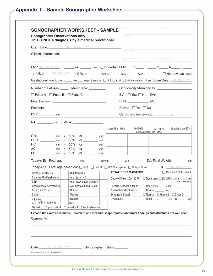
⦁⦁ Sonographer Worksheet – A document used by the sonographer during an obstetrical (OB) ultrasound assessment� Not considered to be an Ultrasound Report� Sample – Appendix 1
⦁⦁ Image documentation – Images which are assessed and retained� These images are stored by the diagnostic imaging site� A list of required images to be assessed and retained which support the required content for an OB ultrasound report is included� Appendix 2
⦁⦁ For the purposes of this document trimesters are defined as:⦁� 1st – up to and including 14wks 0d⦁� 2nd (mid trimester) – 14wks 1d – 26wks 6d⦁� 3rd – after and including 27wks 0d
Licensure & Certification Requirements⦁⦁ Individuals providing OB ultrasound reports and individuals performing OB ultrasounds are required to
have the appropriate licensure and credentials as per the provincial regulatory authorities�
Safety Consideration⦁⦁ While performing an ultrasound, exposure time and acoustic output should be kept to the lowest
levels consistent with obtaining diagnostic information and limited to medically indicated procedures� Appendix 3
2.0 Abnormal or Unexpected Findings
Since the first identification of a clinically significant finding is often not anticipated by the referring care provider and may require immediate attention and case management decisions, verbal communication with the referring care provider, in advance of the final written report, is strongly recommended.
⦁⦁ When abnormal or unexpected findings are identified, the ultrasound examination must be expanded appropriately and all abnormal findings documented, measured (if appropriate) and reported�
⦁⦁ When significant fetal growth and/or fluid volume abnormalities and/or structural malformations are identified, recommendations given in the final report should include a referral to, or consultation with, the Fetal Diagnostic Service (FDS), the Provincial Medical Genetics Program, or other specialists� Appendix 4
⦁⦁ When a fetal soft marker(s) is (are) identified, recommendations given in the final report may include referral to, or consultation with, the Provincial Medical Genetics Program, or other specialists� The website for the BC Prenatal Genetic Screening Program (PGSP) also provides helpful information�
⦁⦁ When a nuchal translucency (NT) measurement is above the established cut off, recommendations given in the final report should include standard comments as determined by the BC Prenatal Genetic Screening Program� Appendix 5
1.0 Definitions, Licensure & Certification Requirements, Safety Consideration

4 Perinatal Services BC
CROWN RUMP LENGTH (CRL)The recommended reference chart (unpublished) for crown rump length (CRL) is from data obtained at the same time as the Lessoway et al, ultrasound fetal biometry charts for a North American Caucasian population study (referenced below) using the same methods and analysis� This chart is currently used by many of the diagnostic imaging sites in BC and the BC Prenatal Genetic Screening Program (PGSP)� Appendix 6 www.perinatalservicesbc.ca/NR/rdonlyres/1AF7DA89-95D7-4195-9284-46B6CBADE3A4/0/CRLChart.pdf
FETAL ANATOMIC MEASUREMENTS*The recommended reference charts for measurement of head circumference (HC), femur length (FL), biparietal diameter (BPD), abdominal circumference (AC) and thorax circumference (TC) are:
⦁⦁ Lessoway V, Schulzer M, Wittmann B, et al� Ultrasound fetal biometry charts for a North American Caucasian population� J Clin Ultrasound 1998 Nov-Dec;26(9):433–53. www.perinatalservicesbc.ca/NR/rdonlyres/12D553CE-16D0-4A3F-93A6-0BF3AE3DE9AC/0/FetalBiometryChart.pdf
NEWBORN GROWTH PARAMETERSThe newborn birth weight, length and head circumference charts used by the BC Perinatal Data Registry (PDR) are from the BC Ministry of Health–Vital Statistics Agency and are based on BC data (1981 to 2000)�www.perinatalservicesbc.ca/ForHealthcareProviders/Resources/BirthWeightCharts/default.htm
AMNIOTIC FLUID VOLUME
Amniotic fluid assessment should be performed using the Single Deepest Pocket (SDP) method� The modernized description for performing an amniotic fluid volume assessment using the Single Deepest Pocket method is based on:
⦁⦁ Chamberlain et al� Ultrasound evaluation of amniotic fluid volume� I� The relationship of marginal and decreased amniotic fluid volumes to perinatal outcome� Am J Obstet Gynecol. 1984;150(3):245–9.
⦁⦁ Chamberlain et al� Ultrasound evaluation of amniotic fluid volume� II� The relationship of increased amniotic fluid volume to perinatal outcome� Am J Obstet Gynecol. 1984;150(3):250–4.
Modernized Description: The largest pocket of fluid free of cord and fetal parts is identified and its depth (cm) measured as close to right angles as possible to the uterine contour� The pocket must be at least 1 cm in width (width being measured perpendicular to the depth axis) at its narrowest point so that it is at least 1 cm wide throughout the measured depth axis� To avoid over-estimation of pocket depth, the transducer should not be over-angled through the pocket relative to the depth axis�
* NOTE: A Working Group on biometric standards for assessing fetal growth was brought together to carefully consider the option of replacing the current, widely used, Lessoway standard with the new Intergrowth 21st standard� The Intergrowth 21st international standard was developed on the basis of recommendations from a WHO expert committee and the study was funded by the Bill and Melinda Gates Foundation� The Working Group carefully examined contemporary data from British Columbia, which included cohorts at risk for growth restriction and macrosomia as well as a low risk “normal” cohort� The analysis showed that adopting the Intergrowth standard would lead to a significant decrease in the frequency of small-for-gestational age diagnoses and a substantial increase in large-for-gestational age diagnoses� The Working Group found this concerning, especially since the under identification of small fetuses has the potential for increasing mortality and serious morbidity, while the excess number of large fetuses identified would drastically increase the need for clinical resources� The Working Group recommends that the Lessoway standard remain the provincial standard� For more details – see Appendix 7.
3.0 Standardized Charts and Formulas

5Standards for Obstetrical Ultrasound Assessments
SINGLE DEEPEST POCKET (SDP) INTERPRETATION*
DEFINITIONS
Oligohydramnios: SDP < 2�0 cm Normal: SDP between 2�0 and 8�0 cm Polyhydramnios: SDP > 8�0 cm
Abnormalities of amniotic fluid using these simple cutoffs should occur less than 3% of time in a controlled population up to term�
⦁⦁ Magann et al� The amniotic fluid index, single deepest pocket, and two diameter pocket in normal human pregnancy� Am J Obstet Gynecol. 2000;182(6):1581 – 1587 (Table II).
Abnormal measurements should not be based on a single measure, but confirmed by repeated measurement�
UMBILICAL ARTERY DOPPLERUmbilical artery Doppler is not a minimum assessment requirement and is only done when clinically indicated� The recommended reference for Systolic/Diastolic (S/D) ratio, Resistance Index and Pulsatility Index when performing umbilical artery Doppler in a free loop of the umbilical cord is:
⦁⦁ Acharya et al� Reference ranges for serial measurements of umbilical artery Doppler indices in the second half of pregnancy� Am J Obstet Gynecol. 2005;192:937 – 44 (Table IV).
BIOPHYSICAL PROFILE (BPP)A biophysical profile (BPP) is not a minimum assessment requirement and is only done when clinically indicated� If a Biophysical Profile (BPP) is requested and facilities and expertise exist to perform this assessment, the ultrasound components should include:
1� Breathing movements
2� Movements
3� Tone
4� Amniotic Fluid Volume
5� Reactive Fetal Heart Rate
⦁⦁ Manning FA� Dynamic ultrasound-based fetal assessment: the fetal biophysical profile score� Clin Obstet Gynecol. 1995;38(1):26–44.
ESTIMATED FETAL WEIGHT (EFW)Although the determination of Estimated Fetal Weight (EFW) is not considered to be a minimum assessment requirement for an OB ultrasound assessment, it is commonly performed and may be requested by the referring HCP to assist in clinical management� Although many formulas exist which predict EFW, there is limited evidence to support any particular formula as being a significantly better predictor of fetal weight�
⦁⦁ Scioscia M, Vimercati A, Ceci O, et al� Estimation of Birth Weight by Two-Dimensional Ultrasonography: A Critical Appraisal of Its Accuracy� Obstetrics & Gynecology January 2008;111(1):57–65.
* NOTE: For further information regarding amniotic fluid volume assessment and interpretation please see Appendix 8.
3.0 Standardized Charts and Formulas, cont’d

6 Perinatal Services BC
4.1 ALL OB Ultrasound ReportsFinal reports must include, but are not limited to, the following:
DEMOGRAPHICS⦁⦁ Examination date
⦁⦁ Patient full name
⦁⦁ Second patient identifier (birth date, hospital identification number, health insurance number)
⦁⦁ Starting date of last menstrual period (LMP) if available
⦁⦁ Date of the first ultrasound after 7weeks 0days, the crown rump length (CRL) and the corresponding gestational age at that time
⦁⦁ Indication for ultrasound scan
⦁⦁ Name of requesting physician/caregiver (preferably with contact information)
⦁⦁ List of caregivers to receive copies
⦁⦁ Date of final report
⦁⦁ Name of interpreting/reporting physician
GESTATIONAL AGE DETERMINATIONIn assigning the gestational age to interpret the current ultrasound scan the following rules are recommended� Appendix 9
1� If the pregnancy is a result of timed ovulation induction (ovulation induction, Invitro fertilization [IVF], etc) then determination of the current gestational age should be based on that information�
2� If a natural conception, the first ultrasound after 7weeks 0days gestation (crown rump length [CRL] equal to and greater than 10 mm) should be used to date the pregnancy�
3� If the first ultrasound is in the 2nd trimester, a composite mean of at least 3 standard biometric parameters (head circumference [HC], biparietal diameter [BPD], abdominal circumference [AC] and femur length [FL]) each converted to the 50th percentile gestation in weeks and days is used to estimate the gestational age�
4� If a multiple pregnancy, the gestational age of the largest fetus should be used as the gestational age for the pregnancy�
Once a gestational age is established, it should remain as such and be the basis for all further gestational age estimates, including the Estimated Date of Delivery (EDD), unless there is compelling clinical evidence to reassign the gestational age.
REPORT SUMMARYThe report summary / conclusion/ comments should include:
⦁⦁ Estimate of the current gestational age based on the rules listed above�
⦁⦁ An assessment of the appropriateness of biometric measurements for the current gestational age�
⦁⦁ Results of the fetal anatomy assessment, if performed�
⦁⦁ Results of amniotic fluid assessment, if performed�
⦁⦁ Factors which limit the ultrasound assessment� E�g� fetal position, fetal size, fetal age, oligohydramnios, maternal body habitus�
⦁⦁ Fetal sex, if determined during the exam� Appendix 10
⦁⦁ Indication if an endovaginal scan was performed�
⦁⦁ Recommendations as appropriate:
4.0 Minimum Required Content
7Standards for Obstetrical Ultrasound Assessments
⦁� When significant fetal growth and/or fluid volume abnormalities and/or structural malformations are identified, recommendations given in the final report should include a referral to, or consultation with, the Provincial Medical Genetics Program, the Fetal Diagnostic Service (FDS), or other specialists� Appendix 4
⦁� When a fetal soft marker(s) is (are) identified, recommendations given in the final report may include referral to, or consultation with, the Provincial Medical Genetics Program, or other specialists� The website for the BC Prenatal Genetic Screening Program (PGSP) also provides helpful information�
⦁� When a nuchal translucency (NT) measurement is above the established cut off, recommendations given in the final report should include standard comments as determined by the BC Prenatal Genetic Screening Program� Appendix 5
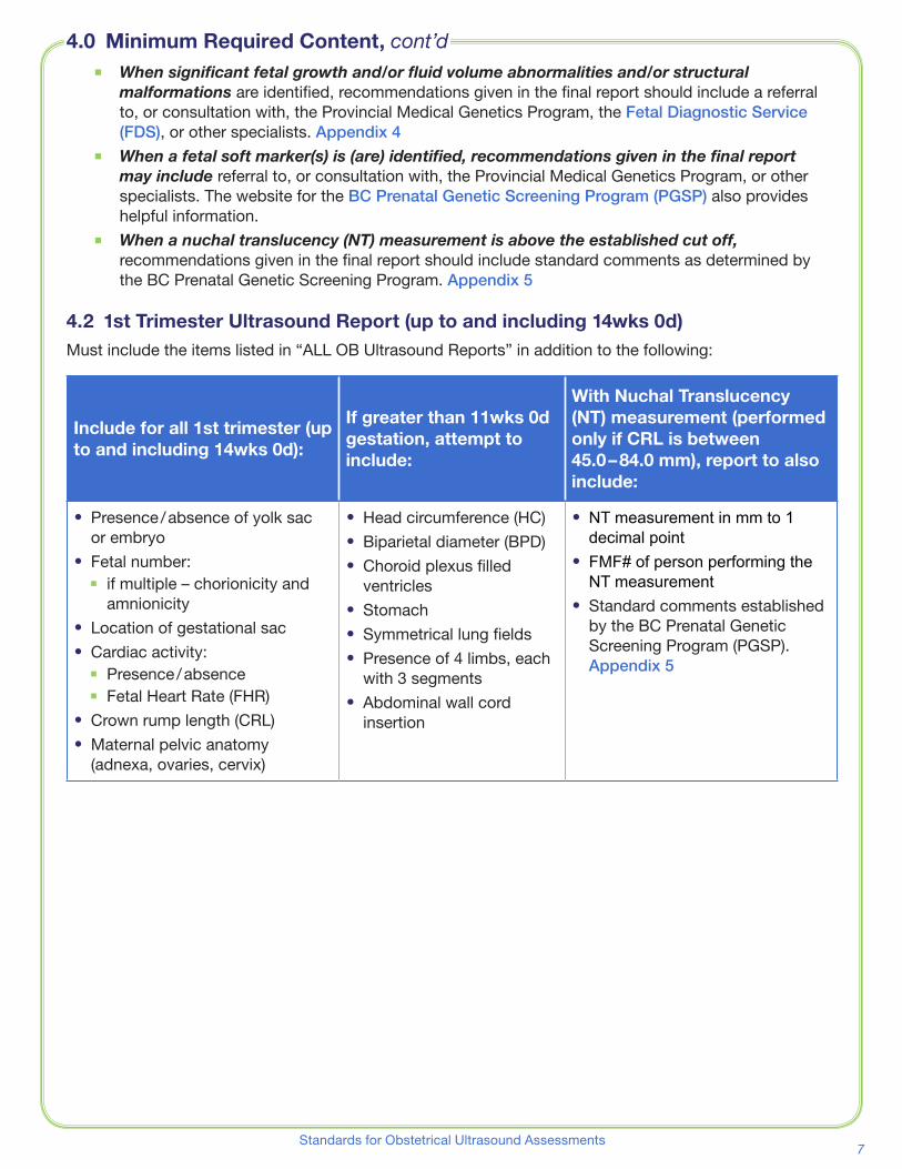
4.2 1st Trimester Ultrasound Report (up to and including 14wks 0d)Must include the items listed in “ALL OB Ultrasound Reports” in addition to the following:
Include for all 1st trimester (up to and including 14wks 0d):
If greater than 11wks 0d gestation, attempt to include:
With Nuchal Translucency (NT) measurement (performed only if CRL is between 45.0 – 84.0 mm), report to also include:
⦁⦁ Presence / absence of yolk sac or embryo
⦁⦁ Fetal number:⦁◾ if multiple – chorionicity and
amnionicity⦁⦁ Location of gestational sac⦁⦁ Cardiac activity:
⦁◾ Presence / absence⦁◾ Fetal Heart Rate (FHR)
⦁⦁ Crown rump length (CRL)⦁⦁ Maternal pelvic anatomy
(adnexa, ovaries, cervix)
⦁⦁ Head circumference (HC)⦁⦁ Biparietal diameter (BPD)⦁⦁ Choroid plexus filled
ventricles⦁⦁ Stomach⦁⦁ Symmetrical lung fields⦁⦁ Presence of 4 limbs, each
with 3 segments⦁⦁ Abdominal wall cord
insertion
⦁⦁ NT measurement in mm to 1 decimal point
⦁⦁ FMF# of person performing the NT measurement
⦁⦁ Standard comments established by the BC Prenatal Genetic Screening Program (PGSP)� Appendix 5
4.0 Minimum Required Content, cont’d
http://www.bcwomens.ca/Services/PregnancyBirthNewborns/PrenatalScreeningDiagnosis/FetalDiagnosis.htm
8 Perinatal Services BC
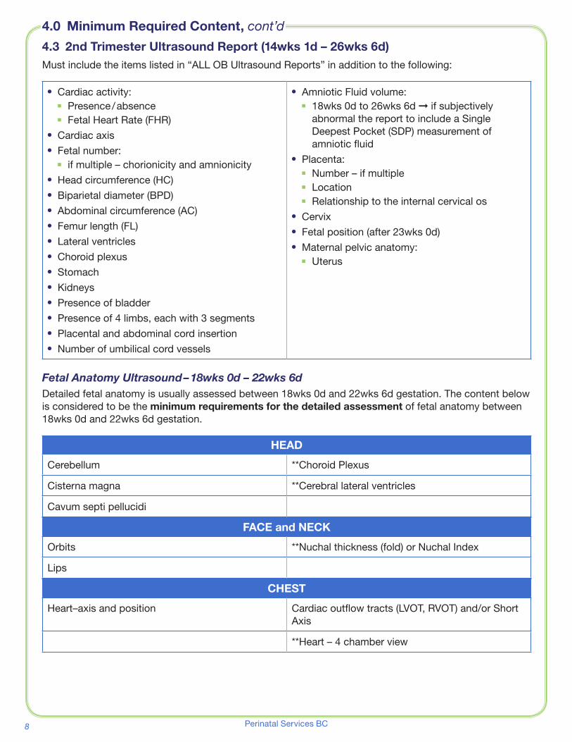
4.3 2nd Trimester Ultrasound Report (14wks 1d – 26wks 6d)Must include the items listed in “ALL OB Ultrasound Reports” in addition to the following:
⦁⦁ Cardiac activity:⦁◾ Presence / absence⦁◾ Fetal Heart Rate (FHR)
⦁⦁ Cardiac axis⦁⦁ Fetal number:
⦁◾ if multiple – chorionicity and amnionicity⦁⦁ Head circumference (HC)⦁⦁ Biparietal diameter (BPD)⦁⦁ Abdominal circumference (AC)⦁⦁ Femur length (FL)⦁⦁ Lateral ventricles⦁⦁ Choroid plexus⦁⦁ Stomach⦁⦁ Kidneys⦁⦁ Presence of bladder⦁⦁ Presence of 4 limbs, each with 3 segments⦁⦁ Placental and abdominal cord insertion⦁⦁ Number of umbilical cord vessels
⦁⦁ Amniotic Fluid volume:⦁◾ 18wks 0d to 26wks 6d ➞ if subjectively
abnormal the report to include a Single Deepest Pocket (SDP) measurement of amniotic fluid
⦁⦁ Placenta:⦁◾ Number – if multiple⦁◾ Location⦁◾ Relationship to the internal cervical os
⦁⦁ Cervix⦁⦁ Fetal position (after 23wks 0d)⦁⦁ Maternal pelvic anatomy:
⦁◾ Uterus
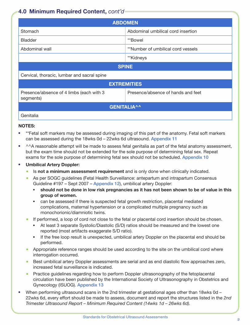
Fetal Anatomy Ultrasound – 18wks 0d – 22wks 6dDetailed fetal anatomy is usually assessed between 18wks 0d and 22wks 6d gestation� The content below is considered to be the minimum requirements for the detailed assessment of fetal anatomy between 18wks 0d and 22wks 6d gestation�
HEAD
Cerebellum **Choroid Plexus
Cisterna magna **Cerebral lateral ventricles
Cavum septi pellucidi
FACE and NECK
Orbits **Nuchal thickness (fold) or Nuchal Index
Lips
CHEST
Heart–axis and position Cardiac outflow tracts (LVOT, RVOT) and/or Short Axis
**Heart – 4 chamber view
4.0 Minimum Required Content, cont’d
9Standards for Obstetrical Ultrasound Assessments
ABDOMEN
Stomach Abdominal umbilical cord insertion
Bladder **Bowel
Abdominal wall **Number of umbilical cord vessels
**Kidneys
SPINE
Cervical, thoracic, lumbar and sacral spine
EXTREMITIES
Presence/absence of 4 limbs (each with 3 segments)
Presence/absence of hands and feet
GENITALIA^^
Genitalia
NOTES:
⦁⦁ **Fetal soft markers may be assessed during imaging of this part of the anatomy� Fetal soft markers can be assessed during the 18wks 0d – 22wks 6d ultrasound� Appendix 11
⦁⦁ ^^A reasonable attempt will be made to assess fetal genitalia as part of the fetal anatomy assessment, but the exam time should not be extended for the sole purpose of determining fetal sex� Repeat exams for the sole purpose of determining fetal sex should not be scheduled� Appendix 10
⦁⦁ Umbilical Artery Doppler:⦁� Is not a minimum assessment requirement and is only done when clinically indicated�⦁� As per SOGC guidelines (Fetal Health Surveillance: antepartum and intrapartum Consensus
Guideline #197 – Sept 2007 – Appendix 12), umbilical artery Doppler:⦁⦁ should not be done in low risk pregnancies as it has not been shown to be of value in this
group of women.⦁⦁ can be assessed if there is suspected fetal growth restriction, placental mediated
complications, maternal hypertension or a complicated multiple pregnancy such as monochorionic/diamniotic twins�
⦁� If performed, a loop of cord not close to the fetal or placental cord insertion should be chosen�⦁⦁ At least 3 separate Systolic/Diastolic (S/D) ratios should be measured and the lowest one
reported (most artifacts exaggerate S/D ratio)�⦁⦁ If the free loop result is unexpected, umbilical artery Doppler on the placental end should be
performed�⦁� Appropriate reference ranges should be used according to the site on the umbilical cord where
interrogation occurred�⦁� Best umbilical artery Doppler assessments are serial and as end diastolic flow approaches zero,
increased fetal surveillance is indicated�⦁� Practice guidelines regarding how to perform Doppler ultrasonography of the fetoplacental
circulation have been published by the International Society of Ultrasonography in Obstetrics and Gynecology (ISUOG)� Appendix 13
⦁⦁ When performing ultrasound scans in the 2nd trimester at gestational ages other than 18wks 0d – 22wks 6d, every effort should be made to assess, document and report the structures listed in the 2nd Trimester Ultrasound Report – Minimum Required Content (14wks 1d – 26wks 6d).
4.0 Minimum Required Content, cont’d

10 Perinatal Services BC
4.4 3rd Trimester Ultrasound Report (27wks 0d – term)Must include items listed in “ALL OB Ultrasound Reports” in addition to the following:
⦁⦁ Cardiac activity:⦁◾ Presence / absence⦁◾ Fetal Heart Rate (FHR)
⦁⦁ Fetal number:⦁◾ If multiple–chorionicity and amnionicity
⦁⦁ Amniotic Fluid volume:⦁◾ Single Deepest Pocket (SDP)
⦁⦁ Fetal position⦁⦁ Placenta:
⦁◾ Number – if multiple⦁◾ Location⦁◾ Relationship to the internal cervical os
⦁⦁ Cervix (if less than 32 wks 0d)
⦁⦁ Head circumference (HC)⦁⦁ Biparietal diameter (BPD)⦁⦁ Abdominal circumference (AC)⦁⦁ Femur length (FL)⦁⦁ Heart – 4 chamber view⦁⦁ Cardiac outflow tracts (LVOT, RVOT) and/or
Short Axis⦁⦁ Stomach⦁⦁ Bladder⦁⦁ Kidneys
NOTES:
⦁⦁ When performing an ultrasound scan in the third trimester and a Fetal Anatomy Ultrasound in the 2nd trimester has not been performed, every effort should be made to assess and adequately document all structures listed in the minimum requirements for the detailed assessment of fetal anatomy between 18wks 0d and 22wks 6d�
⦁⦁ Umbilical Artery Doppler:⦁� Is not a minimum assessment requirement and is only done when clinically indicated�⦁� As per SOGC guidelines (Fetal Health Surveillance: antepartum and intrapartum Consensus
Guideline #197 – Sept 2007 – Appendix 12, umbilical artery Doppler:⦁⦁ should not be done in low risk pregnancies as it has not been shown to be of value in this
group of women.⦁⦁ can be assessed if there is suspected fetal growth restriction, placental mediated
complications, maternal hypertension or a complicated multiple pregnancy such as monochorionic/diamniotic twins�
⦁� If performed, a loop of cord not close to the umbilicus or placental cord insertion should be chosen and a wide enough sample volume chosen to include the umbilical vein as well as the artery�⦁⦁ At least 3 separate Systolic/Diastolic (S/D) ratios should be measured and the lowest one
reported (most artefacts exaggerate S/D ratio)�⦁⦁ If the free loop result is unexpected, umbilical artery Doppler on the placental end should be
performed�⦁� Appropriate reference ranges should be used according to the site on the umbilical cord where
interrogation occurred�⦁� Best umbilical artery Doppler assessments are serial and as end diastolic flow approaches zero,
increased fetal surveillance is indicated�⦁� Practice guidelines regarding how to perform Doppler ultrasonography of the fetoplacental
circulation have been published by the International Society of Ultrasonography in Obstetrics and Gynecology (ISUOG)� Appendix 13
⦁⦁ Fetal growth assessments should not be performed at less than 14 day intervals as the amount of potential fetal growth falls within the range of ultrasound inter-observer error at periods of less than 14 days�
4.0 Minimum Required Content, cont’d
11Standards for Obstetrical Ultrasound Assessments
Determination of Gestational Age by Ultrasound SOGC Clinical Practice Guideline, No� 303, February 2014 – Appendix 9
Fetal Health Surveillance: Antepartum and Intrapartum Consensus Guideline SOGC Clinical Practice Guideline, No� 197 (Replaces No� 90 and No� 112), September 2007 – Appendix 12
Practice guidelines: Use of Doppler ultrasonography in obstetrics International Society of Ultrasound in Obstetrics and Gynecology (ISUOG), 2013 – Appendix 13
CAR Standard for Performing Diagnostic Obstetric Ultrasound Examinations Canadian Association of Radiologists (CAR), September 25, 2010 – Appendix 14
CAR Standard for Communication of Diagnostic Imaging Findings Canadian Association of Radiologists (CAR), September 25, 2010 – Appendix 15
Content of a Complete Routine Second Trimester Obstetrical Ultrasound Examination and Report SOGC Clinical Practice Guideline, No� 223, March 2009 – Appendix 16
Fetal Soft Markers in Obstetric Ultrasound SOGC Clinical Practice Guideline, No� 162, June 2005 (reaffirmed June 2013) – Appendix 17
Practice Guidelines: Performance of first-trimester fetal ultrasound scan International Society of Ultrasound in Obstetrics and Gynecology (ISUOG), 2013
Practice guidelines for performance of the routine mid-trimester fetal ultrasound scan International Society of Ultrasound in Obstetrics and Gynecology (ISUOG), 2010
Practice Guideline for the Performance of Obstetric Ultrasound Examinations American Institute of Ultrasound in Medicine (AIUM), 2007
Diagnostic Imaging Accreditation Standards 2010 Diagnostic Accreditation Program (DAP)
5.0 References

12 Perinatal Services BC
Jill Anglin Sonographer Supervisor, Greig AssociatesBrent Barton Regional Practice Lead – Ultrasound,
Lower Mainland Medical Imaging IntegrationSimon Bicknell BCRS representative
Radiologist, VCHAJacqueline Brown Radiologist, St. Paul’s Hospital, PHABlair Butler MFM – BC Women’s Hospital, PHSAAnita Dircks Project Manager – PSBCDuncan Farquharson MFM – Royal Columbian Hospital, FHA Cathy Fix Sonographer, St. Paul’s Hospital Ultrasound, PHAAlain Gagnon Senior Medical Director – C&W
MFM – BC Women’s HospitalCarrie Green Director Medical Imaging, FHAJudy Harder Sonographer Supervisor, BC Women’s HospitalChristina Kay Primary Maternity Care Lead – Family Practice – PSBCSusan Larson Operations Director, Medical Imaging – BC Women’s HospitalVickie Lessoway Executive Director, BC Ultrasonographers’ SocietyKen Lim Medical Director, Diagnostic/Ambulatory Program, BC Women’s Hospital
MFM – BC Women’s HospitalGerry Marquette Medical Director, Perinatology – PSBCJulie Nicol Radiologist, IHADustin Pendergast Technical Section Head of Ultrasound, VIHA/South RegionDenise Pugash Radiologist, BC Women’s HospitalKim Williams Executive Director, PSBCLeanne Yeates Primary Maternity Care Lead – Midwifery – PSBC Charlotte Yong-Hing BCRS Women’s Imaging Rep.
Radiologist, VCH
The Development Committee would like to thank the radiologists, sonographers, obstetricians, family physicians and midwives who provided valuable comments and feedback�
This document has been endorsed by the BC Radiology Society and the BC Ultrasonographers’ Society and approved by the Provincial Medical Imaging Advisory Committee (MIAC)�
6.0 Development committee members
13Standards for Obstetrical Ultrasound Assessments
Appendix 1 – Sample Sonographer Worksheet
SONOGRAPHER WORKSHEET - SAMPLESonographer Observations only. This is NOT a diagnosis by a medical practitioner
Exam Date: DD – MON – YY
Clinical Information:
Surname Given Name
Address
Phone number
Personal Health Number Physician / Midwife Name
LMP: = wks days ☐ Uncertain LMP G T P A L
1st US on: CRL = mm = wks days ☐ No previous scan
Gestational age today = wks days Based on: ☐ U/S ☐ LMP ☐ IVF conception Last Scan Date:
Number of Fetuses: Membrane: Chorionicity / Amnionicity:
☐ Fetus A ☐ Fetus B ☐ Fetus C EV: ☐ Yes ☐ No EV#
Fetal Position: FHR: BPM
Placenta: Previa: ☐ Yes ☐ No
DVP: cm Cervix (less than 32 wk 0 d): cm
NT: mm FMF #:
Less than 10% 10 – 50% 50 – 90% Greater than 90%(For gestational age today)
CRL mm = 50% for wks
BPD mm = 50% for wks
HC mm = 50% for wks
AC mm = 50% for wks
FL mm = 50% for wks
Today’s Est. Fetal age: wks days ± wks Est. Fetal Weight: gm
Today’s Est. Fetal age based on: ☐ LMP ☐ 1st US ☐ IVF Conception ☐ Today’s Scan EDD:
Cerebral Ventricles Abd. Cord Ins. FETAL SOFT MARKERS ☐ Markers Not Assessed
Cisterna M. / Cerebellum Heart (axis) 4C Choroid Plexus Cyst (CPC) ☐ None seen ☐ Bi / ☐ Uni lateral mmCSP Heart (SAX and/or Outflows) (record largest)
Choroid Plexus/Ventricles Symmetrical Lung Fields Cardiac Echogenic focus ☐ None seen ☐ Present Face (Lips / Orbits) Stomach Nuchal fold (thickness) ☐ Normal mm
Spine Kidneys Echogenic Bowel ☐ Normal ☐ Grade 2 ☐ Grade 3
4 Limbs each with 3 segments
Bladder Pyelectasis ☐ None L mm R mm
3VCGenitalia: ☐ probably M ☐ probably F ☐ not determined
Expand the exam as required. Document and measure, if appropriate, abnormal findings and structures not well seen.
Comments:
Date Sonographer Initials
DD-MON-YY
DD-MON-YY
DD-MON-YY
DD-MON-YY
DD – MON – YY
© Perinatal Services BC AUGUST 2015
14 Perinatal Services BC
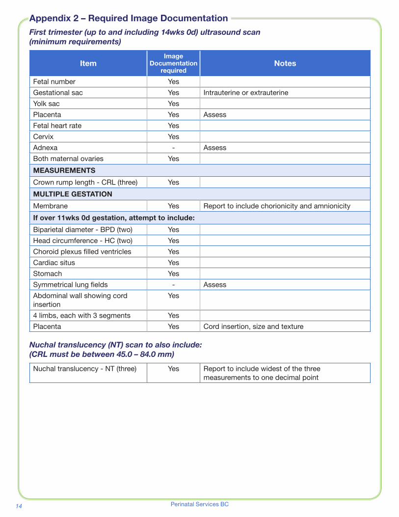
First trimester (up to and including 14wks 0d) ultrasound scan (minimum requirements)
ItemImage
Documentation required
Notes
Fetal number Yes
Gestational sac Yes Intrauterine or extrauterine
Yolk sac Yes
Placenta Yes Assess
Fetal heart rate Yes
Cervix Yes
Adnexa - Assess
Both maternal ovaries Yes
MEASUREMENTS
Crown rump length - CRL (three) Yes
MULTIPLE GESTATION
Membrane Yes Report to include chorionicity and amnionicity
If over 11wks 0d gestation, attempt to include:
Biparietal diameter - BPD (two) Yes
Head circumference - HC (two) Yes
Choroid plexus filled ventricles Yes
Cardiac situs Yes
Stomach Yes
Symmetrical lung fields - Assess
Abdominal wall showing cord insertion
Yes
4 limbs, each with 3 segments Yes
Placenta Yes Cord insertion, size and texture
Nuchal translucency (NT) scan to also include: (CRL must be between 45.0 – 84.0 mm)
Nuchal translucency - NT (three) Yes Report to include widest of the three measurements to one decimal point
Appendix 2 – Required Image Documentation
15Standards for Obstetrical Ultrasound Assessments
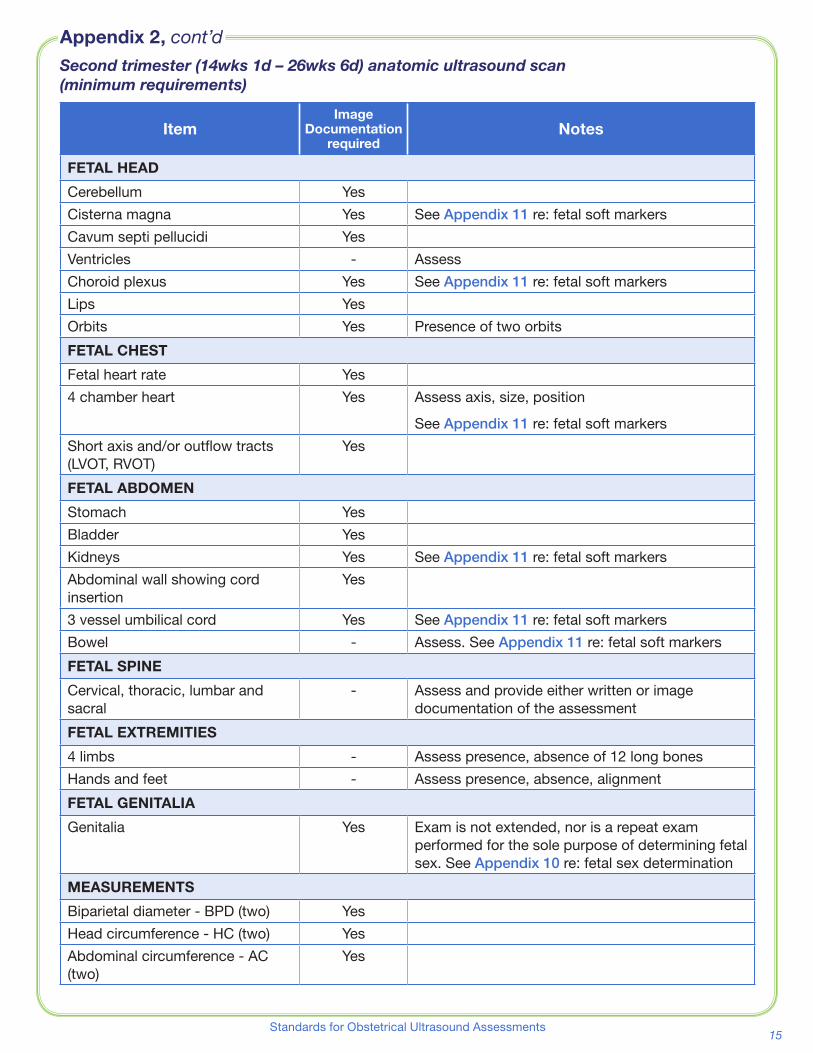
Second trimester (14wks 1d – 26wks 6d) anatomic ultrasound scan (minimum requirements)
ItemImage
Documentation required
Notes
FETAL HEAD
Cerebellum Yes
Cisterna magna Yes See Appendix 11 re: fetal soft markers
Cavum septi pellucidi Yes
Ventricles - Assess
Choroid plexus Yes See Appendix 11 re: fetal soft markers
Lips Yes
Orbits Yes Presence of two orbits
FETAL CHEST
Fetal heart rate Yes
4 chamber heart Yes Assess axis, size, position
See Appendix 11 re: fetal soft markers
Short axis and/or outflow tracts (LVOT, RVOT)
Yes
FETAL ABDOMEN
Stomach Yes
Bladder Yes
Kidneys Yes See Appendix 11 re: fetal soft markers
Abdominal wall showing cord insertion
Yes
3 vessel umbilical cord Yes See Appendix 11 re: fetal soft markers
Bowel - Assess� See Appendix 11 re: fetal soft markers
FETAL SPINE
Cervical, thoracic, lumbar and sacral
- Assess and provide either written or image documentation of the assessment
FETAL EXTREMITIES
4 limbs - Assess presence, absence of 12 long bones
Hands and feet - Assess presence, absence, alignment
FETAL GENITALIA
Genitalia Yes Exam is not extended, nor is a repeat exam performed for the sole purpose of determining fetal sex� See Appendix 10 re: fetal sex determination
MEASUREMENTS
Biparietal diameter - BPD (two) Yes
Head circumference - HC (two) Yes
Abdominal circumference - AC (two)
Yes
Appendix 2, cont’d

16 Perinatal Services BC
ItemImage
Documentation required
Notes
Femur length - FL (two) Yes
Nuchal thickness aka nuchal fold (two)
Yes See Appendix 11 re: fetal soft markers
OTHER
Fetal number Yes
Amniotic Fluid Volume Yes 18wks 0d – 26wks 6d ➞ if subjectively abnormal perform Single deepest pocket assessment (SDP)
Placenta Yes Evaluate location, appearance, cord insertion and relationship to internal cervical os
Fetal position Yes After 23wks 0d
Cervix Yes Assess for internal os funneling and length
MULTIPLE GESTATION
Membrane Yes Report to include chorionicity and amnionicity
Third trimester (27wks 0d – term) ultrasound scan (minimum requirements)
ItemImage
Documentation required
Notes
Fetal number Yes
Fetal heart rate Yes
Amniotic fluid volume Yes Single deepest pocket - SDP
Fetal position Yes
Placenta Yes Evaluate location, appearance, and relationship to internal cervical os
4 chamber heart Yes
Short axis or outflow tracts (LVOT, RVOT)
Yes
Fetal stomach Yes
Fetal bladder Yes
Fetal kidneys Yes
Cervix (if less than 32wks 0d) Yes Assess for internal os funneling and length
MEASUREMENTS
Biparietal diameter - BPD (two) Yes
Head circumference - HC (two) Yes
Abdominal circumference - AC (two)
Yes
Femur length - FL (two) Yes
MULTIPLE GESTATION
Membrane Yes Report to include chorionicity and amnionicity
Appendix 2, cont’d

17Standards for Obstetrical Ultrasound Assessments
Appendix 3 – PW Doppler use in Obstetric Ultrasound
1/2
Acoustic Output Levels in Obstetric Ultrasound Scans 1. Guideline Purpose
The purpose of this guideline is to assure that the ALARA1 Principle is being followed when obstetrical ultrasound scans are performed.
2. Guideline Statement
2.1. Exposure time and acoustic output should be kept to the lowest levels consistent with obtaining diagnostic information and limited to medically indicated procedures. (1)
3. Guideline Principles
The thermal index and mechanical index are not perfect indicators of the risks of thermal and non-thermal bioeffects but currently they are accepted as the most practical and understandable methods of estimating the potential of such risks. Spectral and color Doppler may produce high intensities and routine examination by this modality during the embryonic period is rarely indicated. In addition, because of high acoustic absorption by bone, the potential for heating adjacent tissues must also be kept in mind [in the second and third trimesters]. (2)
3.1. Pulsed Doppler ultrasound should not be used routinely within the fetus. (2,5)
3.2. Pulsed Doppler ultrasound should not be used solely for the purpose of allowing patients to listen to the fetal heart beat.
3.3. Pulsed Doppler ultrasound may be used:
3.3.1. to examine blood flow dynamics in the umbilical arteries
3.3.2. to examine fetal blood vessels in high-risk situations
3.3.3. for certain clinical indications such as to refine risks for trisomies
3.3.4. during fetal echocardiography
3.3.5. to examine maternal uterine vessels
3.4. When performing Doppler ultrasound, the thermal index should be ≤1.0 and the exposure time should be kept as short as possible. (2,4,5)
3.5. When attempting to obtain the fetal heart rate, the AIUM recommends using M-mode first because the time averaged acoustic intensity delivered to the fetus is lower with M-mode than with spectral Doppler. If this is unsuccessful, spectral Doppler ultrasound may be used with the following guidelines:
3.5.1. use spectral Doppler only briefly (4-5 heart beats) (4)
3.5.2. keep the thermal index (TIS for soft tissues in the first trimester and TIB for bones in the second and third trimesters) as low as possible, preferably ≤1.0. (4)
1 As Low As Reasonably Achievable

18 Perinatal Services BC
Appendix 3, cont’d
Acoustic Output Levels in Obstetric Ultrasound Scans
This Regional Guideline was developed by the Lower Mainland Medical Imaging group which has representation from Fraser Health (FHA), Vancouver Coastal Health (VCH), Providence Health Care (PHC) and the Provincial Health Services Authority (PHSA). Approval for inclusion into the Provincial Obstetrical Ultrasound Assessment Standards has been obtained from the Provincial Medical Imaging Executive Committee.
2/2
4. Guideline Scope
This guideline is applicable to all Ultrasound departments.
5. Exceptions
This is a clinical guideline and not a policy. Exceptions may be made at the discretion of the supervising radiologist or obstetrician.
6. References
1. EFSUMB2 - Statement on the Safe use of Doppler Ultrasound During Scans at 11-14 Weeks (or Earlier in Pregnancy) 2013 http://www.efsumb.org/guidelines/ss2013-firsttrimester-doppler.pdf 2. ISUOG3 - Statement on the safe use of Doppler in the 11 to 13+6 week fetal ultrasound examination http://www.isuog.org/NR/rdonlyres/090DC178-48BF-4861-AFF8-3531F8A50FE0/0/ISUOG_Safety_Statement_2011.pdf 3. ISOUG - Safety Statement, 2000 (reconfirmed 2003) http://www.isuog.org/NR/rdonlyres/CC3087DF-08B8-4870-A6CD-B47146568A6B/0/ISUOGSafetystatement2003.pdf 4.AIUM4 - Statement on Measurement of Fetal Heart Rate http://www.aium.org/officialStatements/43 5. AIUM – Statement on the Safe Use of Doppler Ultrasound During 11-14 week scans http://www.aium.org/officialStatements/42 Issued by: Brent Barton, RPL September 12, 2014 Dr. Simon Bicknell, MPL September 12, 2014 Approved by: Medical Imaging Executive Committee September 12, 2014
2 European federation of Societies for Ultrasound in Medicine and Biology 3 International Society of Ultrasound in Obstetrics and Gynecology 4 American Institute of Ultrasound in Medicine
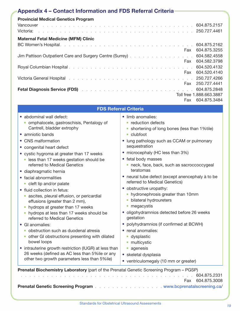
19Standards for Obstetrical Ultrasound Assessments
Provincial Medical Genetics ProgramVancouver � � � � � � � � � � � � � � � � � � � � � � � � � � � � � � � � � � � 604�875�2157Victoria: � � � � � � � � � � � � � � � � � � � � � � � � � � � � � � � � � � � � 250�727�4461
Maternal Fetal Medicine (MFM) ClinicBC Women’s Hospital � � � � � � � � � � � � � � � � � � � � � � � � � � � � � � � 604�875�2162 Fax 604�875�3255Jim Pattison Outpatient Care and Surgery Centre (Surrey) � � � � � � � � � � � � � � � 604�582�4558 Fax 604�582�3798Royal Columbian Hospital � � � � � � � � � � � � � � � � � � � � � � � � � � � � � 604�520�4132 Fax 604�520�4140Victoria General Hospital � � � � � � � � � � � � � � � � � � � � � � � � � � � � � 250�727�4266 Fax 250�727�4441Fetal Diagnosis Service (FDS) � � � � � � � � � � � � � � � � � � � � � � � � � � 604�875�2848 Toll free 1�888�663�3887 Fax 604�875�3484
FDS Referral Criteria
⦁⦁ abdominal wall defect:⦁◾ omphalocele, gastroschisis, Pentalogy of
Cantrell, bladder extrophy⦁⦁ amniotic bands⦁⦁ CNS malformation⦁⦁ congenital heart defect⦁⦁ cystic hygroma at greater than 17 weeks
⦁◾ less than 17 weeks gestation should be referred to Medical Genetics
⦁⦁ diaphragmatic hernia⦁⦁ facial abnormalities
⦁◾ cleft lip and/or palate⦁⦁ fluid collection in fetus:
⦁◾ ascites, pleural effusion, or pericardial effusions (greater than 2 mm),
⦁◾ hydrops at greater than 17 weeks⦁◾ hydrops at less than 17 weeks should be
referred to Medical Genetics⦁⦁ GI anomalies:
⦁◾ obstruction such as duodenal atresia⦁◾ other GI obstructions presenting with dilated
bowel loops⦁⦁ intrauterine growth restriction (IUGR) at less than
26 weeks (defined as AC less than 5%ile or any other two growth parameters less than 5%ile)
⦁⦁ limb anomalies:⦁◾ reduction defects⦁◾ shortening of long bones (less than 1%tile)⦁◾ clubfoot
⦁⦁ lung pathology such as CCAM or pulmonary sequestration
⦁⦁ microcephaly (HC less than 3%)⦁⦁ fetal body masses
⦁◾ neck, face, back, such as sacroccoccygeal teratomas
⦁⦁ neural tube defect (except anencephaly à to be referred to Medical Genetics)
⦁⦁ obstructive uropathy:⦁◾ hydronephrosis greater than 10mm⦁◾ bilateral hydroureters⦁◾ megacystis
⦁⦁ oligohydramnios detected before 26 weeks gestation
⦁⦁ polyhydramnios (if confirmed at BCWH)⦁⦁ renal anomalies:
⦁◾ dysplastic⦁◾ multicystic⦁◾ agenesis
⦁⦁ skeletal dysplasia⦁⦁ ventriculomegaly (10 mm or greater)
Prenatal Biochemistry Laboratory (part of the Prenatal Genetic Screening Program – PGSP) � � � � � � � � � � � � � � � � � � � � � � � � � � � � � � � � � � � � � � � � 604�875�2331 Fax 604�875�3008Prenatal Genetic Screening Program � � � � � � � � � � � � � � � � www.bcprenatalscreening.ca/
Appendix 4 – Contact Information and FDS Referral Criteria

20 Perinatal Services BC
for Nuchal Translucency Ultrasound Measurements
West Tower, 3rd Floor 555 West 12th Avenue, Vancouver, BC, V5Z 3X7
Tel: 604 877 2121 ext 223772 l Fax: 604 872 1987 l www.bcprenatalscreening.ca
Minimum Reporting Standards for Nuchal Translucency Ultrasound Measurements
Initially developed by the BC Radiologists Working Group in conjunction with the NT Working Group (2009), and revised with the Provincial OB Ultrasound Standards Working Group (2015)
All 1st trimester ultrasound reporting standards are described in the Provincial Obstetrical Ultrasound Assessment Standards for BC (Perinatal Services BC, 2012, revised 2015). This includes the reporting standards for a Nuchal Translucency (NT) ultrasound. http://www.perinatalservicesbc.ca/Guidelines/standards/ultrasound-assess-stnds/default.htm The NT ultrasound report is sent to the ordering MD/MW as well as the C&W Prenatal Biochemistry Lab to be incorporated with the patient’s serum results to calculate the Integrated Prenatal Screen (IPS) risk report for the pregnancy. Go to www.bcprenatalscreening.ca for more information on prenatal genetic screening. As per the Provincial Obstetrical Ultrasound Assessment Standards, the minimum required content for a 1st trimester report (up to 14wks 0d) must include:
the items listed in “ALL OB Ultrasound Reports” in addition to the following:
Include for all 1st trimester (up to and including 14wks 0d): Presence/absence of yolk sac
or embryo Fetal number:
- if multiple: chorionicity and amnionicity
Location of gestational sac Cardiac activity:
- presence/absence - fetal heart rate (FHR)
Crown rump length (CRL) Maternal pelvic anatomy
(adnexa, ovaries, cervix)
If greater than 11wks 0d gestation, attempt to include:
Head circumference (HC)
Biparietal diameter (BPD)
Choroid plexus filled ventricles
Stomach Symmetrical lung
fields Presence of 4 limbs,
each with 3 segments Abdominal wall cord
insertion
With Nuchal Translucency (NT) measurement (performed only if CRL is between 45.0 – 84.0 mm), report to also include: NT measurement in mm
to 1 decimal point FMF# of person
performing the NT measurement
Standard comments established by the BC Prenatal Genetic Screening Program (PGSP) See comments below (page 2)
Appendix 5 – PGSP Minimum Reporting Standards

21Standards for Obstetrical Ultrasound Assessments
Appendix 5, cont’d
West Tower, 3rd Floor 555 West 12th Avenue, Vancouver, BC, V5Z 3X7
Tel: 604 877 2121 ext 223772 l Fax: 604 872 1987 l www.bcprenatalscreening.ca
Standard Comments (or Equivalents) for Inclusion on all NT Reports
If NT measurement is.... Standard Comment on NT Report would be....
Below Cut-Off ≤2.9 mm
(2.9 mm or less)
The risk of Down syndrome associated with this NT measurement will be
integrated with other parameters [maternal age, first trimester (PAPP-A)
and second trimester (AFP, uE3, HCG, inhibin A) blood test results] to
generate a pregnancy specific risk level for Down syndrome (trisomy 21).
The C&W Prenatal Biochemistry Laboratory will issue this report after
receiving the second blood sample (to be ideally collected between 15
weeks 2 days – 16 weeks 6 days gestation). See
www.bcprenatalscreening.ca for more info and to download lab
requisition.
A routine detailed fetal ultrasound is recommended at 18 – 20 weeks.
Above Cut-off ≥ 3.0 mm
(greater than or equal to 3.0 mm)
The NT measurement is 3.0 mm or above. An immediate risk assessment
for Down syndrome and trisomy 18 will be determined by C&W Prenatal
Biochemistry Laboratory based on age and the NT only, OR age, NT, and
PAPP-A (as part 1 serum availability permits). If screen risk for Down
syndrome and/or trisomy 18 is positive (≥ 1/300), a lab report will be sent
to the ordering care provider.
If the NT is ≥ 3.5 mm,
add additional comment
An NT measurement greater than or equal to 3.5 mm is a risk factor for a
congenital heart defect, or other genetic conditions. A referral to the Dept.
of Medical Genetics (Vancouver or Victoria), and a detailed fetal ultrasound
and echocardiogram at 18-20 weeks gestation, are recommended.

22 Perinatal Services BC
Appendix 6 – Crown Rump Length (CRL) Chart
CROWN RUMP LENGTH (CRL)
Weeks Days 10%(mm)
50%(mm)
90%(mm)
Weeks Days 10%(mm)
50%(mm)
90%(mm)
6+ 0… 3 4 5 10+ 0… 26 33 41
3 5 7 28 35 43
2… 4 5 8 2… 29 36 44
5 6 9 30 37 46
4… 5 7 10 4… 31 39 48
6 8 11 32 40 50
6… 7 9 12 6… 34 42 51
7+ 0… 7 10 13 11+ 0… 35 44 53
8 11 14 36 45 55
2… 9 12 15 2… 38 47 57
10 13 17 39 48 59
4… 10 14 18 4… 40 50 60
11 15 19 42 52 62
6… 12 16 20 6… 43 54 64
8+ 0… 13 17 21 12+ 0… 45 55 66
14 18 23 46 57 68
2… 14 19 24 2… 47 59 70
15 20 25 49 61 72
4… 16 21 26 4… 50 62 73
17 22 28 52 64 75
6… 18 23 29 6… 53 66 77
9+ 0… 19 24 31 13+ 0… 55 68 79
20 25 32 56 69 81
2… 21 27 33 2… 58 71 83
22 28 35 59 73 85
4… 23 29 36 4… 61 75 86
24 30 38 62 77 88
6… 25 32 40 6… 63 78 90
14+ 0… 65 80 92
Source: Unpublished data - Data was obtained at the same time, using the same methods and analysis as Lessoway V, Schulzer M, Wittman B, et al. Ultrasound fetal biometry charts for a North American Caucasian population. J Clin Ultrasound 1998 Nov-Dec; 26(9):433 – 53.
Reprinted with Permission from Lessoway, V.

23Standards for Obstetrical Ultrasound Assessments
On September 6, 2014 The Lancet published Intergrowth 21st international fetal growth standards for the clinical interpretation of routinely taken ultrasound measurements and for comparisons across populations� (Lancet 2014; 384: 869–79) These international standards were developed on the basis of recommendations from a WHO expert committee and the study was funded by the Bill and Melinda Gates Foundation�
A Working Group on biometric standards for assessing fetal growth was brought together to carefully consider the option of replacing the current, widely used, Lessoway standards with the new Intergrowth 21st standards� The working group noted that the Lessoway standard was created using a rigorous methodology and clinicians in British Columbia are familiar with this standard� However, the Intergrowth standard has several compelling features since it was created using stricter inclusion criteria, careful standardization of ultrasound measurements and sophisticated statistical methods� The Working Group was favourably disposed towards adopting the Intergrowth standard and therefore carried out an analysis to anticipate the clinical consequences of switching from the Lessoway standard to the Intergrowth standard� This involved a retrospective evaluation of the medical records of 3 groups of women previously seen at the BC Women’s Hospital including a) low risk women without known pregnancy complications (N=4440), b) women referred for suspected small-for-gestational age (SGA) fetuses (N=2099), or hypertensive complications (N=1086), and c) women referred for suspected large-for-gestational age (LGA) fetuses (N=1016), or who were diabetic (N=1692),
The analysis showed that the frequency of SGA fetuses identified by the Intergrowth standard would be significantly less than that identified by the Lessoway standard� On the other hand, the Intergrowth standard would lead to a substantial increase in LGA fetuses compared with the Lessoway standard� In short, adopting the Intergrowth standard would lead to a significant change in the frequency of SGA and LGA diagnoses in clinical practice (irrespective of whether the 3rd/97th or the 10th/90th centile was used for diagnosis)� In analyzing a “low risk cohort”, similar results were seen (the LGA was overrepresented using Intergrowth 21 charts)� The Working Group found this concerning, especially since the under identification of small fetuses has the potential for increasing mortality and serious morbidity, while the excess number of LGA fetuses identified would drastically increase the need for clinical resources� An alternative interpretation is that the current standard over-diagnoses SGA fetuses and drastically under-diagnose LGA fetuses (though observed rates of macrosomia at birth contradict the latter proposition)�
After careful consideration, the Working Group recommended against adopting the Intergrowth standard and to continue to use the Lessoway standard for fetal biometry assessments� The group continues to keep abreast of future developments in the field and will also pursue analyses that provide an outcome (mortality and serious morbidity) based assessment of the Intergrowth standard�
Working Group:
Dr� Blair Butler (MFM) Dr� Jennifer Hutcheon (Epidemiologist) Dr� KS Joseph (Epidemiologist) Vickie Lessoway (Sonographer) Dr� Ken Lim (MFM) Dr� Gerry Marquette (MFM)
Appendix 7 – Intergrowth 21st Fetal Growth Standards Review

24 Perinatal Services BC
Amniotic fluid assessment by ultrasound is at best semi-quantitative. It is not a true quantitative measurement and as such reproducibility and accuracy (to the true volume within, especially at the extremes) will be elusive using 2D ultrasound.
Physiological considerations
The assessment of amniotic fluid volume has been an integral component of obstetrical scanning and fetal health surveillance for many years� Abnormalities of amniotic fluid are associated with a variety of adverse maternal, fetal and obstetrical conditions� It is also clear that amniotic fluid volume is dynamic and can rapidly change due to non-pathological processes (maternal hydration, maternal activity/rest, maternal position etc)� There is likely a limit to how much amniotic fluid can be affected by these factors, but this modifiable component, as assessed by AFI, is as much as 45-50 mm range (Ulker, 2012, 2013), (Kilpatrick, 1993)� At the extremes of diagnostic categories (oligohydramnios, polyhydramnios), this is unlikely to alter the diagnosis, but near the thresholds, this may be a factor� This may help explain the inconsistency of results when tests are repeated within hours of each other, or even between different operators� Furthermore, it is unclear whether manipulation of these modifiable factors affects fetal outcomes, or changes the diagnostic value of amniotic fluid assessment�
It would be difficult to control all the physiological factors which may affect the amniotic fluid volume in the typical diagnostic ultrasound lab� Ulker (2013) found that rest in the left lateral position improved the AFI compared to activity (15 min of sitting followed by 5 min of walking repeatedly for at least 1 hour)� There was no change in the AFI in the activity group� Both oral hydration at a rate of 1000 cc/hr and rest in the left lateral position had effects within the first hour of initiation� Maternal hydration can be simply instituted, but maternal rest in the left lateral position for a sufficient duration would be more difficult, given that patients are scanned in a supine (commonly wedged) position� Physiologically, it is unknown if rest in the supine position (as opposed to left lateral) would increase or decrease amniotic fluid volume (due to uterine compression of vena cava and/or aorta leading to decreased placental perfusion)� Therefore, we suggest that women be well hydrated prior to the ultrasound exam to control that physiological factor�
Technical and operator factors
Prior studies have demonstrated that using color Doppler (to exclude cord) may affect the assessment� With more modern technology, this should be less of an issue� A recent study by Sande (2015) suggests the limits of agreement of both AFI and SVDP are quite wide, both between and within observers which again, adds to the inconsistency of results which negatively affects clinical utility� Maternal position during fluid assessment is frequently not described in the literature in regards to single pocket assessments�
Choice of ultrasound measurement of amniotic fluid assessment
It is difficult to estimate volume (3 dimensional) based on one or two dimensional measures (such as AFI or SDP)� Changes in ultrasound imaging quality and technology, and lack of contemporary modern studies contribute to the complexity of this topic� To minimize intervention rates, without compromising fetal outcomes, the Single Deepest Pocket (SDP) method is advocated� This is based on a Cochrane meta-analysis of existing randomized data comparing a single pocket assessment versus the AFI in diagnosing oligohydramnios� In four of the five trials used in the analysis, the technique can be linked back to the original Chamberlain definition� The fifth article is a foreign language journal which could not be easily reviewed� Chamberlain’s original definition of single pocket method leaves some room for interpretation, hence the proposed definition/standard in this document can be best described as modernized and our best interpretation of Chamberlain’s technique�
Chamberlain’s original definition: “ � � � the largest pocket of fluid was measured and recorded� The depth of the pocket was measured at a right angle to the uterine contour � � � the width of the largest pocket of amniotic fluid was measured at a right angle to the depth measurement � � � In all cases except for decreased amniotic fluid, the width of a pocket of amniotic fluid was > 1 cm�”
It should be noted that a linear transducer was used, and no mention is made of transducer scanning plane or orientation� Maternal position is not described� Communication with a co-author revealed that
Appendix 8 – Amniotic Fluid Volume
25Standards for Obstetrical Ultrasound Assessments
Appendix 8 – Amniotic Fluid Volume (cont’d)“{Chamberlain} emphasized that the transducer should not be angled but when possible should follow the contour of the uterus�”
Another way to think of the modernized description is as follows:
Find the largest pocket free of cord and fetal body parts� If that pocket can fit a 2 x 1 cm rectangle whose long axis is approximately at right angles to the uterine wall, then the amniotic fluid assessment is considered normal� If the pocket is so big, that there is fluid/space around an 8 x 1 cm rectangle (oriented as above), then the amniotic fluid assessment is considered to be polyhydramnios�
Terminology
There are different methods to assess the largest pocket of fluid which vary from a single vertical or perpendicular measurement to a two dimensional product of the pocket (Magann, 1994)� The terms used are also varied, and unfortunately, are used interchangeably, adding to the confusion when assessing the literature� For example, Chamberlain’s reference has been described using the following terms: Single deepest pocket (Magann, 2004), Maximum vertical pocket (Williams, 1993), Maximum pool depth (Nwosu, 1994) and Single deepest vertical pocket (Moses, 2004)� In his recent (2015) article, Sande uses the terms “single deepest vertical pool” or “maximum vertical pocket”, but this is described as the “ultrasound probe being held vertical to the uterine contour onto the abdomen and parallel to the maternal sagittal plane”� This does not appear to match the Chamberlain definition, particularly since the minimum width of the pocket is not included� Maternal position is not described�
For this guideline, it was felt that using the term “single deepest pocket” (SDP) was the one most often ascribed to Chamberlain and removes the word “vertical”, which suggests that the ultrasound plane has to be vertical, or perpendicular to the floor, which is not used in Chamberlain’s description�
Origin of definitions of fluid abnormalities
This was based on Chamberlain’s original definitions, and randomised trials used in the Cochrane meta-analysis� The reference ranges are from Magann (2000) (whose group contributed three of the RCTs to the above mentioned Cochrane meta-analysis) and the normal distributions published in that paper, suggest the simple definitions proposed are reasonable to define normal from abnormal�
References
Kilpatrick et al� Maternal hydration increases amniotic fluid index in women with normal amniotic fluid� Obstet Gynecol. 1993 Jan;81(1): 49-52�
Magann et al� Amniotic fluid index vs single deepest pocket technique during modified biophysical profile: a randomized clinical trial� Am J Obstet Gynecol. 2004 August;191(2): 667-668�
Magann et al� Comparative Efficacy of Two Sonographic measurements for the Detection of Aberrations in the Amniotic Fluid Volume and the Effect of Amniotic Fluid Volume on Pregnancy Outcome� Obstet Gynecol. 1994 June;83(6): 959-962�
Magann et al� The amniotic fluid index, single deepest pocket and two-diameter pocket in normal human pregnancy� Am J Obstet Gynecol. 2000 182(6); 1581-87�
Moses et al� A randomized clinical trial of the intrapartum assessment of amniotic fluid volume: Amniotic fluid index versus the single deepest pocket technique� Am J Obstet Gynecol. 2004 190: 1564-70�
Nabhan et al� Amniotic fluid index versus single deepest vertical pocket as a screening test for preventing adverse pregnancy outcome� Cochrane Database of Systemic Reviews 2008, Issue 3�
Nwosu et al� Longitudinal assessment of amniotic fluid index� Br J Obstet Gynaecol. 1993 Sep;100(9): 816-9�
Sande et al� Reproducibility of measuring amniotic fluid index and single deepest vertical pool throughout gestation� Prenat Diagn. 2015 May;35(5): 434-9�

26 Perinatal Services BC
Appendix 8 – Amniotic Fluid Volume (cont’d)Ulker et al� Correlation between the duration of maternal rest in the left lateral decubitus position and the
amniotic fluid volume increase� J Ultrasound Med. 2012 May;31(5): 705-9�
Ulker et al� Effect of maternal hydration on the amniotic fluid volume during maternal rest in the left lateral decubitus position: a randomized prospective study� J Ultrasound Med. 2013 Jun;32(6): 955-61�
Ulker et al� Effects of maternal left lateral position and res5t on amniotic fluid index: a prospective clinical study� J Reprod Med. 2012 May-June;57(5-6): 270-6�
Williams K� Amniotic fluid assessment� Obstet Gynecol Surv. 1993 Dec;48(12): 795-800�
Working Group:Dr� Jason Burrows (MFM) Dr� Leanne Dahlgren (MFM) Dr� Nancy Kent (MFM) Dr� Duncan Farquharson (MFM) Dr� Kirsten Grabowska (MFM) Dr� Jennifer Hutcheon (Epidemiologist)Dr� Ken Lim (MFM) Dr� Kim McDonald (MFM Fellow) Dr� Amy Metcalfe (Epidemiologist)
27Standards for Obstetrical Ultrasound Assessments
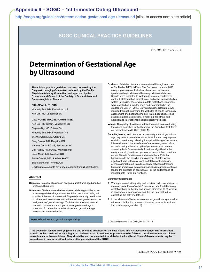
http://sogc.org/guidelines/determination-gestational-age-ultrasound [click to access complete article]
Appendix 9 – SOGC – 1st trimester Dating Ultrasound
FEBRUARY JOGC FÉVRIER 2014 l 171
No. 303, February 2014
SOGC CLINICAL PRACTICE GUIDELINES
Determination of Gestational Age by Ultrasound
This document reflects emerging clinical and scientific advances on the date issued and is subject to change. The information should not be construed as dictating an exclusive course of treatment or procedure to be followed. Local institutions can dictate amendments to these opinions. They should be well documented if modified at the local level. None of these contents may be reproduced in any form without prior written permission of the SOGC.
This clinical practice guideline has been prepared by the Diagnostic Imaging Committee, reviewed by the Family Physician Advisory Committee, and approved by the Executive and Council of the Society of Obstetricians and Gynaecologists of Canada.
PRINCIPAL AUTHORS
Kimberly Butt, MD, Fredericton NB
Ken Lim, MD, Vancouver BC
DIAGNOSTIC IMAGING COMMITTEE
Ken Lim, MD (Chair), Vancouver BC
Stephen Bly, MD, Ottawa ON
Kimberly Butt, MD, Fredericton NB
Yvonne Cargill, MD, Ottawa ON
Greg Davies, MD, Kingston ON
Nanette Denis, RDMS, Saskatoon SK
Gail Hazlitt, RN, RDMS, Winnipeg MB
Lucie Morin, MD, Montreal QC
Annie Ouellet, MD, Sherbrooke QC
Shia Salem, MD, Toronto, ON
Disclosure statements have been received from all contributors .
Keywords: ultrasound, gestational age, datingJ Obstet Gynaecol Can 2014;36(2):171–181
Evidence: Published literature was retrieved through searches of PubMed or MEDLINE and The Cochrane Library in 2013 using appropriate controlled vocabulary and key words (gestational age, ultrasound biometry, ultrasound dating) . Results were restricted to systematic reviews, randomized control trials/controlled clinical trials, and observational studies written in English . There were no date restrictions . Searches were updated on a regular basis and incorporated in the guideline to July 31, 2013 . Grey (unpublished) literature was identified through searching the websites of health technology assessment and health technology-related agencies, clinical practice guideline collections, clinical trial registries, and national and international medical specialty societies .
Values: The quality of evidence in this document was rated using the criteria described in the Report of the Canadian Task Force on Preventive Health Care (Table 1) .
Benefits, harms, and costs: Accurate assignment of gestational age may reduce post-dates labour induction and may improve obstetric care through allowing the optimal timing of necessary interventions and the avoidance of unnecessary ones . More accurate dating allows for optimal performance of prenatal screening tests for aneuploidy . A national algorithm for the assignment of gestational age may reduce practice variations across Canada for clinicians and researchers . Potential harms include the possible reassignment of dates when significant fetal pathology (such as fetal growth restriction or macrosomia) result in a discrepancy between ultrasound biometric and clinical gestational age . Such reassignment may lead to the omission of appropriate—or the performance of inappropriate—fetal interventions .
Summary Statements
1 . When performed with quality and precision, ultrasound alone is more accurate than a “certain” menstrual date for determining gestational age in the first and second trimesters (≤ 23 weeks) in spontaneous conceptions, and it is the best method for estimating the delivery date . (II)
2 . In the absence of better assessment of gestational age, routine ultrasound in the first or second trimester reduces inductions for post-term pregnancies . (I)
Abstract
Objective: To assist clinicians in assigning gestational age based on ultrasound biometry .
Outcomes: To determine whether ultrasound dating provides more accurate gestational age assessment than menstrual dating with or without the use of ultrasound . To provide maternity health care providers and researchers with evidence-based guidelines for the assignment of gestational age . To determine which ultrasound biometric parameters are superior when gestational age is uncertain . To determine whether ultrasound gestational age assessment is cost effective .

28 Perinatal Services BC
Appendix 10 – Fetal Sex Determination Policy
MINISTRY OF HEALTH POLICY COMMUNIQUE 2012
FETAL SEX DETERMINATION BY ULTRASOUND
EFFECTIVE DATE: OCTOBER 22, 2012
POLICY OBJECTIVE The objective of this policy is to achieve: consistency in approach across health authorities to fetal sex determination by ultrasound and disclosure of
fetal sex information to patients; alignment with the College of Physicians and Surgeons of British Columbia’s Guideline on Disclosure of
Fetal Sex; and, alignment with Canadian clinical practice guidelines.
SCOPE This policy applies to obstetrical ultrasound services provided by the regional health authorities and the Provincial Health Services Authority. POLICY
1. At the time of a routine full fetal anatomical assessment, a reasonable attempt will be made by the practitioner performing the ultrasound assessment to assess fetal genitalia within the allotted exam time.
2. The allotted exam time will not be extended for the sole purpose of determining fetal sex and repeat exams will not be scheduled for the sole purpose of determining fetal sex.
3. The sex of the fetus will be recorded as Male, Female or Not Determined in the notes taken during the ultrasound assessment.
4. The practitioner performing the ultrasound assessment is not to release any information about the sex of the fetus directly to the patient.
5. The sex of the fetus shall be on the final report that is issued to the referring practitioner. 6. The patient may request fetal sex information from her referring practitioner.
MINISTRY CONTACT: Executive Director, Hospital and Provincial Services Branch
29Standards for Obstetrical Ultrasound Assessments
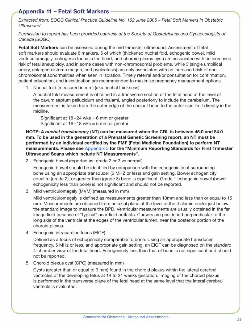
Extracted from: SOGC Clinical Practice Guideline No. 162 June 2005 – Fetal Soft Markers in Obstetric Ultrasound
Permission to reprint has been provided courtesy of the Society of Obstetricians and Gynaecologists of Canada (SOGC)
Fetal Soft Markers can be assessed during the mid trimester ultrasound� Assessment of fetal soft markers should evaluate 8 markers, 5 of which (thickened nuchal fold, echogenic bowel, mild ventriculomegaly, echogenic focus in the heart, and choroid plexus cyst) are associated with an increased risk of fetal aneuploidy, and in some cases with non-chromosomal problems, while 3 (single umbilical artery, enlarged cisterna magna, and pyelectasis) are only associated with an increased risk of non-chromosomal abnormalities when seen in isolation� Timely referral and/or consultation for confirmation, patient education, and investigation are recommended to maximize pregnancy management options�
1� Nuchal fold (measured in mm) (aka nuchal thickness)
A nuchal fold measurement is obtained in a transverse section of the fetal head at the level of the cavum septum pellucidum and thalami, angled posteriorly to include the cerebellum� The measurement is taken from the outer edge of the occiput bone to the outer skin limit directly in the midline�
Significant at 18 – 24 wks = 6 mm or greater Significant at 16 – 18 wks = 5 mm or greater
NOTE: A nuchal translucency (NT) can be measured when the CRL is between 45.0 and 84.0 mm. To be used in the generation of a Prenatal Genetic Screening report, an NT must be performed by an individual certified by the FMF (Fetal Medicine Foundation) to perform NT measurements. Please see Appendix 5 for the “Minimum Reporting Standards for First Trimester Ultrasound Scans which include NT Measurements”.
2� Echogenic bowel (reported as: grade 2 or 3 vs normal)
Echogenic bowel should be identified by comparison with the echogenicity of surrounding bone using an appropriate transducer (5 MHZ or less) and gain setting� Bowel echogenicity equal to (grade 2), or greater than (grade 3) bone is significant� Grade 1 echogenic bowel (bowel echogenicity less than bone) is not significant and should not be reported�
3� Mild ventriculomegaly (MVM) (measured in mm)
Mild ventriculomegaly is defined as measurements greater than 10mm and less than or equal to 15 mm� Measurements are obtained from an axial plane at the level of the thalamic nuclei just below the standard image to measure the BPD� Ventricular measurements are usually obtained in the far image field because of “typical” near-field artifacts� Cursors are positioned perpendicular to the long axis of the ventricle at the edges of the ventricular lumen, near the posterior portion of the choroid plexus�
4� Echogenic intracardiac focus (EICF)
Defined as a focus of echogenicity comparable to bone� Using an appropriate transducer frequency, 5 MHz or less, and appropriate gain setting, an EICF can be diagnosed on the standard 4-chamber view of the fetal heart� Echogenicity less than that of bone is not significant and should not be reported�
5� Choroid plexus cyst (CPC) (measured in mm)
Cysts (greater than or equal to 3 mm) found in the choroid plexus within the lateral cerebral ventricles of the developing fetus at 14 to 24 weeks gestation� Imaging of the choroid plexus is performed in the transverse plane of the fetal head at the same level that the lateral cerebral ventricle is evaluated�
Appendix 11 – Fetal Soft Markers

30 Perinatal Services BC
6� Enlarged cisterna magna (measured in mm)
The cisterna magna is measured on a transaxial view of the fetal head angled 15 degrees caudal to the canthomeatal line� The anterior/posterior diameter is taken between the inferior/posterior surface of the vermis of the cerebellum to the inner surface of the cranium� An enlarged cisterna magna is defined by an anterior/posterior diameter of greater than (or equal to) 10 mm� The measurement will be falsely exaggerated by a steep scan angle through the posterior fossa or dolichocephaly�
7� Pyelectasis (measured in mm)
Mild pyelectasis is defined as a hypoechoic spherical or elliptical space within the renal pelvis that measures greater than (or equal to) 5mm and less than (or equal to) 10 mm� The measurement is taken on a transverse section through the fetal renal pelvis using the maximum anterior-to-posterior measurement� Measurements less than 5 mm are normal, should not be designated as pyelectasis, and should not be reported�
8� Single Umbilical Artery (SUA)
Single umbilical artery is the absence of one of the fetal umbilical cord arteries surrounding the fetal bladder�
Permission to reprint has been provided courtesy of the Society of Obstetricians and Gynaecologists of Canada (SOGC)
Appendix 11, cont’d
31Standards for Obstetrical Ultrasound Assessments
Antepartum and Intrapartum Consensus Guideline
http://sogc.org/guidelines/fetal-health-surveillance-antepartum-and-intrapartum-consensus-guideline/
[click to access complete article]
SOGC CLINICAL PRACTICE GUIDELINE
Fetal Health Surveillance: Antepartum andIntrapartum Consensus Guideline
Abstract
Objective: This guideline provides new recommendations pertainingto the application and documentation of fetal surveillance in theantepartum and intrapartum period that will decrease theincidence of birth asphyxia while maintaining the lowest possiblerate of obstetrical intervention. Pregnancies with and without riskfactors for adverse perinatal outcomes are considered. Thisguideline presents an alternative classification system forantenatal fetal non-stress testing and intrapartum electronic fetalsurveillance to what has been used previously. This guideline isintended for use by all health professionals who provideantepartum and intrapartum care in Canada.
Options: Consideration has been given to all methods of fetalsurveillance currently available in Canada.
Outcomes: Short- and long-term outcomes that may indicate thepresence of birth asphyxia were considered. The associated ratesof operative and other labour interventions were also considered.
Evidence: A comprehensive review of randomized controlled trialspublished between January 1996 and March 2007 wasundertaken, and MEDLINE and the Cochrane Database wereused to search the literature for all new studies on fetalsurveillance both antepartum and intrapartum. The level ofevidence has been determined using the criteria andclassifications of the Canadian Task Force on Preventive HealthCare (Table 1).
Sponsor: This consensus guideline was jointly developed by theSociety of Obstetricians and Gynaecologists of Canada and theBritish Columbia Perinatal Health Program (formerly the BritishColumbia Reproductive Care Program or BCRCP) and was partlysupported by an unrestricted educational grant from the BritishColumbia Perinatal Health Program.
SEPTEMBER JOGC SEPTEMBRE 2007 � S3
SOGC CLINICAL PRACTICE GUIDELINE
This guideline reflects emerging clinical and scientific advances as of the date issued and are subject to change. The information
should not be construed as dictating an exclusive course of treatment or procedure to be followed. Local institutions can dictate
amendments to these opinions. They should be well documented if modified at the local level. None of these contents may be
reproduced in any form without prior written permission of the SOGC.
Key Words: Fetal surveillance, intermittent auscultation, electronic fetal monitoring, umbilical Doppler, uterine artery Doppler, contractionstress test, biophysical profile, fetal movement, antepartum, intrapartum, non-stress test
No. 197 (Replaces No. 90 and No. 112), September 2007
This guideline has been reviewed and approved by theMaternal-Fetal Medicine Committee, the Clinical ObstetricsCommittee, and the Executive and Council of the Society ofObstetricians and Gynaecologists of Canada.
PRINCIPAL AUTHORS
Robert Liston, MD, Vancouver BC
Diane Sawchuck, RN, PhD, Vancouver BC
David Young, MD, Halifax NS
FETAL HEALTH SURVEILLANCE CONSENSUS COMMITTEE
Normand Brassard, MD, Quebec QC
Kim Campbell, RM, Abbotsford BC
Greg Davies, MD, Kingston ON
William Ehman, MD, Nanaimo BC
Dan Farine, MD, Toronto ON
Duncan Farquharson, New Westminster BC
Emily Hamilton, MD, Montreal QC
Michael Helewa, MD, Winnipeg MB
Owen Hughes, MD, Ottawa ON
Ian Lange, MD, Calgary AB
Jocelyne Martel, MD, Saskatoon SK
Vyta Senikas, MD, Ottawa ON
Ann Sprague, RN, PhD, Ottawa ON
Bernd Wittmann, MD, Penticton BC
TRANSLATION
Martin Pothier, SOGC, Ottawa ON
PROJECT OFFICER
Judy Scrivener, SOGC, Ottawa ON
Appendix 12 – SOGC – Fetal Health Surveillance:

32 Perinatal Services BC
use of Doppler ultrasonography in obstetrics
http://www.isuog.org/NR/rdonlyres/3A326B54-B2BF-4621-BDEC-4C0D52380967/0/ISUOGDopplerGuidelines2013.pdf [click to access complete article]
Ultrasound Obstet Gynecol 2013; 41: 233–239Published online in Wiley Online Library (wileyonlinelibrary.com). DOI: 10.1002/uog.12371
isuog.org GUIDELINES
ISUOG Practice Guidelines: use of Doppler ultrasonographyin obstetricsClinical Standards Committee
The International Society of Ultrasound in Obstetricsand Gynecology (ISUOG) is a scientific organization thatencourages sound clinical practice, teaching and researchrelated to diagnostic imaging in women’s healthcare. TheISUOG Clinical Standards Committee (CSC) has a remitto develop Practice Guidelines and Consensus Statementsas educational recommendations that provide health-care practitioners with a consensus-based approach fordiagnostic imaging. They are intended to reflect what isconsidered by ISUOG to be the best practice at the time atwhich they are issued. Although ISUOG has made everyeffort to ensure that Guidelines are accurate when issued,neither the Society nor any of its employees or membersaccepts any liability for the consequences of any inac-curate or misleading data, opinions or statements issuedby the CSC. They are not intended to establish a legalstandard of care because interpretation of the evidencethat underpins the Guidelines may be influenced by indi-vidual circumstances and available resources. ApprovedGuidelines can be distributed freely with the permissionof ISUOG ([email protected]).
SCOPE OF THE DOCUMENT
This document summarizes Practice Guidelines regardinghow to perform Doppler ultrasonography of the feto-placental circulation. It is of utmost importance not toexpose the embryo and fetus to unduly harmful ultrasoundenergy, particularly in the earliest stages of pregnancy. Atthese stages, Doppler recording, when clinically indicated,should be performed at the lowest possible energy levels.ISUOG has published guidance on the use of Dopplerultrasound at the 11 to 13+6-week fetal ultrasoundexamination1. When performing Doppler imaging, thedisplayed thermal index (TI) should be ≤ 1.0 and expo-sure time should be kept as short as possible, usually nolonger than 5–10 min and not exceeding 60 min1.
It is not the intention of these Guidelines to defineclinical indications, specify proper timing of Dopplerexamination in pregnancy or discuss how to interpretfindings or the use of Doppler in fetal echocardiography.The aim is to describe pulsed Doppler ultrasound andits different modalities: spectral Doppler, color flow map-ping and power Doppler, which are commonly used to
study the maternal–fetal circulation. We do not describethe continuous wave Doppler technique, because this isnot usually applied in obstetric imaging; however, incases in which the fetus has a condition leading to veryhigh-velocity blood flow (e.g. aortic stenosis or tricuspidregurgitation) it might be helpful in order to define clearlythe maximum velocities by avoiding aliasing.
The techniques and practices described in these Guide-lines have been selected to minimize measurement errorsand improve reproducibility. They may not be applica-ble in certain specific clinical conditions or for researchprotocols.
RECOMMENDATIONS
What equipment is needed for Doppler evaluation ofthe fetoplacental circulation?
• Equipment should have color flow and spectral waveDoppler capabilities with onscreen display of flowvelocity scales or pulse repetition frequency (PRF) andDoppler ultrasound frequency (in MHz).
• Mechanical index (MI) and TI should be displayed onthe ultrasound screen.
• Ultrasound system should generate a maximum velocityenvelope (MVE) showing the whole spectral Dopplerwaveform.
• MVE should be possible to delineate using automaticor manual waveform traces.
• System software must be able to estimate peak sys-tolic velocity (PSV), end-diastolic velocity (EDV) andtime-averaged maximum velocity from the MVE andto calculate the commonly used Doppler indices i.e.pulsatility (PI) and resistance (RI) indices and sys-tolic/diastolic velocity (S/D) ratio. On the tracing, thevarious points included in the calculations should beindicated, to ensure correctly calculated indices.
How can the accuracy of Doppler measurements beoptimized?
Pulsed wave Doppler ultrasonography
• Recordings should be obtained during absence of fetalbreathing and body movements, and if necessary duringtemporary maternal breath hold.
Copyright 2013 ISUOG. Published by John Wiley & Sons, Ltd. ISUOG GUIDELINES
Appendix 13 – ISUOG Practice Guidelies:
http://www.isuog.org/NR/rdonlyres/3A326B54-B2BF-4621-BDEC-4C0D52380967/0/ISUOGDopplerGuidelines2013.

33Standards for Obstetrical Ultrasound Assessments
for Performing Diagnostic Obstetric Ultrasound Examinations
www.car.ca/uploads/standards guidelines/20101125_en_standard_diagnostic_ob_ultrasound.pdf [click to access complete article]
Tel.: 613 860-3111 Fax: 613 860-3112 310-377 Dalhousie Street, Ottawa, Ontario K1N 9N8 CANADA www.car.ca [email protected]
CAR Standard for Performing Diagnostic Obstetric
Ultrasound Examinations
Approved: September 25, 2010
Katherine Fong; Cliff Levi; Lisl Mayer; Francoise Rypens, Shia Salem, Chair; Ian Suchet; Ants Toi and Witek Zaleski.
The standards of the Canadian Association of Radiologists (CAR) are not rules, but are guidelines that attempt to define principles of practice that should generally produce radiological care. The physician and medical physicist may modify an existing standard as determined by the individual patient and available resources. Adherence to CAR standards will not assure a successful outcome in every situation. The standards should not be deemed inclusive of all proper methods of care or exclusive of other methods of care reasonably directed to obtaining the same results. The standards are not intended to establish a legal standard of care or conduct, and deviation from a standard does not, in and of itself, indicate or imply that such medical practice is below an acceptable level of care. The ultimate judgment regarding the propriety of any specific procedure or course of conduct must be made by the physician and medical physicist in light of all circumstances presented by the individual situation.
Appendix 14 – CAR Standard
34 Perinatal Services BC
for Communication of Diagnostic Imaging Findings
www.car.ca/uploads/standards guidelines/20101125_en_standard_communication_di_findings.pdf [click to access complete article]
Appendix 15 – CAR Standard
Tel.: 613 860-3111 Fax: 613 860-3112 310-377 Dalhousie Street, Ottawa, Ontario K1N 9N8 CANADA www.car.ca [email protected]
CAR Standard for Communication of Diagnostic
Imaging Findings
Approved: September 25, 2010
This standard is partly based on the ACR practice guidelines which we acknowledge. Gregory Butler, Chair; Reza Forghani, Mehran Midia, and Shia Salem
The standards of the Canadian Association of Radiologists (CAR) are not rules, but are guidelines that attempt to define principles of practice that should generally produce radiological care. The physician and medical physicist may modify an existing standard as determined by the individual patient and available resources. Adherence to CAR standards will not assure a successful outcome in every situation. The standards should not be deemed inclusive of all proper methods of care or exclusive of other methods of care reasonably directed to obtaining the same results. The standards are not intended to establish a legal standard of care or conduct, and deviation from a standard does not, in and of itself, indicate or imply that such medical practice is below an acceptable level of care. The ultimate judgment regarding the propriety of any specific procedure or course of conduct must be made by the physician and medical physicist in light of all circumstances presented by the individual situation.

35Standards for Obstetrical Ultrasound Assessments
Second Trimester Obstetrical Ultrasound Examination and Report
http://sogc.org/wp-content/uploads/2013/01/gui223CPG0903.pdf [click to access complete article]
Appendix 16 – SOGC – Content of a Complete Routine
SOGC CLINICAL PRACTICE GUIDELINE
Content of a Complete Routine SecondTrimester Obstetrical Ultrasound Examinationand Report
Abstract
Objective: To review the benefits of and requirements for a completesecond trimester ultrasound and the documentation needed.
Outcomes: A complete second trimester ultrasound providesinformation about the number of fetuses, the gestational age, thelocation of the placenta, and fetal and maternal anatomy.
Evidence: In the production of this document, the American Instituteof Ultrasound in Medicine’s “Practice Guideline for thePerformance of Obstetric Ultrasound Examinations,” the AmericanCollege of Obstetricians and Gynecologists’ practice bulletin,“Ultrasound in Pregnancy,” and the Royal College of Obstetriciansand Gynaecologists’ Working Party Report, “Ultrasound
Screening” were reviewed. PubMed and the Cochrane Databasewere searched using the words “routine second trimesterobstetrical ultrasound.”
Values: The evidence was evaluated using the guidelines developedby the Canadian Task Force on Preventive Health Care.
Benefits, Harms, and Costs: A routine complete second trimesterultrasound between 18 and 22 weeks and a complete ultrasoundreport will provide the best opportunity to diagnose fetal anomaliesand to assist in the management of prenatal care. It will alsoreduce the number of ultrasound examinations done during thesecond trimester for completion of fetal anatomy survey. The costsare those involved with the performance of obstetrical ultrasound.
Validation: This is a revision of previous guidelines; informationfrom other consensus reviews from medical publications has beenused.
Sponsors: The Society of Obstetricians and Gynaecologists ofCanada.
Recommendations
1. Pregnant women should be offered a routine second trimesterultrasound between 18 and 22 weeks’ gestation. (II-2B)
2. Second trimester ultrasound should screen for the number offetuses, the gestational age, and the location of the placenta. (II-1A)
3. Second trimester ultrasound should screen for fetal anomalies. (II-2B)
J Obstet Gynaecol Can 2009;31(3):272–275
SECOND TRIMESTER ULTRASOUND
An ultrasound scan performed between 18 and
22 weeks’ gestation provides the pregnant woman and
her care provider with information about multiple aspects
of her pregnancy.1–4 The obstetrical ultrasound will inform
them of and/or confirm the number of fetuses present, the
gestational age, and the location of the placenta. It will pres-
ent an opportunity to diagnose congenital anomalies
and/or to detect soft markers of aneuploidy and to identify
maternal pelvic pathology.
The occurrence of twins undiagnosed at delivery isextremely rare when women have received a second trimes-ter ultrasound, and the likelihood of postdates inductionand intrauterine growth restriction significantly decreases.1
In the last two decades, the infant death rate from congenital
272 � MARCH JOGC MARS 2009
SOGC CLINICAL PRACTICE GUIDELINE
This clinical practice guideline has been reviewed by theDiagnostic Imaging Committee and approved by the Executiveand Council of the Society of Obstetricians and Gynaecologists ofCanada.
The Society of Obstetricians and Gynaecologists of Canadaacknowledges advisory input from the Canadian Association ofRadiologists pertaining to imaging guidelines in the creation of thisdocument.
PRINCIPAL AUTHORS
Yvonne Cargill, MD, Ottawa ON
Lucie Morin, MD, Montreal QC
DIAGNOSTIC IMAGING COMMITTEE
Lucie Morin (Chair), MD, Montreal QC
Stephen Bly, PhD, Ottawa ON
Kimberly Butt, MD, Fredericton NB
Yvonne Cargill, MD, Ottawa ON
Nanette Denis, RDMS, CRGS, Saskatoon SK
Robert Gagnon, MD, Montreal QC
Marja Anne Hietala-Coyle, RN, Halifax NS
Kenneth Lim, MD, Vancouver BC
Annie Ouellet, MD, Sherbrooke QC
Marie-Hélène Racicot, MD, Montreal QC
Shia Salem, MD, Canadian Association of Radiologists, Toronto ON
Disclosure statements have been received from all members ofthe committee.
Key Words: Routine second trimester ultrasound, ultrasound report
No. 223, March 2009 (Replaces No. 103, May 2001)
This document reflects emerging clinical and scientific advances on the date issued and is subject to change. The information
should not be construed as dictating an exclusive course of treatment or procedure to be followed. Local institutions can dictate
amendments to these opinions. They should be well documented if modified at the local level. None of these contents may be
reproduced in any form without prior written permission of the SOGC.
36 Perinatal Services BC
www.sogc.org/guidelines/public/162E-CPG-June2005.pdf [click to access complete article]
SOGC CLINICAL PRACTICE GUIDELINES
Fetal Soft Markers in Obstetric Ultrasound
Abstract
Objective: To evaluate ultrasound “soft markers” used in fetal geneticscreening.
Options: Ultrasound screening at 16 to 20 weeks is one of the mostcommon genetic screening and (or) diagnostic tests used duringpregnancy. The practical concern for ultrasound screening isfalse-positive and false-negative (missed or not present) results.The use and understanding of ultrasound soft markers and theirscreening relative risks is an important option in the care ofpregnant women. Currently, the presence of a “significant”ultrasound marker adds risk to the likelihood of fetal pathology, butthe absence of soft markers, except in controlled situations, shouldnot be used to reduce fetal risk.
Outcomes: The use of ultrasound in pregnancy has significant healthand economic outcomes for families and the health care system,compared with no ultrasound use. The Society of Obstetricians andGynaecologists of Canada (SOGC) recommends a single “routine”ultrasound evaluation at 16 to 20 weeks in all pregnancies.Patients need to be counselled about the positive and negativefindings that ultrasound may reveal so they are prepared forunexpected pregnancy knowledge and the possibility of furthertesting options being offered.
Evidence: Committee members were asked to review specific softmarker ultrasound topics after consensus was reached on themost commonly published soft markers. Medline and PubMeddatabases were searched for peer-reviewed English articlespublished from 1985 to 2003. Reviews of each soft marker topicwere written by committee members with quality of evidence andclassification of recommendations. These reviews were thencirculated and discussed by the combined committee. Final formatfor the guideline was completed by the committee chairpersons.
Values: The quality of evidence and classification ofrecommendations followed discussion and consensus by thecombined committees of Diagnostic Imaging and Genetics of theSOGC.
Benefits, Harms, Costs: It is not possible at this time to determinethe benefits, harms, and costs of the guideline because this wouldrequire health surveillance and research and health resources notpresently available; however, these factors need to be evaluated ina prospective approach by provincial and tertiary initiatives.Consideration of these issues is in the options and outcomesection of this abstract.
Recommendations:
1. The screening ultrasound at 16 to 20 weeks should evaluate 8markers, 5 of which (thickened nuchal fold, echogenic bowel, mildventriculomegaly, echogenic focus in the heart, and choroid plexuscyst) are associated with an increased risk of fetal aneuploidy, andin some cases with nonchromosomal problems, while 3 (singleumbilical artery, enlarged cisterna magna, and pyelectasis) areonly associated with an increased risk of nonchromosomalabnormalities when seen in isolation (II-2 B).
2. Identification of soft markers for fetal aneuploidy requirescorrelation with other risk factors, including history, maternal age,and maternal serum testing results (II-1 A).
3. Soft markers identify a significant increase in fetal risk for geneticdisease. Timely referral for confirmation, counselling, andinvestigation is required to maximize management options (III-B).
Validation: Peer-reviewed guideline development is part of thecommittee process in addition to SOGC council and editorialreview.
Sponsors: SOGC.
J Obstet Gynaecol Can 2005;27(6):592–612
592 �JUNE JOGC JUIN 2005
SOGC CLINICAL PRACTICE GUIDELINES
PRINCIPAL AUTHORS
Michiel C. Van den Hof, MD, Halifax NS
R. Douglas Wilson, MD, Philadelphia PA
CONTRIBUTING AUTHORS
DIAGNOSTIC IMAGING COMMITTEE
Stephen Bly, PhD, Health Canada Radiation Protection Bureau,Ottawa ON
Robert Gagnon, MD, London ON
Ms. Barbara Lewthwaite, MN, Winnipeg MB
Ken Lim, MD,Vancouver BC
Lucie Morin, MD, Montreal QC
Shia Salem, MD, Toronto ON
GENETICS COMMITTEE
Victoria Allen, MD, Halifax NS
Claire Blight, BN, Halifax NS
Gregory Davies, MD, Kingston ON
Valerie Desilets, MD, Montreal QC
Alain Gagnon, MD, Vancouver BC
Gregory Reid, MD, Winnipeg MB
Anne Summers, MD, North York ON
Phil Wyatt, MD, North York ON
David C. Young, MD, Halifax NS
Key Words: Ultrasound, soft marker, prenatal screening, fetus,aneuploidy, trisomy, genetic
These guidelines reflect emerging clinical and scientific advances as of the date issued and are subject to change. The information
should not be construed as dictating an exclusive course of treatment or procedure to be followed. Local institutions can dictate
amendments to these opinions. They should be well documented if modified at the local level. None of these contents may be
reproduced in any form without prior written permission of the SOGC.
No 162, June 2005
Disclaimer: This guideline was peer reviewed by the SOGC’s Genetics Committee in June 2013, and has been reaffirmed for continued use until further notice.
Appendix 17 – SOGC – Fetal Soft Markers in Obstetric Ultrasound

37Standards for Obstetrical Ultrasound Assessments

While every attempt has been made to ensure that the information contained herein is clinically accurate and current, Perinatal Services BC acknowledges that many issues remain controversial, and therefore may be subject to practice interpretation�© Perinatal Services BC, 2014
Perinatal Services BC West Tower, Suite 350 555 West 12th Avenue Vancouver, BC Canada V5Z 3X7 Tel: 604-877-2121
www.perinatalservicesbc.ca