
Excellnet serological tests in identification of infectious agents

Aquino et al. Infectious Agents and Cancer 2013, 8:37http://www.infectagentscancer.com/content/8/1/37
REVIEW Open Access
MYC chromosomal aberration in differentialdiagnosis between Burkitt and other aggressivelymphomasGabriella Aquino1†, Laura Marra1†, Monica Cantile1, Annarosaria De Chiara1, Giuseppina Liguori1, Maria Pia Curcio1,Rocco Sabatino1, Giuseppe Pannone2, Antonio Pinto3, Gerardo Botti1 and Renato Franco1*
Abstract
Myc oncogenetic deregulation is abundantly described in several solid human cancer and lymphomas. Particularly,Burkitt's lymphoma belongs to the family of B Non Hodgkin aggressive lymphomas. Although it is morphologicallycharacterized, immunophenotypic and cytogenetic diagnosis remains complex. In 2008, the WHO has introduced anew diagnostic class of aggressive B-cell lymphomas with features intermediate between BL and DLBCL. Thisdiagnostic class represents a temporary container of aggressive B-cell lymphomas, not completely belonging to theBL and DLBCL categories. The importance of establishing a correct diagnosis would allow a better prognosticclassification and a better therapeutic approach. In this review, we summarize the main diagnostic approachesnecessary for appropriate diagnoses and we emphasize the importance of cytogenetic analysis of the oncogeneMyc in the histopathological diagnosis and the prognostic/predictive stratification. In this contest, Myc representsthe more involved gene in the development of these lymphomas. Therefore, we analyze the genetic aberrationscausing its over-expression and the concomitant deregulation of molecular pathways related to it. We also proposea FISH approach useful in the diagnosis of these lymphomas.
Keywords: Burkitt Lymphoma, FISH, MYC, Aggressive non-Hodgkin B-cell lymphoma, Diffuse large B cell lymphoma,B-cell lymphoma unclassifiable
IntroductionChromosomal translocations involving the immunoglobu-lin genes are common in B-cell non-Hodgkin lymphomas[1,2]. Some translocations are characterizing specificlymphoma histotypes and are often considered ascancer-initiating events [3]. For instance, t(8;14)(q24;q32), that involves Myc and IgH genes, is generallyconsidered a hallmark of Burkitt Lymphoma (BL), butthis translocation is not the only cytogenetic alterationobserved in this type of lymphoma. BL is an aggressivenon-Hodgkin B-cell lymphoma (B-NLH) characterizedby the most rapidly growing cells [4]. It represents thefirst human tumor associated to a specific viral infection
* Correspondence: [email protected]†Equal contributors1Pathology Unit, "Istituto Nazionale Tumori Fondazione G. Pascale" - Irccs,Naples, ItalyFull list of author information is available at the end of the article
© 2013 Aquino et al.; licensee BioMed CentralCommons Attribution License (http://creativecreproduction in any medium, provided the or
and one of the first with a chromosomal rearrangementactivating an oncogene [5,6]. Recent evidence suggeststhat lipid pathway is altered in BL. Indeed neoplastic cellsare characterizated by the accumulation of lipid vacuoles[7]. Conventionally three clinical variants of BL have beendescribed: endemic (eBL), sporadic (sBL) and HIV–related[8]. Histologically BL shows a “starry sky” appearance,due to death cells and scattered tingible-body-ladenmacrophages present in monomorphic B-cell popula-tion background and a high proliferation rate is alwaysdemonstrated [4]. Although these morphological char-acteristics are observed in the BL, in adults a reliablediagnosis is very difficult to produce, since a subset oflymphomas with morphological features similar to BLare described [9]. Particularly differential diagnosis fromsome cases of diffuse large B cell lymphoma (DLBCL)and from B-cell lymphoma, unclassifiable, often resultsdifficult. Even with the use of current diagnostic criteria,the distinction is not precise; in fact the agreement
Ltd. This is an Open Access article distributed under the terms of the Creativeommons.org/licenses/by/2.0), which permits unrestricted use, distribution, andiginal work is properly cited.
Aquino et al. Infectious Agents and Cancer 2013, 8:37 Page 2 of 9http://www.infectagentscancer.com/content/8/1/37
among expert hematopathologists on the pathologicaldiagnosis of this subset of aggressive B lymphomas isonly 53 percent [10,11]. The distinction between BLand DLBCL is clinically important, because theselymphomas are treated with different chemotherapeuticprotocols and differ in their outcome [12].Adult BL shows a rapdly developing disease, so diagno-
sis and staging are urgent because aggressive high-dosechemo-therapy should be started as soon as possible.Aggressive prophylaxis must be started immediatelyafter diagnosis is confirmed [13]. However the inter-pretation of response is difficult because there isn’t asingle protocol [14]. In addition recently Rituximab hasalso been introduced for treatment of BL and B aggressivelymphomas [15].
ReviewMYC physiologyC-Myc is a transcription factor, playing a role in thecontrol of the cell cycle progression. C-Myc belongs to a
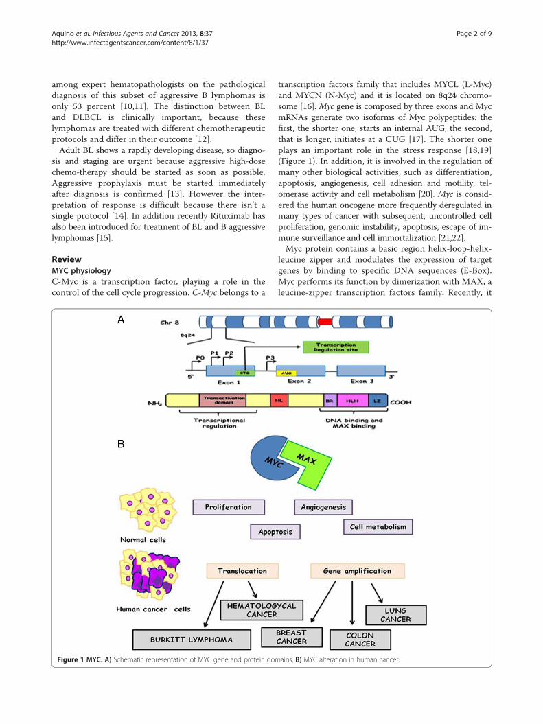
Figure 1 MYC. A) Schematic representation of MYC gene and protein dom
transcription factors family that includes MYCL (L-Myc)and MYCN (N-Myc) and it is located on 8q24 chromo-some [16]. Myc gene is composed by three exons and MycmRNAs generate two isoforms of Myc polypeptides: thefirst, the shorter one, starts an internal AUG, the second,that is longer, initiates at a CUG [17]. The shorter oneplays an important role in the stress response [18,19](Figure 1). In addition, it is involved in the regulation ofmany other biological activities, such as differentiation,apoptosis, angiogenesis, cell adhesion and motility, tel-omerase activity and cell metabolism [20]. Myc is consid-ered the human oncogene more frequently deregulated inmany types of cancer with subsequent, uncontrolled cellproliferation, genomic instability, apoptosis, escape of im-mune surveillance and cell immortalization [21,22].Myc protein contains a basic region helix-loop-helix-
leucine zipper and modulates the expression of targetgenes by binding to specific DNA sequences (E-Box).Myc performs its function by dimerization with MAX, aleucine-zipper transcription factors family. Recently, it
ains; B) MYC alteration in human cancer.
Aquino et al. Infectious Agents and Cancer 2013, 8:37 Page 3 of 9http://www.infectagentscancer.com/content/8/1/37
has been demonstrated that c-Myc expression in GCs(Germinal Center) is lower compared to naive and mem-ory cells. Probably, this low expression could protectagainst Myc-induced genomic instability in the GC [23].
Myc alteration in human cancerMyc oncogenetic deregulation could be induced bypoint mutations, gene amplification, translocation, epi-genetic reprogramming, enhanced translation andincreased protein stability [21]. The effects are C-Mycprotein overexpression, demonstrated in 80% of breastcancers, 70% of colon cancers, 90% of gynecologicalcancers, 50% of hepatocellular carcinomas, 30% of lungcancer and a variety of hematological tumors [24]. Aber-rant Myc expression has also been identified in ProstaticCancer where it has been proposed as a potential prognos-tic factor [25,26] (Figure 1B). Generally Myc gene amplifi-cation has been described as the most frequent molecularalteration in most of solid tumours [21]. In addictionSingle-nucleotide Polymorphisms (SNPs) within 8q24chromosomal region have been found to be associatedwith colorectal, breast, bladder, ovarian and prostatecancers [26,27]. Furthermore, point mutations are re-vealed in Myc N-terminal domain (residues 44-65), inparticular, the most frequently mutated residue is Thr-58.The phosphorylation of this residue has been shown tocontrol c-Myc degradation and mutations, abolishingThr-58, lead to an increased c-Myc half life in BL[28,29]. The translocation t(8;14) has been described asthe most frequent aberration involving Myc gene in BLwith the immunoglobulin heavy chain (IgH) gene aspartner. Less common aberration involves light chainimmunoglobulin genes (Igλ or Igκ) in the translocationst(2;8) and t(8;22) [30]. The activation of the Myc gene at8q24 is considered the main pathogenetic feature of BL,but the contribution of other genetic mutations to thedisease is an important developing point [30]. In additionMyc translocation is not only specifically observed in BLbut it can occur in other hematological malignancies. In-deed Myc rearrangement is observed in 5-10% of diffuselarge B-cell lymphomas and up to 50% of high-grade B-cell lymphomas other than Burkitt lymphomas [31]. Inthese tumours, Myc translocations can also involve non-IG partners [24].
Molecular pathways associated to MYC overexpression inBurkitt lymphomaMyc translocation in BL is considered as a lymphomainitiating event, in other lymphomas it may also occur asa secondary event during disease progression [3]. Welldocumented oncogenetic alterations are associated toother intracellular pathways.Recently C. Love et al. have highlighted a series of
gene mutations in BL. For example, ID3 (inhibitor of
DNA binding protein) gene mutations produce a two-fold higher gene expression in BL cells when comparedto DLBCL [32]. ID3 mutation is associated to increasedG to S phase cell cycle progression correlated to higherexpression of cell cycle pathway genes, such as E2F1,CDK7, MCM10 and with an higher expression of knownMyc target genes. This phenomenon, through ID3 mi-metics, could represent the possibility of a potential thera-peutic approach in BL [32].Using animal models, many studies have shown that
the translocation involving Myc could be mediated bycitidinedeaminase (AICDA) and that it is not activatedby the recombinase (RAG1/2). This suggests the presenceof somatic hypermutation or class switch recombination,that may be detected in normal tissue [33,34].Several papers described an overexpression of NF-kB
(Nuclear Factor kappa-light-chain-enhancer of activatedB cells) both in DLBCL and HL and its lower expressionin BL. Klapproth et al. suggest in a study realized on miceand human that in c-Myc transformated lymphoma cells,NF-kB-pathway is deregulated. Therefore they concludedthat c-MYC overexpression sensitizes cells to NF-κB–in-duced apoptosis, and the absence of NF-κB signaling isan assumption for MYC-mediated tumorigenesis [35].Another study tried to explain the possible role of
IP3K (Inositol 1,4,5-Trisphosphate 3-kinase) and Myc inprimary events of lymphomagenesis, using mousemodels of BL. The results show a significant activationof IP3K pathway, especially in cells where the signal ofNF-kB is off [36]. Recently, IP3K overexpression hasbeen found in human BL, suggesting a functional role inBL pathogenesis [35]. The involvement of NF-kB andIP3K pathways might have implication for the developmentof therapies against MYC-positive tumours. Recent studieshighlight c-Myc influence on the Retinoblastoma (Rb) path-ways. Rb gene family is composed by three (Rb, pRb2/p130and p107) cell cycle regulator protein members. Cinti et al.showed that several genetic alterations disrupt the nuclearlocalization of the retinoblastoma-related gene RB2/p130in human tumor cell lines and primary tumors [37]. Inparticular, mutation of RB2/p130 caused the upregulationof cyclins E1 and A2, involved in cell cycle progressionfrom G1 to S-phase, and the inactivation of the transcrip-tion factor E2F4 (typically increasing during the S phase)[38]. This alteration is more frequent in endemic BL andlesser in sporadic BL. However, RB2/p130 mutation hasnot been included into molecular signatures that distin-guish BL and DLBCL, suggesting that RB2/p130 deregula-tion was common in all B–non-Hodgkin lymphomas(NHLs), and not exclusive for BL pathogenesis [39].The INK4/ARF locus encodes two tumour suppressor
genes, p16 and p14 that distinctively regulate Rb and p53pathways. Human and murine studies have showed thatthe simultaneous c-Myc iperactivation and INK4/ARF

Aquino et al. Infectious Agents and Cancer 2013, 8:37 Page 4 of 9http://www.infectagentscancer.com/content/8/1/37
inactivation are an essential step during the developmentof BL, conferring a further growth advantage and apop-tosis protection to the cells [40,41]. Moreover, c-Myc, p14and p16 are degraded with proteasome-dependent mech-anism, and a less ubiquitination is demonstrated in BL.These evidences suggest that proteasome inhibitors maybe further considered in the treatment of BL [42]. Re-cent studies have highlighted that miRNAs (small noncoding RNA) may have a role in malignant transform-ation in several solid tumors, but little is known abouttheir expression and deregulation in malignant lymph-omas [43]. In particular, hsa-miR-155 was found to behighly expressed in 90% of Hodgkin's lymphomas andin diffuse large B-cell lymphomas. Moreover recentstudies have underlined its role in B-cell differentiation.Furthermore, the miR-17-92 cluster was described as atranscriptional target of c-Myc and it was over-expressedduring lymphomagenesis [44]. Other studies have assessedthat hsa-miR-127 up-regulation in EBV positive BLconfirming different pathogenetic mechanisms betweenEBV-positive and EBV-negative BL [45]. We have sche-matized in Figure 2 all the previously discussed path-ways correlated to Myc gene aberration.
Detection of MYC gene/protein alterationSeveral studies have demonstrated distinctive complexkaryotypes (CK) in BL and DLBCL. Havelange et al.have identified recurrent alterations associated with
Figure 2 Schematic representation of MYC translocation and molecul
Myc rearrangements in 84 aggressive B-cell lymph-omas by multicolor fluorescence in situ hybridization(M-FISH). They concluded that BL karyotypes wereless complex and aneuploid than other lymphomaswith Myc-rearrangement. This condition suggests thatBL with CK are indeed different from other aggressiveMYC-rearranged lymphomas, usually showing widergenetic complexity [46]. Several Comparative genomichybridization (CGH) studies highlight genomic imbal-ances in BL. Garcìa et al. found chromosomes 12q, Xq,22q, 20q, 9q gains, and chromosomes 13q and 4qlosses. Moreover they found high level of amplificationin the regions 1q23-31, 6p12-p25, 8p22-23 [47]. Anywayprevious studies have determined that BL has a simplekaryotype < or=2 additional abnormalities and it is associ-ated to better overall survival respect to other B aggressivelymphomas, this condiction is generally observed in BLs[48]. Gains or amplifications of chromosomes 1q and 7q(respectively 20% and 10% of BL) have been associatedwith worse clinic outcome and also all 13q chromosomalabnormalities have been related to an aggressive behaviour[47,49,50]. Furthermore, recently, a comparative analysisbetween whole-genome oligonucleotide array CGH ana-lysis and FISH in a Burkitt's lymphoma-derived cell lines,showed three minimal critical regions (MCR) localized onChr 1q harboring several genes such as BCA2, PIAS3 andMDM4 and AKT3. These regions appear critically in-volved in BL prognosis [51].
ar pathways involved in lymphomagenesis.

Aquino et al. Infectious Agents and Cancer 2013, 8:37 Page 5 of 9http://www.infectagentscancer.com/content/8/1/37
However, the Myc translocation remains the maincytogenetic signature of BL as shown by its routinely usein several diagnostic algorithms. This investigation isfundamental in differential diagnosis with other lymph-omas morphologically similar to BL but with atypicalimmunophenotype or genetic signatures.Hummel et al. proposed a “BL similarity index”, based
on the analysis of 58 genes, classifying aggressive NHLinto molecular BL (mBL), intermediate cases, and nonmolecular Burkitt. They analysed 220 mature aggressiveB-cell lymphomas, and identified a consistent genepanel characteristic of molecular BL. These genes alsoincluded several target genes of the nuclear factor-κBpathway (i.e., BCL2A1, FLIP, CD44, NFKBIA, BCL3, andSTAT3) that normally distinguished activated B-cell-like(ABC) or germinal-center B-cell-like lymphomas (GC)[52,53]. Through this index yet not all cases with mor-phologic or immunophenotypical features of Burkitt’slymphoma were classified as mBL. Molecular signaturewas strongly supported from the genetic analysis thatdefines three groups: i) the myc-simple group character-ized by IG-Myc fusion and a low number of chromosomalimbalances (complexity score < 6, MYC translocationcould be the primary oncogenic event) largely overlappedwith the molecular BL and associated with a favourableclinical outcome; ii) Myc-complex status (complexity
Figure 3 Above, FISH assay shows the MYC locus rearrangement witRearrangement Probe Kit). A. MYC rearrangement-positive tumor cells,B. Absence of MYC translocation, showing fusion signal patterns; Below, FIGH/MYC/CEP 8 Tri-Color DF FISH Probe Kit): C. The reciprocal t(8;14) in tugreen fusions, and two aqua centromeric signals; D. Absence of reciprocasignals pattern.
score > 6, Myc translocation could be the second onco-genic event) associated with a poor outcome, independ-ently of age and clinical stage corresponding to theintermediate group; iii) Myc negative group including nonmolecular Burkitt cases [53].On the basis of current literature, Bellan et al. sug-
gested a practical approach for BL diagnosis. To distin-guish among BL, DLBCL and the provisional categoryof “unclassifiable B-cell lymphoma”, with intermediatefeatures between DLBCL and BL (BCLU), they usedcytogenetic, molecular and immunohistochemical tech-niques, selecting a large panel of antibodies. In particular,FISH analysis was performed to detect the translocationsinvolving Myc, BCL2 and BCL6 through commerciallyavailable probes [54].Myc translocation could be easily detected through the
use of commercial probes by breaking apart or by dualfusion strategy used for FISH analyses. The first strategyallows, in a single approach, to assess the integrity of theMyc gene, but it does not provide information about thetranslocation partner. Known translocation partners ofMyc gene could be detected using the second strategy(Figure 3). Several commercial probes are available, butthey detect separately the different Myc partners. In factsome Break Apart probes, investigating the presence ofMyc rearrangements in the locus corresponding to Igλ
h a break-apart probe (Vysis LSI MYC Dual Color Break Apartshowing one yellow fusion signal, one orange and one green signals;ISH assay shows the IGH/MYC/CEP8 Tri-Color Dual Fusion probe (Vysismor cells showed a pattern of one orange, one green, two orange/l translocation showed the two aqua, two orange, and two green
Aquino et al. Infectious Agents and Cancer 2013, 8:37 Page 6 of 9http://www.infectagentscancer.com/content/8/1/37
or Igκ, are available. The only commercial dual colourdual fusion Myc probe allows the translocation t (8:14)assessment. There are no commercial probes to investi-gate the non-IG Myc partners. The Myc break apart probecombined with the previously described probes are neces-sary to detect this subset of translocations.Naresh et al. proposed in the diagnostic approach of
BL an immunohistochemistry and FISH scoring system,based on three phases. Particularly, the first phase isbased on the scoring of both the morphological featuresand a small immunohistochemistry panel detections, in-cluding BCL2 and CD10 antibodies. If the cumulativescore is ≥ 5, the diagnosis of BL can be proposed; if thescore is < 3 the diagnosis is not BL. The unresolvedcases with intermediate score should be further scoredthrough a larger panel of immunohistochemistry, suchas ki67 (score 0-1-2), CD38 (score 0-1) and CD44 (score0-1). Only if cumulative scores is ≥8, diagnosis of BL isdefinitively proposed. Finally, If this phase is uncertain,FISH analysis, including Myc\IG translocation andrearrangements of BCL2 and BCL6 should be crucial. Ifcumulative scores is ≥8: diagnosis of BL; if the score isbetween 6 and 7: BL not excluded. Through this approachis possible enable to give lead to a precise diagnosis of BLin more than 90% cases [55].Instead, Salaverria et al. propose a genetic model of
pathogenesis of high-grade B cell lymphomas “gray zone”,related to genetic aberration and age. The gray zone repre-sents hybrid zone between classical BL and classicalDLBCL, and contain “secondary Burkitt-transformedlymphomas” and many indeterminate lymphomas. Cyto-genetically Sporadic BLs in children and adults are similar.Both groups are characterized by the same low genomiccomplexity including the same genetic aberrations. A cor-rect subclassification of mature aggressive B-cell lymph-omas in adults directly influence the therapeutic strategy.This genetic model shows that real adult BLs are very rareand that the BL with more genetic alteration is extremelydifficult to find. In this contest, the distinctive feature ofBL is represented by its low genomic complexity [56].Myc translocation has not only diagnostic value but
it is also a powerful prognostic indicator in severallymphomas.Myc translocation is associated to poor prognosis in
other B aggressive lymphomas. Indeed LI S. et al. showedseveral lymphoma cases with a germinal center B-cellimmunophenotype carryingMyc and BCL2 rearrangementsand clinically aggressive behaviour, independently of theirmorphological appearance [57]. Pei Lin et al. assert that theonly Myc aberration, in unclassifiable B-cell lymphoma,identifies patient subsets, requiring more aggressive therapythan R-CHOP [58].However, Myc can be regulated by other mechanisms
inducing its increased protein expression and its
hyperactivation [59,60]. FISH is unable to detectgenetic deregulation that affects gene expression onthe transcriptional and translational levels unlike im-munohistochemical analysis [61]. For a long time theMyc evaluation by immunohistochemestry has beenhampered by a lack of anti-MYC antibodies that aresuitable to detect the increased protein expression.However, recently a new commercial, Myc antibody(clone Y69; Epitomics, Burlingame, CA) seems to beuseful in the diction of myc overexpression, independ-ently from molecular mechanism. This monoclonalantibody targets the Myc protein-N terminus. ThisMyc antibody shows a typical nuclear staining and ithas been proposed a significant diagnostic cut-off inBL for the immunoscoring when higher than 40%. Severalrecent studies proposed to introduce this antibody in anovel diagnostic alghorithm.Green et al. analyse a group of DLBCL both trough
immunohistochemistry for Myc, BCL2, CD10, BCL6,and MUM1/interferon regulatory factor 4, and FISH forMyc and BCL2. They concluded that FISH analysisidentified Double-Hit Lymphoma (DHL) in 6% ofpatients, while immunohistochemical MYC and BCL2analyses identified a double-hit group that comprised29% of patients. These cases were significantly associatedwith shorter OS (P < .001), and shorter progression-freesurvival (PFS; P < .001), concluding that the only MYCand Bcl2 immunohistochemestry defined a large subsetof DLBCLs strongly associated with poor outcome inpatients treated with R-CHOP [62].Finally, Horn et al. proposed to introduce a novel diag-
nostic approach using Myc antibody. They suggest a com-bined immunohistochemical and FISH score to predictoutcome in DLBCL patients (score 0, when BCL2 <70%and MYC <40%, score 1 MYC and Bcl2 expression near tocut off and score 3, when Bcl2 and MYC expression ismore than cut-off) [63].The routinely diagnostic application of Myc antibody
is not yet applied. Recent studies show Myc antibodyuse in DLBCL where the percentage of Myc translocationis very low [64]. In the future Myc antibody could be usedfor the other B aggressive lymphomas, BL and BCLU, todetect Myc altered expression independently from themutations.
ConclusionDiagnostic algorithmsDetection of the Myc translocation is currently performedby conventional cytogenetics, Southern blot, and polymer-ase chain reaction–based methods. Nevertheless, all thesemethods can fail to detect IG-Myc fusions [65]. The mostreliable method is cytogenetic analysis by fluorescence insitu hybridization (FISH). Nowadays the molecular genet-ics is fully integrated into the routine diagnostic of

Figure 4 The cytogenetics diagnostic algorithm. MYC rearrangement is recommended on lymphoma cases with increased Ki67 expression(> 90%). Molecular Burkitt Lymphoma (BL) is defined only by the presence of the MYC-IG rearrangement, an early and frequent event; DiffuseLarge B-cell Lymphoma (DLBCL) is cytogenetically set by the absence of MYC aberration; Intermediate BL/DLBCL are defined by unstablephenotype, generally MYC rearrangement is a second event and it doesn’t involve IG locus.
Aquino et al. Infectious Agents and Cancer 2013, 8:37 Page 7 of 9http://www.infectagentscancer.com/content/8/1/37
lymphomas. The Gold standard method is the CGH Arraybut this analysis has not been introduced into routinediagnostic laboratories because it is labour-intensiveand it has a high rate of failure [29]. Thus in the fu-ture, the development of a FISH assay for simultan-eous detection of all known Burkitt abnormalities willbe necessary.We summarized a cytogenetic diagnostic “flowchart”
for a better and safer histopathologic diagnosis ofBurkitt (Figure 4). We recommend a first FISH ap-proach using Myc Break Apart probe on lymphomacases with increased (>90%) Ki67, to identify all positivesamples for Myc translocations. BCL2 and BCL6 trans-locations using Break Apart probes will be performedon all negative samples. Finally on the positive speci-mens should evaluate also the presence of IG-Myctranslocation through the use of a Dual colour dualfusion Myc-IGH probe and IGK and IGL Break Apartprobes. Then, BCL2 and BCL6 status should be investi-gated. In our opinion, this is the best approach to avoidmisdiagnosis of molecular BL but it is useful only if it isintegrated with morphologic and immunophenotypicevaluation. The diagnosis of BL and other aggressiveB cell Lymphomas, with or without Myc breakpoints,
represents an important start-point for future clinicaltrials to establish different therapeutical strategies forthese lymphomas. Although the FISH-based algorithmicapproach results an important tool for BL diagnosis, it isnot easily accessible in most of the pathology laboratoriesbecause it is an expensive method and so it remains aspeculative analysis.
Competing interestsThe authors declare that they have no competing interests.
Authors’ contributionsGA and LM drafted the manuscript. All authors read and approved the finalmanuscript.
Author details1Pathology Unit, "Istituto Nazionale Tumori Fondazione G. Pascale" - Irccs,Naples, Italy. 2Medicine and Surgery Department, Foggia University, Foggia,Italy. 3Haematology Unit, "Istituto Nazionale Tumori Fondazione G. Pascale" -Irccs, Naples, Italy.
Received: 21 June 2013 Accepted: 17 September 2013Published: 30 September 2013
References1. Lenz G, Staudt LM: Aggressive lymphomas. N Engl J Med 2010,
362:1417–1429.2. Willis TG, Dyer MJ: The role of immunoglobulin translocations in the
pathogenesis of B-cell malignancies. Blood 2000, 96:808–822.

Aquino et al. Infectious Agents and Cancer 2013, 8:37 Page 8 of 9http://www.infectagentscancer.com/content/8/1/37
3. Seifert M, Scholtysik R, Küppers R: Origin and pathogenesis of B celllymphomas. Methods Mol Biol 2013, 971:1–25.
4. Leoncini L, Raphaël M, Stein H, Harris NL, Jaffe ES, Kluin PM: Burkittlymphoma. In WHO classification of tumours of haematopoietic andlymphoid tissues, Volume 2. 4th edition. Edited by IARC Press.2008:262–264.
5. Dalla Favera R, Bregni M, Erikson J, Patterson D, Gallo RC, Croce CM: HumanC-myc one gene is located on the region of chromosome 8 that istranslocated in Burkitt lymphoma cell. Proc Natl Acad Sci USA 1982,79:7824–7827.
6. Taub R, Kirsch I, Morton C, Lenoir G, Swan D, Tronick S, Aaronson S, Leder P:Translocation of the c-myc gene into the immunoglobulin heavy chainlocus in human Burkitt lymphoma and murine plasmacytoma cells.Proc Natl Acad Sci USA 1982, 79:7837–7841.
7. Ambrosio MR, Piccaluga PP, Ponzoni M, Rocca BJ, Malagnino V, Onorati M,De Falco G, Calbi V, Ogwang M, Naresh KN, Pileri SA, Doglioni C, Leoncini L,Lazzi S: The alteration of lipid metabolism in Burkitt lymphoma identifiesa novel marker: adipophilin. PLoS One 2012, 7:e44315.
8. Newton R, Ziegler J, Beral V, Mbidde E, Carpenter L, Wabinga H, MbulaiteyeS, Appleby P, Reeves G, Jaffe H: Uganda Kaposi’s Sarcoma study group: acase–control study of human immunodeficiency virus infection andcancer in adults and children residing in Kampala, Uganda. Int J Cancer2001, 92:622–627.
9. Perkins AS, Friedberg JW: Burkitt lymphoma in adults. Hematology Am SocHematol Educ Program 2008, 341–348. doi:10.1182/asheducation-2008.1.341.
10. Magrath I, Jaffe ES, Bhatia K: Burkitt’s lymphoma. In Neoplastichematopathology. Edited by Knowles DM. Philadelphia: Lippincott Williams& Wilkins; 2001:953–986.
11. Armitage JO, Armitage JO, the Non-Hodgkin's Lymphoma ClassificationProject: A clinical evaluation of the International Lymphoma study groupclassification of non-Hodgkin’s lymphoma: the non-Hodgkin’sLymphoma classification project. Blood 1997, 89:3909–3918.
12. Pfreundschuh M, Trümper L, Kloess M, Schmits R, Feller AC, Rübe C,Rudolph C, Reiser M, Hossfeld DK, Eimermacher H, Hasenclever D, SchmitzN, Loeffler M: Two-weekly or 3-weekly CHOP chemotherapy with orwithout etoposide for the treatment of elderly patients with aggressivelymphomas: results of the NHL-B2 trial of the DSHNHL. Blood 2004,104:634–641.
13. Molyneux EM, Rochford R, Griffin B, Newton R, Jackson G, Menon G,Harrison CJ, Israels T, Bailey S: Burkitt’s lymphoma. Lancet 2012,379:1234–1244.
14. Magrath IT, Haddy TB, Adde MA: Adults and children with small non-cleaved-cell lymphoma have a similar excellent outcome when treatedwith the same chemotherapy regimen. J Clin Onc 1996, 14:925–934.
15. Thomas DA, Faderl S, O’Brien S, Bueso-Ramos C, Cortes J, Garcia-Manero G,Giles FJ, Verstovsek S, Wierda WG, Pierce SA, Shan J, Brandt M, HagemeisterFB, Keating MJ, Cabanillas F, Kantarjian H: Chemoimmunotherapy withhyper-CVAD plus rituximab for the treatment of adult Burkitt andBurkitt-type lymphoma or acute lymphoblastic leukemia. Cancer 2006,106:1569–1580.
16. Depinho RA, Hatton K, Ferrier P, Zimmerman K, Legouy E, Tesfaye A, CollumR, Yancopoulos G, Nisen P, Alt F: Myc family genes: a dispersed multi-gene family. Ann Clin Res 1986, 18:284–289.
17. Blackwood EM, Eisenman RN: Max: a helix-loop-helix zipper protein thatforms a sequence-specific DNA-binding complex with Myc. Science 1991,251:1211–1217.
18. Spotts GD, Patel SV, Xiao Q, Hann SR: Identification of downstream-initiated c-Myc proteins which are dominant-negative inhibitors oftransactivation by full-length c-Myc proteins. Mol Cell Biol 1997,17:1459–1468.
19. Xiao Q, Claassen G, Shi J, Adachi S, Sedivy J, Hann SR: Transactivation-defective c-MycS retains the ability to regulate proliferation andapoptosis. Genes Dev 1998, 12:3803–3808.
20. Henriksson M, Lüscher B: Proteins of the Myc network: essential regulatorsof cell growth and differentiation. Adv Cancer Res 1996, 68:109–182.
21. Vita M, Henriksson M: The Myc oncoprotein as a therapeutic target forhuman cancer. Semin Cancer Biol 2006, 16:318–330.
22. Dang CV: MYC on the path to cancer. Cell 2012, 149:22–35.23. Klein U, Tu Y, Stolovitzky GA, Keller JL, Haddad J Jr, Miljkovic V, Cattoretti G,
Califano A, Dalla-Favera R: Transcriptional analysis of the B cell germinalcenter reaction. Proc Nti Acad ScI U S A 2003, 100:2639–2644.
24. Gardner LB, Lee LA, Dang CV: C-myc protooncogene. In Encyclopedia ofcancer. Edited by Bertino JR. Amsterdam: Elsevier Science; 2002:555–561.
25. Gurel B, Iwata T, Koh CM, Yegnasubramanian S, Nelson WG, De Marzo AM:Molecular alterations in prostate cancer as diagnostic, prognostic, andtherapeutic agents? Adv Anat Pathol 2008, 15:319–331.
26. Quinn DI, Henshall SM, Sutherland RL: Molecular markers of prostatecancer outcome. Eur J Cancer 2005, 41:858–888.
27. Brisbin AG, Asmann YW, Song H, Tsai YY, Aakre JA, Yang P, Jenkins RB,Pharoah P, Schumacher F, Conti DV, Duggan DJ, Jenkins M, Hopper J,Gallinger S, Newcomb P, Casey G, Sellers TA, Fridley BL: Meta-analysis of8q24 for seven cancers reveals a locus between NOV and ENPP2associated with cancer development. BMC Med Genet 2011, 5(12):156.
28. Bahram F, von der Lehr N, Cetinkaya C, Larsson LG: c-Myc hot spotmutations in lymphomas result in inefficient ubiquitination anddecreased proteasome-mediated turnover. Blood 2000, 95:2104–2110.
29. Coller HA, Grandori C, Tamayo P, Colbert T, Lander ES, Eisenman RN, GolubTR: Expression analysis with oligonucleotide microarrays reveals thatMYC regulates genes involved in growth, cell cycle, signaling, andadhesion. Proc Natl Acad Sci USA 2000, 97:3260–3265.
30. Brady G, MacArthur GJ, Farrell PJ: Epstein-Barr virus and Burkittlymphoma. J Clin Pathol 2007, 60:1397–1402.
31. Philip Kluin et Ed Schuuring: Molecular cytogeneitis of Lymphoma: wheredo we stand in 2010? Histopathology 2011, 58:128–144.
32. Love C, Sun Z, Jima D, Li G, Zhang J, Miles R, Richards KL, Dunphy CH, ChoiWW, Srivastava G, Lugar PL, Rizzieri DA, Lagoo AS, Bernal-Mizrachi L, MannKP, Flowers CR, Naresh KN, Evens AM, Chadburn A, Gordon LI, Czader MB,Gill JI, His ED, Greenough A, Moffitt AB, McKinney M, Banerjee A, Grubor V,Levy S, Dunson DB, et al: The genetic landscape of mutations in Burkittlymphoma. Nat Genet 2012, 44:1321–1325.
33. Guikema JE, Schuuring E, Kluin PM: Structure and consequences of IGHswitch breakpoints in Burkitt lymphoma. J Natl Cancer Inst Monogr 2008,39:32–36.
34. Janz S: Genetic and environmental cofactors of Myc translocations inplasma cell tumor development in mice. J Natl Cancer Inst Monogr 2008,39:37–40.
35. Klapproth K, Sander S, Marinkovic D, Baumann B, Wirth T: The IKK2/NF-{kappa}B pathway suppresses MYC-induced lymphomagenesis. Blood2009, 114:2448–2458.
36. Sander S, Calado DP, Srinivasan L, Köchert K, Zhang B, Rosolowski M, RodigSJ, Holzmann K, Stilgenbauer S, Siebert R, Bullinger L, Rajewsky K: SynergybetweenPI3K signaling and MYC in Burkitt lymphomagenesis. Cancer Cell2012, 22:167–179.
37. Cinti C, Leoncini L, Nyongo A, Ferrari F, Lazzi S, Bellan C, Vatti R, ZamparelliA, Cevenini G, Tosi GM, Claudio PP, Maraldi NM, Tosi P, Giordano A: Geneticalterations of the retinoblastoma-related gene RB2/p130 identifydifferent pathogenetic mechanisms in and among Burkitt’s lymphomasubtypes. Am J Pathol 2000, 156:751–760.
38. Cinti C, Claudio PP, Howard CM, Neri LM, Fu Y, Leoncini L, Tosi GM, MaraldiNM, Giordano A: Genetic alterations disrupting the nuclear localization ofthe retinoblastoma-related gene RB2/p130 in human tumor cell linesand primary tumors. Cancer Res 2000, 60:383–389.
39. Leoncini L, Bellan C, Cossu A, Claudio PP, Lazzi S, Cinti C, Cevenini G, MeghaT, Laurini L, Luzi P, Orcioni GF, Piccioli M, Pileri S, Giardino C, Tosi P,Giordano A: Retinoblastoma-related p107 and pRb2/p130 proteins inmalignant lymphomas: distinct mechanisms of cell growth control. ClinCancer Res 1999, 5:4065–4072.
40. Eischen CM, Weber JD, Roussel MF, Sherr CJ, Cleveland JL: Disruption ofthe ARF-Mdm2-p53 tumor suppressor pathway in Myc-inducedlymphomagenesis. Genes Dev 1999, 13:2658–2669.
41. Schmitt CA, McCurrach ME, De Stanchina E, Wallace-Brodeur RR, Lowe SW:INK4a/ARF mutations accelerate lymphomagenesis and promotechemoresistance by disabling p53. Genes Dev 1999, 13:2670–2677.
42. Roberti A, Rizzolio F, Lucchetti C, De Leval L, Giordano: Ubiquitin-mediatedprotein degradation and methylation-induced gene silencing cooperatein the inactivation of the INK4/ARF locus in Burkitt lymphoma cell lines.Cell Cycle 2011, 10:127–134.
43. Volinia S, Calin GA, Liu CG, Ambs S, Cimmino A, Petrocca F, Visone R, IorioM, Roldo C, Ferracin M, Prueitt RL, Yanaihara N, Lanza G, Scarpa A,Vecchione A, Negrini M, Harris CC, Croce CM: A microRNA expressionsignature of human solid tumors defines cancer gene targets. Proc NatlAcad Sci U S A 2006, 103:2257–2261.

Aquino et al. Infectious Agents and Cancer 2013, 8:37 Page 9 of 9http://www.infectagentscancer.com/content/8/1/37
44. Lawrie CH: MicroRNA expression in lymphoma. Expert Opin Biol Ther 2007,7:1363–1374.
45. De Falco G, Antonicelli G, Onnis A, Lazzi S, Bellan C, Leoncini L: Role of EBVin microRNA dysregulation in Burkitt lymphoma. Semin Cancer Biol 2009,19:401–406.
46. Havelange V, Ameye G, Théate I, Callet-Bauchu E, Mugneret F, Michaux L,Dastugue N, Penther D, Barin C, Collonge-Rame MA, Baranger L, Terré C,Nadal N, Lippert E, Laï JL, Cabrol C, Tigaud I, Herens C, Hagemeijer A,Raphael M, Libouton JM, Poirel HA: Patterns of genomic aberrationssuggest that Burkitt lymphomas with complex karyotype are distinctfrom other aggressive B-cell lymphomas with MYC rearrangement. GenesChromosomes Cancer 2013, 52:81–92.
47. García JL, Hernandez JM, Gutiérrez NC, Flores T, González D, Calasanz MJ,Martínez-Climent JA, Piris MA, Lopéz-Capitán C, González MB, Odero MD,San Miguel JF: Abnormalities on 1q and 7q are associated with pooroutcome in sporadic Burkitt’s lymphoma: a cytogenetic and comparativegenomic hybridization study. Leukemia 2003, 17:2016–2024.
48. Seegmiller AC, Garcia R, Huang R, Maleki A, Karandikar NJ, Chen W: Simplekaryotype and bcl-6 expression predict a diagnosis of Burkitt lymphomaand better survival in IG-MYC rearranged high-grade B-cell lymphomas.Mod Pathol 2010, 23:909–920.
49. Boerma EG, Siebert R, Kluin PM, Baudis M: Translocations involving 8q24 inBurkitt lymphoma and other malignant lymphomas: a historical reviewof cytogenetics in the light of todays knowledge. Leukemia 2009,23:225–234.
50. Poirel HA, Cairo MS, Heerema NA, Swansbury J, Aupérin A, Launay E, SangerWG, Talley P, Perkins SL, Raphaël M, McCarthy K, Sposto R, Gerrard M,Bernheim A, Patte C, FAB/LMB 96 International study committee: Specificcytogenetic abnormalities are associated with a significantly inferioroutcome in children and adolescents with mature B-cell non-Hodgkin’slymphoma: results of the FAB/LMB 96 international study. Leukemia 2009,23:323–331.
51. Toujani S, Dessen P, Ithzar N, Danglot G, Richon C, Vassetzky Y, Robert T,Lazar V, Bosq J, Da Costa L, Pérot C, Ribrag V, Patte C, Wiels J, Bernheim A:High resolution genome-wide analysis of chromosomal alterations inBurkitt’s lymphoma. PLoS One 2009, 4:e7089.
52. Wring G, Tan B, Rosenwald A, Hurt EH, Wiestner A, Staudt LM: A geneexpression- based method to diagnose clinically distinct subgroups ofdiffuse large B cell lymphoma. Proc Natl Acad Sci USA 2003,100:9991–9996.
53. Hummel M, Bentink S, Berger H, Klapper W, Wessendorf S, Barth TF, BerndHW, Cogliatti SB, Dierlamm J, Feller AC, Hansmann ML, Haralambieva E,Harder L, Hasenclever D, Kühn M, Lenze D, Lichter P, Martin-Subero JI,Möller P, Müller-Hermelink HK, Ott G, Parwaresch RM, Pott C, Rosenwald A,Rosolowski M, Schwaenen C, Stürzenhofecker B, Szczepanowski M,Trautmann H, Wacker HH, Molecular Mechanisms in Malignant LymphomasNetwork Project of the Deutsche Krebshilfe, et al: A biologic definition ofBurkitt's lymphoma from transcriptional and genomic profiling. N Engl JMed 2006, 354:2419–2430.
54. Bellan C, Stefano L, De Giulia F, Rogena EA, Lorenzo L: Burkitt lymphomaversus diffuse large B-cell lymphoma: a practical approach. HematolOncol 2010, 28:53–56.
55. Naresh KN, Ibrahim HA, Lazzi S, Rince P, Onorati M, Ambrosio MR, Bilhou-Nabera C, Amen F, Reid A, Mawanda M, Calbi V, Ogwang M, Rogena E,Byakika B, Sayed S, Moshi E, Mwakigonja A, Raphael M, Magrath I, LeonciniL: Diagnosis of Burkitt lymphoma using an algorithmic approach-applicable in both resource-poor and resource-rich countries. Br JHaematol 2011, 154:770–776.
56. Salaverria I, Siebert R: The gray zone between Burkitt’s lymphoma anddiffuse large B-cell lymphoma from a genetics perspective. J Clin Oncol2011, 29:1835–1843.
57. Li S, Lin P, Fayad LE, Lennon PA, Miranda RN, Yin CC, Lin E, Medeiros LJ: B-cell lymphomas with MYC/8q24 rearrangements and IGH@BCL2/t(14;18)(q32;q21): an aggressive disease with heterogeneous histology, germinalcenter B-cell immunophenotype and poor outcome. Mod Pathol 2012,25:145–156.
58. Lin P, Dickason TJ, Fayad LE, Lennon PA, Hu P, Garcia M, Routbort MJ,Miranda R, Wang X, Qiao W, Medeiros LJ: Prognostic value of MYCrearrangement in cases of B-cell lymphoma, unclassifiable, with featuresintermediate between diffuse large B-cell lymphoma and Burkittlymphoma. Cancer 2012, 118:1566–1573.
59. Lenz G, Wright GW, Emre NC, Kohlhammer H, Dave SS, Davis RE, Carty S,Lam LT, Shaffer AL, Xiao W, Powell J, Rosenwald A, Ott G, Muller-HermelinkHK, Gascoyne RD, Connors JM, Campo E, Jaffe ES, Delabie J, Smeland EB,Rimsza LM, Fisher RI, Weisenburger DD, Chan WC, Staudt LM: Molecularsubtypes of diffuse large B-cell lymphoma arise by distinct geneticpathways. Proc Natl Acad Sci USA 2008, 105:13520–13525.
60. Bea S, Zettl A, Wright G, Salaverria I, Jehn P, Moreno V, Burek C, Ott G, PuigX, Yang L, Lopez-Guillermo A, Chan WC, Greiner TC, Weisenburger DD,Armitage JO, Gascoyne RD, Connors JM, Grogan TM, Braziel R, Fisher RI,Smeland EB, Kvaloy S, Holte H, Delabie J, Simon R, Powell J, Wilson WH,Jaffe ES, Montserrat E, Muller-Hermelink HK: Diffuse large B-cell lymphomasubgroups have distinct genetic profiles that influence tumor biologyand improve gene-expression-based survival prediction. Blood 2005,106:3183–3190.
61. Ventura RA, Martin-Subero JI, Jones M, McParland J, Gesk S, Mason DY,Siebert R: FISH analysis for the detection of lymphoma-associatedchromosomal abnormalities in routine paraffin-embedded tissue. J MolDiagn 2006, 8:141–151.
62. Green TM, Young KH, Visco C, Xu-Monette ZY, Orazi A, Go RS, Nielsen O,Gadeberg OV, Mourits-Andersen T, Frederiksen M, Pedersen LM, Møller MB:Immunohistochemical double-hit score is a strong predictor of outcomein patientswith diffuse large B-cell lymphoma treated with rituximabplus cyclophosphamide, doxorubicin, vincristine, and prednisone. J ClinOncol 2012, 30:3460–3467.
63. Horn H, Ziepert M, Becher C, Barth TF, Bernd HW, Feller AC, Klapper W,Hummel M, Stein H, Hansmann ML, Schmelter C, Möller P, Cogliatti S,Pfreundschuh M, Schmitz N, Trümper L, Siebert R, Loeffler M, Rosenwald A,Ott G, German High-Grade Non-Hodgkin Lymphoma Study Group: MYCstatus in concert with BCL2 and BCL6 expression predicts outcome indiffuse large B-cell lymphoma. Blood 2013, 121:2253–2263.
64. Johnson NA, Slack GW, Savage KJ, Connors JM, Ben-Neriah S, Rogic S, ScottDW, Tan KL, Steidl C, Sehn LH, Chan WC, Iqbal J, Meyer PN, Lenz G, WrightG, Rimsza LM, Valentino C, Brunhoeber P, Grogan TM, Braziel RM, Cook JR,Tubbs RR, Weisenburger DD, Campo E, Rosenwald A, Ott G, Delabie J,Holcroft C, Jaffe ES, Staudt LM: Concurrent expression of MYC and BCL2 indiffuse large B-cell lymphoma treated with rituximab pluscyclophosphamide, doxorubicin, vincristine, and prednisone. J Clin Oncol2012, 30:3452–3459.
65. Guikema JE, De Boer C, Haralambieva E, Smit LA, Van Noesel CJ, SchuuringE, Kluin PM: IGH switch breakpoints in Burkitt lymphoma: exclusiveinvolvement of noncanonical class switch recombination. GenesChromosomes Cancer 2006, 45:808–819.
doi:10.1186/1750-9378-8-37Cite this article as: Aquino et al.: MYC chromosomal aberration indifferential diagnosis between Burkitt and other aggressive lymphomas.Infectious Agents and Cancer 2013 8:37.
Submit your next manuscript to BioMed Centraland take full advantage of:
• Convenient online submission
• Thorough peer review
• No space constraints or color figure charges
• Immediate publication on acceptance
• Inclusion in PubMed, CAS, Scopus and Google Scholar
• Research which is freely available for redistribution
Submit your manuscript at www.biomedcentral.com/submit


