PROTOTYPE DEVELOPMENT OF NORMAL ASIAN HUMAN SKULL NURHANIS ...umpir.ump.edu.my/8500/1/cd8191.pdf ·...
24
PROTOTYPE DEVELOPMENT OF NORMAL ASIAN HUMAN SKULL NURHANIS SOFIAH BT ABD GHAFAR Report submitted in fulfillment of the requirements for the awards of the degree of Bachelor of Mechanical Engineering Faculty of Mechanical Engineering UNIVERSITI MALAYSIA PAHANG JUNE 2013
Transcript of PROTOTYPE DEVELOPMENT OF NORMAL ASIAN HUMAN SKULL NURHANIS ...umpir.ump.edu.my/8500/1/cd8191.pdf ·...

PROTOTYPE DEVELOPMENT OF NORMAL
ASIAN HUMAN SKULL
NURHANIS SOFIAH BT ABD GHAFAR
Report submitted in fulfillment of the requirements
for the awards of the degree of
Bachelor of Mechanical Engineering
Faculty of Mechanical Engineering
UNIVERSITI MALAYSIA PAHANG
JUNE 2013

vii
ABSTRACT
Skull which is cover with scalp is skeletal structure of human’s head that protect and
form a cavity for the brain, where the nervous system is. Nowadays, prototype of
human’s skull model needed in wide use such as to educate and train physicians, inform
patient, support R&D and for surgical planning The purpose of this study is to build a
three dimensional of normal Asian human’s skull by using any applicable machine. The
process of replicating human’s skull start with two dimensional Computed
Tomography(CT) data that need to be translate into three dimensional rapid prototyping
data by using MIMICS software. The three dimensional(3D) skull model need to be
observed and measure as the scope of this project focused to specific normal Asian
human. The observation of its morphological features of the skull and linear
measurement had found that the 3D skull model belong to normal Asian human. Then,
the 3D file images will be exported into related software for fabrication of prototype
process. The prototype development had be done by using modeler machine that use
subtractive concept The skull prototype that made up from chemical wood, or its
specific name, miraboard 700 undergoes drop ball experiment for capability
determination of the skull prototype. For validation, the drop ball test experiment result
was compare to result of analytical method. The research comes out with almost the
same value percentage different between the experimental and analytical method.
viii
ABSTRAK
Tengkorak yang diliputi kulit kepala adalah merupakan rangka kepala manusia yang
berfungsi untuk melindungi dan menghasilkan kaviti untuk otak dimana terdapatnya
sistem saraf. Kini, prototaip tengkorak adalah diperlukan secara meluas bagi tujuan
pendidikan, latihan ahli fizik, pemberitahuan kepada pesakit, menyokong kajian dan
pembangunan dan rancangan untuk pembedahan.Tujuan kajian ini adalah untuk
membina sebuah prototaip tengkorak untuk orang Asia yang normal. Proses membina
prototaip ini bermula dengan imej dua dimensi daripada imbasan komputasi
tomografi.yang akan ditukar kepada imej tiga dimensi menggunakan program khusus
yang dikenali sebagai MIMICS. Imej tengkorak tersebut mesti menjalani proses
pemerhatian dan pengukuran bagi memastikan ianya mencapai objektif projek, iaitu
menghasilkan tengkorak untuk orang Asia normal.Seterusnya, imej tersebut akan
dieksport didalam format STL kepada program yang berkaitan untuk proses pembinaan.
Proses pembinaan tengkorak ini telah disiapkan menggunakan mesin modeler, yang
menggunakan proses penolakan. Prototaip tengkorak ini yang diperbuat dripada kayu
kimia, atau nama spesifiknya adalah miraboard 700 akan menjalani eksperimen impak
kepala bagi mengenalpasti kemampuannya berbanding prototaip tengkorak yang lain.
Untuk proses pengesahan, keputusan eksperimen telah dibandingkan dengan keputusan
yang menggunakan kaedah analisis. Kajian ini telah mendapati peratusan perbezaan
yang hampir sama berbanding keputusan eksperimen dan kaedah analisis.
ix
TABLE OF CONTENT
Page
EXAMINER APPROVAL DOCUMENT i
TITLE PAGE ii
SUPERVISOR’S DECLARATION iii
STUDENT’S DECLARATION iv
DEDICATION v
ACKNOWLEDGEMENTS vi
ABSTRACT vii
ABSTRAK viii
TABLE OF CONTENT ix
LIST OF TABLES xiii
LIST OF FIGURES xiv
LIST OF SYMBOLS xvii
LIST OF ABBREVIATIONS xviii
CHAPTER 1 INTRODUCTION
1.1 Background of The Study 1
1.2 Problem Statement 2
1.3 Objective of Study 3
1.4 Scope of Study 3
1.5 Report Arrangement 4
CHAPTER 2 LITERATURE REVIEW
2.1 Introduction 5
2.2 Anatomy of Human Head 5
2.2.1 The Skull 5
2.2.2 The Brain 7
2.3 Assessment of Race from Skull 8
2.3.1 Determination of Asian Race Skull 9
x
2.4 Rapid Prototyping Process 13
2.5 Source of Rapid Prototyping Data for Human Anatomy-
CT Scan/MRI 15
2.6 Accuracy of Rapid Prototyping Skull Model 17
2.7 Landmarks of Skull for Linear Measurement Guidelines 20
2.8 Previous Research of Prototype Skull/Head Development 23
2.8.1 On the use of a patient-specific rapid-prototyped
model to simulate the response of the human head
to impact and comparison with analytical and finite
element models, E.A.C. Johnson and P.G.Young
(2005) 23
2.8.2 Material Modeling and Analysis for the
Development of a Realistic Blast Headform,
S. G. M. Hossain (2010) 28
2.9 Summary 32
CHAPTER 3 METHODOLOGY
3.1 Introduction 33
3.2 Flow Chart 34
3.3 Design of Project Study 35
3.4 Raw Data from CT scan Images 36
3.5 Extract Region of Interest Process 37
3.6 Fabrication: Prototype Development Process 38
3.7 Experiment: Drop Ball Test 42
3.7.2 Equipment used for expriment 42
3.8 Conclusion 45
CHAPTER 4 RESULTS AND DISCUSSIONS
4.1 Introduction 46
4.2 Extraction Region of Interest and Calculation of 3D Images 46
4.2.1 Two Dimensional Images Information 46
xi
4.3 Assessment of Race from Skull Morphological Features 51
4.3.1 Assessment of Race from Front View
Morphological Features 53
4.3.2 Assessment of Race from Side View
Morphological Features. 54
4.3.3 Assessment of Race from Bottom View
Morphological Features 55
4.4 Comparison of Linear Dimension between Studied Skull and
Standard 56
4.5 The Fabrication of Prototype of Normal Asian Human
Skull 60
4.6 Capability of Studied Skull Prototype Compare to
Previous Study 65
4.7 Conclusion 67
CHAPTER 5 CONCLUSION
5.1 Introduction 74
5.2 Conclusion 74
5.3 Future Recommendations 75
REFERENCES 76
APPENDICES
A1 Raw Data CT scans Images 78
A2 Raw Data CT scans Images 79
A3 Raw Data CT scans Images 80
A4 Raw Data CT scans Images 81
A5 Raw Data CT scans Images 82
A6 Raw Data CT scans Images 83
B1 Gantt chart for Final Year Project 1 84
B2 Gantt chart for Final Year Project 2 85

xii
LIST OF TABLES
Table No. Title Page
4.1 Assessment of Race from Front View Morphological
Features 53
4.2 Assessment of Race from Side View Morphological Features 54
4.3 Assessment of Race from Bottom View
Morphological Features 55
4.4 Linear Measurement Comparison between Studied Skulls
(Present study) and Normal Size Malaysian Skull
(Previous Study) 58
4.5 Result of Vibration at Different Height: ABS prototype skull
model 67
4.6 Result of Vibration at Different Height: Miraboard 700
prototype Skull model 67
4.7 Material Properties of Skull Prototype and Ball 71

xiii
LIST OF FIGURES
Figure No. Title Page
2.1 Posterior view of the skull 6
2.2 View of skull from above 6
2.3 View of skull from lateral position 7
2.4 Various part of human brain 8
2.5 Morphological feature of Caucasoid 9
2.6 Morphological feature of Mongoloid 10
2.7 Morphological feature of Negroid 11
2.8 Different characteristic of different race 12
2.9 A figure to demonstrate rapid prototyping process 13
2.10 Contour detection with grey prediction 16
2.11 Human skull layer-CT data 17
2.12 Skulls and Rapid Prototyping replicas 18
2.13 3D plot of the measured posterior curvatures 19
2.14 Landmarks and reference lines of Skull 20
2.15 Illustration of the size and shape differences between
a fossil and a recent skull 22
2.16 Rendered image from MRI data 24
2.17 Images of the completed RP model 24
2.18 Relationship between RP model and FE model 26
2.19 Illustrative representation of the analytical shell model 26
2.20 Silicone gel sample prepared on aluminium cylinders 28
2.21 Development of Brain 29
2.22 Development of initial skull 30
2.23 Development of final version skull 31
2.24 PDMS skin 32

xiv
3.1 Flow Chart of Project 34
3.2 2D scan images 36
3.3 All images are loaded and displayed in three
views in MIMICS 37
3.4 Rapid Prototyping Machine- 3D Printer 40
3.5 Modeller Machine 41
3.6 Experimental Setup for Drop Ball Test 42
3.7 Sensor cable 43
3.8 Accelerometer 43
3.9 DAQ 44
3.10 Preparing for Skull 45
4.1 A parts of MRI images in DICOM format 47
4.2 3D images calculated from MRI raw data. MRI images are
not suitable for extraction process of hard tissues like brain 48
4.3 CT scan raw data of 65 years old female patient 49
4.4 Determination of minimum and maximum value for optimum
segmentation 49
4.5 Calculated 3D image in MIMICS software 50
4.6 3D skull models 51
4.7 Skull landmarks for linear measurement 59
4.8 Skull Part in SolidWork drawing 60
4.9 Isometric view 61
4.10 Cutting flow during fabricating of upper part prototype of skull 62
4.11 Cutting flow during fabricating of lower part prototype of skull 63
4.12 The defect that caused by created cutting data error 64
4.13 Complete prototype of skull 65
4.14 Raw data from DASYLab signal process 66
4.15 Vibration versus time at 0.5m height 67
4.16 Height of ball from skull versus vibration

xv
(previous study of skull) 68
4.17 Height of ball from skull versus vibration
(present study of skull) 68
4.18 Comparison between previous study of skull and
present study of skull 69

xvi
LIST OF SYMBOLS
g Gravitational acceleration
s seconds
E Young Modulus
v Poisson ratio
h Thickness
R radius
m mass
xvii
LIST OF ABBREVIATIONS
CAD Computer Aided Design
RP Rapid Prototyping
2D Two dimensional
3D Three dimensional
FEA Finite Element Analysis
CT Computed Tomography
MRI Magnetic Resonance Image
SLA Stereolitograph
SLS Selective laser sintering
MIMICS Materialise's Interactive Medical Image Control System
DICOM Digital Imaging and Communication in Medicine
DAQ Data Acquisition
FYP1 Final Year Project 1
FYP 2 Final Year Project 2
CHAPTER 1
INTRODUCTION
1.1 PROJECT BACKGROUND
Human head is a very complex structure consisting of numerous objects with
various mechanical properties. Three main components of human’s head are skull, scalp
and brain which have their own role for human’s system. Skull which is cover with
scalp is skeletal structure of human’s head that protect and form a cavity for the brain,
where the nervous system is. Nowadays, prototype of human’s skull model needed in
wide use such as to educate and train physicians, inform patient, support R&D and for
surgical planning.
Leonardo da Vinci was the first to model brain structure by injecting molten wax
into the ventricle of an oxen brain. Since then, neuron anatomical models have shown
utility in neuroscience and medicine in areas such as education, diagnosis, and surgical
planning. Currently, classical modeling techniques are being supplanted by modern
methods that emphasize three-dimensional anatomical relationships using imaging
techniques (Daniel J.Kelley et al., 2007).
The introduction of rapid prototyping technology has allowed three-dimensional
models of human’s structure such as skull, brain and bones to be available. Rapid
prototyping technology was widely used for prototype development of human’s
structure with accurate features and sizes. One of the best rapid prototyping techniques
is stereolitography. Its medical application was first introduced in 1991 at the Clinic for
2
Maxillofacial Surgery in Vienna (Lindner et al., 1995). The current clinical
applications of the SL model are vast and expanding rapidly.
In modern technology, three dimensional reconstructed image derived from
computed tomography (CT) data was the best option available for evaluation and
treatment of surgical problems in dental surgery and various other specialties. (A.Nizam
et al., 2006) Its main uses are for diagnostics and treatment planning in the field of
dental surgery, oncologic surgery, reconstructive surgery and craniofacial surgery
(Kermer, 1999). Other uses of such models are as in advance dental implantology
procedures, documentation of unusual cases, training of junior staffs and students and
patients’ education via excellent communication between surgeons and patients in the
presence of the model (Ramieri et al., 1999).
1.2 PROBLEM STATEMENT
Technology in rapid prototyping was widely used for many bioscience used.
Many natural objects such as skeletal parts, fossil bones or cellular structures can be
replicate by using rapid prototyping. However, working with rapid prototyping (RP)
technologies in the biosciences differs radically from using them in computational aided
design(CAD) environments. In CAD, model is planned and conceived entirely on the
computer screen, then converted to physical reality. In bioscience applications, the
objects already exist physically. Building models of them essentially involves reverse
engineering, starting with acquiring data such as a stack of cross-sectional CT images.
Prior to building a replica, these highly complex data need extensive preprocessing
(Zollikofer C.P.E et al, 1995).
In the field of human and sport engineering, the rapid skull prototype model is used
for education and for experimental study. For example, researcher will use skull
prototype model for head impact experiment. In order to get most accurate result of the
experiment, the skull model must have almost same dimension and properties to the real
human skull. This problem can be overcome by rapid prototyping technology. It will
produce skull prototype model from reformatted CT or MRI image which directs the
3
ultraviolet laser beam to draw layer by layer of the desired structure of skull. In this
present study, prototype of human skull is focusing on Asian race.
1.3 OBJECTIVES OF STUDY
The objectives of this study are:
(i) To develop three dimensional prototype of normal Asian human skull.
(ii) To determine the capability of prototype of normal Asian human skull through
drop ball test experiment
1.4 SCOPES OF STUDY
The following scopes of the study are determined in order to achieve the objectives of
the project:
(i) The human’s skull model must follow the morphology features of Asian skull.
(ii) The human’s skull model must have standard dimension of normal human.
(iii) Prototype of skull model will undergoes drop ball test experiment on force plate
in order to check its capability.
(iv) Two different material skull prototype will undergoes drop ball test which are
chemical wood skull, the present study and the Acrylonitrile butadiene
styrene(ABS) skull, from the previous study
(v) The experiment between the two different materials skull prototype need to be
done in order to determine the capability of the present study skull prototype.
(vi) In order to validate the drop ball test experiment results, this study will used
analytical method.
4
1.5 REPORT ARRANGEMENT
This report is divided into five chapters. The chapter one is the introduction
about the project. It is includes the brief project, problem statement, project objectives
and the scopes of the study.
The chapter two is discussed about literature review. This chapter provided with
introduction of the project design strategies and methods. In here, the general methods
to build three dimensional human skull models have been discussed. Then it also
includes the brief introduction to various methods to determine the accuracy of the skull
model, the way to reconstruct skull model by using rapid prototyping and comparison
with previous research method.
The chapter three is discussed about methodology of the project. Firstly the
design of project study and frame work is studied. Then it moves to the steps before the
STL data of skull will undergoes three dimensional printing processes. . In this study,
the design of the project will separate into two work flow, which by the end of this
project, the results from the two work flow will be comparing.
The chapter four is focusing on preliminary results and discussion. The three
dimensional skull drawing is printed. Then followed by drop ball test for capability
determination. Both of the existing result will be comparing. The results also have been
analyzed.
The chapter five is about the conclusion and recommendations are made based
on the results that have gain in the research. This chapter also mentioned about the
alternative way to improve the way of creating rapid prototyping human’s skull.

CHAPTER 2
LITERATURE REVIEW
2.1 INTRODUCTION
In this chapter, it basically describes more about the studies on rapid prototyping
process of human structures like skull, brain and teeth with different purpose. Human’s
skull rapid prototyping processes which has been done earlier by other researchers. It
also discussed about the method used to check accuracy of the three dimensional rapid
prototyping skull models with the real model by using different ways.
2.2 ANATOMY AND MATERIAL PROPERTIES OF HUMAN HEAD
Human head is a very complex structure with numerous objects that having
many mechanical properties (S. G. M. Hossain, 2010). The head consists of a facial area
and cranial skull surrounded by the scalp. The main components of head are skull,
brain, scalp and cerebrospinal fluid (S. G. M. Hossain, 2010)
2.2.1 The Skull
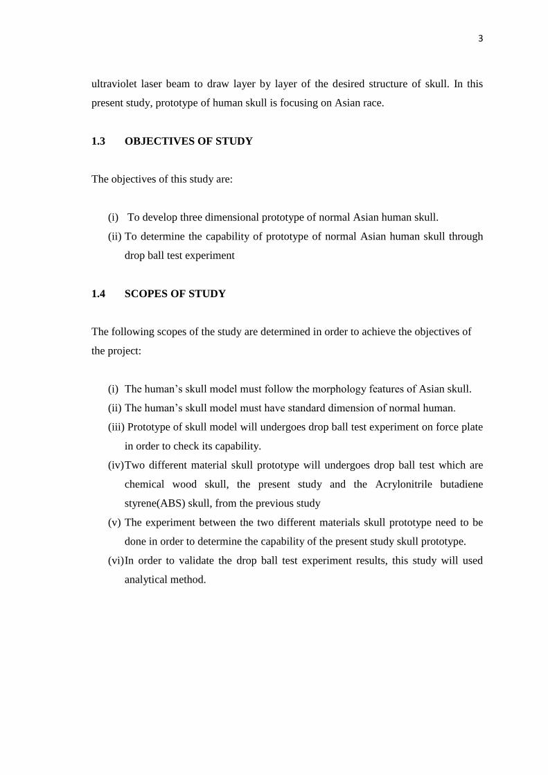
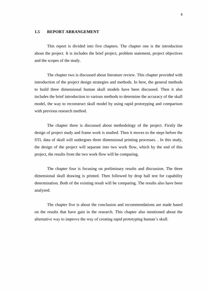
The skeletal structure of the head is divided into three major parts: neurocranium
(housing of the brain), face and base. Neurocranium is made of eight bones: frontal, two
parietal, two occipital, sphenoid and ethmoid ( N. Yoganandan, 2001). The following
figures illustrate these bones making up the human skull (A.Halim,2009)
6
Figure 2.1: Posterior view of the skull
Source: A.Halim (2009)
Figure 2.2: View of skull from above
Source: A.Halim(2009)

7
Figure 2.3: View of skull from lateral position
Source: A.Halim (2009)
2.2.2 The Brain
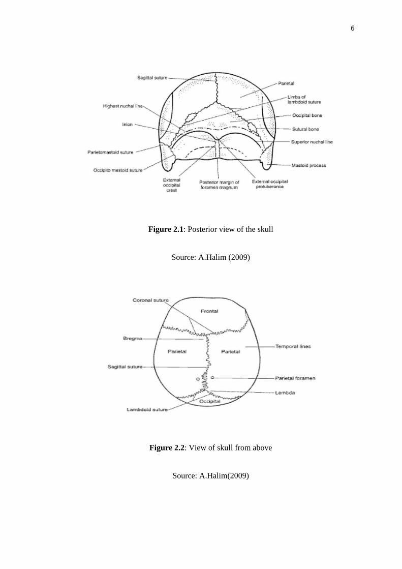
Human brain is made of mainly two types of cells: neurons and glia. Neurons
are the cells that enable the nervous system to carry out all the complex computational
functions. A typical human brain weighing 1.3 kg and with a size of 1.5 liters contains
an estimated number of 20-100 billion neuron cells. The glial cells are described as the
supporting cells for the nervous system; those play an important role of allowing the
nervous system to work properly. The estimated number of glial cells in an average
human nervous system is 10 times the number of neuron cells (S. G. M. Hossain, 2010)
8
Figure 2.4: Various part of human brain
Source: S. G. M. Hossain (2010)
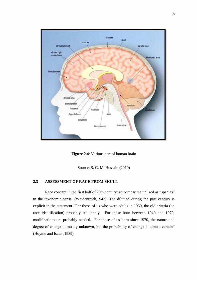
2.3 ASSESSMENT OF RACE FROM SKULL
Race concept in the first half of 20th century: so compartmentalized as “species”
in the taxonomic sense. (Weidenreich,1947). The dilution during the past century is
explicit in the statement “For those of us who were adults in 1950, the old criteria (on
race identification) probably still apply. For those born between 1940 and 1970,
modifications are probably needed. For those of us born since 1970, the nature and
degree of change is mostly unknown, but the probability of change is almost certain”
(Hoyme and Iscan ,1989)
9
2.3.1 Determination of Asian Race skull
Over the years, race determination has relied primarily on the morphologic
features of the skull and facial skeleton since these provide consistently reliable results
in the majority of cases. The three traditional classifications used for race determination
are:-
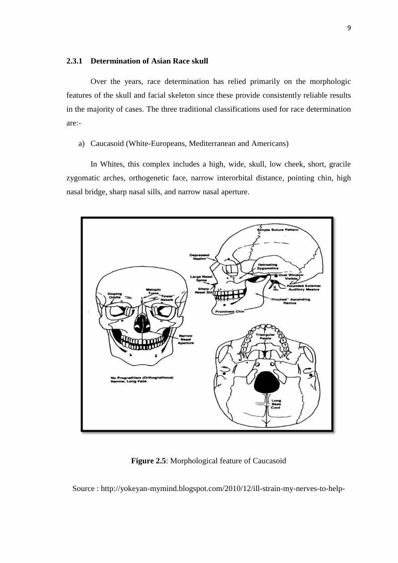
a) Caucasoid (White-Europeans, Mediterranean and Americans)
In Whites, this complex includes a high, wide, skull, low cheek, short, gracile
zygomatic arches, orthogenetic face, narrow interorbital distance, pointing chin, high
nasal bridge, sharp nasal sills, and narrow nasal aperture.
Figure 2.5: Morphological feature of Caucasoid
Source : http://yokeyan-mymind.blogspot.com/2010/12/ill-strain-my-nerves-to-help-
10
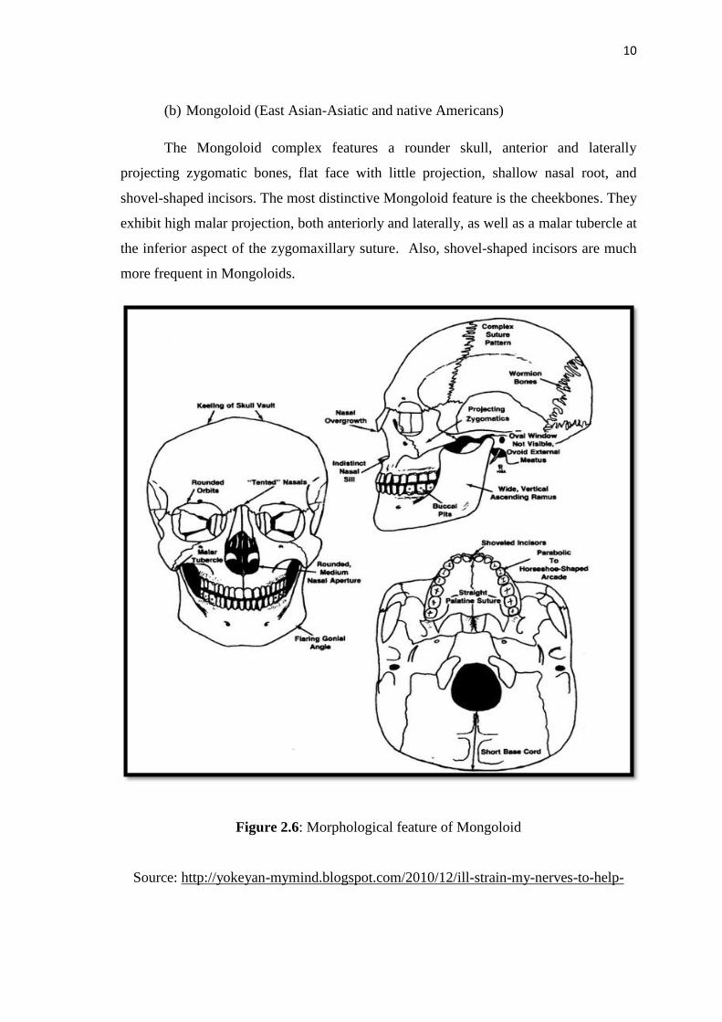
(b) Mongoloid (East Asian-Asiatic and native Americans)
The Mongoloid complex features a rounder skull, anterior and laterally
projecting zygomatic bones, flat face with little projection, shallow nasal root, and
shovel-shaped incisors. The most distinctive Mongoloid feature is the cheekbones. They
exhibit high malar projection, both anteriorly and laterally, as well as a malar tubercle at
the inferior aspect of the zygomaxillary suture. Also, shovel-shaped incisors are much
more frequent in Mongoloids.
Figure 2.6: Morphological feature of Mongoloid
Source: http://yokeyan-mymind.blogspot.com/2010/12/ill-strain-my-nerves-to-help-
11
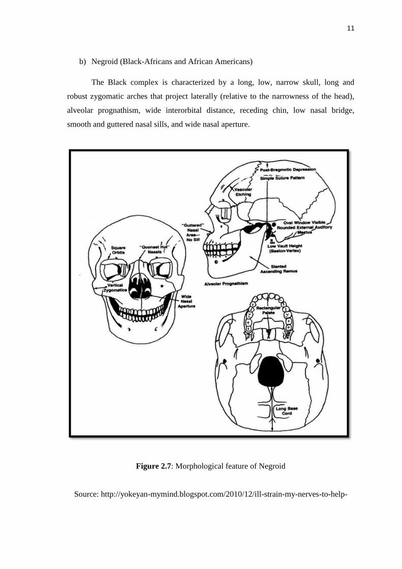
b) Negroid (Black-Africans and African Americans)
The Black complex is characterized by a long, low, narrow skull, long and
robust zygomatic arches that project laterally (relative to the narrowness of the head),
alveolar prognathism, wide interorbital distance, receding chin, low nasal bridge,
smooth and guttered nasal sills, and wide nasal aperture.
Figure 2.7: Morphological feature of Negroid
Source: http://yokeyan-mymind.blogspot.com/2010/12/ill-strain-my-nerves-to-help-

12
Figure 2.8: Different characteristic for different race (a) Caucasoid-Whites tend to have
a high, wide head (b) Negroid- blacks often exhibit long, low crania. (c)
Mongoloid- Mongoloids are more rounded with flat face
Source: P.T. Jayaprakash (2011)