protocol Collecting, rearing, spawning and inducing ...
8
© 2013 Nature America, Inc. All rights reserved. PROTOCOL 916 | VOL.8 NO.5 | 2013 | NATURE PROTOCOLS INTRODUCTION In many respects, the small, estuarine starlet sea anemone (N. vectensis) is an ideal model system for this era of rapid envi- ronmental change. Its geographic distribution spans a tremendous range of natural and anthropogenic environmental variation. It is amenable to ecological and environmental studies in the field. It is also amenable to organismal, cellular, molecular and genomics studies in the laboratory 1,2 . N. vectensis 3 (Fig. 1) is easy to collect and simple and inexpensive to culture in the laboratory 4 . It regu- larly undergoes sexual reproduction, as well as two distinct forms of asexual reproduction under artificial culture conditions 2,4–6 . Its spawning can be synchronized through a combination of regular feedings, light changes and temperature shifts 7,8 . It will reliably and rapidly undergo complete bidirectional regeneration when bisected through the body column, a property that facilitates the development of living clonal stocks 9 . For a marine invertebrate with a planktonic larval stage, the development of Nematostella is rapid (5–7 d from the fertilized egg to a juvenile polyp) and its generation time is short (~50 d from the fertilized egg to a sexually mature adult under ideal culture conditions) 6 . Throughout its life cycle, the anemone is mostly transparent, making it possible to visualize the internal anatomy (and labeling of RNAs and proteins) in whole animals (Figs. 1 and 2). Morphologically, it is a very simple animal that lacks true organs, and it is composed of only two tissue layers, the outer (ectodermal) epidermis and the inner (endo- dermal) gastrodermis (Fig. 2). As a representative of the phylum Cnidaria—which emerged as an independent evolutionary lineage some 50–75 million years before the divergence of protostomes from deuterostomes—Nematostella provides a perspective on early animal evolution that is independent of the so-called ‘higher ani- mals’ upon which most studies of animal development, physiology and genomics are based. As an anthozoan cnidarian, it also serves as an outgroup to inform studies on the closely related reef-building corals, which are crucial to the function and persistence of diverse reef ecosystems and threatened by extinction in many locales across the globe. Here we present protocols for collecting Nematostella, maintaining anemones in the lab, obtaining embryos and develop- ing clonal stocks by inducing regeneration. Animal collection Although Nematostella can be obtained from individual investiga- tors or sporadically from a handful of commercial vendors, it is also easy to collect organisms from coastal estuaries in North America and England. A key advantage of collecting the animals yourself, rather than acquiring them from a colleague or purchasing them through a vendor, is that you can choose to sample from environ- mentally variable or geographically widespread habitats. This is important because you may be interested in the factors that affect the abundance of Nematostella in the wild, or you may be seeking to recover greater functional genetic diversity. Nematostella has been reported at 40 sites along the Atlantic and Gulf coasts of North America (from Nova Scotia to Louisiana), nine sites along the Pacific coast of North America (from California to Washington) and ten sites along the southern coast of England (Supplementary Data and ref. 3). Population genetic analysis indi- cates that its native range is the Atlantic coast of North America; it appears to have been introduced to the Pacific coast and to England 10 . Nematostella can tolerate an extremely wide range of temperatures ( − 1 to 28 °C) 11–13 , salinities (2–52 parts per thousand, p.p.t.) and pH values (from <7 to >9; J.R.F., unpub- lished observations), but its occurrence appears to be restricted to estuarine habitats, especially to salt marsh pools 14 . Within estua- rine habitats, depth and flow appear to be key determinants of its abundance; it occurs at depths <1.0 m, where flow rates are low (e.g., in the range of 0.03–0.10 cm s − 1 ). Nematostella is tolerant of habitats that are degraded by human activity; we have collected this anemone at multiple estuarine habitats in heavily developed urban neighborhoods along the Boston waterfront. One published collection method is to sift the soft sediment from suitable estuarine habitats over a fine screen mesh (~1 mm 2 ) and pick the anemones one-by-one from the screen using for- ceps or a wide-bore transfer pipette 10 . This approach is laborious. In addition, for the inexperienced collector, anemones are diffi- cult to recognize on the screen, as they are practically transparent, and they retract their tentacles into the pharynx when disturbed. A simpler and more efficient method, which is described here, is Collecting, rearing, spawning and inducing regeneration of the starlet sea anemone, Nematostella vectensis Derek J Stefanik, Lauren E Friedman & John R Finnerty Department of Biology, Boston University, Boston, Massachusetts, USA. Correspondence should be addressed to J.R.F. ([email protected]). Published online 11 April 2013; doi:10.1038/nprot.2013.044 Over the past 20 years, the starlet sea anemone, Nematostella vectensis, a small estuarine animal, has emerged as a powerful model system for field and laboratory studies of development, evolution, genomics, molecular biology and toxicology. Here we describe how to collect Nematostella, culture it through its entire sexual life cycle and induce regeneration for the production of clonal stocks. In less than 1 h at a suitable field site, a researcher on foot can collect hundreds of individual anemones. In a few months, it is possible to establish a laboratory colony that will be reliable in generating hundreds or thousands of fertilized eggs on a roughly weekly schedule. By inducing regeneration roughly every 2 weeks, in less than 6 months, one can establish a clonal stock consisting of hundreds of genetically identical anemones. These results can be achieved very inexpensively and without specialized equipment.
Transcript of protocol Collecting, rearing, spawning and inducing ...
©20
13 N
atu
re A
mer
ica,
Inc.
All
rig
hts
res
erve
d.
protocol
916 | VOL.8 NO.5 | 2013 | nature protocols
IntroDuctIonIn many respects, the small, estuarine starlet sea anemone (N. vectensis) is an ideal model system for this era of rapid envi-ronmental change. Its geographic distribution spans a tremendous range of natural and anthropogenic environmental variation. It is amenable to ecological and environmental studies in the field. It is also amenable to organismal, cellular, molecular and genomics studies in the laboratory1,2. N. vectensis3 (Fig. 1) is easy to collect and simple and inexpensive to culture in the laboratory4. It regu-larly undergoes sexual reproduction, as well as two distinct forms of asexual reproduction under artificial culture conditions2,4–6. Its spawning can be synchronized through a combination of regular feedings, light changes and temperature shifts7,8. It will reliably and rapidly undergo complete bidirectional regeneration when bisected through the body column, a property that facilitates the development of living clonal stocks9. For a marine invertebrate with a planktonic larval stage, the development of Nematostella is rapid (5–7 d from the fertilized egg to a juvenile polyp) and its generation time is short (~50 d from the fertilized egg to a sexually mature adult under ideal culture conditions)6. Throughout its life cycle, the anemone is mostly transparent, making it possible to visualize the internal anatomy (and labeling of RNAs and proteins) in whole animals (Figs. 1 and 2). Morphologically, it is a very simple animal that lacks true organs, and it is composed of only two tissue layers, the outer (ectodermal) epidermis and the inner (endo-dermal) gastrodermis (Fig. 2). As a representative of the phylum Cnidaria—which emerged as an independent evolutionary lineage some 50–75 million years before the divergence of protostomes from deuterostomes—Nematostella provides a perspective on early animal evolution that is independent of the so-called ‘higher ani-mals’ upon which most studies of animal development, physiology and genomics are based. As an anthozoan cnidarian, it also serves as an outgroup to inform studies on the closely related reef-building corals, which are crucial to the function and persistence of diverse reef ecosystems and threatened by extinction in many locales across the globe. Here we present protocols for collecting Nematostella, maintaining anemones in the lab, obtaining embryos and develop-ing clonal stocks by inducing regeneration.
Animal collectionAlthough Nematostella can be obtained from individual investiga-tors or sporadically from a handful of commercial vendors, it is also easy to collect organisms from coastal estuaries in North America and England. A key advantage of collecting the animals yourself, rather than acquiring them from a colleague or purchasing them through a vendor, is that you can choose to sample from environ-mentally variable or geographically widespread habitats. This is important because you may be interested in the factors that affect the abundance of Nematostella in the wild, or you may be seeking to recover greater functional genetic diversity.
Nematostella has been reported at 40 sites along the Atlantic and Gulf coasts of North America (from Nova Scotia to Louisiana), nine sites along the Pacific coast of North America (from California to Washington) and ten sites along the southern coast of England (Supplementary Data and ref. 3). Population genetic analysis indi-cates that its native range is the Atlantic coast of North America; it appears to have been introduced to the Pacific coast and to England10. Nematostella can tolerate an extremely wide range of temperatures ( − 1 to 28 °C)11–13, salinities (2–52 parts per thousand, p.p.t.) and pH values (from <7 to >9; J.R.F., unpub-lished observations), but its occurrence appears to be restricted to estuarine habitats, especially to salt marsh pools14. Within estua-rine habitats, depth and flow appear to be key determinants of its abundance; it occurs at depths <1.0 m, where flow rates are low (e.g., in the range of 0.03–0.10 cm s − 1). Nematostella is tolerant of habitats that are degraded by human activity; we have collected this anemone at multiple estuarine habitats in heavily developed urban neighborhoods along the Boston waterfront.
One published collection method is to sift the soft sediment from suitable estuarine habitats over a fine screen mesh (~1 mm2) and pick the anemones one-by-one from the screen using for-ceps or a wide-bore transfer pipette10. This approach is laborious. In addition, for the inexperienced collector, anemones are diffi-cult to recognize on the screen, as they are practically transparent, and they retract their tentacles into the pharynx when disturbed. A simpler and more efficient method, which is described here, is
Collecting, rearing, spawning and inducing regeneration of the starlet sea anemone, Nematostella vectensisDerek J Stefanik, Lauren E Friedman & John R Finnerty
Department of Biology, Boston University, Boston, Massachusetts, USA. Correspondence should be addressed to J.R.F. ([email protected]).
Published online 11 April 2013; doi:10.1038/nprot.2013.044
over the past 20 years, the starlet sea anemone, Nematostella vectensis, a small estuarine animal, has emerged as a powerful model system for field and laboratory studies of development, evolution, genomics, molecular biology and toxicology. Here we describe how to collect Nematostella, culture it through its entire sexual life cycle and induce regeneration for the production of clonal stocks. In less than 1 h at a suitable field site, a researcher on foot can collect hundreds of individual anemones. In a few months, it is possible to establish a laboratory colony that will be reliable in generating hundreds or thousands of fertilized eggs on a roughly weekly schedule. By inducing regeneration roughly every 2 weeks, in less than 6 months, one can establish a clonal stock consisting of hundreds of genetically identical anemones. these results can be achieved very inexpensively and without specialized equipment.

©20
13 N
atu
re A
mer
ica,
Inc.
All
rig
hts
res
erve
d.
protocol
nature protocols | VOL.8 NO.5 | 2013 | 917
to collect the sediment into shallow trays, allow the disturbed sedi-ment to settle and retrieve the anemones with a transfer pipette as they extend their bodies from the substrate and assume a feeding posture15. This procedure is less time consuming, and it results in a higher yield of anemones from the same area of bottom sediment because the anemones are easy to recognize when they have their tentacles extended.
Animal culture and spawningThe difficulty in maintaining and spawning cnidarians in the lab-oratory has been a major impediment to conducting functional studies of their physiology, development, cell biology and molecular genetics. The pioneering work of Hand and Uhlinger4,16 resulted in Nematostella being the first sea anemone to be raised to sexual maturity in laboratory culture. Indeed, although replicating the natural conditions required to induce spawning in many marine invertebrates has proven to be difficult, in Nematostella’s case, a period of laboratory conditioning actually increases the frequency and eliminates the seasonality of spawning4,16,17.
Hand and Uhlinger4 demonstrated that spawning is clearly inhibited by anoxia and possibly by high levels of ammonia—two conditions that can be remedied by water changes. Their published protocol for spawning Nematostella prescribes feeding Artemia to anemones every other day for 6 d (Fig. 3a). On the eighth day, the anemones are fed mussel ovary (Mytilus californicus) and the water is changed. Under this regimen, females spawned an average of 0.80 times per week, and 75% of these spawning events occurred on the day after a water change and mussel feeding. Males spawned 0.72 times per week, with 64% of the spawning events occurring on the day after the water was changed and mussels were provided as food4.
Two findings suggest that water changes are a stronger spawning cue than feeding. When the mussel feeding and water changes were separated by 24, 48 or 72 h, spawning in Nematostella was found to be more tightly correlated with the timing of water changes com-pared with the timing of mussel feeding16. In addition, Uhlinger16 showed that Nematostella can be entrained into spawning cycles through the use of water changes alone, even over the course of several months without food. However, over time, the starved females produced fewer and fewer eggs. Changing the water decreases the concentration of metabolic waste products, whereas it increases the concentration of dissolved oxygen and possibly alters the salinity, all of which could serve to induce spawning in a manner that might mimic the infusion of water into salt marsh pools via tidal inundation or rainfall.
Subsequently to the work of Hand and Uhlinger4, Fritzenwanker and Technau8 demonstrated that changing light and temperature could also induce spawning, with the highest fecundity achieved under a simultaneous exposure to both bright light and elevated temperature (Fig. 3b). An updated version of this protocol prescribes rearing anemones in cool (18 °C), dark conditions for 10–14 d with daily feeding7. At the end of this conditioning period, the water change stimulus from Hand and Uhlinger4 is combined with 13 h of exposure to bright light and elevated temperature (25 °C). Following this treatment, the anemones are maintained under bright light at 18 °C and monitored for spawning every 15 min.
Here we describe a protocol for spawning Nematostella that com-bines all four previously identified spawning cues—water changes, bright light, increased temperature and feeding (Fig. 3c). Consistent with a recent study7, we prescribe a longer period of conditioning (14 d) because, in our experience, individuals that are spawned con-sistently on a 7-d schedule can begin to show diminished fecundity.
1.0 mm
a b
c
0.1 mm
0.1 mm
Apicaltuft
Mesentery
*
*
Gastrovascularcavity
Gastrovascularcavity
Tentacle
Mesentery
Pharynx
Figure 2 | Developmental stages of Nematostella vectensis. (a) Freshly spawned egg mass. Each individual egg suspended in the gelatinous matrix is ~200 µm in diameter. (b) Planula larva 3 d after fertilization. The deep focal plane of this image reveals the division between endodermal and ectodermal tissue layers, as well as the developing pharynx and the mesenteries that span the gap between the pharynx and the body wall. The asterisk indicates the location of the mouth. Opposite the mouth is a prominent grouping of long cilia known as the apical tuft. The planula is an active swimmer, and it moves primarily in the direction of the apical tuft. At this stage, the larva is ~250 µm in length from the base of the apical tuft to the mouth. (c) Juvenile anemone 10 d after fertilization. Typically, at the time of settlement, the juvenile anemone has four tentacles. The deep focal plane of this image reveals the division between endodermal and ectodermal tissue layers, as well as the pharynx and the first four mesenteries. There will eventually be eight mesenteries, and these structures will bear bundles of longitudinal muscle fibers that are used to shorten the animal along its oral-aboral axis. The gametes also develop within the mesenteries.
1 mm
Mouth
Pharynx
MesenteriesPhysa or
foot
Scapus orcolumn
Capitulum orhead
Figure 1 | Adult polyp of Nematostella vectensis. Field-caught animals are typically ~1 cm long from mouth to tip of the physa, but in culture, well-fed individuals can reach several centimeters in length. The body can be divided into three main regions: the head or capitulum; the column or scapus and the foot or physa. Adult polyps typically have 10–16 tentacles.

©20
13 N
atu
re A
mer
ica,
Inc.
All
rig
hts
res
erve
d.
protocol
918 | VOL.8 NO.5 | 2013 | nature protocols
Thus, if two cohorts of animals are placed on a 14-d schedule and staggered by 7 d, it can result in more consistently produc-tive spawning than placing one cohort on a 7-d schedule. Beginning with a few founder individuals of each sex, it is possible to rapidly develop a self-sustaining, sexually reproducing colony of N. vectensis consisting of several hundred individuals on a square meter of laboratory benchtop.
Experimental designBefore collecting Nematostella and attempting to establish a lab-oratory culture, you should carefully design your sampling and culturing protocols so that they are tailored to the questions you wish to answer. For example, if you wish to identify factors that affect the abundance and distribution of Nematostella or to col-lect animals that show different environmental tolerances, you should use a field site harboring many small semi-isolated bodies of water—e.g., a high marsh habitat with numerous small pools separated by dense stands of marsh grass—that differ in key envi-ronmental variables, including temperature, salinity, dissolved oxygen, pH, nitrogen levels and so on, and you must be prepared
for measuring these variables in the field. To accurately estimate the abundance of Nematostella in a small body of water, you must repeatedly sample a consistent amount of sediment from multi-ple spots in the same pool body because the spatial distribution of the anemone can fluctuate substantially over short distances. Furthermore, when establishing the culture you will have to sepa-rate individual animals collected from different locations. If you wish to perform experiment on single clone lines to eliminate genetic variability within a test population, you would then use regeneration to accumulate living clonal stocks for each genotype you wish to maintain. If, in contrast, you are merely interested in establishing a laboratory culture to produce abundant embryos for developmental analysis, you can collect all of your animals from a single spot that reliably harbors the anemones, and you can culture tens or hundreds of genetically diverse males and females together in larger culture dishes.
a
c
bLighting
Temperature
Not specified
16–26 °C(room temperature) 18 °C
24 °C
18 °C
18–20 °C
20–25 °C
1 2 3 4 5 6 7 8 9
1 2 3 4 5 6 7 8 9 10 11 12 13 14 15
70–100%
16
75%
Conditioning
Dark
Dark
Light
Light
Induction
70–100%
Optional Optional
Change 1× per week
Changewater
Feeding
Spawning
Lighting
Temperature
Changewater
Feeding
Spawning
Day 1 2 3 4 5 6 7 8Day
2× per week / timing notspecified
Artemia
Artemia Artemia Artemia
Artemia Artemia ArtemiaMytilus
Day
Figure 3 | Protocols for spawning Nematostella. (a) Hand and Uhlinger4 published the first protocol for spawning Nematostella on a roughly weekly schedule. The protocol involved regular feeding of the anemones with freshly hatched brine shrimp larvae (Artemia), in addition to weekly feedings with mussel ovary (Mytilus) and coordinated water changes. (b) Fritzenwanker and Technau8 eliminated the use of mussel and introduced shifts in temperature and light as important inducers of spawning. (c) The protocol described in the text is modified from Genikhovich and Technau7. This protocol incorporates a 2-week period of conditioning when the animals are maintained in the dark at 18 °C and are fed at least 5 d per week, with weekly water changes. At the conclusion of this conditioning period, the temperature is elevated for a period of ~12–15 h and the animals are placed in bright light. Within a few hours of the conclusion of the elevated temperature treatment, the large majority of animals will typically release eggs and sperm.
MaterIalsREAGENTS
Artificial seawater (ASW; Instant Ocean)Artemia salina cysts (available commercially from multiple vendors)Fresh mussels (optional; e.g., Mytilus edulis, available in fish markets)l-Cysteine (optional; MP Biomedicals, cat. no. 0219464680)Adult anemones (polyps)MgCl2 (Sigma-Aldrich, cat. no. M-8266)
EQUIPMENTHip waders (optional; Dick’s Sporting Goods, cat. no. 4039311)Trowel, spade or Eckman grab (Wildlife Supply, cat. no. 197-C15) for excavating soft sediment
••••••
••
Wide, shallow, plastic containers with tight-fitting lids (e.g., food storage containers) for collecting the sedimentTransfer pipettes for retrieving the anemones (Fisher Scientific, cat. no. 13-711-7M)Small culture dishes filled with 1/3-strength ASW (Pyrex glass food storage bowls with plastic lids are ideal. The lids can be perforated with a hole-punch to allow air to circulate while minimizing evaporation and the possibility of spills)Separatory funnel (~250 ml; Fisher Scientific, cat. no. 13-678-604) supported in a ring-stand (Fisher scientific, cat. no. S47788) ideal for culturing the anemone’s food, brine shrimp nauplii (Artemia salina)
•
•
•
•

©20
13 N
atu
re A
mer
ica,
Inc.
All
rig
hts
res
erve
d.
protocol
nature protocols | VOL.8 NO.5 | 2013 | 919
Incubator (e.g., Fisher Scientific, cat. no. 97-990E) maintained at ~18 °CIncubator maintained at ~25 °CAquarium air pump (for aerating the Artemia cultures) attached via air tubing to an aeration stoneSingle-edged razor blade (Fisher scientific, cat. no. 12-640)Scalpel (Fisher scientific, cat. no. 089275A)Plastic Petri dish, 150 mm (or similar shallow vessel; Fisher scientific, cat. no. R80150)Rotary shaker (optional; see Step 15)
REAGENT SETUPCollection sites Within its range, salt marsh pools are particularly reliable sites for collecting N. vectensis. These pools generally feature still waters and soft, flocculent sediments rich in organic material. The salinity commonly fluctuates between 15 and 30 p.p.t. Such pools can generally be sampled on foot. In low-lying marsh habitats, it is generally preferable to collect samples during low tide, as the pools will be shallower and the regions between pools will not be inundated. ! cautIon Marsh water may contain high levels of potentially pathogenic bacteria, such as Vibrio species and chemical contaminants from nearby point sources of pollution. To reduce exposure to contaminants, wear waders if you decide to stand in the pools, and wear gloves when you collect the sediments, particularly if you have any open wounds. Wash your hands thoroughly after completing the collection process.Artificial seawater For full-strength ASW, dissolve 35.9 g of Instant Ocean per liter of dH2O (35–36 p.p.t.) to produce 2–5 liters of full-strength ASW in total. As per the manufacturer’s recommendations, circulate the newly mixed water overnight with an airstone, which oxygenates the water and
•••
•••
•
allows the CO2 of the water to equilibrate with that of the air, thus stabilizing the pH. Dilute one part full-strength ASW with two parts dH2O to produce 1/3-strength ASW for culturing anemones (11–12 p.p.t.). It is convenient to prepare 5–15 liters of filtered full-strength ASW at one time and store it in a plastic carboy with an integrated spigot so that it can be easily diluted and dispensed into culture vessels as required. Full-strength or 1/3-strength ASW may be stored indefinitely in capped containers at room temperature (20–22 °C). crItIcal Although a laboratory study of Nematostella reproduction provided evidence of peak fecundity at this salinity, not all populations of Nematostella show optimal reproduction at this salinity. Thus, 1/3-strength ASW represents a useful starting point, but the researcher should optimize the salinity when working with a new strain of anemones. At the time it was collected, a population that we isolated from Peggy’s Cove, Nova Scotia, Canada, was sound at a higher salinity and individuals from this location appear to prefer 2/3-strength ASW18.Hatching brine shrimp Add ~0.5 g of Artemia cysts to ~100 ml of full-strength ASW in a separatory funnel. Subject the solution to continuous aeration using an aeration stone. After 24–36 h, cease aeration, allow the hatched nauplii larvae (which are bright orange in color and actively swimming) to become concentrated at the bottom of the funnel. Thereafter, collect the concentrated nauplii, avoiding unhatched cysts, which are not eaten by the anemones. Brine shrimp should be used within 48 h of hatching. After this time, discard any remaining culture.MgCl2 (7%, wt/vol) relaxant Dissolve 7 g of MgCl2 in 100 ml of 1/3 ASW. The resulting solution can be stored indefinitely at room temperature.
proceDureanimal collection ● tIMInG 2 h1| At a suitable estuarine habitat, collect the surface sediment (i.e., the top 3–5 cm) according to option A, if your objective is not to sample consistent areas of the pool. Use option B if you intend to sample such areas.(a) collection of sediment throughout a pool (i) Transfer surface sediment into shallow containers using a trowel or shovel, along with sufficient water to keep the
sediment submerged. crItIcal step Obtain the requisite permit from the relevant local and/or national authorities in your locality for the scientific collection of coastal invertebrates.
(B) collection of sediment at specified, predetermined sites of a pool (i) A dedicated sampling device, such as an Eckman grab, can be used to sample a consistent area. However, in general,
these devices are designed to excavate greater depths than are required to collect Nematostella. To sample a consistent area with a low-tech method, remove the bottom of a tall plastic bucket (such as a 5-gallon paint bucket) using a hacksaw. Push this ‘holey bucket’ a few centimeters deep into the sediment of a shallow pool (where the water level will not overtop the rim of the bucket.) After gently removing most of the water with a siphon or scoop (in an effort to avoid disturbing the substrate), the top few centimeters of sediment can then be excavated from the area circumscribed by the bucket.
2| Seal the containers and transport them back to the laboratory.
3| Remove the lids from the containers. Allow the sediment to settle and the water to clarify for 1 h or overnight. crItIcal step Do not keep the containers indoors for more than a day or two. The composition of the sediment ecosystem will undergo a visible phase shift, with a precipitous die-off of many invertebrates and a pronounced alteration of the microbial community; this is evident in a color shift (e.g., often, a filamentous white fungus will proliferate in the culture, forming a lacy film over the sediment). If you wish to maintain these marsh microcosms in a semi-natural state, in which the Nematostella will continue to thrive for weeks or months, keep the containers outdoors with the lids removed, so that they are exposed to rain and sun (and top them off with dH2O if the water level drops precipitously).
4| Extract the anemones that protrude from the sediment (supplementary Video 1) using a wide-bore transfer pipette and transfer the animals to culture dishes containing 1/3-strength ASW.? trouBlesHootInG
©20
13 N
atu
re A
mer
ica,
Inc.
All
rig
hts
res
erve
d.
protocol
920 | VOL.8 NO.5 | 2013 | nature protocols
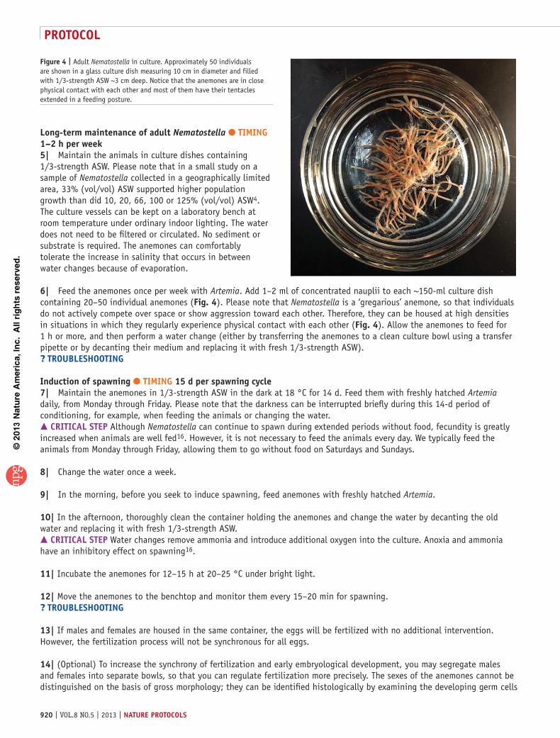
long-term maintenance of adult Nematostella ● tIMInG 1–2 h per week5| Maintain the animals in culture dishes containing 1/3-strength ASW. Please note that in a small study on a sample of Nematostella collected in a geographically limited area, 33% (vol/vol) ASW supported higher population growth than did 10, 20, 66, 100 or 125% (vol/vol) ASW4. The culture vessels can be kept on a laboratory bench at room temperature under ordinary indoor lighting. The water does not need to be filtered or circulated. No sediment or substrate is required. The anemones can comfortably tolerate the increase in salinity that occurs in between water changes because of evaporation.
6| Feed the anemones once per week with Artemia. Add 1–2 ml of concentrated nauplii to each ~150-ml culture dish containing 20–50 individual anemones (Fig. 4). Please note that Nematostella is a ‘gregarious’ anemone, so that individuals do not actively compete over space or show aggression toward each other. Therefore, they can be housed at high densities in situations in which they regularly experience physical contact with each other (Fig. 4). Allow the anemones to feed for 1 h or more, and then perform a water change (either by transferring the anemones to a clean culture bowl using a transfer pipette or by decanting their medium and replacing it with fresh 1/3-strength ASW).? trouBlesHootInG
Induction of spawning ● tIMInG 15 d per spawning cycle7| Maintain the anemones in 1/3-strength ASW in the dark at 18 °C for 14 d. Feed them with freshly hatched Artemia daily, from Monday through Friday. Please note that the darkness can be interrupted briefly during this 14-d period of conditioning, for example, when feeding the animals or changing the water. crItIcal step Although Nematostella can continue to spawn during extended periods without food, fecundity is greatly increased when animals are well fed16. However, it is not necessary to feed the animals every day. We typically feed the animals from Monday through Friday, allowing them to go without food on Saturdays and Sundays.
8| Change the water once a week.
9| In the morning, before you seek to induce spawning, feed anemones with freshly hatched Artemia.
10| In the afternoon, thoroughly clean the container holding the anemones and change the water by decanting the old water and replacing it with fresh 1/3-strength ASW. crItIcal step Water changes remove ammonia and introduce additional oxygen into the culture. Anoxia and ammonia have an inhibitory effect on spawning16.
11| Incubate the anemones for 12–15 h at 20–25 °C under bright light.
12| Move the anemones to the benchtop and monitor them every 15–20 min for spawning.? trouBlesHootInG
13| If males and females are housed in the same container, the eggs will be fertilized with no additional intervention. However, the fertilization process will not be synchronous for all eggs.
14| (Optional) To increase the synchrony of fertilization and early embryological development, you may segregate males and females into separate bowls, so that you can regulate fertilization more precisely. The sexes of the anemones cannot be distinguished on the basis of gross morphology; they can be identified histologically by examining the developing germ cells
Figure 4 | Adult Nematostella in culture. Approximately 50 individuals are shown in a glass culture dish measuring 10 cm in diameter and filled with 1/3-strength ASW ~3 cm deep. Notice that the anemones are in close physical contact with each other and most of them have their tentacles extended in a feeding posture.

©20
13 N
atu
re A
mer
ica,
Inc.
All
rig
hts
res
erve
d.
protocol
nature protocols | VOL.8 NO.5 | 2013 | 921
in the mesenteries of reproductive adults17. However, it is more practical to simply segregate males and females into separate culture dishes once they have been observed during spawning (Fig. 2a and ANTICIPATED RESULTS). After sex segregation and after anemones have begun to spawn, use a clean pipette to transfer the egg masses into a clean bowl containing 1/3-strength ASW (the volume of the egg mass plus accompanying ASW will be ~5 ml). Use a separate clean pipette to transfer 10–20 ml of water from a male-only bowl (where spawning has occurred) into the bowl containing the eggs, which will serve as a fertilization chamber. With no further intervention, fertilization will commence within minutes and will be complete within a few hours. crItIcal step Perform this fertilization step relatively quickly as the eggs will only remain viable and capable of being fertilized for a few hours after spawning.
15| (Optional) If you wish to monitor or manipulate zygotes immediately after fertilization, it is convenient to remove the gelatinous mass surrounding the eggs. This goal can be achieved by incubating the eggs for 10–15 min in 4% (wt/vol) l-cysteine prepared in 1/3-strength ASW with gentle agitation on a rotary shaker8.
16| Transfer the fertilized eggs (or entire egg masses) into individual wells of 12-well culture plates filled with 2 ml of 1/3-strength ASW at room temperature to allow embryogenesis to proceed. It is not necessary to feed the developing embryos or larvae. Within 2–3 d of fertilization, the planula larva will be actively swimming about the culture dish propelled by a fairly uniform layer of cilia and a prominent apical tuft, which forms opposite to the mouth (Fig. 2b). Within 5–7 d, two to four tentacles will have developed, and the juvenile polyps will spontaneously settle to the bottom of the culture plate. Newly metamorphosed polyps can be fed with freshly hatched Artemia, but their ability to feed is increased if the Artemia have been pulverized with a mortar and pestle.
17| Thoroughly clean the containers housing the recently spawned anemones to remove the remaining gametes and/or developing embryos. Clean the glassware by scrubbing and repeatedly rinsing with water (no detergents). Perform the final rinse with 1/3-strength ASW. Replace the culture water with a fresh 1/3-strength ASW.
18| Return the anemones to conditions of 18 °C temperature and darkness and repeat Steps 7 and 8 to condition the animals for subsequent spawning. If weekly or daily spawning is desired, multiple anemone cultures can be established that are timed to spawn on a rotating schedule.
Induction of regeneration ● tIMInG 10–14 d19| Introduce a single adult anemone in a 150-mm plastic Petri dish containing 1/3-strength ASW.
20| Allow the animal to fully extend its body column and tentacles. Alternatively, an extended, relaxed posture can be induced by treating the animal with 7 mM MgCl2 prepared in 1/3-strength ASW.
21| At a point located approximately midway between the mouth and the tip of the foot, firmly apply a single-edged razor blade to the animal, pinning it against the bottom of the dish.
22| Draw a scalpel blade along the edge of the razor blade, slicing the anemone cleanly in two approximately equal-length parts, one bearing the foot and the other bearing the mouth and tentacles.
23| Maintain the bisected animal in 1/3-strength ASW for 5–7 d (Fig. 5), allowing the cut ends of the animal to regenerate missing structures. The animal need not be fed during the regeneration process. Individuals may be subjected to different environmental conditions during regeneration as a test of how they tolerate environmental stress.? trouBlesHootInG
a
d e
b c
Figure 5 | Nematostella regenerating missing head structures. (a–e) The same anemone is shown at 1 (a), 2 (b), 3 (c), 4 (d) and 5 d (e) after the head and the oral portion of the body column were resected using a scalpel.

©20
13 N
atu
re A
mer
ica,
Inc.
All
rig
hts
res
erve
d.
protocol
922 | VOL.8 NO.5 | 2013 | nature protocols
? trouBlesHootInGTroubleshooting advice can be found in table 1.
● tIMInGSteps 1–4, animal collection: 2 hSteps 5 and 6, long-term maintenance of adult Nematostella: 1–2 h per weekSteps 7–18, induction of spawning: 15 dSteps 19–23, induction of regeneration: 10–14 d
taBle 1 | Troubleshooting table.
step problem possible reason solution
4 Failure to obtain anemones while collecting
The abundance of Nematostella varies substan-tially from site to site within a given estuary. Some pools may harbor hundreds or thousands of anemones per square meter, whereas other pools in the same marsh may be devoid of anemones
Try sampling of multiple pools rather than taking multiple samples from a single pool. If practical, attempt to collect animals at an estuary where they have already been reported, and seek advice from someone with firsthand knowledge of the site. Nematostella populations achieve the greatest densities in the summer. Attempt your collections between May and October
6 The anemones are stressed, as evidenced by the fact that they keep their bodies contracted, with their tentacles withdrawn and often produce copious mucus
A decline in water quality is a likely cause Transfer the anemones to a new container with fresh 1/3-strength ASW
12 Failure to observe egg masses after following the prescribed regimen of feeding and temperature changes for several weeks
Your culture may lack females or, if egg masses are being produced but the eggs are not being fertilized (they will fail to undergo cell divisions and they will begin to degrade within hours of being laid), it may lack males
The sex of Nematostella is not evident to the naked eye. The sex of the reproductive individuals can be determined by histological examination of the mesenteries, which house the developing germ cells17
aThe food source the anemones are being provided is not rich enough
Try mincing some fresh mussel, especially ovary, and use forceps to feed small pieces to individual anemones4. The water should be changed within a few hours of feeding mussels to the anemones, as the water quality will tend to degrade quickly because of the presence of uneaten food
23 An inappropriate structure is generated or no regeneration at all occurs
If mesenteries protrude from the cut-site following wound closure, regeneration can be substantially delayed or the animal may not even regenerate. If an anemone is bisected very close to the mouth, a second ‘head’ may form at the cut end of the animal, resulting temporarily in a two-headed individual that lacks a foot19. If the animal is only partially bisected, a second head or a second foot may form at the site of the wound19
Obtain a clean cut approximately midway along the animal’s primary body axis, always below the level of the pharynx
aBe aware that some natural populations, such as that of Crane Beach in Massachusetts14, have been shown to consist primarily or entirely of single-sex clones derived by asexual reproduction6—indeed, a study of genetic structure in ten English lagoons showed, using random amplification of polymorphic DNA (RAPD), that 61% of individuals shared a single multilocus genotype, which ranged in frequency from 0.01 to 1.00 across sites20.

©20
13 N
atu
re A
mer
ica,
Inc.
All
rig
hts
res
erve
d.
protocol
nature protocols | VOL.8 NO.5 | 2013 | 923
antIcIpateD resultsHundreds of N. vectensis individuals can be collected from less than 1 square meter of soft substrate using the present protocol. If anemones are present, they will readily protrude a few millimeters above the substrate and extend their tentacles in a feeding posture (supplementary Video 1). The animals can be identified with the naked eye; however, it can be helpful to use a dissecting microscope or a magnifying glass aided by oblique lighting in order to detect small individuals. Sediment and debris may adhere to the body of freshly collected anemones. These contaminants can be removed by gently pipetting the animals up and down in clean ASW using a wide-bore transfer pipette. The anemones tend to have a slight brown tint when first collected, but they will gradually assume a pink tint as they feed on Artemia. It may take a field-caught animal a few days before it will readily eat the Artemia.
With regular feedings and water changes as described above, 50–100 animals can be comfortably accommodated in a 250-ml culture bowl (Fig. 4). Healthy adults will be nearly fully elongated for most of the time, and their tentacles will be fully extended in a feeding posture.
Once you create the proper conditions, spawning should occur 1–6 h after the anemones are removed from the light and heat. A typical female anemone can produce several hundred eggs, which are contained within a jelly. The jelly-encased egg masses are negatively buoyant. The water in cultures containing male anemones that have spawned will often turn turbid, which is a sign that the males have released their gametes. When reared at room temperature, the first cleavage of the eggs occurs about 120 min after fertilization8. However, when embryogenesis is carried out at 18 °C, the first cleavage occurs about 50 min later, on average ~170 min after fertilization8.
When adult polyps are bisected, the head-bearing fragment should fully regenerate its missing foot within 24–36 h. The foot-bearing fragment will begin erupting tentacles within 36 h; by 72–96 h, a fully functioning head will form, complete with mouth, pharynx and tentacles, as well as the ability to capture prey and feed.
Note: Supplementary information is available in the online version of the paper.
acknowleDGMents This research was supported by National Science Foundation grant no. MCB-0924749 (T.D. Gilmore and J.R.F.). D.J.S. was supported by a Predoctoral Fellowship Award to Promote Diversity in Health-Related Research from the National Institutes of Health (no. F31 GM095289-01). D.J.S. was also supported by a Warren-McLeod Graduate Fellowship in Marine Biology.
autHor contrIButIons The collection and regeneration protocols were developed by J.R.F. All authors participated in writing the manuscript and optimizing the protocols.
coMpetInG FInancIal Interests The authors declare no competing financial interests.
Reprints and permissions information is available online at http://www.nature.com/reprints/index.html.
1. Reitzel, A.M., Ryan, J.F. & Tarrant, A.M. Establishing a model organism: a report from the first annual Nematostella meeting. BioEssays 34, 158–161 (2012).
2. Darling, J.A. et al. Rising starlet: the starlet sea anemone, Nematostella vectensis. BioEssays 27, 211–221 (2005).
3. Stephenson, T.A. The British Sea Anemones Vol. II. (The Ray Society, 1935).
4. Hand, C. & Uhlinger, K. The culture, sexual and asexual reproduction, and growth of the sea anemone Nematostella vectensis. Biol. Bull. 182, 169–176 (1992).
5. Hand, C. & Uhlinger, K.R. Asexual reproduction by transverse fission and some anomalies in the sea anemone Nematostella vectensis. Invert. Biol. 114, 9–18 (1995).
6. Hand, C. & Uhlinger, K. The unique, widely distributed sea anemone, Nematostella vectensis Stephenson: A review, new facts, and questions. Estuaries 17, 501–508 (1994).
7. Genikhovich, G. & Technau, U. Induction of spawning in the starlet sea anemone Nematostella vectensis, in vitro fertilization of gametes, and dejellying of zygotes. Cold Spring Harb. Protoc. http://dx.doi.org/10.1101/pdb.prot5281 (2009).
8. Fritzenwanker, J.H. & Technau, U. Induction of gametogenesis in the basal cnidarian Nematostella vectensis (Anthozoa). Dev. Genes Evol. 212, 99–103 (2002).
9. Burton, P.M. & Finnerty, J.R. Conserved and novel gene expression between regeneration and asexual fission in Nematostella vectensis. Dev. Genes Evol. 219, 79–87 (2009).
10. Reitzel, A., Darling, J., Sullivan, J. & Finnerty, J. Global population genetic structure of the starlet anemone Nematostella vectensis: multiple introductions and implications for conservation policy. Biol. Invasions 10, 1197–1213 (2008).
11. Frank, P.G. & Bleakney, J.S. Asexual reproduction, diet, and anomalies of the anemone Nematostella vectensis in Nova Scotia. Canadian Field Naturalist 92, 259–263 (1978).
12. Sheader, M., Suwailem, A.M. & Rowe, G.A. The anemone, Nematostella vectensis, in Britain: considerations for conservation management. Aquatic Conserv.: Mar. Freshw. Ecosyst. 7, 13–25 (1997).
13. Williams, R.B. Nematostella vectensis. in The IUCN Invertebrate Red Data Book 43–46 (International Union for Conservation of Nature and Natural Resources, 1983).
14. Darling, J.A., Reitzel, A.M. & Finnerty, J.R. Regional population structure of a widely introduced estuarine invertebrate: Nematostella vectensis Stephenson in New England. Mol. Ecol. 13, 2969–2981 (2004).
15. Reitzel, A. et al. Physiological and developmental responses to temperature by the estuarine sea anemone Nematostella vectensis: evidence for local adaptation to high temperatures. Mar. Ecol. Prog. Ser. http://dx.doi.org/10.3354/meps10282 (2012).
16. Uhlinger, K.R. Sexual Reproduction and Early Development in the Estuarine Sea Anemone, Nematostella vectensis Stephenson, 1935. PhD thesis (University of California, Davis, 1997).
17. Frank, P. & Bleakney, J.S. Histology and sexual reproduction of the anemone Nematostella vectensis Stephenson 1935. J. Nat. Hist. 10, 441–449 (1976).
18. Sullivan, J.C. et al. Two alleles of NF-κB in the sea anemone Nematostella vectensis are widely dispersed in nature and encode proteins with distinct activities. PloS ONE 4, e7311 (2009).
19. Reitzel, A.R., Burton, P., Krone, C. & Finnerty, J.R. Comparison of alternate developmental trajectories in the starlet sea anemone Nematostella vectensis (Stephenson): embryogenesis, regeneration, and two forms of asexual fission. Invert. Biol. 126, 99–112 (2007).
20. Pearson, C.V., Rogers, A.D. & Sheader, M. The genetic structure of the rare lagoonal sea anemone, Nematostella vectensis Stephenson (Cnidaria; Anthozoa) in the United Kingdom based on RAPD analysis. Mol. Ecol. 11, 2285–2293 (2002).