Proteins – Monomer: Amino acids – Polymer: Polypeptide (aka protein) – Key Elements: C, H, N,...
26
Proteins – Monomer: Amino acids – Polymer: Polypeptide (aka protein) – Key Elements: C, H, N, “R” (R varies) Copyright © 2008 Pearson Education, Inc., publishing as Pearson Benjamin Cummings Proteins are: a) Hydrophobic b) Hydrophilic c) Could be either d) Not sure
-
Upload
linette-foster -
Category
Documents
-
view
217 -
download
0
Transcript of Proteins – Monomer: Amino acids – Polymer: Polypeptide (aka protein) – Key Elements: C, H, N,...
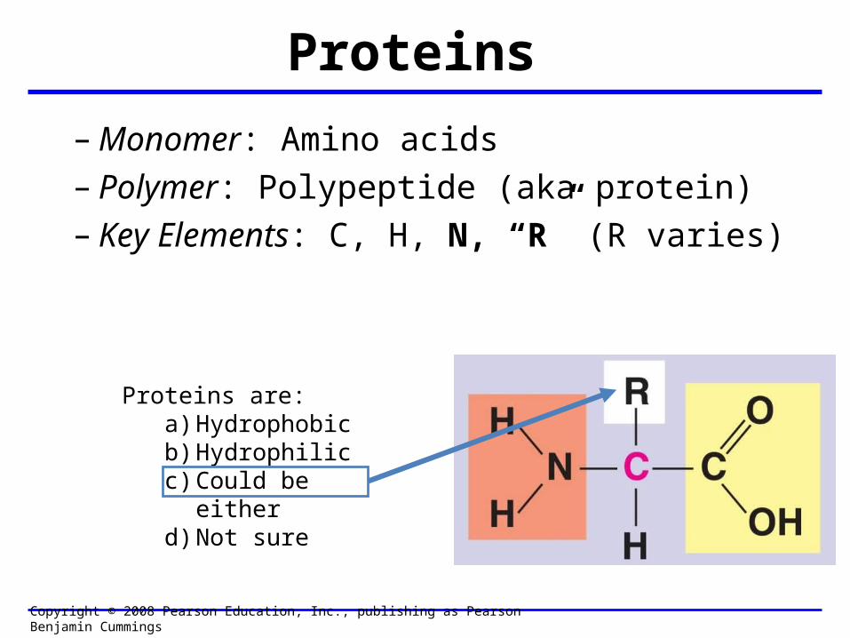
Proteins– Monomer: Amino acids– Polymer: Polypeptide (aka protein)– Key Elements: C, H, N, “R” (R varies)
Copyright © 2008 Pearson Education, Inc., publishing as Pearson Benjamin Cummings
Proteins are:a) Hydrophobicb) Hydrophilicc) Could be eitherd) Not sure
Protein Functions
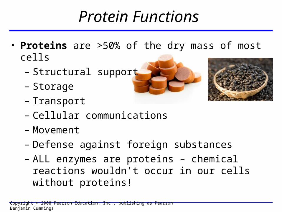
• Proteins are >50% of the dry mass of most cells– Structural support– Storage– Transport– Cellular communications– Movement– Defense against foreign substances– ALL enzymes are proteins – chemical reactions wouldn’t
occur in our cells without proteins!
Copyright © 2008 Pearson Education, Inc., publishing as Pearson Benjamin Cummings

Proteins have many diverse functions; they are the most functionally diverse type of macromolecules
1. Structural support (e.g. silk of spider webs)
2. Storage of energy & nitrogen (e.g. egg albumin)
3. Transport of substances within organisms(e.g. hemoglobin) or across membranes (e.g. aquaporins)
4. Signaling: long-distance (e.g. insulin) or short-distance; gene-regulatory proteins; receptor proteins
Functions of Proteins: Examples

Functions of Proteins: Examples
Proteins have many diverse functions; they are the most functionally diverse type of macromolecules
5. Defense against invading pathogens (e.g. antibodies in immune system)
Antibody protein Protein from flu virus
6. Movement (e.g. muscle proteins) ATP Actin and Myosin

Fig. 8.16
Enzymes speed up chemical reactions
7. Metabolic catalysts (enzymes)

Beans and otherlegumes
Corn (maize)and other grains
Lysine
Essential amino acids
Tryptophan
Isoleucine
Leucine
Phenylalanine
Threonine
Valine
MethionineFig. 41.2
So, a diet of only wheat bread and corn would not be sufficient.

Peptide bond is C-N bond between neighboring amino acids formed by removal of H2O (dehydration synthesis).
Since amino acids in proteins are bonded together by peptide bonds, proteins are also called polypeptides.
Peptidebond
Peptidebond
Side chains
Backbone
Fig. 5.18

Levels of protein structure
Fig. 5.21
PrimaryStructure
SecondaryStructure
TertiaryStructure
pleated sheet
Examples ofamino acidsubunits
helix
QuaternaryStructure
imagecent.com; alison.knitsmiths.us; bigtopshirtshop.com

Exact sequence of amino acids is called the primary structure of a protein.
Fig. 5.21
Amino acidsubunits
25
20
15
10
5
1
Primary Structure

Levels of protein structure
Fig. 5.21
PrimaryStructure
SecondaryStructure
TertiaryStructure
pleated sheet
Examples ofamino acidsubunits
helix
QuaternaryStructure

Fig. 5.21
Secondary Structure
pleated sheet
helix
Secondary structure:Helix or pleated sheet.

Pleated sheet protein often used for structural purposes
Spider’s abdominal glandssecrete silk fibers made
of structural proteinwith pleated sheets.
Fig. 5.21

Levels of protein structure
Fig. 5.21
PrimaryStructure
SecondaryStructure
TertiaryStructure
pleated sheet
Examples ofamino acidsubunits
helix
QuaternaryStructure

Polypeptidebackbone
Tertiary structure results from various kinds of interactions between atoms of the side chains (R groups).
Fig. 5.21

Levels of protein structure
Fig. 5.21
PrimaryStructure
SecondaryStructure
TertiaryStructure
pleated sheet
Examples ofamino acidsubunits
helix
QuaternaryStructure

Polypeptidechain
Chains
HemeIron
Chains
CollagenHemoglobin Fig. 5.21
Quarternary structure is
the interaction of different subunits.

Changes in primary structure can have a profound effect on protein function.
Primarystructure
Secondaryand tertiarystructures
Function
Quaternarystructure
Molecules donot associatewith oneanother; eachcarries oxygen.
Normalhemoglobin(top view)
subunit
Normal hemoglobin
7654321
GluVal His Leu Thr Pro Glu
Fig. 5.22

Primarystructure
Secondaryand tertiarystructures
Function
Quaternarystructure
Molecules doNot stickto eachother; eachcarries oxygen
Normalhemoglobin
subunit
Normal hemoglobin
7654321
GluVal His Leu Thr Pro Glu
Molecules crystallize into fiber; capacity to carry oxygengreatly reduced
Sickle-cellhemoglobin
Sickle-cell hemoglobin
7654321
ValVal His Leu Thr Pro Glu
Fig. 5.22
Exposedhydrophobicregion

Nonpolar R groups: hydrophobic
Glycine Alanine Valine Leucine Isoleucine
Fig. 5.17Amino acid R (rest) groups
Arginine HistidineAspartic acid Glutamic acid Lysine
ElectricallyCharged
R groups:hydrophilic

Primarystructure
Secondaryand tertiarystructures
Function
Quaternarystructure
Molecules doNot stickto eachother; eachcarries oxygen
Normalhemoglobin
subunit
Normal hemoglobin
7654321
GluVal His Leu Thr Pro Glu
Molecules crystallize into fiber; capacity to carry oxygengreatly reduced
Sickle-cellhemoglobin
Sickle-cell hemoglobin
7654321
ValVal His Leu Thr Pro Glu
Fig. 5.22
Exposedhydrophobicregion

Normal red bloodcells are full ofindividualhemoglobinmolecules, each carrying oxygen.
Fibers of abnormalhemoglobin deformred blood cells intosickle shape.
10 µm 10 µm
Fig. 5.22

The Genetics of Sickle Cell Anemia
• Sickle cell anemia is a recessive disorder.
• AA (homozygous dominant) don’t have symptoms.
• aa (homozygous recessive) have severe symptoms, including:
• Low body oxygen levels• Severe pain• Swelling of hands and feet• Frequent infections (spleen)• Delayed growth/development• Vision problems
• What about heterozygotes (Aa)?• Usually lack negative effects, but high stress situations can trigger symptoms
Healthyyounow.com

Why hasn’t natural selection eliminated sickle cell anemia?
Some clues:• 1 in 10 African Americans is a carrier for sickle cell anemia• Rates of sickle cell anemia are also higher in people with Mediterranean, Middle Eastern, and Indian ancestry

• Individuals with two copies of the altered gene suffer many complications (pain, infections, stroke, etc.), but are
resistant to malaria. •Individuals with one copy suffer few complications and
are also resistant.
Sickle-cell disease inheritance.http://en.wikipedia.org/wiki/Sickle-cell_disease
0–2.5%
Distribution ofmalaria caused byPlasmodium falciparum(a parasitic unicellular eukaryote)
Frequencies of thesickle-cell allele
2.5–5.0%
7.5–10.0%
5.0–7.5%
>12.5%
10.0–12.5%
Fig. 23.17
“Heterozygote Advantage”

Nonpolar R groups: hydrophobic
Glycine Alanine Valine Leucine Isoleucine
Fig. 5.17Amino acid R (rest) groups
Arginine HistidineAspartic acid Glutamic acid Lysine
ElectricallyCharged
R groups:hydrophilic

Today’s Exit Ticket
The bonds creating the primary structure of a protein are called 1)___________ and form between a 2)___ atom in one amino acid and a 3)____ atom in another amino acid.
The bonds creating the secondary structure of a protein are called 4)__________ and form between 5)___________.
The bonds creating the tertiary structure of a protein can be covalent, ionic, or hydrogen bonds, and form between 6)_______________.
7) Describe the quaternary structure of a protein.