Protein s in Serum and Urineulbld.lf1.cuni.cz/file/2531/proteins-serum-urine1617-theory.pdf ·...
19
ÚSTAV LÉKAŘSKÉ BIOCHEMIE A LABORATORNÍ DIAGNOSTIKY 1. LF UK Proteins in Serum and Urine General Medicine Lenka Fialová & Martin Vejražka translated and edited by Jan Pláteník 2016/2017
Transcript of Protein s in Serum and Urineulbld.lf1.cuni.cz/file/2531/proteins-serum-urine1617-theory.pdf ·...
ÚSTAV LÉKAŘSKÉ BIOCHEMIE A LABORATORNÍ DIAGNOSTIKY 1. LF UK
Proteins in Serum and Urine
General Medicine
Lenka Fialová & Martin Vejražka
translated and edited by Jan Pláteník
2016/2017
Proteins in serum and urine
1
1 Proteins in serum
Blood plasma or serum1 contains many different proteins, originating from various cells.
Biosynthesis of most of the serum proteins localizes to the liver; small part comes from
other tissues such as lymphocytes (immunoglobulins) and enterocytes (e.g. apoprotein B-
48). Degradation takes place in hepatocytes and the monocyto-phagocytic system, where
proteins are degraded mainly following formation a complex (e.g. antigen-antibody,
hemopexin-haptoglobin, and lipoproteins). Another way of removal of serum proteins
represents their excretion by the kidney and gastrointestinal tract.
The proteins in plasma serve numerous functions:
• keep colloidal-osmotic pressure of the intravascular fluid
• transport of many compounds (e.g. hormones, vitamins, lipids, bilirubin, drugs)
• keep acid-base balance
• nutrition
• blood clotting and fibrinolysis
• defense reactions (humoral immunity):
- specific immunity (immunoglobulins)
- non-specific immunity (complement, acute phase response proteins)
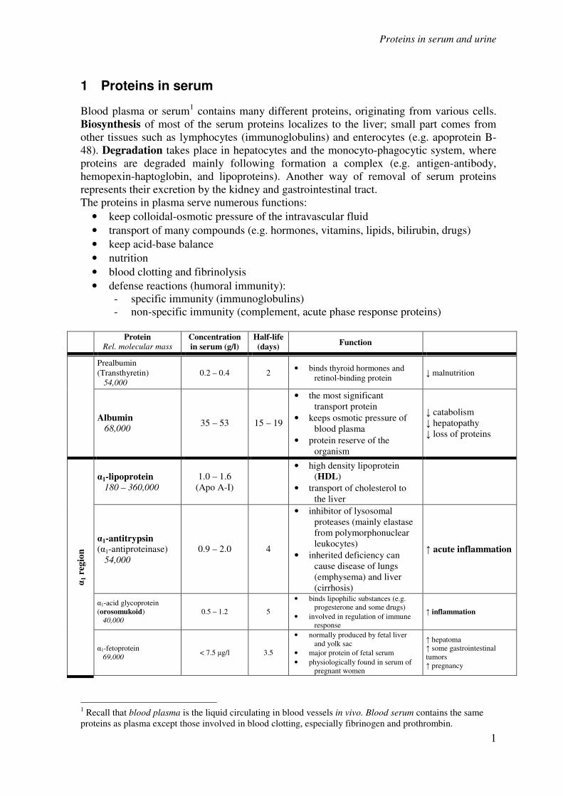
Protein Rel. molecular mass
Concentration
in serum (g/l)
Half-life
(days) Function
Prealbumin
(Transthyretin)
54,000
0.2 – 0.4 2 • binds thyroid hormones and
retinol-binding protein ↓ malnutrition
Albumin
68,000 35 – 53 15 – 19
• the most significant
transport protein
• keeps osmotic pressure of
blood plasma
• protein reserve of the
organism
↓ catabolism
↓ hepatopathy
↓ loss of proteins
α1 r
egio
n
α1-lipoprotein
180 – 360,000
1.0 – 1.6
(Apo A-I)
• high density lipoprotein
(HDL)
• transport of cholesterol to
the liver
α1-antitrypsin (α1-antiproteinase)
54,000
0.9 – 2.0 4
• inhibitor of lysosomal
proteases (mainly elastase
from polymorphonuclear
leukocytes)
• inherited deficiency can
cause disease of lungs
(emphysema) and liver
(cirrhosis)
↑ acute inflammation
α1-acid glycoprotein
(orosomukoid)
40,000
0.5 – 1.2 5
• binds lipophilic substances (e.g.
progesterone and some drugs)
• involved in regulation of immune
response
↑ inflammation
α1-fetoprotein
69,000 < 7.5 µg/l 3.5
• normally produced by fetal liver
and yolk sac
• major protein of fetal serum
• physiologically found in serum of
pregnant women
↑ hepatoma
↑ some gastrointestinal
tumors
↑ pregnancy
1 Recall that blood plasma is the liquid circulating in blood vessels in vivo. Blood serum contains the same
proteins as plasma except those involved in blood clotting, especially fibrinogen and prothrombin.
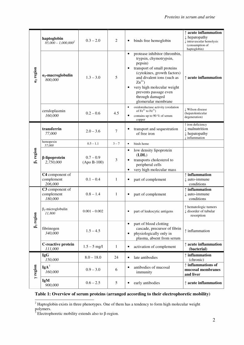
Proteins in serum and urine
2
α2 r
egio
n
haptoglobin 85,000 – 1,000,0002
0.3 – 2.0 2 • binds free hemoglobin
↑ acute inflammation
↓ hepatopathy ↓ intravascular hemolysis
(consumption of
haptoglobin)
α2-macroglobulin
800,000 1.3 – 3.0 5
• protease inhibitor (thrombin,
trypsin, chymotrypsin,
pepsin)
• transport of small proteins
(cytokines, growth factors)
and divalent ions (such as
Zn2+
)
• very high molecular weight
prevents passage even
through damaged
glomerular membrane
↑ acute inflammation
ceruloplasmin
160,000 0.2 – 0.6 4.5
• oxidoreductase activity (oxidation
of Fe2+ to Fe3+)
• contains up to 90 % of serum
copper
↓ Wilson disease
(hepatolenticular
degeneration)
β1 r
egio
n
transferrin
77,000 2.0 – 3.6 7
• transport and sequestration
of free iron
↑ iron deficiency
↓ malnutrition
↓ hepatopathy ↓ inflammation
hemopexin
57,000 0.5 – 1.1 3 – 7 • binds heme
β-lipoprotein
2,750,000
0.7 – 0.9
(Apo B-100) 3
• low density lipoprotein
(LDL)
• transports cholesterol to
peripheral cells
• very high molecular mass
C4 component of
complement
206,000
0.1 – 0.4 1 • part of complement
↑ inflammation
↓ auto-immune
conditions
β2 r
egio
n
C3 component of
complement
180,000
0.8 – 1.4 1 • part of complement
↑ inflammation
↓ auto-immune
conditions
β2-microglobulin
11,800 0.001 – 0.002 • part of leukocytic antigens
↑ hematologic tumors
↓ disorder of tubular
resorption
fibrinogen
340,000 1.5 – 4.5
• part of blood clotting
cascade, precursor of fibrin
• physiologically only in
plasma, absent from serum
↑ inflammation
C-reactive protein 111,000
1.5 – 5 mg/l 1 • activation of complement ↑ acute inflammation
(bacterial)
γ r
egio
n
IgG
150,000 8.0 – 18.0 24 • late antibodies
↑ inflammation
(chronic)
IgA3
160,000 0.9 – 3.0 6
• antibodies of mucosal
immunity
↑ inflammations of
mucosal membranes
and liver
IgM 900,000
0.6 – 2.5 5 • early antibodies ↑ acute inflammation
Table 1: Overview of serum proteins (arranged according to their electrophoretic mobility)
2 Haptoglobin exists in three phenotypes. One of them has a tendency to form high molecular weight
polymers. 3 Electrophoretic mobility extends also to β-region.
Proteins in serum and urine
3
1.1 Albumin
Albumin represents 55 – 65 % of the total serum protein (average albumin concentration in
plasma is 40 g/l). It is synthesized in the liver and its production depends on the intake of
amino acids.
• Albumin substantially contributes to maintenance of the plasma oncotic pressure.
Levels of albumin below 20 g/l are associated with edema.
• It serves as a carrier for transport of bilirubin, heme, steroids, thyroxin, free fatty acids,
bile acids, metals, drugs, and many other substances.
• It forms a protein reserve in the body and serves as a source of amino acids, especially
the essential ones, for many tissues. In malnutrition the albumin concentration decreases.
However, serum albumin is not suitable as an early marker of protein malnutrition, since
under the condition of amino acid deficiency also albumin catabolism slows down; and
albumin is mobilized from the interstitial space in order to keep the plasma oncotic
pressure. From these and other reasons (long half-life, large pool in the body) decrease in
albumin does not fully reflect the extent of protein malnutrition.
1.2 Acute phase proteins
The acute phase response is a physiologic process that occurs due to local or systemic
inflammation, tissue injury due to trauma or surgery, or tumor proliferation. In many other
situations the reaction is present in a less pronounced form, such as the condition following
an extreme physical exercise, acute heart stroke or around delivery.
Simply stated, the acute phase reaction is triggered by conditions that include destruction
of cells, reversible cell damage followed by reparation, or metabolic activation of cells
participating in the immune response.
During the acute phase reaction the involved cells produce many mediators and signaling
molecules, which in the liver (and to a lesser extent elsewhere) induce rapid changes in
protein synthesis. The proteins, whose plasmatic concentrations change markedly (over
25%) under this condition are called the acute phase proteins (acute phase reactants,
APRs). Some proteins increase in plasma (positive acute phase reactants); whereas others
decrease (negative acute phase reactants).
The APRs represent a heterogeneous collection of proteins. Nevertheless, most of them can
be classified according to its function to one of the following groups:
• Components of immune reactions: Some APRs directly participate in removal of the
agent that caused inflammation. Other proteins function in removal of damaged cells, or
modulate the immune reaction. The examples are C-reactive protein, the proteins of
complement, especially C3 and C4, tumor necrosis factor α (TNF-α), interleukin 1 (IL-
1) and interleukin 6 (IL-6).
• Protection from collateral tissue damage: During the acute phase the phagocytes and
dying cells release many substances with a potential to destroy the causative agent and
‘dissolve’ the affected tissue. Proteolytic enzymes and reactive oxygen species are the
main substances acting in this manner. Their effects must be limited to keep the
collateral damage to healthy tissue as low as possible. That is why the APRs include
protease inhibitors (α1-antitrypsin, α1-antichymotrypsin, α2-macroglobulin), and also
proteins that inhibit production and availability of reactive oxygen species, very
Proteins in serum and urine
4
often by binding and stabilizing transition metals and their complexes (haptoglobin,
hemopexin, ferritin, ceruloplasmin).
• Transport of waste by-products of inflammation: in addition to haptoglobin and
hemopexin mentioned above probably also serum amyloid A (SAA) protein would be in
this category.
• Coagulation factors and proteins participating in tissue regeneration: e.g.
fibrinogen.
Function of some of the positive acute phase proteins remains unknown, although the
protein may be significant in clinical examinations (e.g., procalcitonin, PCT).
The positive acute phase proteins can be divided according to the time dynamics of their
increase into three groups:
• Early response acute phase proteins have very short biological half-life; changes in
their levels begin to appear 6 – 10 hours after the primary insult, and peak during the
second or third day. The typical representatives are C-reactive protein (CRP) and serum
amyloid A protein (SAA). More recently, procalcitonin (PCT) is examined.
• Intermediate response acute phase proteins increase 12 – 36 hours after beginning of
the disease, and peak by the end of the first week. α1-acidic glycoprotein
(orosomucoid), α1-antitrypsin, haptoglobin, and fibrinogen belong to this group.
• Delayed response acute phase proteins are represented by C3 and C4 proteins of the
complement and ceruloplasmin. Changes in their levels are observable 48 – 72 hours
after the insult and reach maximum in 6 – 7 days.
The C-reactive protein (CRP) is one of the most significant APR’s. This protein serves as
an opsonin. It has been named after its ability to precipitate with C-polysaccharide of
Pneumococcus. The CRP increases as early as in 4 hours following the acute phase onset
and during the first two days its concentration may rise more than 100times. Maximal CRP
concentration is reached in 24 – 48 hours; its half-life is also about 24 hours.
The physiological concentration of CRP in plasma is usually below 2-8 mg/l. Rapid and
robust CRP increase, typically above 60 mg/l, occurs especially in acute bacterial
infections. In contrast, viral infections show relatively low levels of CRP (usually below
40 mg/l). Estimation of plasmatic CRP levels thus helps in decision whether an antibiotic
therapy should be started. Successful antibiotic therapy should lead to fast fall of CRP
level; in contrast, persistent infection causes lasting CRP elevation.
Examination of CRP can reveal an infection following surgery. The third day after an
operation the CRP should rapidly normalize. Persistent elevation or only a partial decrease
followed by a new elevation suggests presence of an infection or another inflammatory
complication.
A moderate increase in CRP is also associated with heart stroke. Slightly elevated CRP
levels (usually around 10 mg/l) are considered as a sign of risk of cardiovascular disease.
CRP monitoring is also useful in auto-immune diseases.
A disadvantage of CRP is its low specificity. Unlike procalcitonin it does not inform about
severity of organ damage, but only about presence of the infection. The two markers thus
complement each other.
Procalcitonin (PCT) is an acute phase protein that has recently come into focus both in
research and clinical practice. It is a protein that has 116 amino acids (MW 13,000) and is
Proteins in serum and urine
5
physiologically formed by C cells of thyroid gland as a precursor of calcitonin. Especially
during generalized bacterial infections it is also produced by other cells, mainly
monocytes, macrophages and neurocrine cells, and its plasmatic concentration swiftly
rises. The exact physiological significance of procalcitonin is rather unclear; it probably
participates in regulation of inflammation and has analgesic effects. Half-life of
procalcitonin is one day and after an immune stimulation its serum concentration rises
about 20times during 2-3 hours. An increase is observed only in generalized bacterial,
mycotic and protozoic infections, but not in viral infections. A less pronounced elevation
is seen in polytraumatic injury, burns and large abdominal surgery.
Negative acute phase reactants are proteins whose levels decrease during an acute stress
decrease. Albumin, prealbumin and transferrin behave in this manner. For monitoring of
the acute phase response, they are less significant than the positive reactants; rather, these
proteins are examined as markers of liver protein synthesis and malnutrition.
1.3 Immunoglobulins
Antibodies (immunoglobulins) are specific proteins of blood plasma with electrophoretic
mobility β - γ. They are produced by plasmatic cells as humoral immune response to a
particular antigen. The immunoglobulin molecule is able to bind specifically the antigen
against which the immunoglobulin was directed; an immune complex results. Other
functions of immunoglobulins encompass fixation of complement, binding neutrophilic
leukocytes and macrophages, and activation of phagocytosis. There are 5 classes of
immunoglobulins: IgG, IgM, IgA, IgD, and IgE. The basic structure of immunoglobulin
molecule consists of two identical heavy (H) chains, designated according to the classes
as γ, µ, α, δ, and ε; and two light (L) chains κ and λ that all the classes have in common.
Each immunoglobulin molecule contains the light chains of either κ or λ type.
The primary infection with bacteria or protozoa elicits during 2 – 3 days production of IgM
antibodies, which is 5 – 7 days later switched to production of IgG of the same specificity.
A repeated infection causes a rapid increase in IgG but only a small elevation of IgM.
Marked changes in the amount and spectrum of plasma immunoglobulins are best revealed
by electrophoresis; and can manifest as:
• Hypogammaglobulinemia: occurs due to high loss of immunoglobulins in urine or
intestine. Another relevant cause is a decrease in immunoglobulin production, which can
affect one or all the immunoglobulin classes. These defects in humoral immunity can be
primary or secondary and result in severe immune deficits manifesting themselves as
severe recurrent infections.
• Hypergammaglobulinemia:
• Polyclonal: develops as a response to antigen stimulation, in which
several clones of plasmatic cells are activated producing more immunoglobulins
of one or more classes. It is also called polyclonal gammapathy. It is found in
chronic infections, liver and auto-immune diseases. In electrophoretic
examination a broad blurred band in the γ-globulin region is found.
• Monoclonal: a single clone of plasmatic cells produces monoclonal
immunoglobulin (paraprotein) formed by a single type of heavy and the light
chain. The physico-chemical homogeneity of the paraprotein leads to a marked
sharp narrow band on electrophoresis, usually in the region of β-γ globulins.
Monoclonal immunoglobulins most often belong to the IgG and IgM class. They
Proteins in serum and urine
6
are produced either as a complete immunoglobulin molecule, or as separated
light chains (the Bence-Jones protein), or heavy chains. The Bence-Jones
protein easily passes through glomerular filter to the urine. Finding of
monoclonal immunoglobulin is symptomatic for a malign disease
plasmocytoma (multiple myeloma, tumor originating from a single clone of
plasmatic cells), or for a benign monoclonal gammapathy.
1.4 Laboratory investigation of serum proteins
The basic examination of proteins in serum or plasma is an estimation of their total
concentration, called ‘total serum protein’. If a pathologic value is found, or in other
indicated cases, further detailed examination follows, encompassing electrophoresis of
serum proteins, immunofixation and targeted estimation of concentration of selected
proteins.
1.4.1 Total serum protein
Estimation of total protein is a common and affordable test providing basic information on
protein synthesis, utilization and excretion. Many diseases alter the spectrum of serum
proteins, but only some of them alter also the total protein level.
Changes in total serum protein:
Hypoproteinemia (serum protein is low):
Absolute: amount of serum protein is decreased due to
a) high loss by:
kidney
gastrointestinal tract (e.g. intestinal inflammation)
skin (burns)
bleeding
to the ‘third space’ (e.g. abdominal cavity in ascites)
b) low protein biosynthesis (chronic liver diseases)
c) insufficient intake of protein (malnutrition)
Relative: in hyperhydration state the actual amount of serum protein is actually
unchanged, but the protein is diluted due to retention of water and salts. Concentrations of
all particular proteins are decreased proportionally.
Hyperproteinemia (serum protein is high):
Absolute: usually caused by an increased production of certain specific proteins,
such as immunoglobulins in plasmocytoma.
Relative: comes in dehydration state due to insufficient intake or excessive loss of
fluids (diarrhoe, vomiting). The total amount of protein is preserved, but apparent
concentrations of all particular proteins are proportionally increased.
Principle of total protein estimation:
The biuret reaction is used. Proteins form a violet complex with cupric salts in alkali,
suitable for photometric estimation. The resulting complex of Cu2+
ions with peptidic
bonds strongly absorbs light at 540-560 nm. The color intensity is proportional to the
concentration of protein. In general, substances containing at least two groups –CO-NH2 or
Proteins in serum and urine
7
–CO-NH- give the biuret reaction, i.e. the reaction is not specific for proteins. The simplest
compound giving the reaction with cupric salts in alkaline medium is biuret NH2-CO-NH-
CO-NH2 (dimer of urea), hence the name of the reaction. Amino acids and dipeptides do
not react.
Reference values (S-protein): 65 – 85 g/l
1.4.2 Albumin
Estimation of serum albumin is a common test, suitable especially in diseases of the liver,
kidney and assessment of nutritional state. Low serum albumin can accompany chronic
inflammations and increased catabolism in some disease states. Other causes of low or
high albumin are similar as for total serum protein.
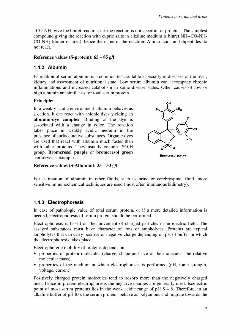
Principle:
In a weakly acidic environment albumin behaves as
a cation. It can react with anionic dyes yielding an
albumin-dye complex. Binding of the dye is
associated with a change in color. The reaction
takes place in weakly acidic medium in the
presence of surface-active substances. Organic dyes
are used that react with albumin much faster than
with other proteins. They usually contain -SO3H
group. Bromcresol purple or bromcresol green
can serve as examples.
Reference values (S-Albumin): 35 – 53 g/l
For estimation of albumin in other fluids, such as urine or cerebrospinal fluid, more
sensitive immunochemical techniques are used (most often immunoturbidimetry).
1.4.3 Electrophoresis
In case of pathologic value of total serum protein, or if a more detailed information is
needed, electrophoresis of serum protein should be performed.
Electrophoresis is based on the movement of charged particles in an electric field. The
assayed substances must have character of ions or ampholytes. Proteins are typical
ampholytes that can carry positive or negative charge depending on pH of buffer in which
the electrophoresis takes place.
Electrophoretic mobility of proteins depends on:
• properties of protein molecules (charge, shape and size of the molecules, the relative
molecular mass);
• properties of the medium in which electrophoresis is performed (pH, ionic strength,
voltage, current).
Positively charged protein molecules tend to adsorb more than the negatively charged
ones, hence in protein electrophoresis the negative charges are generally used. Isoelectric
point of most serum proteins lies in the weak acidic range of pH 5 – 6. Therefore, in an
alkaline buffer of pH 8.6, the serum proteins behave as polyanions and migrate towards the
Proteins in serum and urine
8
anode (+). Albumin bears the highest negative charge among serum proteins, and so it
moves to the anode with the highest speed.
Electrophoresis can be performed on various kinds of mechanical support. Historically, the
earliest support used was a chromatographic paper. Nowadays the protein electrophoresis
in clinical laboratories is most often done on cellulose acetate sheets or in an agarose gel.
In case of electrophoresis on paper or cellulose acetate the proteins migrate in a buffer
solution that is outside the support structure; therefore, the protein molecules separate
according to their charges. Likewise, in agarose gel of rather low concentration the protein
separation would be based on charge.4
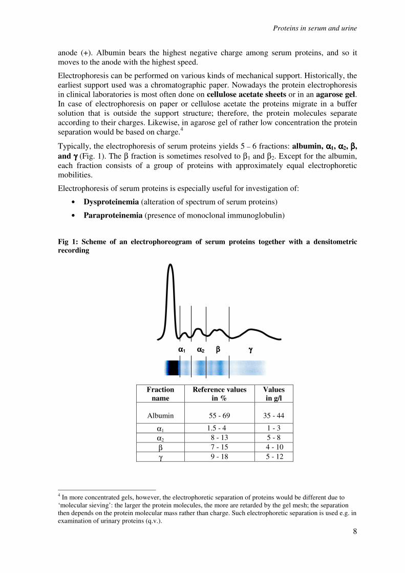
Typically, the electrophoresis of serum proteins yields 5 – 6 fractions: albumin, αααα1, αααα2, ββββ,
and γγγγ (Fig. 1). The β fraction is sometimes resolved to β1 and β2. Except for the albumin,
each fraction consists of a group of proteins with approximately equal electrophoretic
mobilities.
Electrophoresis of serum proteins is especially useful for investigation of:
• Dysproteinemia (alteration of spectrum of serum proteins)
• Paraproteinemia (presence of monoclonal immunoglobulin)
Fig 1: Scheme of an electrophoreogram of serum proteins together with a densitometric
recording
4 In more concentrated gels, however, the electrophoretic separation of proteins would be different due to
‘molecular sieving’: the larger the protein molecules, the more are retarded by the gel mesh; the separation
then depends on the protein molecular mass rather than charge. Such electrophoretic separation is used e.g. in
examination of urinary proteins (q.v.).
Fraction
name
Reference values
in %
Values
in g/l
Albumin
55 - 69
35 - 44
α1 1.5 - 4 1 - 3
α2 8 - 13 5 - 8
β 7 - 15 4 - 10
γ 9 - 18 5 - 12
αααα1 αααα2 ββββ γγγγ

Proteins in serum and urine
9
Description of particular fractions on electrophoresis of serum proteins
• Prealbumin zone In this region prealbumin (transthyretin) is found; but its band is very faint and difficult to evaluate.
• Albumin zone
Albumin forms a remarkable, rather wide, and well delineated zone. It appears weaker if albumin
concentration falls below 30 g/l. Seldom observed doubling of the albumin band can be symptomatic of a
genetic structural aberration of albumin in heterozygotes – bisalbuminemia – or happens due to binding of an
exogenous substance, such as penicillin, to albumin.
• Interzone between albumin and αααα1-globulins
Faint homogeneous staining of this area is caused mainly by αααα1-lipoproteins (HDL). The αααα1-acidic
glycoprotein displays the same electrophoretic mobility, but its contribution to the interzone staining is
minimal.
• Zone of αααα1-globulins
The α1-globulin zone is affected predominantly by presence of αααα1-antitrypsin. Acute inflammations cause
visible thickening of this zone. Genetic variability of α1-antitrypsin is clinically significant, and can manifest
also in electrophoresis as weakening or even disappearance of this zone and/or alteration of its position.
• Zone of αααα2-globulins
This zone consists mainly of two proteins - αααα2-macroglobulin and haptoglobin.
Changes in concentration of α2-macroglobulin are of minor diagnostic significance. Haptoglobin forms 6
phenotypes that differ in electrophoretic mobility. The electrophoretic examination of serum proteins,
however, does not allow identification of particular haptoglobin phenotype.
• Interzone between αααα2 and ββββ1-globulins Normally this zone is only faintly stained. In case of hemolysis the resulting complexes of hemoglobin-
haptoglobin form a band in this region.
• Zone of ββββ1-globulins
Shape and intensity of staining of the β1-globulin zone is affected almost exclusively by the presence of
transferrin. The intensity of this zone correlates well with total iron binding capacity of blood plasma. In
case of anemia due to iron deficiency as well as in pregnancy the synthesis of transferrin increases and so
also the β1-globulin zone intensity seen on serum electrophoreogram. Another protein with β1-elektrophoretic
mobility hemopexin is poorly stained with the commonly used protein stains and so changes of its
concentration do not affect appearance of serum electrophoreogram.
• Interzone between ββββ1 and ββββ2-globulins
The immunoglobulin IgA is found here, causing homogeneous staining of this region. Next, ββββ-lipoprotein
(LDL) forms a distinct band in this zone, whose visibility depends on its concentration.
• Zone of ββββ2-globulins
• The C3 component of complements contributes to this zone. The staining intensity of β2-globulins band,
however, is a poor indicator of C3 concentration.
• Zone of γγγγ-globulins
Appearance of the γ-globulin zone is affected by concentrations of the four subclasses of immunoglobulin
IgG. Elevation of IgG manifests as more intense staining and widening of this zone. Immunoglobulin IgM is
found rather close to the start. Increase in IgM concentration, isolated or accompanying the IgG elevation,
cannot be seen on electrophoresis.
Proteins in serum and urine
10
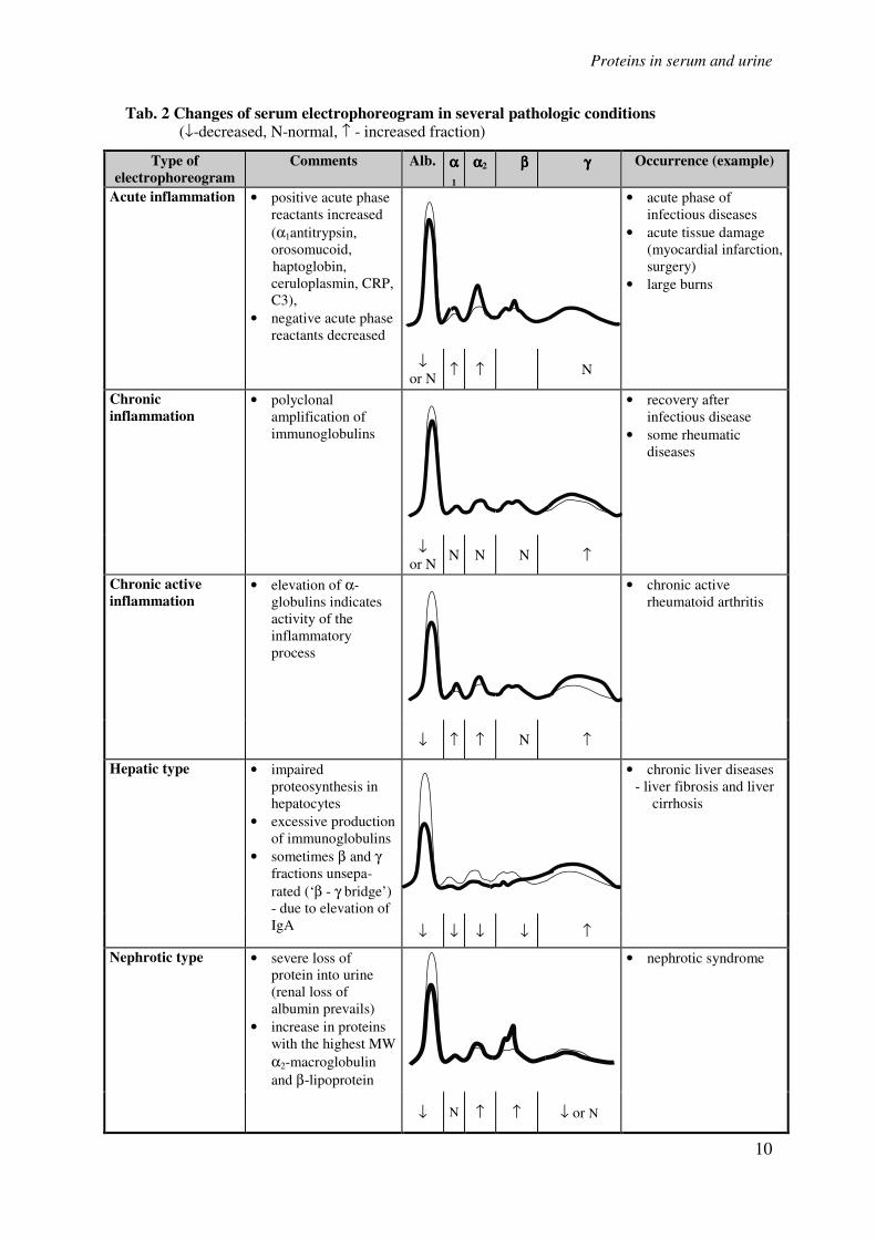
Tab. 2 Changes of serum electrophoreogram in several pathologic conditions (↓-decreased, N-normal, ↑ - increased fraction)
Type of
electrophoreogram
Comments Alb. αααα1
αααα2 ββββ γγγγ Occurrence (example)
Acute inflammation • positive acute phase
reactants increased
(α1antitrypsin,
orosomucoid,
haptoglobin,
ceruloplasmin, CRP,
C3),
• negative acute phase
reactants decreased
• acute phase of
infectious diseases
• acute tissue damage
(myocardial infarction,
surgery)
• large burns
↓ or N
↑ ↑ N
Chronic
inflammation • polyclonal
amplification of
immunoglobulins
• recovery after
infectious disease
• some rheumatic
diseases
↓ or N
N N N ↑
Chronic active
inflammation • elevation of α-
globulins indicates
activity of the
inflammatory
process
• chronic active
rheumatoid arthritis
↓ ↑ ↑ N ↑
Hepatic type • impaired
proteosynthesis in
hepatocytes
• excessive production
of immunoglobulins
• sometimes β and γ
fractions unsepa-
rated (‘β - γ bridge’)
- due to elevation of
IgA
• chronic liver diseases
- liver fibrosis and liver
cirrhosis
↓ ↓ ↓ ↓ ↑
Nephrotic type • severe loss of
protein into urine
(renal loss of
albumin prevails)
• increase in proteins
with the highest MW
α2-macroglobulin
and β-lipoprotein
• nephrotic syndrome
↓ N ↑ ↑ ↓ or N
Proteins in serum and urine
11
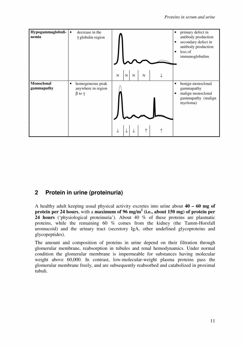
Hypogammaglobuli-
nemia • decrease in the
γ globulin region
• primary defect in
antibody production
• secondary defect in
antibody production
• loss of
immunoglobulins
N N N N ↓
Monoclonal
gammapathy • homogeneous peak
anywhere in region
β to γ
• benign monoclonal
gammapathy
• malign monoclonal
gammapathy (malign
myeloma)
↓ ↓ ↓ ↑ ↑
2 Protein in urine (proteinuria)
A healthy adult keeping usual physical activity excretes into urine about 40 – 60 mg of
protein per 24 hours, with a maximum of 96 mg/m2 (i.e., about 150 mg) of protein per
24 hours (‘physiological proteinuria’). About 40 % of these proteins are plasmatic
proteins, while the remaining 60 % comes from the kidney (the Tamm-Horsfall
uromucoid) and the urinary tract (secretory IgA, other undefined glycoproteins and
glycopeptides).
The amount and composition of proteins in urine depend on their filtration through
glomerular membrane, reabsorption in tubules and renal hemodynamics. Under normal
condition the glomerular membrane is impermeable for substances having molecular
weight above 60,000. In contrast, low-molecular-weight plasma proteins pass the
glomerular membrane freely, and are subsequently reabsorbed and catabolized in proximal
tubuli.
Proteins in serum and urine
12
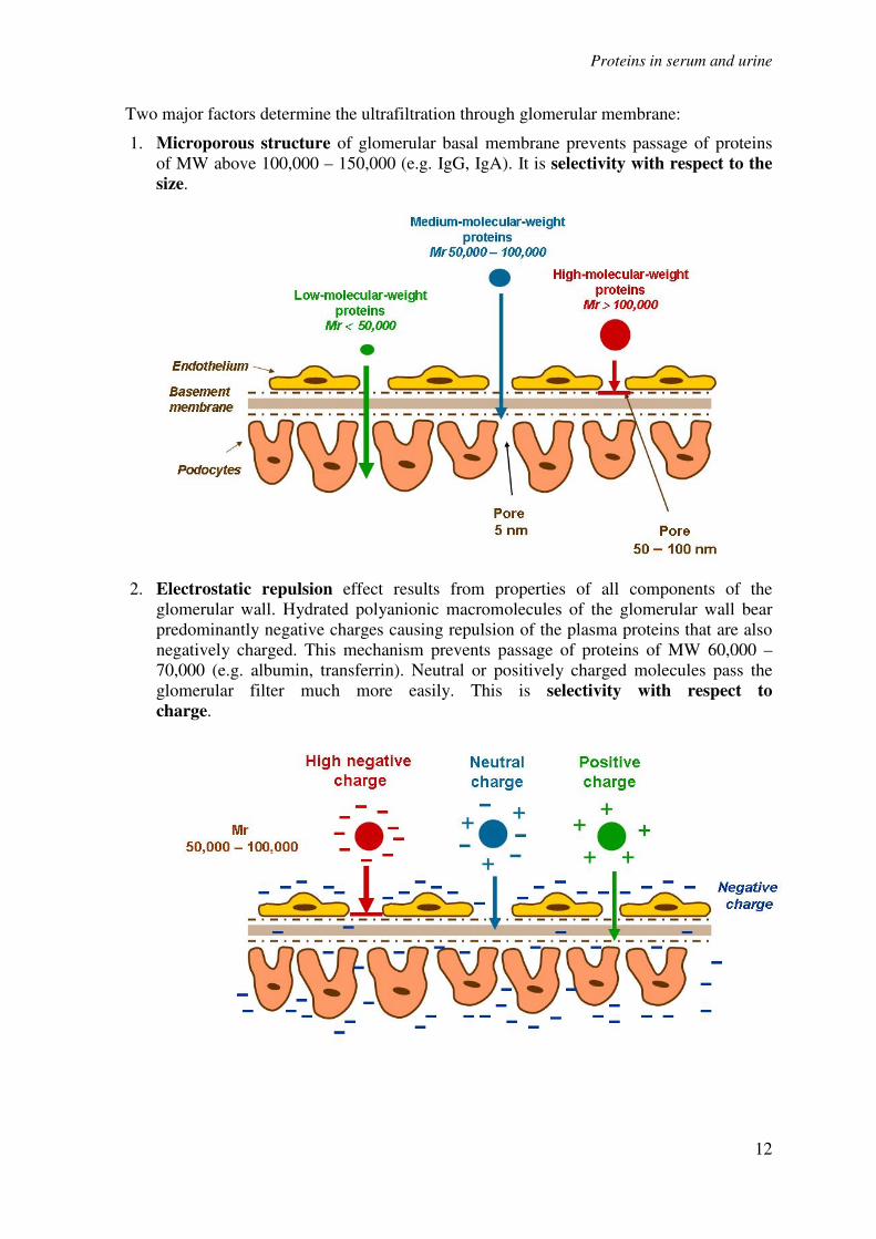
Two major factors determine the ultrafiltration through glomerular membrane:
1. Microporous structure of glomerular basal membrane prevents passage of proteins
of MW above 100,000 – 150,000 (e.g. IgG, IgA). It is selectivity with respect to the
size.
2. Electrostatic repulsion effect results from properties of all components of the
glomerular wall. Hydrated polyanionic macromolecules of the glomerular wall bear
predominantly negative charges causing repulsion of the plasma proteins that are also
negatively charged. This mechanism prevents passage of proteins of MW 60,000 –
70,000 (e.g. albumin, transferrin). Neutral or positively charged molecules pass the
glomerular filter much more easily. This is selectivity with respect to
charge.
Proteins in serum and urine
13
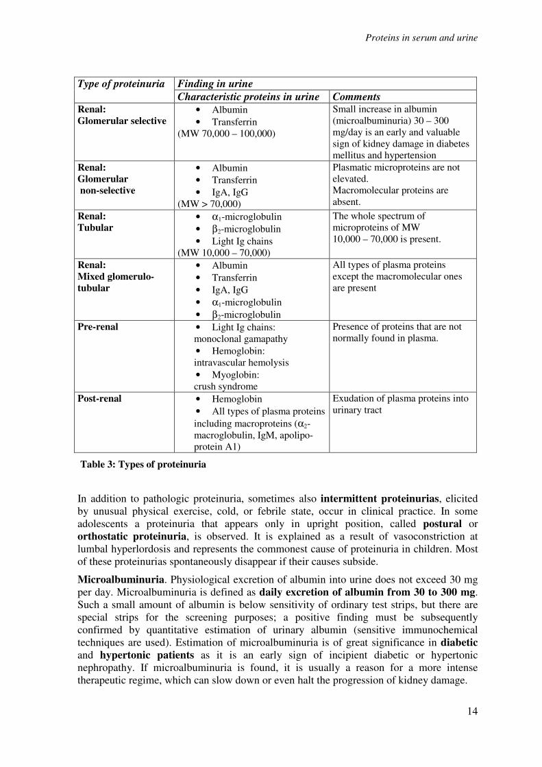
2.1 Classification of proteinuria
Occurrence of proteins in urine in the amount exceeding 150 mg per 24 hours is called a
proteinuria. It is a common sign of kidney diseases and classification of proteinuria is of
high diagnostic value. Proteinurias can be divided into three basic groups:
• Pre-renal proteinuria (“overflow” proteinuria) originates at high concentrations of
low-molecular-weight plasma proteins that even under normal condition pass into the
ultrafiltrate. Glomerular permeability for proteins need not be impaired. Tubular
reabsorption may be intact as well, but due to protein over-load some proteins escape into
urine. Pre-renal proteinuria occurs e.g. in cases of severe intravascular hemolysis, when
hemoglobin appears in the urine (hemoglobinuria); or in the crush syndrome (traumatic
damage to skeletal muscles), when myoglobin is found in urine (myoglobinuria). Presence
of immunoglobulin light chains (Bence-Jones protein) points to myeloma. The pre-renal
proteinuria often accompanies some acute inflammatory and necrotizing diseases. Tissue
degradation products as well as some small acute-phase reactants (e.g. orosomucoid) are
excreted.
• Renal proteinuria is further divided to:
- Glomerular proteinuria results from an increased permeability of the
glomerular membrane for protein. In selective glomerular proteinuria the
negative charge of glomerular membrane is lost, which results in increased
excretion of the middle-size proteins of MW 70,000 – 100,000, such as albumin
and transferrin, while large proteins are preserved. In non-selective glomerular
proteinuria the selectivity with respect to the size is impaired as well, and in
addition to the middle-size proteins also species with MW above 100,000, such
as IgG, penetrate into urine. In glomerular proteinuria the daily loss of protein
usually exceeds 2 g.
- Tubular proteinuria is caused by decreased reabsorption of normally filtrated
proteins. High excretion of low-molecular-weight proteins (microproteins) of
MW 10,000 – 70,000 (β2-microglobulin, α1-microglobulin, free Ig light chains),
which are normally reabsorbed in tubules, is found. A common cause is
damage to tubular cells by some nephrotoxic drugs (cytostatics, some
antibiotics) or heavy metals (Hg, Pb, Cd). Tubular proteinuria can also
accompany some prerenal proteinurias (paraproteinuria, myoglobinuria) due to
competition of the pathologic protein and other filtrated proteins for tubular
transport processes. Tubular proteinuria usually results in only moderate loss of
protein, typically 0.3 – 1.5 g/24 hours.
- Mixed proteinuria is a combination of non-selective glomerular and tubular
proteinuria. It is found in advanced stages of kidney diseases as a sign of
damage to all parts of the nephrons.
• Post-renal proteinuria results from bleeding, tumors and inflammations in the
urinary tract; the urine is directly contaminated by blood plasma. This condition is reliably
identified by presence of plasma proteins of very high molecular weight (α2-
macroglobulin, IgM), which do not pass the glomerular membrane even in the non-
selective glomerular proteinurias.
Proteins in serum and urine
14
Type of proteinuria Finding in urine
Characteristic proteins in urine Comments Renal:
Glomerular selective • Albumin
• Transferrin
(MW 70,000 – 100,000)
Small increase in albumin
(microalbuminuria) 30 – 300
mg/day is an early and valuable
sign of kidney damage in diabetes
mellitus and hypertension
Renal:
Glomerular
non-selective
• Albumin
• Transferrin
• IgA, IgG
(MW > 70,000)
Plasmatic microproteins are not
elevated.
Macromolecular proteins are
absent.
Renal:
Tubular • α1-microglobulin
• β2-microglobulin
• Light Ig chains
(MW 10,000 – 70,000)
The whole spectrum of
microproteins of MW
10,000 – 70,000 is present.
Renal:
Mixed glomerulo-
tubular
• Albumin
• Transferrin
• IgA, IgG
• α1-microglobulin
• β2-microglobulin
All types of plasma proteins
except the macromolecular ones
are present
Pre-renal • Light Ig chains:
monoclonal gamapathy
• Hemoglobin:
intravascular hemolysis
• Myoglobin:
crush syndrome
Presence of proteins that are not
normally found in plasma.
Post-renal • Hemoglobin
• All types of plasma proteins
including macroproteins (α2-
macroglobulin, IgM, apolipo-
protein A1)
Exudation of plasma proteins into
urinary tract
Table 3: Types of proteinuria
In addition to pathologic proteinuria, sometimes also intermittent proteinurias, elicited
by unusual physical exercise, cold, or febrile state, occur in clinical practice. In some
adolescents a proteinuria that appears only in upright position, called postural or
orthostatic proteinuria, is observed. It is explained as a result of vasoconstriction at
lumbal hyperlordosis and represents the commonest cause of proteinuria in children. Most
of these proteinurias spontaneously disappear if their causes subside.
Microalbuminuria. Physiological excretion of albumin into urine does not exceed 30 mg
per day. Microalbuminuria is defined as daily excretion of albumin from 30 to 300 mg.
Such a small amount of albumin is below sensitivity of ordinary test strips, but there are
special strips for the screening purposes; a positive finding must be subsequently
confirmed by quantitative estimation of urinary albumin (sensitive immunochemical
techniques are used). Estimation of microalbuminuria is of great significance in diabetic
and hypertonic patients as it is an early sign of incipient diabetic or hypertonic
nephropathy. If microalbuminuria is found, it is usually a reason for a more intense
therapeutic regime, which can slow down or even halt the progression of kidney damage.
Proteins in serum and urine
15
2.2 Laboratory examination of protein in urine
The laboratory examination of proteinuria aims at
1) Qualitative demonstration of protein in urine. In practice two methods are in use:
• Diagnostic strips
• Test with sulfosalicylic acid.
2) Quantitative estimation of protein in urine. If a pathologic proteinuria is found, a daily
loss of proteins in urine is measured. The urine is collected for 24 hours, mixed and a
sample is taken in which the protein concentration is measured. The urine protein
concentration in g/l multiplied by volume of urine in liters gives the daily loss of protein.
Evaluation of daily protein loss:
Protein in urine: Degree of proteinuria:
< 0.150 g/24 hours Physiological proteinuria
< 1 g/24 hours Moderate proteinuria (most often tubular)
1.0-3.5 g/24 hours Intermediate proteinuria
> 3.5 g/24 hours Severe proteinuria
> 10 g/24 hours Proteinuria associated with severe
nephrotic syndrome
3) Determination of the type of proteinuria. Electrophoretic techniques that resolve urinary
proteins according to their molecular weights provide semiquantitative information on the
spectrum of excreted proteins, enabling classification of the proteinuria. Quantitative
estimation of selected particular proteins in urine, such as albumin, α1-microglobulin, β2-
microglobulin, and IgG, is also valuable.
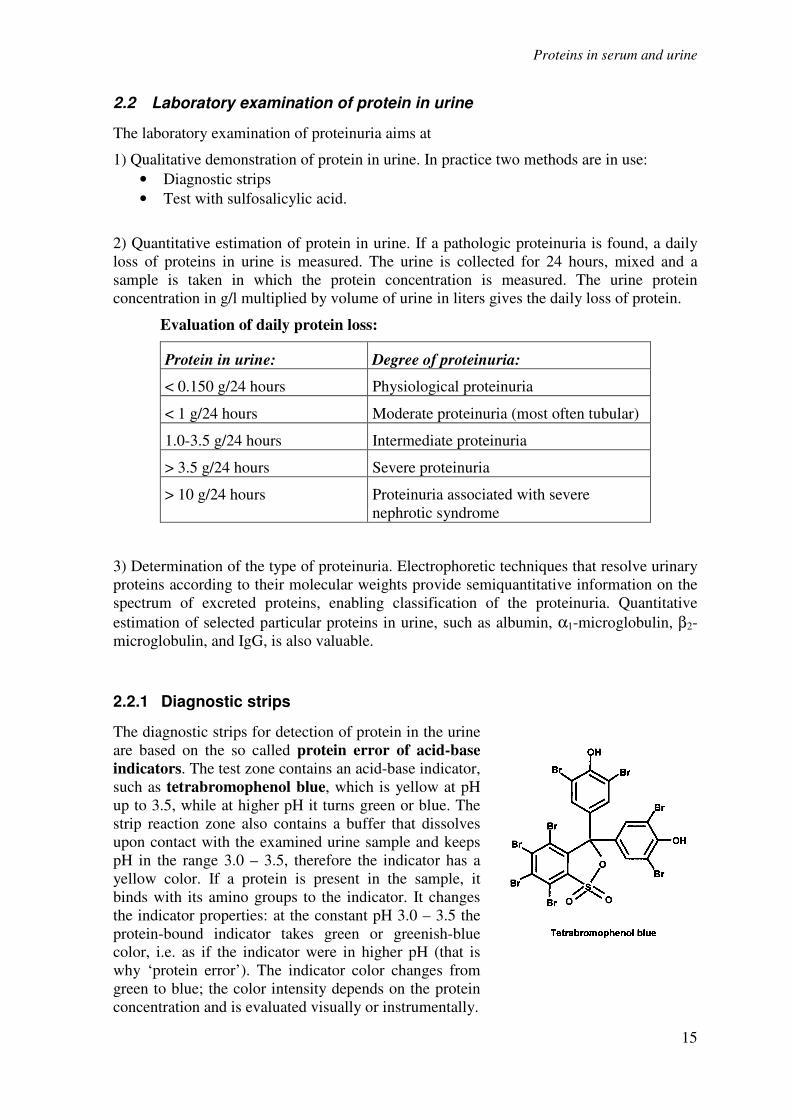
2.2.1 Diagnostic strips
The diagnostic strips for detection of protein in the urine
are based on the so called protein error of acid-base
indicators. The test zone contains an acid-base indicator,
such as tetrabromophenol blue, which is yellow at pH
up to 3.5, while at higher pH it turns green or blue. The
strip reaction zone also contains a buffer that dissolves
upon contact with the examined urine sample and keeps
pH in the range 3.0 – 3.5, therefore the indicator has a
yellow color. If a protein is present in the sample, it
binds with its amino groups to the indicator. It changes
the indicator properties: at the constant pH 3.0 – 3.5 the
protein-bound indicator takes green or greenish-blue
color, i.e. as if the indicator were in higher pH (that is
why ‘protein error’). The indicator color changes from
green to blue; the color intensity depends on the protein
concentration and is evaluated visually or instrumentally.
Proteins in serum and urine
16
With very alkaline urine samples (pH above 8), or if the urine is highly concentrated, the
test can give false positive results (due to exhaustion of the buffer in the strip reaction
zone). In these cases the sample should be acidified with diluted acetic acid to pH 5 – 6
and the test repeated. A false positive result can also appear due to high concentration of
some substances with amino groups (contamination of the sample vessel with some
disinfectants) that bind on the indicator in a similar way like the proteins do.
A disadvantage of the diagnostic strips is their different sensitivity towards particular
proteins: the strips react very well with albumin, indicating its presence in urine from 0.1 –
0.5 g/l; whereas their sensitivity to globulins, glycoproteins and the Bence-Jones protein is
much lower. These strips are not suitable for detection of microalbuminuria, in which
albumin concentration ranges between 20 and 200 mg/l (daily albumin loss 30 – 300
mg/24 hours). Special test strips for screening of microalbuminuria are available; they are
based on an immunochemical reaction.
2.2.2 Test with sulfosalicylic acid
Addition of sulfosalicylic acid to protein-containing urine sample results in protein
denaturation and cloudy appearance or even precipitate. The reaction is fairly sensitive as it
detects proteinuria from 0.1 – 0.2 g/l. Differences in sensitivity towards particular proteins
are not so pronounced as in the diagnostic strips; sulfosalicylic acid precipitates globulins
as well.
The test gives false positive results in cases of urinary excretion of some X-ray contrast
substances, penicillin, sulfonamides, salicylic acid, and antidiabetic drugs.
Semiquantitative evaluation of the sulfosalicylic acid test:
Appearance: Evaluation Approximate protein concentration:
Opalescence Traces 0.05 – 0.1 g/l
Slight turbidity (transparent,
a text behind the tube is
legible)
+ 0.1 – 0.2 g/l
Opaque turbidity (not
transparent, without flakes) ++ 0.5 – 1.0 g/l
Milky turbidity with flakes +++ 2.0 – 5.0 g/l
Cheese-like precipitate ++++ above 5.0 g/l
2.2.3 Quantitative estimation of protein in urine
The quantitative measurement of protein in urine is technically rather demanding. Three
groups of techniques are used:
1. Denaturation of proteins e.g. with trichloroacetic acid, sulfosalicylic acid, followed
by turbidimetric measurement of the degree of turbidity.
Proteins in serum and urine
17
2. Colorimetric methods with or without denaturation, e.g. the biuret method.
3. Methods based on dyes that bind to proteins in the sample, such as Coomassie
Brilliant Blue G250, or pyrogallol red.
Nowadays, the turbidimetric and pyrogallol red methods are preferred, since they are
suitable for automatic analyzers.
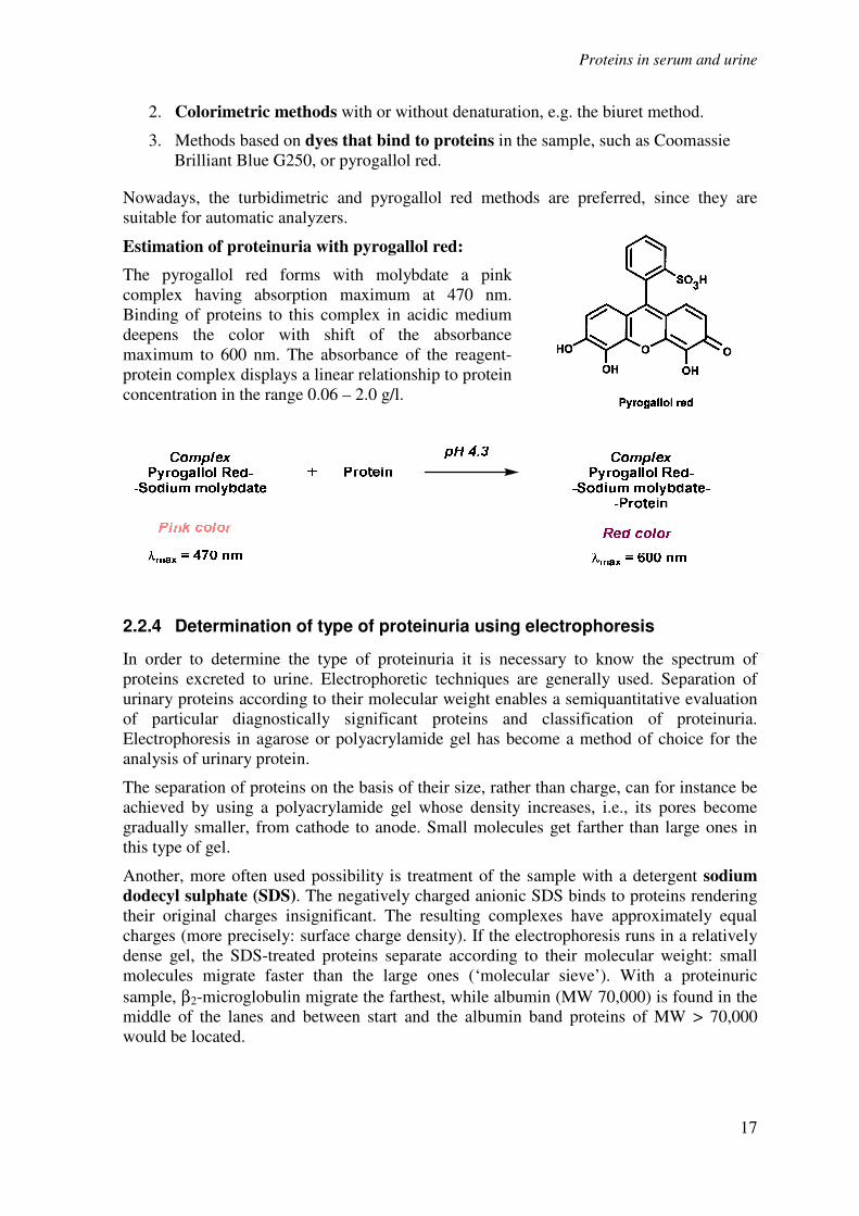
Estimation of proteinuria with pyrogallol red:
The pyrogallol red forms with molybdate a pink
complex having absorption maximum at 470 nm.
Binding of proteins to this complex in acidic medium
deepens the color with shift of the absorbance
maximum to 600 nm. The absorbance of the reagent-
protein complex displays a linear relationship to protein
concentration in the range 0.06 – 2.0 g/l.
2.2.4 Determination of type of proteinuria using electrophoresis
In order to determine the type of proteinuria it is necessary to know the spectrum of
proteins excreted to urine. Electrophoretic techniques are generally used. Separation of
urinary proteins according to their molecular weight enables a semiquantitative evaluation
of particular diagnostically significant proteins and classification of proteinuria.
Electrophoresis in agarose or polyacrylamide gel has become a method of choice for the
analysis of urinary protein.
The separation of proteins on the basis of their size, rather than charge, can for instance be
achieved by using a polyacrylamide gel whose density increases, i.e., its pores become
gradually smaller, from cathode to anode. Small molecules get farther than large ones in
this type of gel.
Another, more often used possibility is treatment of the sample with a detergent sodium
dodecyl sulphate (SDS). The negatively charged anionic SDS binds to proteins rendering
their original charges insignificant. The resulting complexes have approximately equal
charges (more precisely: surface charge density). If the electrophoresis runs in a relatively
dense gel, the SDS-treated proteins separate according to their molecular weight: small
molecules migrate faster than the large ones (‘molecular sieve’). With a proteinuric
sample, β2-microglobulin migrate the farthest, while albumin (MW 70,000) is found in the
middle of the lanes and between start and the albumin band proteins of MW > 70,000
would be located.
Proteins in serum and urine
18
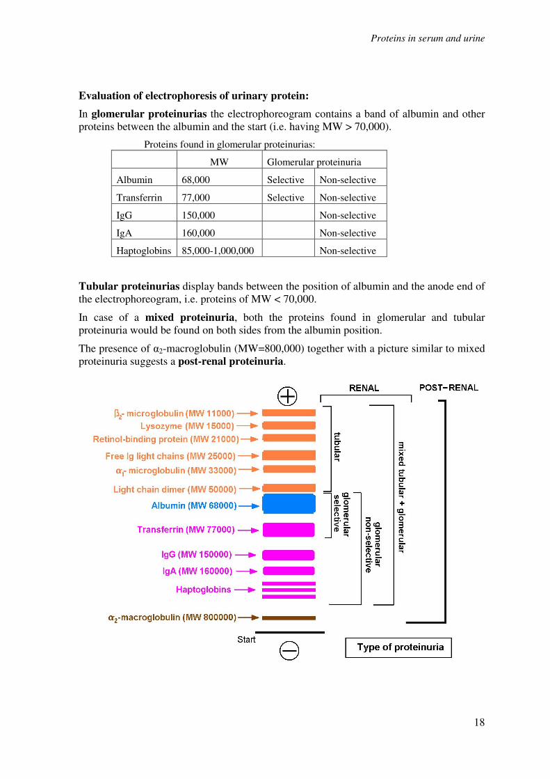
Evaluation of electrophoresis of urinary protein:
In glomerular proteinurias the electrophoreogram contains a band of albumin and other
proteins between the albumin and the start (i.e. having MW > 70,000).
Proteins found in glomerular proteinurias:
MW Glomerular proteinuria
Albumin 68,000 Selective Non-selective
Transferrin 77,000 Selective Non-selective
IgG 150,000 Non-selective
IgA 160,000 Non-selective
Haptoglobins 85,000-1,000,000 Non-selective
Tubular proteinurias display bands between the position of albumin and the anode end of
the electrophoreogram, i.e. proteins of MW < 70,000.
In case of a mixed proteinuria, both the proteins found in glomerular and tubular
proteinuria would be found on both sides from the albumin position.
The presence of α2-macroglobulin (MW=800,000) together with a picture similar to mixed
proteinuria suggests a post-renal proteinuria.