Protein Glycosylation in Helicobacter pylori: …publish.uwo.ca/~ccreuzen/Publications/2011 -...
18
Protein Glycosylation in Helicobacter pylori: Beyond the Flagellins? Patrick S. Hopf, Rachel S. Ford, Najwa Zebian, Alexandra Merkx-Jacques, Somalinga Vijayakumar, Dinath Ratnayake, Jacqueline Hayworth, Carole Creuzenet* Infectious Diseases Research Group, Department of Microbiology and Immunology, The University of Western Ontario, London, Ontario, Canada Abstract Glycosylation of flagellins by pseudaminic acid is required for virulence in Helicobacter pylori. We demonstrate that, in H. pylori, glycosylation extends to proteins other than flagellins and to sugars other than pseudaminic acid. Several candidate glycoproteins distinct from the flagellins were detected via ProQ-emerald staining and DIG- or biotin- hydrazide labeling of the soluble and outer membrane fractions of wild-type H. pylori, suggesting that protein glycosylation is not limited to the flagellins. DIG-hydrazide labeling of proteins from pseudaminic acid biosynthesis pathway mutants showed that the glycosylation of some glycoproteins is not dependent on the pseudaminic acid glycosylation pathway, indicating the existence of a novel glycosylation pathway. Fractions enriched in glycoprotein candidates by ion exchange chromatography were used to extract the sugars by acid hydrolysis. High performance anion exchange chromatography with pulsed amperometric detection revealed characteristic monosaccharide peaks in these extracts. The monosaccharides were then identified by LC-ESI-MS/MS. The spectra are consistent with sugars such as 5,7-diacetamido-3,5,7,9-tetradeoxy-L-glycero-L- manno-nonulosonic acid (Pse5Ac7Ac) previously described on flagellins, 5-acetamidino-7-acetamido-3,5,7,9-tetradeoxy-L- glycero-L-manno-nonulosonic acid (Pse5Am7Ac), bacillosamine derivatives and a potential legionaminic acid derivative (Leg5AmNMe7Ac) which were not previously identified in H. pylori. These data open the way to the study of the mechanism and role of protein glycosylation on protein function and virulence in H. pylori. Citation: Hopf PS, Ford RS, Zebian N, Merkx-Jacques A, Vijayakumar S, et al. (2011) Protein Glycosylation in Helicobacter pylori: Beyond the Flagellins? PLoS ONE 6(9): e25722. doi:10.1371/journal.pone.0025722 Editor: Olivier Neyrolles, Institut de Pharmacologie et de Biologie Structurale, France Received May 27, 2011; Accepted September 9, 2011; Published September 30, 2011 Copyright: ß 2011 Hopf et al. This is an open-access article distributed under the terms of the Creative Commons Attribution License, which permits unrestricted use, distribution, and reproduction in any medium, provided the original author and source are credited. Funding: This work was supported in part by operating grant MOP-62775 from the Canadian Institutes of Health Research (CIHR, http://www.cihr-irsc.gc.ca) and in part by funding from the Schulich School of Medicine and Dentistry of the University of Western Ontario (http://www.schulich.uwo.ca/) to Dr. Creuzenet. Dr. Creuzenet was the recipient of a University Faculty Award from the Natural Sciences and Engineering Research Council of Canada, and of a Premier’s Research Excellence Award (Ontario). A. Merkx-Jacques was the recipient of a Canadian Digestive Health Foundation/CIHR Doctoral Research Award. J. Hayworth was the recipient of a summer scholarship from the Canadian Association of Gastroenterologists, and P. Hopf was the recipient of an Ontario Graduate Scholarship. The funders had no role in study design, data collection and analysis, decision to publish, or preparation of the manuscript. Competing Interests: The authors have declared that no competing interests exist. * E-mail: [email protected] Introduction Helicobacter pylori chronically infects 50% of the world’s population and causes gastritis, gastric ulcers and cancers [1,2]. There is a ,6-fold increased risk of gastric cancer after H. pylori infection, and gastric cancer is the second most common cause of death from cancer [3]. Therefore, H. pylori causes a huge burden on the economy and health care system. The efficacy of current treatments is threatened by the emergence of antibiotic resistance [4,5]. Hence, novel treatments are urgently needed. Abundant research has identified a large array of virulence factors in H. pylori, including lipopolysaccharide, adhesins, toxins, urease, the Cag pathogenicity island and flagella (Reviewed in [6]). The flagellins (FlaA, FlaB) that make up the flagellar filament are O- glycosylated, and their glycosylation is essential for flagella production and virulence [7,8,9,10]. Likewise, glycosylation of the flagellins occurs and is also necessary for flagella formation in the closely related Campylobacter jejuni [11,12,13]. Protein glyco- sylation has now been described for many other prokaryotic proteins. While the precise role of glycosylation on the function of bacterial proteins is not well understood, glycosylation appears to contribute to the virulence of a number of pathogens [14,15,16,17,18,19,20] and potentially to tolerance in human symbionts [21]. Bacterial protein glycosylation is very diverse in terms of the size, composition and structure of the oligosaccharides present, and the sugars can be derived from O-antigen synthesis pathways or stem from dedicated pathways (Reviewed in [22]). The cellular location and mechanism of glycosylation also vary, encompassing transfer of complex glycans onto their target protein in the periplasm, or stepwise addition of single sugars in the cytoplasm. To date, C. jejuni is the only known bacterium with both N- and O- protein glycosylation pathways. The O-glycosylation pathway targets the flagellins that are modified by pseudaminic acid (PA) derivatives and is conserved in H. pylori [13,23,24]. In contrast, the N- glycosylation pathway appears unique and was proposed to glycosylate ,38 C. jejuni proteins by a diacetamidobacillosamine (DAB)-containing heptasaccharide [25,26]. Both pathways have been characterized at the biochemical level [23,24,27,28,29,30,31,32,33]. Of relevance to this study, the PA biosynthesis pathway is initiated by the UDP-GlcNAc C6 dehydratase FlaA1 (HP0840) and the aminotransferase HP0366. While abundant literature is available on protein glycosylation in C. jejuni, little is known about glycosylation of proteins other PLoS ONE | www.plosone.org 1 September 2011 | Volume 6 | Issue 9 | e25722
Transcript of Protein Glycosylation in Helicobacter pylori: …publish.uwo.ca/~ccreuzen/Publications/2011 -...

Protein Glycosylation in Helicobacter pylori: Beyond theFlagellins?Patrick S. Hopf, Rachel S. Ford, Najwa Zebian, Alexandra Merkx-Jacques, Somalinga Vijayakumar,
Dinath Ratnayake, Jacqueline Hayworth, Carole Creuzenet*
Infectious Diseases Research Group, Department of Microbiology and Immunology, The University of Western Ontario, London, Ontario, Canada
Abstract
Glycosylation of flagellins by pseudaminic acid is required for virulence in Helicobacter pylori. We demonstrate that, in H.pylori, glycosylation extends to proteins other than flagellins and to sugars other than pseudaminic acid. Several candidateglycoproteins distinct from the flagellins were detected via ProQ-emerald staining and DIG- or biotin- hydrazide labeling ofthe soluble and outer membrane fractions of wild-type H. pylori, suggesting that protein glycosylation is not limited to theflagellins. DIG-hydrazide labeling of proteins from pseudaminic acid biosynthesis pathway mutants showed that theglycosylation of some glycoproteins is not dependent on the pseudaminic acid glycosylation pathway, indicating theexistence of a novel glycosylation pathway. Fractions enriched in glycoprotein candidates by ion exchange chromatographywere used to extract the sugars by acid hydrolysis. High performance anion exchange chromatography with pulsedamperometric detection revealed characteristic monosaccharide peaks in these extracts. The monosaccharides were thenidentified by LC-ESI-MS/MS. The spectra are consistent with sugars such as 5,7-diacetamido-3,5,7,9-tetradeoxy-L-glycero-L-manno-nonulosonic acid (Pse5Ac7Ac) previously described on flagellins, 5-acetamidino-7-acetamido-3,5,7,9-tetradeoxy-L-glycero-L-manno-nonulosonic acid (Pse5Am7Ac), bacillosamine derivatives and a potential legionaminic acid derivative(Leg5AmNMe7Ac) which were not previously identified in H. pylori. These data open the way to the study of the mechanismand role of protein glycosylation on protein function and virulence in H. pylori.
Citation: Hopf PS, Ford RS, Zebian N, Merkx-Jacques A, Vijayakumar S, et al. (2011) Protein Glycosylation in Helicobacter pylori: Beyond the Flagellins? PLoSONE 6(9): e25722. doi:10.1371/journal.pone.0025722
Editor: Olivier Neyrolles, Institut de Pharmacologie et de Biologie Structurale, France
Received May 27, 2011; Accepted September 9, 2011; Published September 30, 2011
Copyright: � 2011 Hopf et al. This is an open-access article distributed under the terms of the Creative Commons Attribution License, which permitsunrestricted use, distribution, and reproduction in any medium, provided the original author and source are credited.
Funding: This work was supported in part by operating grant MOP-62775 from the Canadian Institutes of Health Research (CIHR, http://www.cihr-irsc.gc.ca) andin part by funding from the Schulich School of Medicine and Dentistry of the University of Western Ontario (http://www.schulich.uwo.ca/) to Dr. Creuzenet.Dr. Creuzenet was the recipient of a University Faculty Award from the Natural Sciences and Engineering Research Council of Canada, and of a Premier’s ResearchExcellence Award (Ontario). A. Merkx-Jacques was the recipient of a Canadian Digestive Health Foundation/CIHR Doctoral Research Award. J. Hayworth was therecipient of a summer scholarship from the Canadian Association of Gastroenterologists, and P. Hopf was the recipient of an Ontario Graduate Scholarship. Thefunders had no role in study design, data collection and analysis, decision to publish, or preparation of the manuscript.
Competing Interests: The authors have declared that no competing interests exist.
* E-mail: [email protected]
Introduction
Helicobacter pylori chronically infects 50% of the world’s
population and causes gastritis, gastric ulcers and cancers [1,2].
There is a ,6-fold increased risk of gastric cancer after H. pylori
infection, and gastric cancer is the second most common cause of
death from cancer [3]. Therefore, H. pylori causes a huge burden
on the economy and health care system. The efficacy of current
treatments is threatened by the emergence of antibiotic resistance
[4,5]. Hence, novel treatments are urgently needed. Abundant
research has identified a large array of virulence factors in H.
pylori, including lipopolysaccharide, adhesins, toxins, urease, the
Cag pathogenicity island and flagella (Reviewed in [6]). The
flagellins (FlaA, FlaB) that make up the flagellar filament are O-
glycosylated, and their glycosylation is essential for flagella
production and virulence [7,8,9,10]. Likewise, glycosylation of
the flagellins occurs and is also necessary for flagella formation in
the closely related Campylobacter jejuni [11,12,13]. Protein glyco-
sylation has now been described for many other prokaryotic
proteins. While the precise role of glycosylation on the function of
bacterial proteins is not well understood, glycosylation appears
to contribute to the virulence of a number of pathogens
[14,15,16,17,18,19,20] and potentially to tolerance in human
symbionts [21].
Bacterial protein glycosylation is very diverse in terms of the
size, composition and structure of the oligosaccharides present,
and the sugars can be derived from O-antigen synthesis pathways
or stem from dedicated pathways (Reviewed in [22]). The cellular
location and mechanism of glycosylation also vary, encompassing
transfer of complex glycans onto their target protein in the
periplasm, or stepwise addition of single sugars in the cytoplasm.
To date, C. jejuni is the only known bacterium with both N- and O-
protein glycosylation pathways. The O-glycosylation pathway targets
the flagellins that are modified by pseudaminic acid (PA) derivatives
and is conserved in H. pylori [13,23,24]. In contrast, the N-
glycosylation pathway appears unique and was proposed to
glycosylate ,38 C. jejuni proteins by a diacetamidobacillosamine
(DAB)-containing heptasaccharide [25,26]. Both pathways have been
characterized at the biochemical level [23,24,27,28,29,30,31,32,33].
Of relevance to this study, the PA biosynthesis pathway is initiated by
the UDP-GlcNAc C6 dehydratase FlaA1 (HP0840) and the
aminotransferase HP0366.
While abundant literature is available on protein glycosylation
in C. jejuni, little is known about glycosylation of proteins other
PLoS ONE | www.plosone.org 1 September 2011 | Volume 6 | Issue 9 | e25722

than flagellins in H. pylori. We observed that inactivation of the PA
pathway by disruption of the flaA1 gene affects virulence factor
production beyond the lack of flagellum [10]. In this manuscript,
we show that this is also the case upon disruption of hp0366. This
suggests that, in contrast to the current dogma, PA-dependent
protein glycosylation is not limited to the flagellins in H. pylori. Our
data suggest that the PA pathway also targets proteins other than
the flagellins. Furthermore, our data indicate that a novel PA-
independent glycosylation pathway exists in H. pylori and
glycosylates numerous proteins. Mass spectrometry analyses
performed on sugars extracted from candidate glycoproteins
provided hits consistent with novel carbohydrates not previously
identified in H. pylori. Combined with the identification of the
novel glycoprotein candidates, this work will eventually also allow
us to determine the role of glycosylation on protein function, an
area that has remained elusive for bacterial proteins.
Results
Disruption of the PA biosynthesis pathway affects theproduction of multiple virulence factors
Mutants with a disrupted PA biosynthesis pathway were
constructed to determine the impact of the PA pathway on the
production of non-flagellar virulence factors. The flaA1 mutant, in
which the first step in PA biosynthesis is disrupted (Fig 1), was
described previously [10]. A hp0366 mutant, in which the second
step of the PA pathway is disrupted (Fig 1), was constructed for this
study in a similar manner, via insertion of a kanamycin resistance
cassette in hp0366. This mutant grew at rates comparable with
wild-type (WT) (Data not shown). Like the flaA1 mutant, the
hp0366 mutant was non-motile (Data not shown) and did not
produce flagella (Fig 2A) although it still produced flagellins (Data
not shown). Inactivation of flaA1 or hp0366 also affected LPS
synthesis. Specifically, the flaA1 mutant produced altered O-
antigen [10] and the hp0366 mutant lacked O-antigen (Fig 2B).
This is despite the fact that the O-antigen sugars of the
lipopolysaccharide (LPS) do not comprise PA and are synthesized
by enzymes distinct from FlaA1 and HP0366 (Fig 1)
[34,35,36,37,38,39]. Also, both mutants had reduced urease
activity ((Fig 2C and [10]) as demonstrated by direct comparison
of the OD565nm values obtained for the wild-type and mutant
extracts used at the same concentrations. All phenotypes were
gene-specific and could be complemented by introduction of the
corresponding gene in trans (Fig 2 and [10]).
Furthermore, H. pylori is known to adhere to and invade gastric
cells [40,41,42,43]. Adhesion leads to alterations of cellular
signaling that are important for pathogenicity [44,45,46]. In
C. jejuni, protein glycosylation affects interactions of the bacterium
with intestinal cells [47,48]. Therefore the impact of inactivation of
hp0366 on adhesion and invasion of host cells was tested. Under
our experimental conditions, ,8% and 1.5% of the WT inoculum
adhered to and invaded gastric cells, respectively. These data are
on par with the literature and reflect the fact that invasion is very
inefficient [49,50]. Inactivation of hp0366 resulted in reduced
invasion of gastric AGS cells, while no effect was observed on
adherence (Fig 2D). Under the conditions used for adhesion and
invasion assays, urease activity and flagella-mediated activity do
not play any role since a neutral pH is maintained throughout the
experiments and the bacteria are forced to make contact with the
epithelial cells via centrifugation. Furthermore, the lack of effect of
hp0366 disruption on adhesion indicates that the flagella do not
serve as adhesins. Therefore, the disruption of the PA pathway,
and not the lack of flagellum assembly, appears to be directly
responsible for the differences seen in bacterial invasion in the
hp0366 mutant. Although the reason is not clear, the introduction
of the complementation plasmid in the WT strain led to decreased
invasion. Therefore, complementation for this phenotype was
assessed by comparing the levels of invasion of the WT and
mutant strains, each harboring the complementation vector. No
significant differences could be observed between the two
complemented strains, whereas statistically significant differences
were observed between the original strains.
Altogether, these data indicate that, in H. pylori, the PA pathway
is essential for the production of multiple virulence factors (urease,
LPS) beyond the flagellum, and is also important for interactions
with host cells. The fact that the flaA1 and hp0366 mutants, that
both lack assembled flagella but have disruptions at different steps
of the PA pathway, present different phenotypes in terms of LPS
synthesis suggests that the effects observed on virulence factor
production are related to inactivation of the PA pathway rather
than to the lack of flagellum assembly.
Pleiotropic or transcriptional effects do not explain themultiple effects of the PA pathway on production ofvirulence factors
Beyond the previously reported lack of membrane-associated
UreA and UreB in the flaA1 mutant [10], no significant differences
were observed in the inner and outer membrane proteins of the
flaA1 and hp0366 mutants compared with the wild-type (WT) (Data
not shown). This indicates that the differences of production of
surface-associated virulence factors are not due to pleiotropic effects
that would have resulted in the instability of the bacterial cell wall.
Also, the coordinated impact of PA synthesis disruption on
several apparently independent virulence factors may involve
transcriptional regulation. In H. pylori, joint transcriptional
regulation of urease and flagellum production occurs via the
flagellar flbA gene [51,52], and hp0366 belongs to the flbA regulon
[53]. Therefore, we measured the levels of expression of genes
involved in virulence factor production in the flaA1 and hp0366
mutants by quantitative real time PCR (qRT-PCR). The genes
tested comprised genes for urease and its accessory proteins, LPS
biosynthetic enzymes (synthases, transferases, O-antigen ligase)
and the flagellar regulator FlbA.
Figure 1. Schematic representation of key steps of the PA andLPS biosynthesis pathways. The main enzymes responsible for thesynthesis of sugar nucleotides are indicated, including the dehydrataseFlaA1 and the aminotransferase HP0366 relevant to this work. The linksbetween both pathways are highlighted, namely a shared precursorUDP-GlcNAc, and the potential glycosylation of LPS assembly enzymesby PA.doi:10.1371/journal.pone.0025722.g001
Protein Glycosylation in Helicobacter pylori
PLoS ONE | www.plosone.org 2 September 2011 | Volume 6 | Issue 9 | e25722

Concerning urease production, the only significant variation was
a slight down-regulation of the urease accessory gene cluster
observed in the flaA1 mutant only (Fig 3), while both mutants show
reduced urease enzymatic activity. Therefore, transcriptional
regulation does not explain fully the decrease in urease activity of
the two mutants. Concerning LPS biosynthetic genes, no significant
decrease was observed in the transcription of any of the genes tested
in the hp0366 mutant, and a slight up-regulation of the O-antigen
ligase gene waaL was observed in the flaA1 mutant only. Therefore,
transcriptional regulation does not explain the lack of O-antigen of
the hp0366 mutant or the reduced amount of O-antigen of the flaA1
mutant. Finally, for the flagellar genes, there were no transcriptional
differences in the flaA1 mutant, and there was a ,50 fold up-
regulation of flaB in the hp0366 mutant, which, if anything, should
contribute to enhanced flagellum production. This does not
correlate with the observation that both mutants were aflagellate.
Note that the differential transcriptional regulation of flaA and flaB
observed in the hp0366 mutant is consistent with the fact that
flagellin genes are under the control of different alternative sigma
factors: sigma 28 for FlaA and sigma 54 for FlaB [53].
Overall, these data suggest that transcriptional regulation is not
causing the observed interconnection of flagella, LPS and urease
production in the PA mutants. In light of the fact that the observed
phenotypes of the flaA1 and hp0366 mutants are gene specific and
do not result from pleiotropic nor transcriptional regulatory
effects, we proposed that the PA pathway may glycosylate proteins
involved in the production of virulence factors other than the
flagellins, and that their glycosylation is necessary for their
function. These include, for example, enzymes involved in LPS
assembly (Fig 1). Therefore, the experiments described below were
aimed at demonstrating the existence of such proteins, and
characterizing their carbohydrate content.
ProQ-emerald staining suggests that H. pylori producesnumerous glycoproteins
To investigate whether H. pylori produces additional glycopro-
teins (GPs), the total proteins were labeled with the fluorescent Cy5
dye, resolved by 2D SDS-PAGE, and the GPs were detected by
ProQ-emerald staining after oxidation of the sugars (Fig 4). While
Figure 2. Impact of disruption of hp0366 on virulence factor production and interactions with gastric cells. Panel A: Analysis offlagellum production by electron microscopy. Arrows point at flagella. Panel B: Analysis of LPS production. LPS was extracted from the WT, mutantand complemented strain (Compl.) and analyzed by SDS-PAGE and silver staining. Panel C: Analysis of the urease activity of WT, mutant andcomplemented (Compl.) H. pylori. Urease activity was measured using phenol red as an indicator [92]. Experiments were done with 3 differentconcentrations of soluble protein extracts and the same trend was observed. The data shown were obtained with the highest amount of proteintested and are the average of three replicas. Panel D: Adherence and invasion of H. pylori WT, mutant and complemented strains to gastric AGS cells.The data are expressed in % of adherence or invasion of WT H. pylori. The data are the average of 3 independent experiments. Compl. indicates thatthe wild-type hp0366 gene was introduced in the strain of interest on a shuttle plasmid. The adherence of WT amounted to 8–10% of the inoculum.No statistical differences (t-test) were observed between WT and mutant strain for adherence. Invasion was measured after elimination of non-internalized bacteria by gentamycin treatment. Invasion represented ,1.5% of the inoculum for the WT. Statistical differences were observedbetween the WT and mutant (shown by asterisk, t-test, p,0.001).doi:10.1371/journal.pone.0025722.g002
Protein Glycosylation in Helicobacter pylori
PLoS ONE | www.plosone.org 3 September 2011 | Volume 6 | Issue 9 | e25722

the H. pylori genome only encodes ,1600 ORFs, 2,300 protein spots
were detected by Cy5 labeling. This suggests that numerous
proteins exist in multiple isoforms due to post-translational
modifications. Out of these, 756 spots were detected using the
ProQ-emerald reagent. The fact that some abundant proteins did
not react with the ProQ-emerald stain whereas some low
abundance proteins did react indicates that the ProQ-emerald
reagent is specific for glycoproteins. However several highly
abundant proteins reacted non-specifically with ProQ-emerald,
likely due to the stringent conditions used for the oxidation of the
sugars. To eliminate false positive proteins, the ratio of intensities of
the ProQ-emerald and Cy5 signals was calculated for a subset of 100
Figure 3. qRT-PCR analysis of the impact of the flaA1 or hp0366 inactivation on transcription of genes involved in urease, LPS andflagellum production. All data were normalized to the level of expression of the housekeeping gene hp1045, the acetyl-CoA synthase.Differences are considered significant for .5 fold difference compared with WT. The interval for non significant variation (NSV) is delimited by dottedlines. Error bars are for duplicate experiments done using the same sample of RNA. The same experiment was also repeated on RNA diluted 1/10 (v/v)and the same trend was observed (not shown). UreA/B: operon encoding both UreA and UreB. UreIEFGH: operon encoding 5 urease accessoryproteins. FucST: fucose synthase. FucT: fucose transferase. GalT: galactose transferase. WaaL: O-antigen ligase. FlaA and FlaB: flagellins A and B. FlhA:flagella basal body and transcriptional regulator, also known as FlbA.doi:10.1371/journal.pone.0025722.g003
Figure 4. 2D gel electrophoresis analysis of H. pylori glycoproteins using Cy5 labeling of total proteins and ProQ-emerald labelingof glycoproteins. The proteins from WT H. pylori were stained with Cy5 and were resolved by 2D gel electrophoresis. Glycoproteins were stainedwith ProQ-emerald. Abundant proteins gave a high background reactivity by ProQ-emerald labeling (ex: UreB). To eliminate false positive proteins,the ratio of the ProQ-emerald and Cy5 signals was calculated and only proteins that showed a high ratio were considered GP candidates. The ratioscalculated are indicated for a few spots shown as an example in the zoomed figures. Spots A and B are provided as examples of non glycosylatedproteins. Signals for Cy5 and ProQ-emerald are in arbitrary units. Contributions from the gel background have already been subtracted. Note that theanalysis was limited to a subset of 100 proteins that were present in sufficient amounts to allow their identification by MS ultimately. The 12 spotshighlighted on the figure (in circles and diamond) are the ones with the highest ratios in this subset and represent GPs. Additional GPs may bepresent.doi:10.1371/journal.pone.0025722.g004
Protein Glycosylation in Helicobacter pylori
PLoS ONE | www.plosone.org 4 September 2011 | Volume 6 | Issue 9 | e25722

proteins that were present in high enough amount (based on Cy5
signal) to allow downstream identification by MS. Spots that had
very low ratios (0 – 0.09) correspond to very weakly ProQ-emerald
labeled proteins that probably stained in a non specific manner and
were not considered for further analysis. These represented the vast
majority of the spots analyzed (,80%). When the distribution of the
ProQ/Cy5 ratios of all other spots was plotted, a bimodal
distribution was observed, with a group of spots exhibiting ProQ/
Cy5 ratios ,0.16 and another group with ratios between 0.17 and
0.33. The spots showing intermediate ratios (0.10–0.16, first group)
could potentially correspond to proteins glycosylated at very low
levels. The higher ratios (0.17 and 0.33) observed in the last group of
spots suggest a higher level of glycosylation of these candidate
glycoproteins. Therefore, only proteins with a high ProQ/Cy5 ratio
(.0.17) were considered GP candidates worthy of further
investigation (See Table insert in Fig 3 for example). A t-Test
indicated a significant difference (p,0.001) between these GP
candidates (mean ProQ/Cy5 ratio 0.2335) and the non GP proteins
(mean ProQ/Cy5 ratio 0.1125) using this threshold value of 0.17.
This method revealed the existence of 12 candidate GPs (Fig 4).
Their identities as obtained by MALDI-MS are listed in Table 2.
Most of the hits ran at a higher molecular weight (,0.2 to
,7.2 kDa) than predicted based on their amino acid sequence,
consistent with post translational modification. Note that the same
hit was often obtained for two independent and well resolved
spots, which is consistent with the heterogeneity of protein
glycosylation that can give rise to multiple protein isoforms. Also,
two strong hits were obtained for each of spots 14 and 15,
including a common hit for the putative FliT flagellar chaperone.
Overall, this analysis led to the identification of 9 GP candidates
within the 12 ProQ-emerald-reactive spots.
The ProQ-emerald-based discovery of 9 potential GPs suggests
that glycosylation is not limited to the flagellins. This warrants
further investigation of these GP candidates and of their sugar
content, since some of the detected proteins may contain a single
sugar while others may contain multiple reacting sugars at
multiple protein sites. While concomitant identification of GPs
and their glycans is possible by MS directly from 2D gel protein
spots [54,55,56], interpretation of the data is very complex for
bacterial GPs where the nature of the sugars and their
organization is not known. Furthermore, the GP candidates
highlighted by ProQ-emerald staining are not very abundant
proteins. Therefore, we employed a cellular fractionation strategy
to facilitate the recovery of larger amounts of putative GPs and
allow the characterization of their sugars.
Cellular fractionation shows that the soluble and outermembrane fractions both contain GP candidates
Total proteins were separated into their soluble and membrane
complements by ultracentrifugation, and the presence of putative
GPs was assessed in each fraction by digoxigenin-3-O-succinly-e-aminocaproic acid hydrazide (DIG) labeling after periodate
oxidation [57]. The oxidation step generates carbonyls from
sugars that contain vicinal alcohol groups. The DIG-hydrazide
reacts with the carbonyls that were formed during the oxidation
step, or with free carbonyls that may be present in some sugars
prior to oxidation. The reaction between the carbonyls and the
hydrazide results in covalent labeling of the sugars (and therefore
of the glycoproteins) by DIG. The DIG label was detected by anti-
DIG Western blotting. An anti-flagellin Western blot was also
performed to determine whether non-flagellin GPs were present.
This analysis showed the presence of DIG-reactive candidate GPs
other than flagellins, each in the soluble and total membrane
fractions (Fig 5A bands S1–S4 and Fig 5B bands M1 to M5).
Similarly to the ProQ-emerald staining, the intensity of the DIG
reaction may vary with the degree of protein glycosylation and
with the type of sugar present on the candidate GP.
Fractionation of the membrane fraction in the inner and outer
membranes (IM and OM, respectively) was performed by
differential solubilization of the inner membrane with lauryl
sarcosine [58]. This method has been successfully employed for
H. pylori [59,60,61]. Efficient separation was indicated by the
different protein patterns of both fractions as assessed by Ponceau
red staining (Fig 5C), the detection of LPS Lewis Y O-antigens in
the OM fraction only (Data not shown), and the detection of
activity of the IM marker lactate dehydrogenase [62] mostly in the
IM fraction (Data not shown).
The candidate GPs were labeled with biotin-hydrazide instead of
DIG-hydrazide (commercial supply discontinued during the course
of our studies). The principle of labeling is identical to that of DIG-
hydrazide so that the data are directly comparable. Biotin-hydrazide
labeling showed that most of the detectable membrane GP
candidates were observed in the OM (Fig 5C). The OM GPs
included the flagellins, which was consistent with the presence of a
membranous sheath around the flagella [63]. The other abundant
and highly reactive GP candidates present in the OM had molecular
weights (MW) corresponding to M2 and M5, where M2 appeared as
a doublet. M5 appears different from the flagellins as it migrates in
between the two flagellin bands (Fig 4C). In addition, a candidate
GP that was barely detectable in total membranes appeared
enriched in the separate membrane fractions (see band M6). To
ascertain that the biotin reactivity observed in the OM samples arose
from sugars comprised in glycoproteins and not from other sugars
such as the lipopolysaccharides, the OM samples were treated with
proteinase K prior to biotin labeling. In this case, no signal was
observed upon anti-biotin Western blotting (Data not shown).
In the IM fraction, reactivity was also observed at MW
corresponding to bands M2 and M5. These may correspond to
additional candidate GPs, or may represent low amounts of
contamination of the IM fraction by OM proteins despite the
multiple rounds of solubilization and ultracentrifugation per-
formed to separate both membrane fractions, and overall efficient
separation achieved.
Existence of several protein glycosylation pathways in H.pylori
The first indication that the PA biosynthesis pathway may not
be the only protein glycosylation pathway in H. pylori was obtained
by comparative analysis of the 2D ProQ-stained GP patterns of
the WT and PA pathway mutant, hp0366. The global GP profiles
appeared identical (Data not shown), and the ProQ/Cy5 ratios of
our candidate GPs were not significantly altered by inactivation of
hp0366 (Table 2, unpaired t-Test p value .0.05 when comparing
the ProQ/Cy5 ratios of GP candidates in wild-type and hp0366
mutant). The only exceptions were the two spots that comprised
GP candidate #9, whose ProQ/Cy5 ratios decreased 1.5 to 2
folds, respectively, indicating that the glycosylation of GP #9 may
be HP0366-dependent.
Because the stringent oxidation conditions used for ProQ-
emerald staining of 2D gels generate background signal which may
mask low abundance GP candidates, comparative analysis of GP
profiles of WT and flaA1 and hp0366 mutants was also performed
using DIG-hydrazide labeling after cellular fractionation. Analysis
of soluble fractions of WT and flaA1 and hp0366 mutants via DIG
labeling demonstrated the existence of candidate GPs whose
glycosylation is not affected by disruption of the PA pathway
(Fig 6A bands S2–S4). This suggests that a PA-independent
glycosylation pathway exists in H. pylori. In addition, this analysis
Protein Glycosylation in Helicobacter pylori
PLoS ONE | www.plosone.org 5 September 2011 | Volume 6 | Issue 9 | e25722

revealed that the glycosylation of at least one abundant and soluble
candidate GP is affected by disruption of the PA pathway (Fig 6A,
band S1). This PA-dependent band does not correspond to any of
the flagellins based on anti-flagellin Western blotting. This suggests
that the PA glycosylation pathway targets at least one non-flagellar
soluble GP in H. pylori. Finally, three soluble GP candidates not
previously detected in the wild-type strain were detected in the PA
mutants (Fig 6A, bands S5, S6, S7). The appearance of these
bands suggests co-regulation or cross-talk between the PA-
dependent and PA-independent pathways highlighted, so that
disruption of the PA pathway increases production of specific GPs
via the PA-independent pathway.
Indications for the existence of several glycosylation pathways
were also obtained from the analysis of the membrane fractions.
First, disruption of the PA pathway results in a significantly reduced
DIG-hydrazide reactivity of the membrane GP candidates at equal
protein loadings, indicating that the PA pathway targets several
membrane proteins. Second, several GP candidates were still
present in the mutants, suggesting that their glycosylation is not PA
dependent. Third, an altered candidate GP profile was obtained
upon purification of the wild-type membrane GPs by phenyl
boronate affinity chromatography (PBA), which is specific for sugars
containing vicinal diols (Fig 5B). Namely, some DIG-reactive
material was not recovered after passage through the PBA matrix.
The differences observed in the pre- and post- PBA fractions suggest
that the GP candidates carry different sugars that have different
affinity for the PBA matrix. This implies the existence of several
glycosylation pathways in H. pylori. Specifically, bands M2 and M4
were not (or only poorly) recovered after PBA chromatography
while bands M3 and M5 were consistently recovered. Interestingly,
the PBA chromatography allowed enrichment of the samples in two
candidate GPs (M7 and M8) that were not apparent by analysis of
total membranes or even by analysis of IM or OM fractions.
Globally, our analysis of the membranes reveals the existence of 8
GP candidates in addition to the 2 flagellins, and indicates that their
sugar complement may be different based on differential reactivity
with the PBA matrix.
Lastly, bands M3, M7 and M8 were present in the PBA elutions
of the membrane fractions of the PA mutants, indicating that they
likely arise from a PA-independent pathway (Fig 6B). In contrast,
less signal was observed for band M5 in the flaA1 mutant but not
in the hp0366 mutant (Fig 6B). This suggests the existence of an
alternate glycosylation pathway that branches off downstream of
FlaA1 but upstream of HP0366.
In summary, these data highlight that H. pylori may have 3
glycosylation pathways: the PA pathway, a PA-independent
pathway, and a third pathway that branches off the PA pathway
downstream of FlaA1. This third pathway may simply reflect the
existence of a redundant aminotransferase activity for the PA
pathway in H. pylori. Moreover, these data suggest that the PA
pathway also targets non flagellar proteins.
Identification of DIG- or Biotin-reactive GPsTo identify the soluble DIG-reactive GP candidates, the
proteins were first separated by anion exchange chromatography,
Figure 5. Analysis of the GPs from the soluble and membrane fractions of the WT H. pylori strain by SDS-PAGE gel and DIG labeling.The cells were lysed by mechanical disruption, separated into the soluble (Panel A) and membrane (Panel B) fractions by ultracentrifugation. Themembrane fraction was also separated in its inner and outer membrane components (Panel C, IM and OM, respectively). The proteins were labeledwith DIG- (Panels A and B) or biotin- (panel C) hydrazide (+) or not (2), and separated on a SDS-PAGE gel. The GPs were detected by anti-DIG Westernblotting (Panels A and B, red in overlay) or streptavidin Western blotting (Panel C). An anti-flagellin Western blot (green in overlay) was alsoperformed to show that most GPs detected are not flagellins. M/T: molecular weight marker in which the glycoprotein transferrin was added to serveas a positive control for the DIG/Biotin blots. T: transferrin only. BHz: biotin hydrazide. The most DIG/biotin-reactive GPs are labeled on each panel.The anti-biotin blot of the OM is provided at two levels of exposure to allow visualization of the low reactivity band M6 and the doublet constitutingthe high reactivity band M2.doi:10.1371/journal.pone.0025722.g005
Protein Glycosylation in Helicobacter pylori
PLoS ONE | www.plosone.org 6 September 2011 | Volume 6 | Issue 9 | e25722

and the unbound proteins were then separated on a cation
exchange chromatography column. The fractions were screened
for their DIG- or Biotin- reactivity by dot-blot and Western blot.
This yielded 4 different anion exchange chromatography fractions
(A to D) that contained 1 to 4 DIG-reactive GP candidates each
(Fig 7). Fractions C and D contained a GP candidate that had the
expected molecular weight (MW) of one of the PA-independent
band S2 (,50 kDa, Fig 6A). Fractions A and B contained a
candidate GP that had the expected MW of the PA-dependent
band S1 (,52 kDa, Fig 6A). This suggests that these bands
comprised several non resolved candidate GPs, or may contain
one GP glycosylated at varying degrees. This is possible since, as
seen for flagellins, glycosylation does not always cause a significant
MW variation. Also, a few DIG-reactive bands (S4 and S7) did not
stain with Ponceau S red, suggesting that these proteins are present
in trace amounts but may be heavily glycosylated.
Identification of the GP candidates by MS analysis of excised
and trypsinolyzed DIG-reactive bands from the ion exchange
fractions mentioned above was attempted for 4 of the 7 soluble GP
candidate bands. This led to 2–3 protein hits per band, reflecting
the presence of multiple proteins in each band. For example, band
S2 shown in fraction C on Fig 7 provided hits for the general F0F1
ATPase (b subunit) and for glutamine synthase with similar
coverage. Therefore, the definitive identification of these candi-
dates will require direct analysis of their glycopeptides, which is
beyond the scope of this study.
The cation exchange fractions all contained only 2 bands (at
,62 and 65 kDa) when analyzed by 1D-SDS-PAGE and
Coomassie staining (Data not shown). The most abundant band
(at ,62 kDa) was observed over a wide range of eluting NaCl
concentrations and was highly reactive with Biotin hydrazide
(Fig 8, left panel). Based on its migration, this band potentially
corresponds to band S6 observed previously in the mutants, which
may have been masked by the high reactivity of the nearby
flagellin band in total soluble protein fractions of the wild-type
strain (Fig 6A). Cation exchange chromatography fractions
collected at different NaCl concentrations along the elution
gradient were run on 1D SDS-PAGE gels, and the biotin-
hydrazide-reactive band was excised and identified by MS as
catalase in all fractions analyzed (HP0875, MW 58,7 kDa, 60–
65% coverage). No other protein hit was obtained. The very
strong biotin-hydrazide reactivity observed in all fractions indicate
that catalase may be glycosylated and that its glycosylation may be
heterogeneous. The difference between the calculated MW and
the observed MW (,62kDa) has been observed previously with
catalase over-expressed in E. coli [64] and is therefore not due to
glycosylation but to anomalous migration on SDS-PAGE gels.
Because catalase is involved in resistance to oxidative stress, it
could be the target of non specific oxidation that would give rise to
non specific glyco-staining. This non specific staining would occur
in the absence of the periodate oxidation step that is used to
oxidize the sugars. To ascertain that this was not the case, we
compared the levels of biotin-hydrazide labeling obtained in the
absence or presence of a periodate oxidation step. We observed
that the signals obtained after periodate oxidation of the sugars
were significantly greater in all fractions compared with the
background reactivity present in the absence of periodate
oxidation (Fig 8, right panel). Therefore, we can conclude that
catalase is a glycoprotein candidate that exhibits heterogeneous
glycosylation.
Figure 6. Comparative analysis of the GPs of the soluble and membrane fractions between the WT and flaA1 and hp0366 mutants.Panel A: soluble proteins. Panel B: membrane proteins. The soluble and membrane fractions were prepared as described in Figure 4. Themembrane fractions were run through a phenyl boronic acid (PBA) column (Post-PBA) or not (Pre-PBA) before DIG labeling and SDS-PAGE analysis.Because of the higher DIG-reactivity of the WT membrane fraction compared with the mutants despite equal protein loadings, the WT is shown attwo different levels of exposure to allow identification of the bands. For all panels: T: transferrin. F: flagellins. +: DIG labeling. 2: no DIG labeling.doi:10.1371/journal.pone.0025722.g006
Protein Glycosylation in Helicobacter pylori
PLoS ONE | www.plosone.org 7 September 2011 | Volume 6 | Issue 9 | e25722
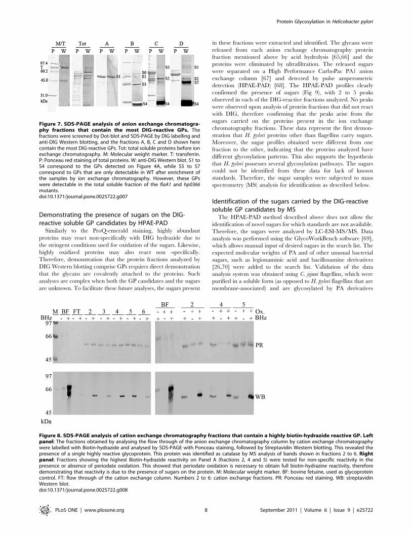
Demonstrating the presence of sugars on the DIG-reactive soluble GP candidates by HPAE-PAD
Similarly to the ProQ-emerald staining, highly abundant
proteins may react non-specifically with DIG hydrazide due to
the stringent conditions used for oxidation of the sugars. Likewise,
highly oxidized proteins may also react non -specifically.
Therefore, demonstration that the protein fractions analyzed by
DIG Western blotting comprise GPs requires direct demonstration
that the glycans are covalently attached to the proteins. Such
analyses are complex when both the GP candidates and the sugars
are unknown. To facilitate these future analyses, the sugars present
in these fractions were extracted and identified. The glycans were
released from each anion exchange chromatography protein
fraction mentioned above by acid hydrolysis [65,66] and the
proteins were eliminated by ultrafiltration. The released sugars
were separated on a High Performance CarboPac PA1 anion
exchange column [67] and detected by pulse amperometric
detection (HPAE-PAD) [68]. The HPAE-PAD profiles clearly
confirmed the presence of sugars (Fig 9), with 2 to 5 peaks
observed in each of the DIG-reactive fractions analyzed. No peaks
were observed upon analysis of protein fractions that did not react
with DIG, therefore confirming that the peaks arise from the
sugars carried on the proteins present in the ion exchange
chromatography fractions. These data represent the first demon-
stration that H. pylori proteins other than flagellins carry sugars.
Moreover, the sugar profiles obtained were different from one
fraction to the other, indicating that the proteins analyzed have
different glycosylation patterns. This also supports the hypothesis
that H. pylori possesses several glycosylation pathways. The sugars
could not be identified from these data for lack of known
standards. Therefore, the sugar samples were subjected to mass
spectrometry (MS) analysis for identification as described below.
Identification of the sugars carried by the DIG-reactivesoluble GP candidates by MS
The HPAE-PAD method described above does not allow the
identification of novel sugars for which standards are not available.
Therefore, the sugars were analyzed by LC-ESI-MS/MS. Data
analysis was performed using the GlycoWorkBench software [69],
which allows manual input of desired sugars in the search list. The
expected molecular weights of PA and of other unusual bacterial
sugars, such as legionaminic acid and bacillosamine derivatives
[26,70] were added to the search list. Validation of the data
analysis system was obtained using C. jejuni flagellins, which were
purified in a soluble form (as opposed to H. pylori flagellins that are
membrane-associated) and are glycosylated by PA derivatives
Figure 7. SDS-PAGE analysis of anion exchange chromatogra-phy fractions that contain the most DIG-reactive GPs. Thefractions were screened by Dot-blot and SDS-PAGE by DIG labelling andanti-DIG Western blotting, and the fractions A, B, C and D shown herecontain the most DIG-reactive GPs. Tot: total soluble proteins before ionexchange chromatography. M: Molecular weight marker. T: transferrin.P: Ponceau red staining of total proteins. W: anti-DIG Western blot. S1 toS4 correspond to the GPs detected on Figure 4A, while S5 to S7correspond to GPs that are only detectable in WT after enrichment ofthe samples by ion exchange chromatography. However, these GPswere detectable in the total soluble fraction of the flaA1 and hp0366mutants.doi:10.1371/journal.pone.0025722.g007
Figure 8. SDS-PAGE analysis of cation exchange chromatography fractions that contain a highly biotin-hydrazide reactive GP. Leftpanel: The fractions obtained by analysing the flow through of the anion exchange chromatography column by cation exchange chromatographywere labelled with Biotin-hydrazide and analysed by SDS-PAGE with Ponceau staining, followed by Streptavidin Western blotting. This revealed thepresence of a single highly reactive glycoprotein. This protein was identified as catalase by MS analysis of bands shown in fractions 2 to 6. Rightpanel: Fractions showing the highest Biotin-hydrazide reactivity on Panel A (fractions 2, 4 and 5) were tested for non-specific reactivity in thepresence or absence of periodate oxidation. This showed that periodate oxidation is necessary to obtain full biotin-hydrazine reactivity, thereforedemonstrating that reactivity is due to the presence of sugars on the protein. M: Molecular weight marker. BF: bovine fetuine, used as glycoproteincontrol. FT: flow through of the cation exchange column. Numbers 2 to 6: cation exchange fractions. PR: Ponceau red staining. WB: streptavidinWestern blot.doi:10.1371/journal.pone.0025722.g008
Protein Glycosylation in Helicobacter pylori
PLoS ONE | www.plosone.org 8 September 2011 | Volume 6 | Issue 9 | e25722

similarly to the H. pylori flagellins. Hits for the expected PA
derivatives were obtained with this control sample (Data not
shown).
As for the H. pylori GP candidate samples, hits were obtained for
two pseudaminic acid derivatives (Pse5Ac7Ac and Pse5Am7Ac) in
fraction A, and the tandem MS analysis patterns were consistent
with this assignment (Fig 10 A and B). Fraction A also had hit
whose MS/MS pattern was consistent with a legionaminic acid
derivative Leg5AmNMe7Ac (Fig 10C). As well, hits were obtained
for two bacillosamine derivatives (Bac2Ac and Bac2Ac4Ac) in
fraction D and the monoacetylated form was confirmed by MS/
MS analysis (Fig 10D). The concomitant presence of hexose was
also demonstrated by MS/MS in this fraction (Fig S1). Note that
Pse and Bac derivatives are known to arise from distinct
biosynthetic pathways in C. jejuni [27,28,30,32], therefore further
supporting our conclusion that H. pylori has several glycosylation
pathways.
Finally, hits with MS/MS patterns consistent with 3-deoxy-D-
manno-octulosonic acid (Kdo) were also obtained for fractions C
and D (Fig S1). Kdo is typically exclusively found in the LPS that is
associated with the OM. These fractions did not contain any
fucose, a signature component of H. pylori LPS. The soluble
fraction where Kdo was detected was obtained after two rounds of
ultracentrifugation of the cellular lysate to eliminate membrane
components. The supernatant was further subjected to ion
exchange chromatography and ammonium sulfate precipitation
prior to release of the sugars from GP-containing fractions for MS
analyses. Therefore, the presence of contaminating sugars arising
from the LPS of the OM is highly unlikely. This was nevertheless
assessed by SDS-PAGE and silver staining after subjecting the
fractions to proteinase K treatment under conditions typically used
to prepare LPS [71]. No traces of LPS could be detected by this
method. Therefore, the Kdo likely originates from glycoproteins.
Overall, sugars could be extracted from our enriched soluble
glycoprotein fractions that were DIG-reactive but not from non
DIG-reactive fractions, and the MS/MS patterns of these sugars
were consistent with novel sugars never shown to be present in
H. pylori before. The preparation of the fractions involved
extensive removal of membrane components by repeated ultra-
centrifugation steps, as well as selective precipitation of proteins by
ammonium sulfate, followed by ion exchange chromatography.
Therefore, the extracted sugars were originally protein-associated.
These data clearly demonstrate the presence of glycoproteins
within our DIG-reactive protein fractions. These data also
demonstrate the presence of a variety of sugars on H. pylori
glycoproteins, which may arise from the PA pathway (PA
derivatives), the LPS biosynthesis pathway (Kdo) or also from a
novel pathway (Bac derivatives).
Discussion
Protein glycosylation in H. pylori extends well beyond theflagellins
The pathogenesis of H. pylori is not well understood despite
abundant research on its virulence factors. The data presented
herein show that the PA biosynthesis pathway, which was hitherto
thought to be exclusively devoted to flagellin glycosylation, affects
the production of other virulence factors, including lipopolysac-
charide and urease. While interconnection between protein
glycosylation and LPS synthesis pathways has been reported
before in several cases [72,73,74], to the best of our knowledge, the
genes responsible for the synthesis of sugars used for protein
glycosylation and LPS synthesis were shared in all examples. In the
current study, the sugar synthesis genes are distinct in each
pathway (Fig 1) and therefore, establishing a link between both
pathways is not quite as straightforward. This led us to propose
that proteins involved in virulence factor production – including
LPS - may be glycosylated in a PA-dependent manner (Fig 1), and
that glycosylation may be important for their activity.
Our data clearly support the existence of multiple glycoproteins
beyond the flagellins, and therefore suggest that protein glycosyl-
ation is not limited to flagellins in H. pylori. We were able to
demonstrate glycosylation of several novel proteins, and several
other GPs of lower abundance were also detected by DIG labeling
and ProQ-emerald staining and remain to be identified. Aside
from this study, the only hint that H. pylori may produce multiple
GPs was recently published based on a global metabolic profiling
study [75], but the GP candidates were neither isolated or
characterized, and the actual presence of sugars was not
demonstrated. It was also proposed earlier that RecA may be
post-translationally modified, potentially through glycosylation,
although no direct evidence was provided (no glyco-specific stain
and no sugar information) [76]. Therefore, our study is the first to
demonstrate labeling of multiple GP candidates by glyco-specific
stains and to provide unambiguous MS-based proof of the
existence of the GP-associated sugars for non-flagellar proteins
in H. pylori.
Despite its very small genome, H. pylori appears to devote
considerable resources to protein glycosylation. This may pertain
to the particularly inhospitable environment in which H. pylori
resides, or to its ability to cause chronic infections. Beyond the
direct role of glycosylation on virulence factor production,
glycosylation may allow masking of antigenic epitopes of OM
proteins, thereby minimizing immune responses and contributing
to the chronicity of H. pylori infections.
Figure 9. HPAE-PAD analysis of sugars released from GPs byacid treatment. Controls included the baseline (trace a), sugarstandards (trace b), and acid extracts from a known glycoprotein(bovine fetuine, trace c) or from a non-DIG reactive ion exchangeprotein fraction (trace d). Acid extracts from DIG-reactive ion exchangeprotein fractions presented several potential sugar peaks (traces e to hfor fractions D, C, B and A, respectively).doi:10.1371/journal.pone.0025722.g009
Protein Glycosylation in Helicobacter pylori
PLoS ONE | www.plosone.org 9 September 2011 | Volume 6 | Issue 9 | e25722

Identity of the novel H. pylori GP candidatesMS analysis of ProQ-emerald-sensitive GP candidates from 2D
SDS-PAGE gels led to the identification of 9 candidate GPs. Note
that identification was only done for the most abundant GP
candidates and that ProQ-emerald staining may not detect all
GPs, depending on the nature of their sugars. The identified
proteins are either cytoplasmic or associated with the inner
membrane, suggesting that protein glycosylation may be impor-
tant for bacterial physiology and not only for direct interactions
with the host. Glycosylation of proteins associated with inner
membrane has also been reported recently for Bacteroides fragilis
[21,77]. Two of the identified H. pylori GP candidates were for two
soluble subunits of the general F0F1 ATPase (a and b subunits),
which was also amongst the hits obtained from an excised band for
a DIG-labeled soluble GP candidate (Band S2, fraction C, Fig 6).
Although there is no known precedent for this, this suggests that
the H. pylori F0F1 ATPase is glycosylated, and that glycosylation
may be important for its function. This may reflect a specific
ability of H. pylori to survive in an acidic environment. Tagging of
the identified GP candidates is underway to allow their
purification and direct analysis of glycopeptides by MS.
One abundant glycoprotein candidate that reacts strongly with
Biotin-hydrazide in a periodate oxidation–dependent manner
and appears to have a heterogeneous glycosylation was identified
unambiguously as catalase. This finding of catalase glycosylation
has never been reported before and studies geared at mapping
the glycosylation sites via glycopeptide analyses will be under-
taken to allow determining the role of glycosylation on catalase
production, stability or function. The low abundance of the other
soluble or membrane GP candidates, or their contamination by
non-glycosylated proteins has prevented us from identifying them
unambiguously. Their identification will require direct analysis of
Figure 10. Mass spectrometry analysis of the sugars extracted from GP candidates by acid hydrolysis. The sugars were analyzed by LC-MS/MS. In each panel, the molecular structure of the expected sugar is shown above the MS/MS spectrum, with the expected fragmentation patternand associated mass loss. The MS/MS spectra are annotated with the total mass loss intervals and with mass loss combinations that lead to the size ofthe observed peaks. All spectra are shown as sodium adducts. Panel A: Pse5Am7Ac. Panel B: Pse5Ac7Ac. Panel C: Leg5AmNMe7Ac. Panel D: Bac2Ac.doi:10.1371/journal.pone.0025722.g010
Protein Glycosylation in Helicobacter pylori
PLoS ONE | www.plosone.org 10 September 2011 | Volume 6 | Issue 9 | e25722

their glycopeptides, which will also map their glycosylation site.
Based on the observation that the PA mutants show altered or no
O-antigen, it is anticipated that LPS biosynthetic enzymes may
be glycosylated by the PA pathway. Therefore, these low
abundance GP candidates may comprise LPS biosynthetic
enzymes.
Note that periodate-based staining of glycoproteins is prone to
non-specific staining. The issue was taken into account in our
analyses of the ProQ-emerald-stained proteins and in the sample
preparation and analytical methods. All labeling and blotting
reactions comprised appropriate negative controls, and the
signals for the soluble fraction were obtained on thoroughly
delipidated fractions to exclude the presence of contaminating
LPS. The samples had undergone repeated rounds of ulcen-
tracentrifugation, as well as precipitation of proteins by
ammonium sulfate. Moreover, it was verified by proteinase K
treatment and silver staining that the observed signals arose from
proteins.
Identity of the novel H. pylori sugars present on GPcandidates
The MS identification of the sugars extracted from the GP
candidates was a further demonstration that the signals obtained
by labeling of the GP candidates with glycan-specific stains were
genuinely indicative of protein glycosylation. In terms of the sugars
identified, the presence of PA derivatives is consistent with the PA-
dependency of one of our GP candidates (band S1). Of note is that
only Pse5Ac7Ac has been described for H. pylori to date [78]. The
presence of the Pse5Am7Ac derivative as identified in this study
demonstrates that H. pylori has the ability to further modify its PA.
Note that the MS/MS spectra are consistent with these
assignments for PA derivatives but determining the absolute
configuration of these sugars would require investigations by
NMR. Moreover, the discovery of Bac derivatives in H. pylori is
entirely novel as Bac has never been reported in H. pylori. Bac is
used for protein glycolysation in C. jejuni [26] and, like for PA, its
biosynthesis is initiated by C6-dehydration of UDP-GlcNAc
[32,79]. Genome mining did not reveal any other UDP-GlcNAc
dehydratase beyond FlaA1 in H. pylori. This suggests that FlaA1 is
shared by both pathways (PA and Bac synthesis) in H. pylori. This is
consistent with our biochemical data [31]. Note that the Bac-
containing fraction (Fraction D) contained several candidate GPs,
including the most reactive S4 and S7 and the faint S2 and S5. S4
and S2 were still DIG-reactive in the absence of FlaA1. This would
argue that their glycosylation is not FlaA1 dependent. S5 and S7
were only present at very low levels in WT samples, and significant
detection of S7 was only observed after enrichment by ion
exchange chromatography. In contrast to the WT, bands S5 and
S7 were abundant in the flaA1/HP0366 mutation background,
suggesting the existence of an alternate glycosylation pathway that
gets up-regulated when FlaA1 or HP0366 is inactivated. The
presence of this alternative pathway could lead to heterogeneous
glycosylation in the WT strain. Based on our MS-based
observation that 1D bands comprise several proteins each, it is
possible that the DIG signals from a 1D band arose from protein(s)
carrying sugars dependent on FlaA1 (such as Bac) and protein(s)
carrying sugars independent from FlaA1. The presence of the
latter sugars would lead to an apparent non-dependence on FlaA1
of the glycosylation of all the proteins comprised within the 1D
band.
In C. jejuni, a Bac derivative (Bac2Ac4Ac) is present on GPs at
the base of a heptasaccharide that also comprises GalNAc and Glc
[26]. In our samples, the Bac derivatives were also found in
association with hits for hexoses, hexosamines and N-acetyl
hexosamines, which suggests that H. pylori may also be able to
assemble a C. jejuni-like complex polysaccharide for protein
glycosylation.
Apart from Kdo, none of the identified sugars are part of the
LPS, which further reinforces the notion that the sugars stem from
glycoproteins. However, as explained above, the observation that
the PA pathway mutants produced altered LPS led us to examine
whether protein glycosylation may affect LPS synthesis. A further
potential connection between the LPS machinery and protein
glycosylation comes from our observation that Kdo was found
associated with our GP candidates. All precautions were taken to
prevent contamination of the samples by LPS, and the fractions do
not contain any of the other LPS signature sugars, therefore
reinforcing the notion that the observed Kdo was bound to a GP.
This could indicate that H. pylori can also use the LPS synthesis
machinery to glycosylate proteins, as seen in other bacterial
species. H. pylori is known to remove a Kdo residue from its LPS
via a dedicated Kdo hydrolase [80,81], but the fate of the removed
Kdo is unknown. We will therefore examine whether the extracted
Kdo can be transferred onto the GP candidates, and what is the
role of this process.
Presence of multiple glycosylation pathways in H. pyloriThe sugar identifications and comparisons of GP candidate
profiles between WT H. pylori and PA mutants suggest that
H. pylori harbours several glycosylation pathways: the classical PA
pathway that generates the PA derivatives highlighted by MS
analyses, a pathway that branches downstream of FlaA1 but
upstream of HP0366 and likely generates the Bac derivatives as
per analogy with the C. jejuni Bac synthesis pathway, and a totally
PA-independent pathway that generates the other sugars identi-
fied. The mapping of the glycosylation sites of our GP candidates
will determine whether the new H. pylori glycosylation pathways
highlighted by our data involve N- or O-glycosylation. While
eukaryotic N-glycosylation requires the NxS/T sequon, an
extended consensus sequon D/ExNxS/T was identified for
bacterial N-glycosylation [82,83] but is not absolute [84,85].
Several of the GP candidates identified so far by MS from ProQ-
emerald stained gels harbor a general (eukaryotic like) N-
glycosylation sequon (Table 2), but none harbored an extended
bacterial one.
In conclusion, this is the first report of the existence and
identification of multiple GP candidates and multiple glycosylation
pathways in H. pylori. These pathways result in the production of
several novel sugars never previously demonstrated in H. pylori and
that could be putatively identified via MS/MS after extraction
from DIG-reactive delipidated protein fractions. While no direct
assignment of the novel sugars to a specific glycoprotein candidate
could be established at this stage, the a priori knowledge gained on
the glycosylation pathways present in H. pylori based on the
identification of the extracted sugars should help characterize the
new glycosylation pathways biochemically and their targets in the
future. We have identified an alternative aminotransferase that
may participate in one of the new glycosylation pathways and will
investigate its effects both via a biochemical and mutagenesis
approach. This will also allow assessing the relative importance of
the various glycosylation pathways in H. pylori. Likewise, the
mapping of the glycosylation sites of our GP candidates will be
performed to allow the investigation of the role of protein
glycosylation on the function of select proteins via mutagenesis of
their glycosylation sites, and on the pathogenicity of the bacterium
via analysis of virulence factor production in these glycosylation
deficient mutants.
Protein Glycosylation in Helicobacter pylori
PLoS ONE | www.plosone.org 11 September 2011 | Volume 6 | Issue 9 | e25722

Materials and Methods
Bacterial growth conditionsH. pylori strain NCTC 11637 (aka ATCC 43504) was a kind gift
from Dr S. Logan (NRC, Ottawa, Canada) and corresponds to the
original isolate described in [86,87]. The bacteria were routinely
grown to confluence on agar plates containing 37 g/l Brain Heart
Infusion (BHI) media (Becton Dickinson), 2.5 g/l yeast extract
(YE) (Bioshop), 0.05% sodium pyruvate, 10% horse serum, and
the antibiotics amphotericin B (4 mg/ml), trimethoprim (5 mg/ml)
and vancomycin (10 mg/ml) under microaerophilic conditions (5%
O2, 10% CO2, and 85% N2) at 37oC. When necessary, selection
with 5 mg/ml kanamycin was applied.
Escherichia coli (DH5a) was grown at 37uC in Luria-Bertani
medium with selection with 100 mg/ml ampicillin or 30 mg/ml
kanamycin when necessary.
Preparation of the hp0366 knockout mutantTo prepare the knockout construct, the hp0366 gene was first
amplified by PCR from genomic DNA of H. pylori strain 26695
using Expand polymerase (Roche Diagnostics) under conditions
recommended by the manufacturer. The primers used were
HP0366P5 AGGGTCCATGGGTTTGAAAGAGTTTGCTTA-
TAGC and HP0366P2 GCGTCGGATCCTCATTCTATTT-
TAAAACTC, which were designed based on genomic data [88]
and contained NcoI and BamHI sites (underlined), respectively. The
PCR product was cut with NcoI and BamHI and inserted into the
pET23 derivative [89] that had been previously cleaved with the
same enzymes. After transformation in E. coli DH5a and selection
on ampicillin 100 mg/ml, the plasmid DNA was recovered and the
gene was fully sequenced. Sequencing was performed at the
Robarts DNA sequencing facility (London, Ontario).
Inverse PCR was then performed on the pET construct described
above using primers HP0366P3 CGCTCTCATGGCATGCTC
and HP0366P4 CTGTTAAACACTAACGCATG and Pfu poly-
merase (Stratagene) following the manufacturer’s recommenda-
tions. The PCR product was ligated with a kanamycin resistance
cassette that had been PCR amplified from the pHel3 vector
(obtained from Dr R. Haas) [90] as described before [10]. The
construct was introduced into H. pylori strain NCTC 11637 by
electroporation [8,52] and mutant candidates were selected in the
presence of 5 mg/ml kanamycin. The mutants were checked for
proper gene disruption by PCR and Southern blotting using
standard procedures.
Preparation of the complemented strainsThe hp0366 gene and its promoter was PCR amplified from
chromosomal DNA from strain 26695 using primers HP0366P2
(see above) and HP0366P1 GAAGTGGAGGATAAGATG and
cloned into the BamHI and EcoRV sites of the pHel2 shuttle vector
(obtained from Dr R. Haas) [90] using standard procedures. The
construct was introduced into the wild-type strain NCTC 11637 or
hp0366 mutant strain by electroporation as described previously
[10]. The plasmid was extracted from H. pylori, transformed into
calcium chloride competent E. coli DH5a, extracted again and
checked for integrity by restriction digestion to allow for the
selection of complemented strains worthy of further phenotypic
characterization.
Lipopolysaccharide (LPS) analysesLPS was prepared according to [71]. Briefly summarized, cell
pellets (50 ml, wet cell volume) were resuspended in 200 ml of 1 M
Tris buffer, pH 6.8 containing 2% SDS, 4% b-mercaptoethanol
and 10% glycerol. After boiling for 30 min, the samples were
treated for 1 h with 100 mg of proteinase K at 56oC. The samples
were then analyzed on 15.6% SDS-PAGE gels and detection was
performed by silver staining [91].
Electron microscopy (EM)The H. pylori cells were harvested from BHI plates and stained
with 2% ammonium molybdate for EM analysis. EM was done at
the EM facility of the Department of Microbiology and
Immunology at the University of Western Ontario.
Urease activity measurementsH. pylori pellets of the wild-type NCTC 11637 strain, the hp0366
mutant and the complemented strain were dissolved in breaking
buffer (20 mM sodium phosphate, pH 7.4, 1 mM EDTA) and
lysed by mechanical disruption using glass beads. After centrifu-
gation for 5 min at 12,000 x g, the supernatant was removed and
its soluble protein content was measured using the Biorad protein
determination reagent. The supernatants were diluted to 0.9, 1.8
and 2.7 g/l in breaking buffer and assayed for urease production
using phenol red as an indicator [92]. Briefly summarized, 10 ml of
supernatant at 0.9, 1.8 or 2.7 g/l in total proteins were added to
100 ml of 0.33 mM urea, 0.001% phenol red in 5 mM sodium
phosphate at pH 6.7, supplemented with 0.15 M NaCl. The
OD565nm which indicates urease activity, was measured at regular
intervals over the course of two hours. Because of the norma-
lization of the samples according to protein content, direct
comparison of the OD565nm values provides a relative comparison
of the levels of urease activity produced by the WT and the
mutant, although the absolute levels of urease activity was not
calculated. Measurements were done in triplicates for each
supernatant protein concentration.
Tissue culture experimentsGastric epithelial cells (AGS cells) were kindly provided by Dr P.
Sherman (Hospital for Sick Children, Toronto). Approximately
300,000 cells per well were grown for ,24 h in 24-well plates until
confluent, reaching about 600,000 cells per well. The cells were
infected for 5 h with wild-type NCTC 11637 or mutant H. pylori
that had been grown for 48 h on BHI plates. Approximately
66107 cfus of H. pylori were added, resulting in a multiplicity of
infection of 1 AGS cell per 100 bacteria. The plates were spun
briefly (5,0006g for 5 min at room temperature) to maximize
contact between the bacteria and the cell monolayer. To
determine total bacterial cell association (adhering and internal-
ized bacteria), the AGS cells were washed 3 times, lysed with
0.08% saponin (Sigma) for 10 min and viable bacterial counts
were determined by plating serial dilutions. To determine the
number of internalized bacteria, the AGS cells were treated with
200 mg/ml gentamycin for 2 h to kill extracellular bacteria. The
cells were then washed and treated as above to determine bacterial
viable counts. Three independent sets of experiments were done,
with triplicates within each experiment. The data represent the
average of all experiments. The data were analyzed by unpaired t-
test with P,0.001 as the limit for significance.
2D gel electrophoresis and ProQ-emerald stainingH. pylori cells grown on BHI plates for 2 days were harvested by
centrifugation, washed in saline and kept frozen at 220uC until
needed. A cell pellet containing ,0.5 mg of proteins (as per
Biorad protein determination assay) was resuspended in 100 ml
lysis buffer (30 mM Tris-HCl, pH 8.8, 2 M thiourea, 7M urea and
4% CHAPS) and sent to Applied Biomics (California, project
manager Dr G. Fu) for labeling and 2D gel analyses. Total
Protein Glycosylation in Helicobacter pylori
PLoS ONE | www.plosone.org 12 September 2011 | Volume 6 | Issue 9 | e25722

proteins (0.5 mg) of H. pylori were labeled with the Cy5 dye (280
pmol) for 30 min to achieve minimal labeling of proteins (1
labeled lysine per protein on average). The reaction was
quenched by addition of 7 nmol of lysine. The 2D gels (pH 3
to 10, 12% gel) were loaded with a mixture of 30 mg of Cy5-
labeled proteins with 120 mg of unlabeled sample that had been
dissolved in lysis buffer. Total proteins were detected in the Cy5
channel (excitation 650 nm, emission 670 nm). Staining with
ProQ Emerald 488 (Invitrogen) was performed according to
supplier’s instructions, and the ProQ-emerald stain was detected
at 520 nm (excitation at 510 nm). The DeCyder software was
used to detect and quantitate the Cy5-labeled and ProQ-reactive
spots. Background intensities for each signal were subtracted from
the signal of each spot before the ProQ/Cy5 signal ratios were
calculated. An unpaired t-Test was performed for statistical
analysis of the GP candidates (ProQ/Cy5 ratio .0.17) versus non
GP proteins (equal variance for both populations, two-tailed p
value, significance set at p,0.001).
In-gel Digestion, Mass Spectrometry, and Databasesearch for ProQ-emerald-reactive GP candidates
These experiments were also performed by Applied Biomics
(California). Proteins of interest were digested in-gel with modified
porcine trypsin protease (Trypsin Gold, Promega). The digested
tryptic peptides were desalted by Zip-tip C18 (Millipore). Peptides
were eluted from the Zip-tip with 0.5 ul of matrix solution (alpha-
cyano-4-hydroxycinnamic acid (5 mg/ml in 50% acetonitrile,
0.1% trifluoroacetic acid, 25 mM ammonium bicarbonate)) and
spotted on the MALDI plate (model ABI 01-192-6-AB). MALDI-
TOF mass spectrometry (MS) and TOF/TOF tandem MS/MS
were performed on an ABI 4700 mass spectrometer (Applied
Biosystems, Framingham, MA). MALDI-TOF mass spectra were
acquired in reflectron positive ion mode, averaging 4000 laser
shots per spectrum. TOF/TOF tandem MS fragmentation spectra
were acquired for each protein, averaging 4000 laser shots per
fragmentation spectrum on each of the 10 most abundant ions
present in each sample (excluding trypsin autolytic peptides and
other known background ions).
Both the resulting peptide mass and the associated fragmenta-
tion spectra were submitted to GPS Explorer workstation
equipped with MASCOT search engine (Matrix science) to search
the National Center for Biotechnology Information non-redun-
dant (NCBInr) database. Searches were performed without
constraining protein molecular weight or isoelectric point, with
variable carbamidomethylation of cysteine and oxidation of
methionine residues, and with one missed cleavage allowed in
the search parameters. Candidates with either protein score C.I.%
or Ion C.I.% greater than 95 were considered significant.
MS identification of proteins from the soluble proteinfraction (SPF)
GP candidate bands were cut out from Coomassie stained SDS-
PAGE gels and subjected to in-gel trypsin digestion. The extracted
peptides were analyzed by LC-MS/MS using standard procedures
at the Don Rix Mass spectrometry facility of the University of
Western Ontario.
RNA isolationTotal RNA isolation was performed with the GE Healthcare
RNAspin Mini RNA isolation kit. The cells were harvested
directly in the lysis buffer provided by the manufacturer and
isolation was carried out as recommended by the manufacturer
except that lysozyme-mediated lysis was performed. The lysate
was filtered and loaded onto the RNA-binding column.
Contaminating DNA was removed by on-column treatment with
DNAseI and total RNA was eluted in RNAse free water. An
additional round of DNAseI treatment was performed off column
using RNase-free DNaseI (from Roche) according to supplier’s
instructions. The treated RNA was quantitated in a SmartSpec
3000 spectrophotometer by reading the absorbance at 260 nm,
and using A260 = 1 for 40 mg/ml as a reference. The RNA was
stored at 220uC until needed.
Reverse-transcriptionThe isolated total RNA was reverse transcribed into cDNA
using iScriptTMcDNA Synthesis Kit (Biorad). Reverse transcrip-
tion was done with random hexamer primers in order to allow
downstream amplification of all genes of interest from a single
cDNA preparation. Following the manufacturers instructions and
using a total volume of 40 mL, total cDNA was synthesized from
30 mL of RNA, diluted 1/10 or 1/100, using iScriptRNaseH+
reverse transcriptase. Negative controls were also created using
RNase-free water instead of the reverse transcriptase.
qRT-PCRThe sequence of the primers used for qRT-PCR are indicated
in Table 1. Each reaction contained 2.25 mL cDNA, 7.5 mL 2X
Perfecta SYBR Green SuperMix (Quanta Biosciences), and
0.67 mL each of 6.67 pmol/mL forward and reverse primers in a
total reaction volume of 15 mL. Real time analysis was done with a
Rotor-Gene 6000 Series 1.7 (Corbett). Real-time PCR conditions
were: initial denaturation and enzyme activation at 95uC for
10 min, followed by 45 cycles of denaturation at 95uC for 15 s,
primer annealing at 52uC for 45 s, and elongation at 60uC for
60 s. The last cycle was followed by a final denaturation at 95uCfor 15 s, and acquisition of a melt curve starting with 60uC for
60 s, and raising the temperature every 15 s by 0.3uC to a final
temperature of 95oC. A fluorescence reading was taken after each
increase.
Culture conditions and preparation of soluble andmembrane protein fractions
Bacteria grown on BHI plates for 48h were harvested in 1 ml of
cold PBS (10 mM sodium phosphate, 140 mM NaCl) per plate.
The bacteria were spun down at 4,000xg. for 15 min at 4oC, and
gently resuspended in the same volume of cold PBS. The optical
density at 600 nm of the suspension was taken on a SmartSpec
3000 spectrophotometer (Bio-Rad) and normalized to 0.6. The
suspended bacteria were aliquoted into 12 ml fractions, repre-
senting 10–13 plates of growth, re-pelleted, and the cell pellet was
frozen at 220uC for future protein analysis.
Each pellet was resuspended in 12 ml of Tris buffered saline
(TBS: 100 mM Tris pH 8.5, 50 mM NaCl). The cells were lysed
in a French press cell (GlenMills) at 1500 psi on ice. Cellular debris
were pelleted (14,000xg, 30min, 4oC). The supernatant was
centrifuged at 100,000xg for 80 min at 4uC in an Optimax
ultracentrifuge with a TLA-110 rotor (Beckman Coulter). The
supernatant containing the soluble proteins was transferred to new
tubes, and the ultracentrifugation was repeated. The final
supernatant was reserved as the soluble protein fraction (SPF).
The pellet from the first ultracentrifugation step, containing
membrane proteins, was resuspended in TBS buffer and the
ultracentrifugation was repeated. The pellet that had been
ultracentrifuged twice was washed in TBS buffer and reserved as
the membrane protein fraction (MPF).
Protein Glycosylation in Helicobacter pylori
PLoS ONE | www.plosone.org 13 September 2011 | Volume 6 | Issue 9 | e25722

Inner and outer membrane preparationWild-type H. pylori was grown for 2 days on 45 BHI plates, as
described above. The cells were harvested in saline (0.85%
NaCl), spun down at 4,000xg for 30 min at 4uC and re-washed
once in 25 ml of saline. The pellet was resuspended in saline to
have an OD600 = 5. The cells were lysed by passage through a
French press (GlenMills) four times at 1500psi on ice. Cellular
debris were pelleted by centrifugation at 5,000xg for 1 h at 4uC.
Insoluble proteins were removed from the supernatant by a 1 h
spin at 13,000xg at 4uC. Membrane proteins were pelleted from
the supernatant by ultracentrifugation at 100,000xg for 1 h at
4uC. The membrane pellet was washed in 500 ml saline by
ultracentrifugation in a micro-ultracentrifuge (Optimax ultra-
centrifuge, Beckman Coulter, TLA-110 rotor). To solubilize
inner membrane proteins, the membrane pellet was resuspended
in 300 ml of PBS (45 mM sodium phosphate, 135 mM NaCl,
pH 7.2) with 1% N-lauroylsarcosine (Sigma) and mixed on a
nutator for 1 h at room temperature. Outer membrane proteins
were pelleted down by ultracentrifugation at 100,000xg for 1 h at
4uC. To refine the separation, the supernatant containing inner
membrane proteins was ultracentrifuged 3 more times until no
pellet was visible. Similarly, the outer membrane pellet was
subjected to another around of N-lauroyl sarcosine treatment
and ultracentrifugation to remove contaminating IM proteins.
The final outer membrane pellet was resuspended in 300 ml
MilliQ water.
DIG-hydrazide labeling of immobilized glycoproteinsThe protein content of samples was assessed by Bradford assay
(Biorad reagent). Samples were normalized to 20 mg of proteins,
separated by 1D SDS-PAGE and transferred to nitrocellulose
membrane. The membrane was washed 3 times in 50 ml of PBS
(50 mM sodium phosphate, 150 mM NaCl, pH 6.5) for 10 min
each. The glycans were oxidized by covering the membrane in
20 ml of 10 mM sodium periodate in 100 mM sodium acetate
pH 5.5 in the dark for 20 min. The residual sodium periodate was
washed off with a second series of 3 washes with 50 ml of PBS. To
label the proteins, 10 ml of 1 mM DIG-hydrazide (digoxigenin-3-
O-succinly-e-aminocaproic acid hydrazide, Roche) in 100 mM
sodium periodate pH 5.5 was added and incubated for 1 h.
Residual DIG-hydrazide was neutralized and removed by 3
washes with 50 ml of TBS (50 mM Tris, 140 mNaCl, pH 7.2). All
procedures were carried out at room temperature.
DIG-hydrazide labeling of glycoproteins in solutionApproximately 20 mL of 1 mg/mL protein solution was used per
in-solution labeling reaction. The labeling was perfomed in 0.1M
sodium acetate pH 5.5, with oxidation by incubation with 1.6 mM
sodium periodate for 20 min at room temperature in the dark.
The reaction was stopped by addition of 20 mM (final
concentration) of sodium disulfite, and incubating for 5 min at
room temperature. DIG-labeling of the proteins was performed by
addition of 1 mM (final) DIG-hydrazide. After 1 h incubation at
Table 1. List of genes whose expression was investigated by qRT-PCR.
Gene Function Primer names Primer sequences
UreA Urease A (HP0073, of ureA-B HP0073P1 GTTAGACAAGTTGATGCTCC
operon) HP0073P2 CCATCAGGAAACATCGCTTC
UreI Accessory protein for urease HP0071P1 CCACCCTACAGCCCCTG
(HP0071of ureIEFGH operon) HP0071P2 CGCAGCAGGAATCGTGTTG
FucST Fucose synthase (HP0044) HP0044P5 (AAGACGGATCC)ATGAAAGAAAA
AATCGCTTTAATCAC
HP0044P7 CTGTAGGCTTAGTGGTAGCGA
FucT a-1,3- fucosyl transferase HPFucTP1 CTGGGGGAGGAGCCGTG
(HP0379) HPFucTP2 GGGTGCGTGTGCAAGTATC
GalT b-1,4- galactosyl transferase HP0826P1 GCCACCCACCACCAAGCGC
(HP0826) HP0826P2 CACCCAAGCTCCCCCAAG
WaaL O-antigen ligase (HP1039) HP1039P9 (GGAATTC)GTGTTGAAAGAGCGTT
TGAAAGCC
HP1039P4 (GAAGATCT)AAACATGTTAGGGAA
GATGCT
FlaA Flagellin A (HP0601) FlaA5 ATGGCTTTTCAGGTCAATAC
FlaA4 CATCCATAGCCTTATCCGC
FlaB Flagellin B (HP0115) HP0115P1 GGATAAATACCAATATCGCC
HP0115P2 CATCCATCGCTTTATCTGC
FlhA Basal body and transcriptional HP1041P1 GGGCCTAGTGCGGTGAG
regulator (HP1041) HP1041P2 CCGCATCAATCGCCATTTG
CoAST AcCoA synthetase (HP1045) HP1045P1 GTCATTATCTATATGCCCAT
HP1045P2 CTGGCTTGAGCATGTAAGG
The functions of the genes are indicated, as well as the names and sequences of the primers used. Nucleotides in brackets correspond to polynucleotide tails andrestriction sites included for other cloning purposes.doi:10.1371/journal.pone.0025722.t001
Protein Glycosylation in Helicobacter pylori
PLoS ONE | www.plosone.org 14 September 2011 | Volume 6 | Issue 9 | e25722

room temperature, the reaction was stopped by neutralizing the
pH with the addition of J V of 4X SDS-PAGE loading buffer.
The proteins were separated by 1D SDS-PAGE gel, transferred on
nitrocellulose for anti-DIG Western blot.
Detection of DIG-labeled glycoproteins by anti-DIGWestern blot
The membrane was washed in de-ionized water and total
proteins were stained by PonceauS. The membrane was de-
stained in TBS, and blocked with 20 ml of 10% (w/v) skim milk
for 1 hour at room temperature and then refrigerated overnight.
The membrane was washed twice for 10 min each in 50 ml of
TBSTT (50 mM Tris, 140 mM NaCl, 2% Triton X-100, 0.5%
Tween-20, pH 7.2), once for 10 min in 50 ml of TBS and then
equilibrated for 10 min in 50 ml of PBS. The membrane was
probed with 20 ml of 1:5000 mouse anti-DIG primary antibody
(Roche) and by anti-mouse AlexaFluor-680 conjugate secondary
antibody (Molecular Probes) in PBS for 60 min in the dark. The
membrane was washed twice in 50ml of TBSTT for 10 min and
once in 50 ml of TBS for 10 min. The membrane was scanned at
700 nm on a Licor Odyssey infra-red scanner.
Biotin-hydrazide labeling of glycoproteinsAs commercial supplies of DIG-hydrazide were discontinued
during the course of these studies, labeling of GPs was also
performed using biotin hydrazide (BHz). Samples from cell
fractionation were diluted 1:1 in 200 mM sodium acetate,
pH 5.5. Samples were oxidized using 10 mM sodium periodate
(Sigma) for 20 min at room temperature in the dark. The reaction
was stopped by addition of 16 mM meta-bisulphite solution made
in 200 mM sodium acetate, pH 5.5 and incubated at room
temperature for 5 min. The labeling reaction was performed by
adding biotin hydrazide (Sigma) to 0.1 mM and the mixture was
incubated at room temperature for 1 h. The labeled samples were
separated by SDS-PAGE and transferred onto nitrocellulose. The
membrane was blocked with 10% Horse Serum (Gibco) in MilliQ
water, and the biotin-labeled GPs were detected by probing the
membrane with 10 ml of 1 mg/ml streptavidin-AlexaFluor-680
Table 2. Identification of candidate glycoproteins by ProQ-emerald staining.
GP #Spot #(MWa)
ProQ/Cy5WT
ProQ/Cy5hp0366 ORF Function %b MWb N-glyc sequond
1 14 0.25 0.19 HP1588 Conserved unknown 51 28.4 None
2 (28.5) HP0879 Flagellar chaperone FliT 37 28.3 2, G
3 15 0.25 0.19 HP1043 Transcriptional regulator 64 25.5 1, G
2 (28.5) HP0879 Flagellar chaperone FliT 53 28.3 2, G
4 33 nd nd HP1132 F0F1 ATP synthase subunit 73 51.5 1, G
(52.1) b
5 61 0.24 0.25 HP0026 Citrate synthase GltA 44 48.3 1, G
(53.6)
5 62 0.28 0.28 HP0026 Citrate synthase GltA 49 48.3 1, G
(53.6)
6 70 0.21 0.18 HP1134 F0F1 ATP synthase subunit 32 55.1 1, G
(62.3) a
7 72 0.19 0.17 HP0900 Hydrogenase HypB 51 27.3 1, G
(30.6)
7 73 0.20 0.20 HP0900 Hydrogenase HypB 40 27.3 1, G
(30.6)
8 77 0.33 0.23 HP1037 X-Pro dipeptidase 32 40.8 None
(42.6)
8 80 0.19 0.22 HP1037 X-Pro dipeptidase 44 40.8 None
(42.6)
9 78 0.20 0.15 HP1555 Elongation factor EF-Ts 54 39.7 None
(44.3)
9 79 0.24 0.13 HP1555 Elongation factor EF-Ts 50 39.7 None
(44.3)
Total proteins were labeled with Cy5, resolved by 2D gel electrophoresis and glycoproteins were labeled by ProQ-emerald staining. The ratios of ProQ-emerald to Cy5staining of the wild-type and hp0366 mutant strains and the protein identities are indicated for the 12 spots identified as GP candidates so far. Because the same proteinhits were obtained for different spots, this results in the identification of 9 GP candidates. The spot numbers refer to Figure 3.aaverage molecular weight calculated from 2 independent ProQ-emerald-stained gels.b% coverage of the protein sequence for MS identification.cmolecular weight as per amino acid sequence, in the absence of glycosylation.dN-glyc sequon: each ORF was examined for the presence of a N-glycosylation sequon of the general NxS/T type or extended bacterial type D/ExNxS/T where x is any
amino acid except proline. The number of sequons is indicated, as well as their type: G, for general.nd: ratio not determined due to smear from abundant neighbouring spot upon Cy5 detection.doi:10.1371/journal.pone.0025722.t002
Protein Glycosylation in Helicobacter pylori
PLoS ONE | www.plosone.org 15 September 2011 | Volume 6 | Issue 9 | e25722

conjugate (Molecular Probes) in PBS for 30 minutes in the dark.
The membrane was washed twice in 50 ml of TBSTT for 10 min
and once in 50 ml of TBS for 10 min. The membrane was
scanned at 700 nm on a Licor Odyssey infra-red scanner to detect
the fluorescent streptavidin conjugate.
Phenyl boronate enrichment of glycoproteinsThe membrane protein fraction (MPF) obtained as described
above from a 12 ml H. pylori suspension at OD600 of 0.6 was
solubilized in 12 ml of 2% Triton-X 100 in PBS. The MPF was
filtered through a 0.45 mm filter and was loaded on a 2 ml Prosep
PB (phenyl boronate, Millipore) affinity column. Before use, the
PBA (phenyl boronate affinity) matrix was flushed with water, 5
column volumes (CV) each of 0.1 M acetic acid, water, 8 M Urea,
water, 0.1 M acetic acid in 20% ethanol and finally washed in 5
CV of running buffer (0.5% Triton-X 100 in PBS). The column
was washed with 5 CV of running buffer, and elution was
performed with 361 CV of 0.2 M Tris buffer (pH 7.4) containing
0.5% triton for 5 min each. The fractions were concentrated by
ammonium sulfate precipitation (see below) before SDS-PAGE
analysis and Western blotting.
Ion exchange chromatographyThe soluble protein fraction (SPF) of WT H. pylori was
fractionated by strong anion and cation exchange chromatogra-
phy on a Mono Q 5/50 GL and Mono S HR 5/50 columns (1 ml
CV, 5 mm650 mm; GE Healthcare). Columns were installed on
an AKTA Explorer FPLC equipped with a 10 ml Super Loop (GE
Healthcare). The system was operated at a flow rate of 1 ml / min.
First, the SPF was run through the MonoQ column. The MonoQ
column was equilibrated for 10 CV with loading buffer (50 mM
NaCl in 20 mM Tris pH 8.5) prior to sample injection. The
samples were eluted with a 10 CV isocratic gradient with loading
buffer followed by a 40 CV linear gradient to 100% elution buffer
(1M NaCl in 20 mM Tris pH 8.5). The flow through collected
during anion exchange chromatography was dialyzed in 50 mM
NaCl in 20mM sodium phosphate pH 6.5, and fractionated on the
MonoS column that had been equilibrated in the same buffer. The
samples were eluted with 10 CV of loading buffer followed by a 40
CV linear gradient to 1 M NaCl in 20 mM sodium phosphate
pH 6.5.
Ammonium sulfate precipitation of protein containingsamples
Each 1 ml protein fraction obtained from ion exchange
chromatography was brought to saturation with the addition of
0.8 g ammonium sulfate. After full dissolution of the ammonium
sulfate crystals, protein precipitation was allowed to occur for 2 h
at 4oC. The samples were pelleted at 14,000xg for 30 min, and the
precipitate was resuspended in 1ml of water.
Release of monosaccharides from glycoproteins by acidhydrolysis
These samples were normalized to 1 g/l total protein as assessed
by Bradford assay (Bio-Rad). The monosaccharides from glycan-
containing proteins were cleaved off with 2 M trifluoroacetic acid
(TFA) at 110uC for 4 hours as described previously [65,66]. The
TFA was removed in a speed-vac (Eppendorf), the volume was
adjusted to 1 ml with water, and the hydrolyzed sugars were
separated from the proteins by ultrafiltration through a Nanosep
Omega centrifugal device (Pall) with a 3.5 kDa cutoff. The sugars
were then analyzed by High Performance Anion Exchange with
pulse amperometric detection (HPAE-PAD) and ESI-MS/MS.
HPAE-PAD analysisThese analyses were carried out on an ICS-3000 ion
chromatography system (Dionex) equipped with an electrochem-
ical detection cell with AgCl reference electrode operated with a
quadraform potential waveform specific for sugar detection. The
column was a 4x50 mm CarboPac PA1 column (Dionex) operated
at 30uC and 1 ml / min. The system was operated with
Chromeleon software. The column was washed for 20 min with
200 mM NaOH, and equilibrated for 20 min with 16 mM
NaOH. Ten ml of sugar samples (adjusted to 16 mM NaOH in
water) were injected, and elution was performed by a 20 min
isocratic gradient of 16 mM NaOH, followed by a 20 min linear
gradient of 0 mM to 320 mM sodium acetate in 16 mM NaOH
and a 10 min linear gradient from 320 mM to 1 M sodium acetate
and from 16 mM to 100 mM NaOH. Sugar standards were
100 mM solutions of a-L (+) fucose (Sigma), D (+) mannose
(Sigma), D (+) glucosamine hydrochloride (Nutritional Biochem-
icals Corporation), and D (+) dextrose (Sigma).
Analysis of released glycans by LC-ESI MS/MSSamples were analyzed by MS at the Don Rix proteomics facility
of the University of Western Ontario. The LC-MS/MS system
consisted of a Waters CapLC with a reversed phase Phenomenex
Jupiter Proteo 90 A column (15061.0 mm, 4 mm) coupled to a Q-
TOF (micro, Waters) mass spectrometer. Online LC was performed
with 0.01% TFA in water (Solvent A) and 95% acetonitrile with
0.01% TFA (Solvent B). Following injection of 5 mL of sample, the
sugars were separated with a 30 min linear gradient from 1% to
50% B followed by a 15 min gradient from 50% to 100% B. Finally,
an isocratic gradient of 100% B was held for 10 min. LC-ESI-MS
analysis was performed in both the positive ion mode and the
negative ion mode with an orifice voltage of 80 V and a scan range
of 200–800 m/z. Collision-induced dissociation was performed
with argon as the collision gas and a variable collision energy of 20–
30 eV over a mass range of 80–600 m/z. Data were acquired and
analyzed by MassLynx 4.0 (Micromass).
Sugar MS data analysesThe hits obtained in MassLynx 4 were screened for potential
sugar hits using GlycoWorkbench, an MS glycan analysis software
that allows users to input non usual sugars to the database [69].
Sugars manually added to the database were PA, bacillosamine
and legionaminic acid, as well as all their currently known
bacterial derivatives. The sugar hits obtained were screened again
in MassLynx for LC and MS peak quality to generate a refined list
of probable sugars. This list was then used as an inclusion list for
LC-ESI MS/MS experiments. For any MS/MS hits, the sizes of
the fragments observed in the tandem mass spectrum were
calculated and compared to a theoretical fragmentation pattern
calculated for the sugar of interest.
Supporting Information
Figure S1 Mass spectrometry analysis of the sugarsextracted from GP candidates by acid hydrolysis. The
sugars were analyzed by LC-MS/MS. In each panel, the
molecular structure of the expected sugar is shown above the
MS/MS spectrum, with the expected fragmentation pattern and
associated mass loss. The MS/MS spectra are annotated with the
total mass loss intervals and with mass loss combinations that lead
to the size of the observed peaks. All spectra are shown as sodium
adducts. Panel A: Hexose. Panel B: 3-deoxy-D-manno-octulosonic
acid (Kdo).
(TIF)
Protein Glycosylation in Helicobacter pylori
PLoS ONE | www.plosone.org 16 September 2011 | Volume 6 | Issue 9 | e25722

Acknowledgments
We thank V. Soo for help with cloning of hp0366 in the pET vector, A.
Goden for preparing bacterial RNA and performing preliminary RT-PCR
experiments, I. Grisky for help with tissue culture experiments, and A.
Wong for assistance with figure preparation. We thank Dr S. Koval and J.
Sholdice for advice and technical assistance for electron microscopy, Drs
G. Lajoie and S. Liu for processing and advice for MS analyses, and Dr S.
Trent for helpful discussion on Kdo.
Author Contributions
Conceived and designed the experiments: PSH RSF AMJ CC. Performed
the experiments: PSH RSF NZ AMJ SV DR JH. Analyzed the data: PSH
RSF NZ AMJ CC. Wrote the paper: CC. Manuscript editing: AMJ RF
NZ.
References
1. Warren J, Marshall B (1983) Unidentified curved bacilli on gastric epithelium in
active chronic gastritis. Lancet 1: 1273–1275.
2. Uemura N, Okamoto S, Yamamoto S, Matsumura N, Yamaguchi S, et al.(2001) Helicobacter pylori infection and the development of gastric cancer.
N Engl J Med 345: 784–789.
3. Forman D, Burley VJ (2006) Gastric cancer: global pattern of the disease and anoverview of environmental risk factors. Best Pract Res Clin Gastroenterol 20:
633–649.
4. Albert TJ, Dailidiene D, Dailide G, Norton JE, Kalia A, et al. (2005) Mutation
discovery in bacterial genomes: metronidazole resistance in Helicobacter pylori. NatMethods 2: 951–953.
5. Raymond J, Lamarque D, Kalach N, Chaussade S, Burucoa C (2010) High level
of antimicrobial reistance in French Helicobacter pylori ioslates. Helicobacter 15:21–27.
6. Fischer W, Prassl S, Haas R (2009) Virulence mechanisms and persistence
strategies of the human gastric pathogen Helicobacter pylori. Curr Top MicrobiolImmunol 337: 129–171.
7. Josenhans C, Vossebein L, Friedrich S, Suerbaum S (2002) The neuA/flmD gene
cluster of Helicobacter pylori is involved in flagellar biosynthesis and flagellinglycosylation. FEMS Microbiol Lett 210: 165–172.
8. Suerbaum S, Josenhans C, Labigne A (1993) Cloning and genetic character-
ization of the Helicobacter pylori and Helicobacter mustelae flaB flagellin genes andconstruction of H. pylori flaA- and flaB-negative mutants by electroporation-
mediated allelic exchange. J Bacteriol 175: 3278–3288.
9. Logan SM, Kelly JF, Thibault P, Ewing CP, Guerry P (2002) Structuralheterogeneity of carbohydrate modifications affects serospecificity of Campylo-
bacter flagellins. Mol Microbiol 46: 587–597.
10. Merkx-Jacques A, Obhi RK, Bethune G, Creuzenet C (2004) The Helicobacter
pylori flaA1 and wbpB genes control lipopolysaccharide and flagellum synthesis
and function. J Bacteriology 186: 2253–2265.
11. Doig P, Kinsella N, Guerry P, Trust TJ (1996) Characterization of a post-translational modification of Campylobacter flagellin: identification of a sero-
specific glycosyl moiety. Mol Microbiol 19: 379–387.
12. Goon S, Kelly JF, Logan SM, Ewing CP, Guerry P (2003) Pseudaminic acid, themajor modification on Campylobacter flagellin, is synthesized via the Cj1293
gene. Mol Microbiol 50: 659–671.
13. Thibault P, Logan SM, Kelly JF, Brisson JR, Ewing CP, et al. (2001)
Identification of the carbohydrate moieties and glycosylation motifs inCampylobacter jejuni flagellin. J Biol Chem 276: 34862–34870.
14. Arora SK, Bangera M, Lory S, Ramphal R (2001) A genomic island in
Pseudomonas aeruginosa carries the determinants of flagellin glycosylation. ProcNatl Acad Sci U S A 98: 9342–9347.
15. Castric P, Cassels FJ, Carlson RW (2001) Structural characterization of the
Pseudomonas aeruginosa 1244 pilin glycan. J Biol Chem 276: 26479–26485.
16. Guerry P, Doig P, Alm RA, Burr DH, Kinsella N, et al. (1996) Identification and
characterization of genes required for post-translational modification of
Campylobacter coli VC167 flagellin. Mol Microbiol 19: 369–378.
17. Kuo C, Takahashi N, Swanson AF, Ozeki Y, Hakomori S (1996) An N-linkedhigh-mannose type oligosaccharide, expressed at the major outer membrane
protein of Chlamydia trachomatis, mediates attachment and infectivity of themicroorganism to HeLa cells. J Clin Invest 98: 2813–2818.
18. Power PM, Roddam LF, Dieckelmann M, Srikhanta YN, Tan YC, et al. (2000)
Genetic characterization of pilin glycosylation in Neisseria meningitidis. Microbi-ology 146(Pt 4): 967–979.
19. Schirm M, Kalmokoff M, Aubry A, Thibault P, Sandoz M, et al. (2004) Flagellin
from Listeria monocytogenes Is Glycosylated with {beta}-O-Linked N-Acetylglucosamine. J Bacteriol 186: 6721–6727.
20. Stimson E, Virji M, Makepeace K, Dell A, Morris HR, et al. (1995)
Meningococcal pilin: a glycoprotein substituted with digalactosyl 2,4-diaceta-mido-2,4,6-trideoxyhexose. Mol Microbiol 17: 1201–1214.
21. Fletcher CM, Coyne MJ, Villa OF, Chatzidaki-Livanis M, Comstock LE (2009)
A general O-glycosylation system important to the physiology of a major humanintestinal symbiont. Cell 137: 321–331.
22. Nothaft H, Szymanski CM (2010) Protein glycosylation in bacteria: sweeter than
ever. Nat Rev Microbiol 8: 765–778.
23. Schoenhofen IC, McNally DJ, Brisson JR, Logan SM (2006) Elucidation of theCMP-pseudaminic acid pathway in Helicobacter pylori: synthesis from UDP-N-
acetyl-glucosamine by a single enzymatic reaction. Glycobiology.
24. Somalinga V, Obhi R, Creuzenet C (2005) HP0366 is required for secretion/biosynthesis of several virulence factors in Helicobacter pylori. Proceedings of the
105th general meeting of the American Society for Microbiology June 5-9th,
Atlanta (USA) ASM press Session No 133/B Abstract B-143. pp 53–54.
25. Linton D, Allan E, Karlyshev AV, Cronshaw AD, Wren BW (2002)
Identification of N-acetylgalactosamine-containing glycoproteins PEB3 and
CgpA in Campylobacter jejuni. Mol Microbiol 43: 497–508.
26. Young NM, Brisson JR, Kelly J, Watson DC, Tessier L, et al. (2002) Structure of
the N-linked glycan present on multiple glycoproteins in the Gram-negative
bacterium, Campylobacter jejuni. J Biol Chem 277: 42530–42539.
27. Creuzenet C (2004) Characterization of CJ1293, a new UDP-GlcNAc C6
dehydratase from Campylobacter jejuni. FEBS Lett 559: 136–140.
28. Obhi RK, Creuzenet C (2005) Biochemical characterization of the Campylobacter
jejuni Cj1294, a novel UDP-4-keto-6-deoxy-GlcNAc aminotransferase that
generates UDP-4-amino-4,6-dideoxy-GalNAc. J Biol Chem 280: 20902–20908.
29. Demendi M, Creuzenet C (2009) Cj1123c (PglD), a multi-facetted acetyltrans-
ferase from Campylobacter jejuni. Biochemistry and Cell Biology 87: 469–483.
30. Vijayakumar S, Merkx-Jacques A, Ratnayake D, Gryski I, Obhi RK, et al.
(2006) Cj1121c, a novel UDP-4-keto-6-deoxy-GlcNAc C4 aminotransferase
essential for protein glycosylation and virulence in Campylobacter jejuni. Journal of
Biological Chemistry 281: 27733–27743.
31. Creuzenet C, Schur MJ, Li J, Wakarchuk WW, Lam JS (2000) FlaA1, a new
bifunctional UDP-GlcNAc C6 Dehydratase/ C4 reductase from Helicobacter pylori.
J Biol Chem 275: 34873–34880.
32. Schoenhofen IC, McNally DJ, Vinogradov E, Whitfield D, Young NM, et al.
(2006) Functional characterization of dehydratase/aminotransferase pairs from
Helicobacter and Campylobacter: enzymes distinguishing the pseudaminic acid
and bacillosamine biosynthetic pathways. J Biol Chem 281: 723–732.
33. Ishiyama N, Creuzenet C, Miller WL, Demendi M, Anderson EM, et al. (2006)
Structural studies of FlaA1 from Helicobacter pylori reveal the mechanism for
inverting 4,6-dehydratase activity. J Biol Chem 281: 24489–24495.
34. Kwon DH, Woo JS, Perng CL, Go MF, Graham DY, et al. (1998) The effect of
galE gene inactivation on lipopolysaccharide profile of Helicobacter pylori. Curr
Microbiol 37: 144–148.
35. McGowan CC, Necheva A, Thompson SA, Cover TL, Blaser MJ (1998) Acid-
induced expression of an LPS-associated gene in Helicobacter pylori. Mol Microbiol
30: 19–31.
36. Wang G, Rasko DA, Sherburne R, Taylor DE (1999) Molecular genetic basis for
the variable expression of Lewis Y antigen in Helicobacter pylori: analysis of the
alpha (1,2) fucosyltransferase gene. Mol Microbiol 31: 1265–1274.
37. Wang G, Boulton PG, Chan NW, Palcic MM, Taylor DE (1999) Novel
Helicobacter pylori alpha1,2-fucosyltransferase, a key enzyme in the synthesis of
Lewis antigens. Microbiology 145(Pt 11): 3245–3253.
38. Wang G, Ge Z, Rasko DA, Taylor DE (2000) Lewis antigens in Helicobacter pylori:
biosynthesis and phase variation. Mol Microbiol 36: 1187–1196.
39. Wu B, Zhang Y, Wang PG (2001) Identification and characterization of GDP-d-
mannose 4,6-dehydratase and GDP-l-fucose snthetase in a GDP-l-fucose
biosynthetic gene cluster from Helicobacter pylori. Biochem Biophys Res Commun
285: 364–371.
40. Mahdavi J, Sonden B, Hurtig M, Olfat FO, Forsberg L, et al. (2002) Helicobacter
pylori SabA adhesin in persistent infection and chronic inflammation. Science
297: 573–578.
41. Ilver D, Arnqvist A, Ogren J, Frick IM, Kersulyte D, et al. (1998) Helicobacter
pylori adhesin binding fucosylated histo-blood group antigens revealed by
retagging. Science 279: 373–377.
42. Odenbreit S, Till M, Hofreuter D, Faller G, Haas R (1999) Genetic and
functional characterization of the alpAB gene locus essential for the adhesion of
Helicobacter pylori to human gastric tissue. Mol Microbiol 31: 1537–1548.
43. Amieva MR, Salama NR, Tompkins LS, Falkow S (2002) Helicobacter pylori enter
and survive within multivesicular vacuoles of epithelial cells. Cell Microbiol 4:
677–690.
44. Censini S, Lange C, Xiang Z, Crabtree JE, Ghiara P, et al. (1996) cag, a
pathogenicity island of Helicobacter pylori, encodes type I-specific and disease-
associated virulence factors. Proc Natl Acad Sci U S A 93: 14648–14653.
45. Censini S, Stein M, Covacci A (2001) Cellular responses induced after contact
with Helicobacter pylori. Curr Opin Microbiol 4: 41–46.
46. Montecucco C, Rappuoli R (2001) Living dangerously: how Helicobacter pylori
survives in the human stomach. Nat Rev Mol Cell Biol 2: 457–466.
47. Kakuda T, DiRita VJ (2006) Cj1496c encodes a Campylobacter jejuni glycoprotein
that influences invasion of human epithelial cells and colonization of the chick
gastrointestinal tract. Infect Immun 74: 4715–4723.
Protein Glycosylation in Helicobacter pylori
PLoS ONE | www.plosone.org 17 September 2011 | Volume 6 | Issue 9 | e25722

48. Karlyshev AV, Everest P, Linton D, Cawthraw S, Newell DG, et al. (2004) The
Campylobacter jejuni general glycosylation system is important for attachment tohuman epithelial cells and in the colonization of chicks. Microbiology 150:
1957–1964.
49. Kwok T, Backert S, Schwarz H, Berger J, Meyer TF (2002) Specific entry ofHelicobacter pylori into cultured gastric epithelial cells via a zipper-like mechanism.
Infect Immun 70: 2108–2120.50. Tan S, Tompkins LS, Amieva MR (2009) Helicobacter pylori usurps cell polarity to
turn the cell surface into a replicative niche. PLoS Pathog 5: e1000407.
51. McGee DJ, Coker C, Testerman TL, Harro JM, Gibson SV, et al. (2002) TheHelicobacter pylori flbA flagellar biosynthesis and regulatory gene is required for
motility and virulence and modulates urease of H. pylori and Proteus mirabilis.J Med Microbiol 51: 958–970.
52. McGee DJ, May CA, Garner RM, Himpsl JM, Mobley HL (1999) Isolation ofHelicobacter pylori genes that modulate urease activity. J Bacteriol 181: 2477–2484.
53. Niehus E, Gressmann H, Ye F, Schlapbach R, Dehio M, et al. (2004) Genome-
wide analysis of transcriptional hierarchy and feedback regulation in the flagellarsystem of Helicobacter pylori. Mol Microbiol 52: 947–961.
54. Keykhosravani M, Doherty-Kirby A, Zhang C, Brewer D, Goldberg HA, et al.(2005) Comprehensive identification of post-translational modifications of rat
bone osteopontin by mass spectrometry. Biochemistry 44: 6990–7003.
55. Zhang C, Doherty-Kirby A, Huystee Rv R, Lajoie G (2004) Investigation ofcationic peanut peroxidase glycans by electrospray ionization mass spectrometry.
Phytochemistry 65: 1575–1588.56. Dell A, Morris HR (2001) Glycoprotein structure determination by mass
spectrometry. Science 291: 2351–2356.57. Josenhans C, Ferrero RL, Labigne A, Suerbaum S (1999) Cloning and allelic
exchange mutagenesis of two flagellin genes of Helicobacter felis. Mol Microbiol
33: 350–362.58. Filip C, Fletcher G, Wulff JL, Earhart CF (1973) Solubilization of the
cytoplasmic membrane of Escherichia coli by the ionic detergent sodium-laurylsarcosinate. J Bacteriol 115: 717–722.
59. Baik SC, Kim KM, Song SM, Kim DS, Jun JS, et al. (2004) Proteomic analysis
of the sarcosine-insoluble outer membrane fraction of Helicobacter pylori strain26695. J Bacteriol 186: 949–955.
60. Carlsohn E, Nystrom J, Karlsson H, Svennerholm AM, Nilsson CL (2006)Characterization of the outer membrane protein profile from disease-related
Helicobacter pylori isolates by subcellular fractionation and nano-LC FT-ICR MSanalysis. J Proteome Res 5: 3197–3204.
61. Doig P, Trust TJ (1994) Identification of surface-exposed outer membrane
antigens of Helicobacter pylori. Infect Immun 62: 4526–4533.62. Osborn MJ, Gander JE, Parisi E, Carson J (1972) Mechanism of assembly of the
outer membrane of Salmonella typhimurium. Isolation and characterization ofcytoplasmic and outer membrane. J Biol Chem 247: 3962–3972.
63. Geis G, Suerbaum S, Forsthoff B, Leying H, Opferkuch W (1993) Ultrastructure
and biochemical studies of the flagellar sheath of Helicobacter pylori. J MedMicrobiol 38: 371–377.
64. Odenbreit S, Wieland B, Haas R (1996) Cloning and genetic characterization ofHelicobacter pylori catalase and construction of a catalase-deficient mutant strain.
J Bacteriol 178: 6960–6967.65. Neeser JR, Schweizer TF (1984) A quantitative determination by capillary gas-
liquid chromatography of neutral and amino sugars (as O-methyloxime
acetates), and a study on hydrolytic conditions for glycoproteins andpolysaccharides in order to increase sugar recoveries. Anal Biochem 142: 58–67.
66. Eggert FM, Jones M (1985) Measurement of neutral sugars in glycoproteins asdansyl derivatives by automated high-performance liquid chromatography.
J Chromatogr 333: 123–131.
67. Cooper GA, Rohrer JS (1995) Separation of neutral asparagine-linkedoligosaccharides by high-pH anion-exchange chromatography with pulsed
amperometric detection. Anal Biochem 226: 182–184.68. Clarke AJ (1993) Compositional analysis of peptidoglycan by high-performance
anion-exchange chromatography. Anal Biochem 212: 344–350.
69. Ceroni A, Maass K, Geyer H, Geyer R, Dell A, et al. (2008) GlycoWorkbench: atool for the computer-assisted annotation of mass spectra of glycans. J Proteome
Res 7: 1650–1659.70. Schoenhofen IC, Vinogradov E, Whitfield DM, Brisson JR, Logan SM (2009)
The CMP-legionaminic acid pathway in Campylobacter: biosynthesis involvingnovel GDP-linked precursors. Glycobiology 19: 715–725.
71. Hitchcock PJ, Brown TM (1983) Morphological heterogeneity among Salmonella
lipopolysaccharide chemotypes in silver-stained polyacrylamide gels. J Bacteriol
154: 269–277.
72. Tabei SM, Hitchen PG, Day-Williams MJ, Merino S, Vart R, et al. (2009) An
Aeromonas caviae genomic island is required for both O-antigen lipopolysaccharide
biosynthesis and flagellin glycosylation,. J Bacteriol 191: 2851–2863.
73. Lindhout T, Lau PC, Brewer D, Lam JS (2009) Truncation in the core
oligosaccharide of lipopolysaccharide affects flagella-mediated motility in
Pseudomonas aeruginosa PAO1 via modulation of cell surface attachment.
Microbiology 155: 3449–3460.
74. Miller WL, Matewish MJ, McNally DJ, Ishiyama N, Anderson EM, et al. (2008)
Flagellin glycosylation in Pseudomonas aeruginosa PAK requires the O-antigen
biosynthesis enzyme WbpO,. J Biol Chem 283: 3507–3518.
75. Koenigs MB, Richardson EA, Dube DH (2009) Metabolic profiling of
Helicobacter pylori glycosylation. Mol Biosyst 5: 909–912.
76. Fischer W, Haas R (2004) The RecA protein of Helicobacter pylori requires a
posttranslational modification for full activity. J Bacteriol 186: 777–784.
77. Fletcher CM, Coyne MJ, Comstock LE (2010) Theoretical and experimental
characterization of the scope of protein O-glycosylation in bacteroides fragilis. J Biol
Chem.
78. Schirm M, Soo EC, Aubry AJ, Austin J, Thibault P, et al. (2003) Structural,
genetic and functional characterization of the flagellin glycosylation process in
Helicobacter pylori. Mol Microbiol 48: 1579–1592.
79. McNally DJ, Hui JP, Aubry AJ, Mui KK, Guerry P, et al. (2006) Functional
Characterization of the Flagellar Glycosylation Locus in Campylobacter jejuni 81-
176 Using a Focused Metabolomics Approach. J Biol Chem 281: 18489–18498.
80. Stead C, Tran A, Ferguson D, Jr., McGrath S, Cotter R, et al. (2005) A novel 3-
deoxy-D-manno-octulosonic acid (Kdo) hydrolase that removes the outer Kdo
sugar of Helicobacter pylori lipopolysaccharide. J Bacteriol 187: 3374–3383.
81. Stead CM, Zhao J, Raetz CR, Trent MS (2010) Removal of the outer Kdo from
Helicobacter pylori lipopolysaccharide and its impact on the bacterial surface. Mol
Microbiol.
82. Kowarik M, Young NM, Numao S, Schulz BL, Hug I, et al. (2006) Definition of
the bacterial N-glycosylation site consensus sequence. Embo J 25: 1957–1966.
83. Chen MM, Glover KJ, Imperiali B (2007) From peptide to protein: comparative
analysis of the substrate specificity of N-linked glycosylation in C. jejuni.
Biochemistry 46: 5579–5585.
84. Gross J, Grass S, Davis AE, Gilmore-Erdmann P, Townsend RR, et al. (2008)
The Haemophilus influenzae HMW1 adhesin is a glycoprotein with an unusual N-
linked carbohydrate modification. J Biol Chem 283: 26010–26015.
85. Grass S, Lichti CF, Townsend RR, Gross J, St Geme JW, 3rd (2010) The
Haemophilus influenzae HMW1C protein is a glycosyltransferase that transfers
hexose residues to asparagine sites in the HMW1 adhesin. PLoS Pathog 6:
e1000919.
86. Marshall B, Royce H, Annear D, Goodwin C, Perarman J, et al. (1984) Original
isolation of Campylobacter pyloris from human gastric mucosa. Microbios Lett 25:
83–88.
87. Goodwin C, Armstrong J, Chilvers T, Peters M, Collins M, et al. (1989) Transfer
of Campylobacter pylori and Campylobacter mustelae to Helicobacter gen. nov. as
Helicobacter pylori comb. nov. and Helicobacter mustelae comb. nov., respectively.
International Journal of Systematic Bacteriology 39: 397–405.
88. Tomb JF, White O, Kerlavage AR, Clayton RA, Sutton GG, et al. (1997) The
complete genome sequence of the gastric pathogen Helicobacter pylori. Nature 388:
539–547.
89. Newton DT, Mangroo D (1999) Mapping the active site of the Haemophilus
influenzae methionyl-tRNA formyltransferase: residues important for catalysis and
tRNA binding. Biochem J 339(Pt 1): 63–69.
90. Heuermann D, Haas R (1998) A stable shuttle vector system for efficient genetic
complementation of Helicobacter pylori strains by transformation and conjugation.
Mol Gen Genet 257: 519–528.
91. Fomsgaard A, Freudenberg MA, Galanos C (1990) Modification of the silver
staining technique to detect lipopolysaccharide in polyacrylamide gels. J Clin
Microbiol 28: 2627–2631.
92. Clyne M, Labigne A, Drumm B (1995) Helicobacter pylori requires an acidic
environment to survive in the presence of urea. Infect Immun 63: 1669–1673.
Protein Glycosylation in Helicobacter pylori
PLoS ONE | www.plosone.org 18 September 2011 | Volume 6 | Issue 9 | e25722


![The serum immunoglobulin G glycosylation signature of ......dysplasia, and finally adenocarcinoma [6]. Helicobacter pylori is a bacterium that infects the gastric epithelium of approximately](https://static.fdocuments.net/doc/165x107/5f4637eb1f92b745a0349ef3/the-serum-immunoglobulin-g-glycosylation-signature-of-dysplasia-and-inally.jpg)