Protein Folding and Peptide Ligand Binding - Two Sides of ... · 2.1 Protein folding and peptide...
29
Protein Folding and Peptide Ligand Binding - Two Sides of the Same Coin Maike Christine Jürgens Degree project in biology, Master of science (2 years), 2010 Examensarbete i biologi 45 hp till masterexamen, 2010 Institutionen för biologisk grundutbildning och Institutionen för medicinsk biokemi och mikrobiologi, Uppsala universitet Handledare: Dr. Per Jemth
Transcript of Protein Folding and Peptide Ligand Binding - Two Sides of ... · 2.1 Protein folding and peptide...

Protein Folding and Peptide LigandBinding - Two Sides of the Same Coin
Maike Christine Jürgens
Degree project in biology, Master of science (2 years), 2010Examensarbete i biologi 45 hp till masterexamen, 2010Institutionen för biologisk grundutbildning och Institutionen för medicinsk biokemi och mikrobiologi,Uppsala universitetHandledare: Dr. Per Jemth

2
Contents 1. Summary 3 2. Introduction 4 2.1 Protein folding and peptide ligand binding: large- and small-scale protein dynamics 4 2.2 High-resolution protein (un)folding 4 2.3 Intramolecular allostery in a small protein domain 5 2.4 Aims 6 3. Results 8 3.1 EnHD X-ray protein folding studies 8 3.2 Allosteric peptide recognition in a small protein domain 9 4. Discussion 14 4.1 High-resolution protein folding studies 14 4.2 Allosteric peptide recognition in a small protein domain 14 4.3 Is allostery common in PDZ domains? 15 5. Materials and Methods 18 5.1 Bacterial strains and plasmids 18 5.2 Cloning procedures 18 5.3 Protein overexpression and purification 20 5.4 Equilibrium denaturation of EnHD wild type and Cys-mutants 23 5.5 Binding experiments of YKQSSV to PSD-95 PDZ3 wild type and mutants 23 6. Acknowledgements 25 7. References 26

3
1. Summary
Biological macromolecules are not static building blocks of life but intriguing dynamic modules governing central processes of biological existence. Therefore, it is essential for a profound understanding of cellular and physiological processes to comprehend protein dynamics at different scales ranging from small-scale motions like allosteric communication to large-scale conformational changes like protein folding reactions. Here, two model proteins were chosen to look at those protein dynamics from different angles.
The DNA-binding protein engrailed homeodomain from Drosophila melanogaster was chosen to develop an X-ray scattering based method for investigation of protein folding. Labelling of specific, engineered Cys residues in the protein with the strongly scattering heavy metal mercury will allow visualisation of protein (un)folding reactions in solution using scattering of X-rays. The scattering technique will permit distance measurements between the heavy metal atoms and thus determination of a high-resolution picture of the (un)folding event. The Cys mutants were successfully purified, and they were fully folded under native conditions although their stability was lower compared to the wild type protein. Labelling of the mutants with mercury could so far not be achieved, but trials are ongoing.
In contrast, small-scale-dynamics allosteric communication can be investigated with double mutant cycle experiments, which report on the interaction energy between two residues within a protein. This method was chosen to look into allosteric recognition of a peptide ligand of the PSD (post-synaptic density)-95 PDZ (PSD-95/discs large homologue/zonula occludens) 3 domain. PDZ domains are highly abundant protein-protein interaction domains that are commonly found in scaffold proteins involved in the organisation of signalling pathways for instance. Positive coupling energies between a mutated peptide ligand residue and some residues within the protein were measured, and hence there is allosteric communication within this small protein domain. Residues that couple are not only found in direct proximity to the peptide ligand but also in secondary structure and loop regions further away from the peptide-binding pocket. These findings experimentally corroborate previous computational and nuclear magnetic resonance studies on allosteric networks in PDZ domains. Still, the different methods applied to identify networks make out different residues as part of the network in the same PDZ domain. Moreover, network residues identified differ also between different PDZ domains. This suggests that allosteric networks, at least in PDZ domains, are sequence specific rather than fold-dependent. Furthermore, data on different mutated residues in the peptide ligand, other PDZ domains, and results from in silico analysis suggest that two different but interconnected networks are responsible for discrimination between different peptide ligands in PDZ domains.
4
2. Introduction 2.1 Protein folding and peptide ligand binding: large- and small-scale protein
dynamics The Levinthal paradox (Levinthal, 1968) describes the fascinating conundrum of protein folding whereby proteins fold much faster than a random sampling of the complete conformational space would take. Since this intriguing discovery was made, thousands of experiments have been carried out trying to understand the mystery of how a two-dimensional polypeptide chain (the denatured or unfolded state) can reproducibly fold into a single, defined three-dimensional structure (the native or folded state) (Anfinsen, 1973; Fersht, 1995; Itzhaki et al., 1995; Matouschek et al., 1989; Serrano et al., 1992c). Owing to an enormous amount of research (Bryngelson et al., 1995; Fersht, 2008; Fersht et al., 1992; Matouschek et al., 1989; Onuchic et al., 1997), it is now possible to characterise and describe a protein folding reaction in quite some detail. However, reasons for why, for example, even related proteins display different folding kinetics still remain elusive (Chi et al., 2007; Daggett and Fersht, 2003b; Ferguson et al., 1999; Gianni et al., 2003). Also, factors influencing the related problem of ligand binding events, e.g. features determining specificity and affinity can in part be identified and described, but a general model describing any binding event is still missing. Common to those unsolved problems is the question of how both protein folding and ligand binding events are influenced by large-scale (e.g. secondary stucture and side-chain movements) or small-scale (e.g. vibrational motions) dynamics. 2.2 High-resolution protein (un)folding studies using X-ray scattering Classical experiments on protein folding kinetics and protein-protein or protein-ligand interactions, both processes that involve protein dynamics, are usually performed as bulk measurements on an ensemble of proteins and at a rather low resolution (Daggett and Fersht, 2003a; Fersht, 2008). During those experiments, protein folding reactions are either controlled by temperature changes or by the addition of denaturants such as urea or guanidinium hydrochloride. Next to nuclear magnetic resonance (NMR) experiments, which also provide high-resolution structural information, X-ray crystallography is one of the first choices when investigating biological molecules at atomic resolution. Unfortunately, a dynamic protein folding reaction cannot be observed in protein crystals, and therefore those experiments have to be carried out with protein in solution. NMR is capable of providing this information (Korzhnev and Kay, 2008; Religa et al., 2005) but only in certain cases and only for molecules with a limited molecular weight. Nevertheless, also X-rays can be used to investigate proteins in solution during so-called small-angle X-ray scattering experiments (SAXS) (Jacques and Trewhella, 2010). A new way of investigating protein folding at high resolution is time-resolved X-ray scattering during which the (un)folding reaction a wild type or heavy metal atom-labelled protein will be observed in solution using X-rays. As the visibility of atoms to X-rays is proportional to the number of electrons in the atom, elements like hydrogen, carbon and oxygen that mainly build up biological macromolecules barely scatter X-ray radiation. Amplification of scattering can either be achieved by repeatedly arranging molecules in the same orientation as in protein crystals or by introducing strong scatterers like heavy metal

5
atoms. Thus, by binding heavy-metal atoms to specific residues in a protein, it is possible to explicitly measure the distance between those two residues, for instance in a temperature dependent manner, giving detailed information on the protein structure during the thermal unfolding process. For example, during time-resolved X-ray scattering, the temperature-dependent change in distances between atoms can be measured in a time-resolved fashion. At different temperatures, the protein solution is exposed to X-ray radiation and a continuous diffraction pattern is collected. A cross section of this diffraction pattern is then plotted as an intensity curve against reciprocal distance (Å-1). A shift of peaks when comparing different temperatures for a protein folding reaction indicates for example a change in the distance between atoms giving rise to this peak (Andersson et al., 2008).
Since mutations like the Cys-mutations introduced into the protein here can destabilize the protein, protein stability will be checked using a naturally present fluorescent probe, a Trp-residue in the protein. Trp-flourescence intensity can be used to measure the degree of protein (un)folding as the fluorescence intensity of Trp depends on the environment. After excitation at about 280 nm, a Trp-residue in the protein emits light (fluoresces) with a peak around 330 nm. The intensity of the peak depends on the environment of the Trp-residue. As the protein unfolds, the Trp-side chain looses contact with other side chains in the protein and is more and more exposed to the surrounding solution. For EnHD, this leads to an increase in Trp-fluorescence. 2.3 Intramolecular allostery in a small protein domain An extra order of magnitude is added to protein dynamics when looking at protein-protein and protein-ligand interactions, which often involve allosteric effects (Cui and Karplus, 2008). Peptide-ligand binding events can be regarded as an extension to protein folding when the peptide-ligand forms secondary structure elements after binding as seen in the PDZ (PSD-95/discs large homologue/zonula occludens 1)-C-terminal peptide interaction (Doyle et al., 1996; Nourry et al., 2003). Such a ligand binding reaction not only involves factors like affinity, specificity and cooperativity but also allostery (Cui and Karplus, 2008). Allostery is a means of passing on information from a site of ligand-binding to a distal site within the protein. It is long investigated and debated in small protein domains like the PDZ domain (Figure 1 (B)) (Cui and Karplus, 2008; Jemth and Gianni, 2007), but its presence is widely accepted for communication processes in multidomain proteins and protein complexes (Cui and Karplus, 2008). PDZ domains are very common protein-protein interaction domains that bind to the C-terminus of their interaction partners (Kim et al., 1995; Kim and Sheng, 2004; Kornau et al., 1995). They are small scaffold protein domains (about 80 to 90 amino acids) that are for example involved in anchoring transmembrane proteins (Kim and Sheng, 2004; Sheng and Sala, 2001). These domains were first described in PSD-95, Discs Large and Zonula occludens-1 protein and have since then been discovered in many other proteins with various functions (Kim and Sheng, 2004). Since there are more than two hundred PDZ domains in various proteins in each cell and even more C-termini as potential interaction partners (Doyle et al., 1996; Stiffler et al., 2007), it is crucial for the PDZ domain to discriminate between its target sequence and other similar, but non-target sequences. The fact that this discrimination works very well is fascinating and intriguing but cannot be explained satisfactorily (Jemth and Gianni, 2007; Stiffler et al., 2007). It is believed by some researchers that only residues in the binding pocket and its proximity determine the specificity of PDZ domains (Chi et al., 2008; Stiffler et al., 2007)

6
whereas others proposed the involvement of a network of residues, hence allostery, to be involved in discrimination between “right” and “wrong” peptides (Fuentes et al., 2004; Gianni et al., 2006; Kong and Karplus, 2009; Lockless and Ranganathan, 1999). Several, mainly computational studies have been undertaken to investigate the existence of such networks in PDZ and other small protein domains but with various and contradicting outcomes (Chi et al., 2008; Gianni et al., 2006; Ho and Agard, 2010; Kong and Karplus, 2009; Lockless and Ranganathan, 1999). So-called double-mutant cycle experiments can help to elucidate if it actually is a network of residues in the whole protein domain that is involved in peptide-ligand recognition or rather some residues in and around the binding pocket (Schreiber and Fersht, 1995). Double-mutant cycle experiments were invented to be able to estimate the interaction (or coupling) energy, ΔΔΔGC, between two residues in a protein (Fersht et al., 1992). To assess the effect of the interaction between two residues within the protein, both residues are mutated to amino acids with shorter side chains but similar chemical properties and stereochemistry, for example, a Val can be exchanged for an Ala to check for the effect of the interaction of the two γ-methyl-groups in Val (Fersht et al., 1992; Serrano et al., 1992a; Serrano et al., 1992b, c). As it is very well known that even a single mutation can alter the stability of a protein (Serrano et al., 1992a; Serrano et al., 1992b), it is not clear which part of the change in stability is due to the single mutations when introducing two mutations into the protein and which part actually accounts for the interaction energy between the two residues under investigation. Therefore, the stability of the wild type protein, the two single mutants and the double mutant are compared to analyse the effect of the single and double mutations. Thus, the “true” interaction energy between two side chains in a protein, which can be short- (direct interaction) or long- range (allosteric communication) can be approximated (Fersht et al., 1992). In this study, the existence of an allosteric network in a small protein domain, the PSD-95 PDZ3 domain, was probed by investigating its ligand binding reactions. PSD-95 is positioned close to the postsynaptic membrane in neurons where it is involved in recruiting different membrane proteins like receptors and ion channels to the postsynaptic membrane (Kim and Sheng, 2004). 2.4 Aims Aim 1 Wild type and mercury-labelled engrailed homeodomain (EnHD, Figure 1 (A)) was used to study thermal unfolding using time-resolved X-ray scattering to acquire high-resolution information on the protein structure at different temperatures. The aim was to acquire high-resolution data on folding reaction of EnHD by measuring the distance between two engineered Cys- and mercury-labelled residues at different temperatures. Furthermore, incorporation of the newly acquired high-resolution data into previous models gives an even more detailed picture of the sequence of folding events and the dynamics of the protein folding reaction. Aim 2 During double-mutant cycle experiments the ligand, in this case a six-amino-acid peptide

7
(YKQTSV), and the PSD-95 PDZ3 domain were mutated to probe the coupling energies between the Thr residue in the peptide and residues within the protein. If no coupling had been observed it had pointed towards specificity mediated only by residues in and around the binding pocket. If, in contrast, such coupling can be observed with residues even “far” away from the ligand binding pocket as is the case for PTP-BL (protein tyrosine phosphatase BL) PDZ2, it would indicate the presence of a network of residues in the protein and hence side chain dynamics that contributes to recognition of the peptide-ligand and thus specificity.
Figure 1: Crystal structures of EnHD (A) and PSD-95 PDZ3 (B). Structures are coloured from blue to red from N- to C-terminus. The C-terminal peptide bound to PSD-95 PDZ3 is coloured in red. Structures are from PDB entries 1ENH and 1TP3 for EnHD or PSD-95 PDZ3, respectively.

8
3. Results 3.1 EnHD- X-ray protein folding studies
Here, the protein engrailed homeodomain (EnHD), which is well characterised from a protein folding point of view (Mayor et al., 2003; Religa et al., 2005), was used to obtain protein samples for time-resolved X-ray scattering experiments. EnHD is a DNA-binding domain from Drosophila melanogaster involved in gene regulation (Clarke et al., 1994). The folding reaction of EnHD is very well investigated and thoroughly characterised (Mayor et al., 2003; Religa et al., 2007; Religa et al., 2005) and thus gives a means of validation of the new method (time-resolved X-ray scattering) developed.
Since EnHD consists of three α-helices, one residue in each helix was chosen for mutation. Combination of two of those residues results in three different mutants, EnHD R18C Q33C, EnHD R18C Q50C and EnHD Q33C Q50C (Figure 2) that can be labelled with mercury.
Figure 2: Different double Cys-mutants of EnHD. (A): EnHD R18C Q33C; (B): EnHD R18C Q50C; (C): EnHD Q33C Q50C. Structures are coloured from N- (blue) to C-terminus (red) with mutated residues in dark blue. PDB-ID: 1ENH. C – cysteine, Q – glutamine, R – arginine.
The mutated residues were chosen such that a longer distance between the two heavy metal atoms corresponds to a more denatured state whereas a short distance resembles the folded state. Surface residues were chosen to avoid perturbation of the hydrophobic core (the area of the protein that in general does not contain any polar or charged side chains, usually the centre of the protein), and therefore minimize the influence of the mutation and labelling on protein stability. Protein stability of the Cys-mutants was analysed and compared to the stability of the wild type protein (Figure 3). The Cys-mutants were destabilized compared to the wild type protein as indicated by the lower urea-midpoint (urea concentration at which 50% of the protein is still folded) but they were still fully folded under native conditions. The urea-midpoint is a measure for protein stability as a lower urea-midpoint corresponds to lower protein stability. Binding of mercury ions to the Cys residues has not been achieved so far.
9
0
1 104
2 104
3 104
4 104
5 104
6 104
0 2 4 6 8 10
WT
R18C Q33C
R18C Q50C
Q33C Q50C
flu
ore
scen
ce i
nte
nsi
ty (
a.u
.)
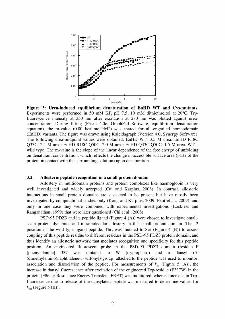
[urea] (M) Figure 3: Urea-induced equilibrium denaturation of EnHD WT and Cys-mutants. Experiments were performed in 50 mM KPi pH 7.5, 10 mM dithiothreitol at 20°C. Trp-fluorescence intensity at 350 nm after excitation at 280 nm was plotted against urea-concentration. During fitting (Prism 4.0c, GraphPad Software, equilibrium denaturation equation), the m-value (0.80 kcal·mol-1·M-1) was shared for all engrailed homeodomain (EnHD) variants. The figure was drawn using Kaleidagraph (Version 4.0, Synergy Software). The following urea-midpoint values were obtained: EnHD WT: 3.5 M urea; EnHD R18C Q33C: 2.1 M urea; EnHD R18C Q50C: 2.0 M urea; EnHD Q33C Q50C: 1.5 M urea. WT – wild type. The m-value is the slope of the linear dependence of the free energy of unfolding on denaturant concentration, which reflects the change in accessible surface area (parte of the protein in contact with the surrounding solution) upon denaturation. 3.2 Allosteric peptide recognition in a small protein domain
Allostery in multidomain proteins and protein complexes like haemoglobin is very well investigated and widely accepted (Cui and Karplus, 2008). In contrast, allosteric interactions in small protein domains are suspected to be present but have mostly been investigated by computational studies only (Kong and Karplus, 2009; Petit et al., 2009), and only in one case they were combined with experimental investigations (Lockless and Ranganathan, 1999) that were later questioned (Chi et al., 2008).
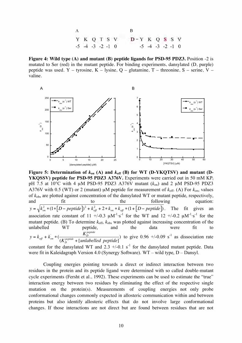
PSD-95 PDZ3 and its peptide ligand (Figure 4 (A)) were chosen to investigate small-scale protein dynamics and intramolecular allostery in this small protein domain. The -2 position in the wild type ligand peptide, Thr, was mutated to Ser (Figure 4 (B)) to assess coupling of this peptide residue to different residues in the PSD-95 PDZ3 protein domain, and thus identify an allosteric network that mediates recognition and specificity for this peptide position. An engineered fluorescent probe in the PSD-95 PDZ3 domain (residue F [phenylalanine] 337 was mutated to W [tryptophan]) and a dansyl (5-(dimethylamino)naphthalene-1-sulfonyl)-group attached to the peptide was used to monitor association and dissociation of the peptide. For measurements of kon (Figure 5 (A)), the increase in dansyl fluorescence after excitation of the engineered Trp-residue (F337W) in the protein (Förster Resonance Energy Transfer - FRET) was monitored, whereas increase in Trp-fluorescence due to release of the dansylated peptide was measured to determine values for koff (Figure 5 (B)).
10
Figure 4: Wild type (A) and mutant (B) peptide ligands for PSD-95 PDZ3. Position -2 is mutated to Ser (red) in the mutant peptide. For binding experiments, dansylated (D, purple) peptide was used. Y – tyrosine, K – lysine, Q – glutamine, T – threonine, S – serine, V – valine.
0
50
100
150
200
250
0 5 10 15 20 25
kobs
(s-1
) WT
kobs
(s-1
) Ser
kobs (
s-1
)
[dansylated peptide] (µM)
y = sqrt(m1^ 2*(1-m0)^ 2+m2^...
ErrorValue0.3180811.076m1 2.15998.3078m2
NA136.2ChisqNA0.99669R
y = sqrt(m1^ 2*(1-m0)^ 2+m2^...
ErrorValue0.2324310.841m1 1.43293.1613m2
NA219.08ChisqNA0.99726R
A B
0
1
2
3
4
5
6
7
0 10 20 30 40 50 60
kobs
(s-1
) WT
kobs
(s-1
) Ser
kobs (
s-1
)
[YKQTSV] (µM)
Figure 5: Determination of kon (A) and koff (B) for WT (D-YKQTSV) and mutant (D-YKQSSV) peptide for PSD-95 PDZ3 A376V. Experiments were carried out in 50 mM KPi pH 7.5 at 10°C with 4 µM PSD-95 PDZ3 A376V mutant (kon) and 2 µM PSD-95 PDZ3 A376V with 0.5 (WT) or 2 (mutant) µM peptide for measurement of koff. (A) For kon, values of kobs are plotted against concentration of the dansylated WT or mutant peptide, respectively, and fit to the following equation:
!
y = kon2" (1" D# peptide[ ])2 + koff
2 + 2" kon " koff " (1+ D# peptide[ ]) . The fit gives an association rate constant of 11 +/-0.3 µM-1·s-1 for the WT and 12 +/-0.2 µM-1·s-1 for the mutant peptide. (B) To determine koff, kobs was plotted against increasing concentration of the unlabelled WT peptide, and the data were fit to
!
y = koff + kon'" (
KD
peptide
(KD
peptide+ [unlabelled peptide]
) to give 0.96 +/-0.09 s-1 as dissociation rate
constant for the dansylated WT and 2.3 +/-0.1 s-1 for the dansylated mutant peptide. Data were fit in Kaleidagraph Version 4.0 (Synergy Software). WT – wild type, D – Dansyl.
Coupling energies pointing towards a direct or indirect interaction between two residues in the protein and its peptide ligand were determined with so called double-mutant cycle experiments (Fersht et al., 1992). These experiments can be used to estimate the “true” interaction energy between two residues by eliminating the effect of the respective single mutation on the protein(s). Measurements of coupling energies not only probe conformational changes commonly expected in allosteric communication within and between proteins but also identify allosteric effects that do not involve large conformational changes. If those interactions are not direct but are found between residues that are not

11
interacting directly, an allosteric network is likely to be involved. For binding of the peptide ligand mutated in the -2 position to PSD-95 PDZ3 coupling energies and hence an interaction between this peptide position and several positions within the protein were detected (Table 1).
The cross-correlation between ΔΔΔGC and Cα-Cα distance for binding of the peptide D-YKQSSV to PSD-95 PDZ3 mutants is shown in Figure 6 (A) and Table 1. The low R-values for fitting of an equation of the type y = c + k·x indicate no correlation between Cα-Cα distance and ΔΔΔGC and hence no distance-dependence of the coupling energy. For this peptide, no correlation between distance and coupling energy can be detected suggesting that discrimination of residues in position -2 in the peptide ligand involves an allosteric network in PSD-95 PDZ3. Possible network residues mapped onto the PSD-95 PDZ3 structure are shown in Figure 7.
Table 1: Apparent association (kon) and dissociation (koff) rate constants for binding of D-YKQSSV to PSD-95 PDZ3 mutants.
Mutant
Cα-Cα
distance
(Å)
kon, µM-1·
s-1 koff, s-1 KD, µM
∆∆∆GC
kon,
kcal·mol-1
∆∆∆GC
koff,
kcal·mol-1
∆∆∆GC
KD,
kcal·mol-1
Wild type - 9.8 (0.2) 8.8 (0.3) 0.89 0 0 0
I314V 19.9 10 (0.2) 9.3 (0.3) 0.92 0.12 -0.010 0.21
I316A 18.4 9.6 (0.1) 5.8 (0.01) 0.60 0.12 -0.17 0.29
L323A 11.5 13 (0.32) 37 (1.6) 2.8 0.22 -0.15 0.37
F325A 8.5 15 (0.45) 15 (0.2) 1.0 0.12 -0.28 0.40
I327V 5.6 11 (0.3) 25 (0.3) 2.3 0.14 -0.31 0.45
V328A 7.0 11 (0.3) 27 (0.4) 2.4 0.11 -0.087 0.20
E331A 13.1 5.4 (0.57) 38 (8) 7.0 -0.34 -0.87 0.53
I336A 10.7 24 (1.1) 161 (78) 6.8 0.67 -0.085 0.75
I336V 10.7 9.1 (0.1) 23 (0.8) 2.5 0.028 0.016 0.013
I338A 9.3 8.3 (0.4) 8 (0.2) 1.0 0.013 -0.063 0.077
F340A 10.1 9.1 (0.4) 15 (0.4) 1.7 0.061 -0.38 0.44
I341A 12.0 8.3 (0.4) 47 (10) 5.7 0.1 -0.35 0.45
I341V 12.0 11 (0.2) 12 (0.2) 1.0 0.16 -0.15 0.31
P346G 16.3 12 (0.2) 12 (0.4) 1.0 0.041 -0.13 0.17
A347G 14.8 7.8 (0.4) 15 (0.1) 1.9 0.061 -0.28 0.34
L353A 16.5 11 (0.4) 6.6 (0.1) 0.6 0.069 -0.27 0.34
V362A 15.8 8.9 (0.06) 7.2 (0.03) 0.8 0.065 -0.12 0.19
H372A 8.2 21 (0.83) 266 (20) 12.9 0.48 0.26 0.22
E373G 8.1 10 (0.2) 21 (0.9) 2.1 0.017 -0.12 0.14
E373A 8.1 9.6 (0.2) 25 (1.9) 2.6 0.058 -0.068 0.13
A375G 9.6 8.1 (0.3) 20 (0.6) 2.5 0.045 -0.12 0.17

12
A376G 6.8 12 (0.3) 31 (1.0) 2.7 0.12 -0.19 0.31
A376V 6.8 12 (0.2) 2.3 (0.1) 0.2 0.078 -0.45 0.53
I377G 10.0 11 (0.2) 7.6 (0.3) 0.7 0.0070 -0.28 0.29
I377A 10.0 11 (0.6) 8.7 (0.2) 0.8 0.050 0.15 -0.10
A378G 12.0 12 (0.3) 5.8 (0.3) 0.5 0.13 -0.25 0.39
K380G 10.3 8.1 (0.2) 12 (0.5) 1.5 -0.059 -0.18 0.12
K380A 10.3 7.9 (0.3) 17 (1.8) 2.1 0.11 -0.080 0.19
A382G 15.1 11 (0.1) 5.1 (0.1) 0.5 0.10 -0.11 0.21
V386A 17.3 10 (0.2) 7.0 (0.2) 0.67 0.12 -0.19 0.31
I388V 17.0 10 (0.2) 11 (0.1) 1.1 0.057 -0.017 0.073
A390G 17.4 10 (0.5) 13 (3.5) 1.2 -0.067 0.047 -0.11
Y392A 19.1 10 (0.3) 14 (1.2) 1.4 0.080 -0.063 0.14
KD was calculated from kon and koff (koff/kon) and ΔΔΔGC values were calculated as described in “5. Materials and Methods”. The Cα-Cα distances were measured in PyMOL (DeLano, 2002), PDB-ID: 1TP3, for the distances between the Cα of the mutated residue in the protein and Cα of the mutated residue in the peptide. Protein residues coupling with position -2 (Thr/Ser mutation) in the peptide are marked in bold. Fitting errors for kon and koff are given in brackets.
-1.5
-1
-0.5
0
0.5
1
1.5
0 5 10 15 20 25
!!!GC (k
on)
!!!GC (k
off)
!!!GC (K
D)
y = 0.17 - 0.0060x R= 0.15
y = -0.20 + 0.0029x R= 0.060
y = 0.37 - 0.0088x R= 0.19
!!!
GC (
kcal·m
ol-1
)
C"#C
" distance (Å)
-1.5
-1
-0.5
0
0.5
1
1.5
-1 0 1 2 3 4
!!!GC (k
on)
!!!GC (k
off)
!!!GC (K
D)
y = 0.049 + 0.056x R= 0.36
y = -0.077 - 0.022x R= 0.23
y = 0.13 + 0.078x R= 0.41
!!!
GC (
kca
l·mo
l-1)
!!Gstability
(kcal·mol-1
)
A B
Figure 6: Distance- and stability dependence of the coupling energy (ΔΔΔGC) for kon, koff and KD. (A) The Cα-Cα distances were measured in PyMOL (DeLano, 2002),PDB-ID: 1TP3 for the distances between the Cα of the mutated residue in the protein and Cα of the mutated residue in the peptide. Values are listed in Table 1. (B) Only residues with no direct interaction to the mutated peptide residue (wild type, I314V, I316A, I336A/V, I338A, F340A, I341A/V, P346G, A347G, V362A, V386A, I388V, A390G and Y392A) are included. Coupling energies are as in Table 1. Data for protein stability was kindly provided by Celestine Chi. KD was calculated from kon and koff (koff/kon) and ΔΔΔGC values were calculated as described in “5. Materials and Methods”. Data were fitted in Kaleidagraph Version 4.0 (Synergy Software). The cut-off (+/- 0.37 kcal·mol-1) for coupling is marked with a straight line in (A) and (B).

13
Furthermore, low R-values indicate no correlation between coupling energy and
protein stability for PSD-95 PDZ3, hence no dependence of ΔΔΔGC on protein stability (Figure 6 (B)). Therefore, protein stability does not influence the interaction between the Thr/Ser residue in the peptide and the different investigated residues in the protein domain.
Taken together, the results on mutant peptide binding to different mutant variants of PSD-95 PDZ3 hint towards the involvement of a network of residues within the domain that is involved in recognition of the cognate peptide.
Figure 7: Residues coupling to Thr/Ser at the -2 position in the ligand peptide for PSD-95 PDZ3. PSD-95 PDZ3 is coloured in dark blue, the peptide ligand is coloured in cyan and coupling residues in the PDZ domain as well as the -2 position in the peptide are marked in red. The Figure was drawn with PyMOL (DeLano, 2002), PDB-ID 1TP3.

14
4. Discussion
4.1 High-resolution protein folding studies Protein dynamics cover a wide spatial range from large-scale motions during protein
folding reactions or local unfolding events to small-scale vibrational changes. Protein (un)folding is commonly monitored using Circular Dichroism spectroscopy, changes in Trp-fluorescence intensity or FRET experiments either at equilibrium or in a time-resolved manner. Although those experiments have contributed enormously to the understanding of protein folding (Daggett and Fersht, 2003a; Fersht, 2008; Religa et al., 2005), more experiments are needed to dissect a protein folding reaction at different levels of detail. Time-resolved X-ray scattering experiments, during which the distance between two specific residues can be monitored, will provide a more detailed, high-resolution picture of the folding reaction. Here, the aim was to obtain double EnHD Cys-mutants labelled with mercury for such scattering experiments. Although the Cys-mutants are destabilized compared to the wild type protein, they are still folded under native conditions and the decrease in stability should thus not impair performance of experiments. Difficulties with the mercury-labelling reaction are partly due to the fact that, so far, it was not possible to measure the extent of labelling. Once this problem will be solved, different labelling conditions can be tested to obtain double mercury-labelled EnHD Cys-mutants for time-resolved X-ray scattering experiments and eventually understand protein dynamics at high-resolution.
4.2 Allosteric peptide recognition in a small protein domain
Another means of studying protein dynamics and allosteric communication at a rather high level of resolution is a double-mutant cycle experiment, during which the interaction energy between two residues within a protein or between a protein and its peptide ligand can be analysed (Fersht et al., 1992; Serrano et al., 1992a). Here, this method was used to study the involvement of an allosteric network in the PSD-95 PDZ3 domain in peptide recognition for Thr/Ser discrimination at position -2 in the ligand peptide. It was found that there indeed is an allosteric network involved in recognition of the peptide ligand as coupling between residues in the peptide-ligand and in the protein domain is observed. This coupling is not only seen for the -2 position in the peptide but also for Val0 (Linda C. Montemiglio, S. Raza Haq, Maike C. Jürgens, Celestine Chi, Åke Engström, Maurizio Brunori, Stefano Gianni and Per Jemth, manuscript in preparation). In contrast to other PDZ and small protein domains, this recognition process does not involve any large conformational changes upon ligand binding as there are no such conformational changes seen in structural and biochemical studies of ligand-bound and -free PSD-95 PDZ3 (Doyle et al., 1996; Gianni et al., 2006). It has been acknowledged previously that allostery without a “visible” conformational change but only involving changes in, for example, the vibrational energy of a side chain can exist (Cooper and Dryden, 1984). Such small-scale-dynamics allostery has been described before in computational studies of PDZ domains (Kong and Karplus, 2009; Lockless and Ranganathan, 1999; Petit et al., 2009) and in NMR experiments (Fuentes et al., 2004; Fuentes et al., 2006, 2008) but coupling of peptide ligand residues to residues in PDZ domains has not been investigated experimentally.
The fact that the coupling is distance-independent points towards an allosteric network involved in peptide-recognition of the -2 peptide position. Experimental data on PSD-95

15
PDZ3 obtained before (Chi et al., 2008) argued against the existence of such a network as no coupling was observed between possible network residues. However, the double mutant cycle experiments previously performed only included pairs between residue H372A and ten residues within the protein and hence did not look into coupling between peptide residues and the PDZ domain. Interestingly, in the current study no coupling between the investigated peptide residues and H372A could be observed although the Thr2-hydroxyl forms an H-bond with H372A in the crystal structure (Doyle et al., 1996). This may explain why no coupling was observed between H372A and other residues in the protein domain since H372 does not respond to binding of the peptide ligand in an allosteric fashion.
Furthermore, most ΔΔΔGC’s in this dataset were, intriguingly, positive. A lower KD for binding of the peptide to the mutant protein compared to the wild type protein would result in a negative free energy of binding. ΔΔΔGC is then positive if ΔΔGKD is more negative for the wild type peptide compared to the mutant peptide as the difference in KD (kon, koff) between the PSD-95 PDZ3 mutants is more pronounced for the wild type peptide than for the mutant peptide. Hence, the effect of making a mutation in the peptide influences kon,koff or KD less when the protein is also mutated, indicating that the PSD-95 PDZ3 domain becomes more promiscuous towards its target peptide upon mutation. At the same time this provides a means for specificity change of PDZ domains during evolution where the domain first becomes more promiscuous before specificity for a different peptide ligand is established by further mutation. 4.3 Is allostery common in PDZ domains?
Network residues identified in PSD-95 PDZ3 can also be compared to network residues found in another PDZ domain, PTP-BL PDZ2 (Montemiglio et al., manuscript in preparation). Some of the network residues found in both proteins are at homologous positions, whereas each PDZ domain also uses unique network residues that are specific for that PDZ domain. This comparison of the networks between PSD-95 PDZ3 and PTP-BL PDZ2 suggests that allosteric networks in PDZ domains are sequence-specific since both proteins use different networks for peptide-ligand recognition. Moreover, from the present data and previous computer simulations (Kong and Karplus, 2009) it can be concluded that two different but not entirely independent networks of residues are involved in recognition of peptide residues in the 0 and -2 position. Some of the residues that are part of the network recognizing Val0 are also part of the Thr2-recognizing network (L323A, I336A, and F340A), whereas other residues like F325A (position 2) or P346G (position 0) are only found in one of the networks (this study and Montemiglio et al., manuscript in preparation). This also excludes the possibility of the existence of one network that is extended for recognition of the full peptide compared to the very C-terminus only, as this would require the presence of unique network residues for the -2 position only. If an extended network were involved in peptide recognition for Thr2, this would imply that all network residues found for Val0 also are found in the network for Thr2, but there should not be any residue that couples to Val0 but not to Thr2. Since this was not the case for PSD-95 PDZ3, two different but interconnected (some residues are found in both networks) networks are involved in allosteric peptide ligand recognition in PSD-95 PDZ3. For the whole binding and recognition process, one can envision that first the C-terminal Val-residue binds to the ligand-binding pocket as recently shown as an intermediate state in PDZ-peptide interaction (Cámara-Artigas et al., 2010;

16
Elkins et al., 2010). This initial binding and subsequent or concomitant change in residue dynamics could be followed by a second binding step during which the other peptide residues, including position -2, are bound (Figure 8).
Figure 8: Proposed binding and recognition mechanism for peptide ligand binding to PSD-95 PDZ3. (A) The peptide is mutated in the 0 position and therefore can not induce any “recognition signal” (half circles) in the PDZ domain. Binding is unsuccessful. (B) Binding of the wild type peptide induces two recognition signals, one for the 0 position and a second signal in the subsequent binding step when the -2 position is approved. Binding of the peptide is successful. (C) Binding of a peptide mutated in the -2 position induces an allosteric signal in the first binding step but the second binding step fails to induce further signals. Therefore, as in (A), the peptide is released from the binding pocket and binding is not successful. The peptide C-terminus is denoted “C”. Red symbolises a PDZ domain in an unbound conformation, yellow shows a PDZ domain after the recognition signal for the extreme C-terminus and green for a PDZ domain in its fully bound state. Dark blue – petide mutated at position 0, light blue – peptide mutated at position -2, green – wild type peptide.
In contrast to PTP-BL PDZ2 (Jemth and Gianni, 2007), no correlation between the
free energy of binding and protein stability could be established for PSD-95 PDZ3. It was proposed before that PSD-95 PDZ3 is a rather rigid PDZ-domain that does not undergo any conformational change upon peptide binding (Chi et al., 2008; Gianni et al., 2006). A stability-dependence of the free energy of binding for PTP-BL PDZ2 may suggest that this domain becomes more rigid upon peptide binding (Gianni et al., 2006; Jemth and Gianni, 2007) whereas this effect is not seen for PSD-95 PDZ3, which already is relatively stiff before the encounter with the peptide ligand (Gianni et al., 2006; Jemth and Gianni, 2007). This assumption is corroborated by the fact that coupling energies, ΔΔΔGC, are in general higher for PTP-BL PDZ2 than for PSD-95 PDZ3 which could reflect the fact that next to changes in

17
side chain dynamics a stiffening (comparable to the last step in a folding mechanism for example) of the PTP-BL PDZ2 domain, which also shows conformational changes upon peptide binding (Gianni et al., 2006), contributes to the measured coupling energy.
Taken together, based on these results a mechanism may be proposed for peptide binding, recognition and discrimination during which the peptide is the allosteric effector and at the same time the presence of the peptide is required to transmit the allosteric signal through the protein domain. This hypothesis is applicable if the peptide-binding event is seen as a coupled folding and binding event (the C-terminal peptide ligand forms a β-strand extending the already present β-sheet) comparable to coupled folding and binding events of so-called intrinsically disordered proteins (Boehr et al., 2009; Dyson and Wright, 2005; Wright and Dyson, 1999). During this process, the allosteric effector binds, and at the same time the folding of the PDZ domain is “completed” (stiffening effect, positive ΔΔΔGCs). Transmission of the signal reporting this event is distributed through the protein domain via the allosteric network(s) identified here involving small-scale changes in side-chain dynamics.

18
5. Materials and Methods
5.1 Bacterial strains and plasmids 2x TY medium (5 g·l-1 NaCl, 10 g·l-1 yeast extract, 16 g·l-1 tryptone) was used for
Escherichia coli cell cultures. For 2x TY plates, 15 g·l-1 agar were added. Plates and media were supplemented with the antibiotics ampicillin (100 µg·ml-1, Astra Zeneca, Sweden) and chloramphenicol (34 µg·ml-1, Calbiochem, USA) when required unless specified otherwise.
Chemically competent Escherichia coli XL1 Blue cells (Stratagene, USA) were used for cloning procedures, and E. coli BL21(DE3)pLysS (Invitrogen, USA) cells were used for protein overexpression.
For heat-shock transformation, cells were thawed on ice before addition of 2 µl plasmid-DNA to 15 µl or 50 µl chemically competent E. coli BL21 or XL1 Blue cells, respectively. After incubation on ice for 15 min, cells were heat-shocked at 42°C for 45 s and incubated on ice for an additional 3 min. 200 to 250 µl 2x TY medium were added before incubation at 37°C for 1 h. Cells were plated on 2x TY plates and incubated at 37°C overnight. Plasmids used in this study are listed in table 2. Table 2: Plasmids used in this work Plasmid Features Source EnHD wild type pSEA 100 coding for residues 1
to 59 of EnHD Tomasz L. Religa and Alan R. Fersht.
EnHD R18C, EnHD Q50C and EnHD Q33C Q50C
Same as for EnHD wild type with introduced point mutations
Lisa Elfström
EnHD R18C Q33C, EnHD R18C Q50C
Same as for EnHD wild type with introduced point mutations
This study
PSD-95 PDZ3 wild type and mutants
Modified pRSET vector coding for residues 306 to 401 of PSD-95 PDZ3 (including the respective mutation)
Celestine Chi, Nicoletta Calosci
For all plasmids, protein overexpression can be induced by the addition of IPTG (isopropyl ß-D-thiogalactopyranoside). 5.2 Cloning procedures 5.2.1 EnHD Cys mutants
EnHD mutants EnHD R18C, EnHD Q50C and EnHD Q33C Q50C were generated by Lisa Elfström. Primer sequences for primers used to generate those and all further mutants are listed in table 3. Table 3: Primer sequences to generate EnHD Cys-mutants. Primer name Sequence R18C fw 5’-TTGGCCCGCCTCAAGTGCGAATTCAACGAGAAT-3’ R18C rv 5’-ATTCTCGTTGAATTCGCACTTGAGGCGGGCCAA-3’

19
Q33C fw 5’-GAGCGGAGACGCCAGTGCCTGAGCAGCGAGCTC-3’ Q33C rv 5’-GAGCTCGCTGCTCAGGCACTGGCGTCTCCGCTC-3’ Q50C fw 5’-ATCAAGATCTGGTTCTGCAACAAGCGCGCCAAG-3’ Q50C rv 5’-CTTGGCGCGCTTGTTGCAGAACCAGATCTTGAT-3’ R18C fw/rv introduces a point mutation from R (Arg) to C (Cys) in position 18 in EnHD, Q33C fw/rv introduces a Q (Gln) to C mutation in position 33, and Q50C fw/rv puts the same mutation in position 50 in EnHD. fw- forward primer, rv – reverse primer.
The pipetting scheme for PCRs performed is noted in table 4, and the PCR protocol is listed in table 5.
Presence of PCR products was checked on a 1% agarose gel (expected size 3360 bp) in 0.5x TBE buffer (45 mM Tris-borate, 1 mM EDTA (ethylenediaminetetraacetic acid)), and PCR products were subsequently digested with DpnI (5 U, Stratagene) for 4 h at 37°C. Amplified DNA was then transformed into chemically competent E. coli XL1 blue cells as described above.
Plasmids from successfully transformed cells were purified with the Plasmid Mini Kit (Omega Bio-Tek, VWR, Sweden) and sequenced (Uppsala Genome Centre, Uppsala University). Table 4: PCR reaction ingredients to generate EnHD R18C Q50C from either EnHD R18C or EnHD Q50C as template or to generate EnHD R18C Q33C from EnHD R18C using the respective primers. Ingredient Amount Template 0.5 µl
Primer forward 10 pmol (Q50C and Q33C primer) or 8 pmol (R18C primer), respectively
Primer reverse 10 pmol or 8 pmol, respectively DMSO (dimethyl sulfoxide) 2% (v/v) Pfu buffer (10x, Stratagene) 1x dNTP (2.5 mM stock) 0.15 mM Pfu Turbo (Stratagene) 1.25 U H2O Add to 50 µl Table 5: PCR protocol to generate EnHD mutants. Step Temperature in °C Time in s 1 95 120 2 95 45 3 58,59,61 45 4 72 300 5 72 600 35 cycles (steps 2 to 4) were performed. 59°C and 61°C were used as annealing temperatures for templates EnHD R18C and EnHD Q50C, respectively. 58°C was used as annealing temperature for the EnHD R18C template to generate R18C Q33C.

20
5.3 Protein overexpression and purification
5.3.1 Protein overexpression For protein overexpression, successfully transformed E. coli BL21(DE3)pLysS cells
were grown in 2x TY medium overnight. The overnight culture was used to inoculate the main culture containing 50 µg·ml-1ampicillin in a 1:100 ratio. Protein overexpression was induced at an OD600 of ~ 0.6 to 1 with 1 mM IPTG (isopropyl ß-D-thiogalactopyranoside , Saveen Werner AB, Sweden).
For EnHD (engrailed homeodomain), protein overexpression could only be achieved when expressing for 4 h at 37°C, whereas PSD-95 PDZ3 was expressed for ~ 16 h at 30°C. Cells were harvested by centrifugation (7,500 g, 4°C, 15 min) and resuspended in 20 mM KPi pH 8.0 (EnHD) or 50 mM Tris-HCl pH 8.5, 400 mM NaCl, respectively.
Before protein purification, cells were lysed by sonication on ice for 5 min (Sonics, Vibracell, CiAB, Sollentuna, Sweden; amplitude 60%), and the lysate was cleared by centrifugation (48,000 g, 4°C, 1 h).
5.3.2 Protein purification EnHD wild-type purification
Plasmid DNA coding for EnHD wild type protein was a gift from Tomasz L. Religa and Alan R. Fersht. EnHD wild type protein was purified according to Mayor et al. (2000). In brief, the crude lysate (filtered, 0.45 and 0.2 µm filters) was loaded onto a SourceQ column (GE Healthcare, CV [column volume] 20 ml) equilibrated with 20 mM KPi pH 8.0. The protein appeared in the flow-through. The flow-through was subsequently loaded onto an S-column (GE Healthcare, CV 20 ml) pre-equilibrated with 20 mM KPi pH 8.0 and eluted in a gradient (0 to 1 M NaCl in 20 mM KPi pH 8.0 in 10 CV) after washing with 3 CV 20 mM KPi pH 8.0. Fractions containing EnHD were identified by 15% SDS-PAGE and stored at -20°C before further purification on a reversed phase column (C18 column, Vydac, USA).
Before loading onto the reversed phase column equilibrated with 0.1% TFA (trifluoroacetic acid, Applied Biosystems, UK), the thawed sample was acidified to denature the protein. After washing with 5 CV 0.1% TFA, the protein was eluted in a gradient from 0 to 80% acetonitrile (Merck, Germany) in 0.1% TFA and fractions containing EnHD were freeze-dried.
EnHD Cys-mutants purification Since EnHD Cys-mutants were found in the pellet and not in the supernatant after cell
lysis, the pellet was resuspended in 20 mM KPi pH 8.0, 8 M urea and incubated for 1 h at room temperature while shaking. After centrifugation for 1 h (48,000 g, 4°C) the supernatant was filtered (0.45 µm and 0.2 µm filters, successively) and loaded onto an S-column (GE Healthcare, CV 20 ml) pre-equilibrated with 20 mM KPi pH 8.0. The column was washed thoroughly with 20 mM KPi pH 8.0 to remove urea (and refold the protein bound to the column) before elution in a gradient from 0 to 1 M NaCl in 20 mM KPi pH 8.0, 5 mM DTT (dithiothreitol, VWR, Sweden) in 10 CV. Fractions containing EnHD Cys-mutant were

21
identified by SDS-PAGE, pooled and dialysed against 20 mM KPi pH 8.0, 5 mM DTT at 4°C overnight.
To remove further impurities, the protein was loaded onto a Heparin column (GE Healthcare, CV 1 ml) pre-equilibrated with 20 mM KPi pH 8.0, 5 mM DTT after addition of 5 mM (final concentration) DTT to the protein solution. After washing with 5 CV 20 mM KPi pH 8.0, 5 mM DTT, the protein was eluted in a stepwise gradient with 0.2, 0.4, 0.6, and 1 M NaCl in 20 mM KPi pH 8.0, 5 mM DTT. Fractions were analysed by 15% SDS-PAGE and those containing EnHD Cys-mutants were stored at -20°C until further purification by reversed phase chromatography as described for the wild type protein.
PSD-95 PDZ3 purification PSD-95 PDZ3 mutants were either provided by S. Raza Haq or Celestine N. Chi or
purified according to Chi et al., (2006). Briefly, the supernatant after cell lysis was filtered (0.45 µm and 0.2 µm filter,
Sarstedt, Germany) and loaded onto a Ni2+-Sepharose column pre-equilibrated with 50 mM Tris-HCl pH 8.5, 400 mM NaCl. After washing with 100 ml of the same buffer, the protein was eluted in 250 mM imidazole, and fractions containing PSD-95 PDZ3 were identified by 15% SDS-PAGE. The protein was dialysed against 50 mM Tris-HCl pH 7.5 overnight at 4°C and loaded onto a Q-column (GE Healthcare, Sweden, CV [column volume] 20 ml) equilibrated with the same buffer. The column was washed with 3 CV 50 mM Tris-HCl pH 7.5, and the protein was eluted in a gradient from 0 to 1 M NaCl in 50 mM Tris-HCl in 12.5 CV. Fractions containing pure PSD-95 PDZ3 were identified by SDS-PAGE, pooled and used for further experiments.
5.3.3 15% sodium dodecyl sulfate polyacrylamide gel electrophoresis Protein samples for SDS-PAGE (sodium dodecyl sulfate polyacrylamide gel
electrophoresis) were prepared with 3x Laemmli loading dye (Table 6) so that 15 µl sample were loaded onto the SDS-gel. Table 6: 3x Laemmli loading dye. Component Amount 1 M Tris-HCl pH 6.8 2.4 ml 20% (w/v) SDS (Sigma, Germany) 3 ml 100% (v/v) glycerol (Merck, Germany) 3 ml 2-mercaptoethanol (VWR, Sweden) 1.6 ml bromophenol blue (Fluka, Switzerland) 0.006 g H2O Add to 10 ml
Solutions for the stacking and separating gel were prepared according to table 7. Table 7: SDS-gel preparation. Ingredient Stacking gel Separating gel Acrylamide solution (30% acrylamide/0.8% bisacryl- amide [AppliChem, Germany]) 0.75 ml 5 ml
80% (v/v) glycerol - 1.25 ml 0.5 M Tris-HCl pH 6.8, 0.4% (w/v) SDS 1.25 ml - 1.5 M Tris-HCl pH 8.8, 0.4% (w/v) SDS - 2.5 ml

22
APS (ammonium persulfate; 40% (w/v)) 8 · 10-3 ml 20 · 10-3 ml TEMED (N, N, N’, N’- Tetramethylethan-1,2-diamine; Sigma, Germany) 8 · 10-3 ml 20 · 10-3 ml
H2O 3 ml 1.2 ml
For Coomassie staining, 250 ml staining solution (0.1% (w/v) Coomassie [VWR, Sweden], 10% (v/v) acetic acid [Merck, Germany], 40% (v/v) ethanol [Solveco, Sweden]) was added to the gel, heated briefly and left shaking for about 15 min. For destaining, the staining solution was removed; the gel rinsed briefly with H2O, and destaining solution (10% (v/v) acetic acid, 40% (v/v) ethanol) was added. After heating for 30 s and shaking for 15 to 30 min, bands started to be visible. Complete destaining was achieved by incubating the gel in either destaining solution or H2O overnight.
5.3.4 Determination of protein concentration Protein concentration was determined measuring the absorbance at 280 nm on a
Nanodrop (Saveen Werner AB, Sweden) using extinction coefficients calculated with the ExPASy Protparam tool (Gasteiger et al., 2005). Protein sequences used for calculation and the respective extinction coefficients are listed under “General information on proteins”.
5.3.5 General information on proteins Amino acid sequences for the different proteins investigated are listed in table 8 and relevant features can be found in table 9. Table 8: EnHD and PSD-95 PDZ3 amino acid sequences. Protein Sequence EnHD EKRPRTAFSSEQLARLKREFNENRYLTERRRQQLSSELGLNEAQIKIWFQ
NKRAKIKKS PSD-95 PDZ3
MHHHHHPRGSREPRRIVIHRGSTGLGFNIVGGEDGEGIWISFILAGGPADL SGELRKGDQILSVNGVDLRNASHEQAAIALKNAGQTVTIIAQYKPEEYSR FE
For EnHD, residues mutated to Cys in the different double Cys-mutants are marked in bold. Trp (W) residues used for fluorescence experiments are marked in red. Table 9: Molecular Weight, extinction coefficient, no of amino acids, and theoretical pI for EnHD wild type and mutants, and PSD-95 PDZ3 wild type. Mutant Molecular
Weight Extinction coefficient
No of amino acids
Theoretical pI
EnHD wild type 7453.5 Da 6990 cm-1 M-1 59 10.67 EnHD R18C Q33C
7375.4 Da 6990 cm-1 M-1 (assuming all Cys are reduced)
59 10.02
EnHD R18C Q50C
7375.4 Da 6990 cm-1 M-1 (assuming all Cys are reduced)
59 10.02

23
EnHD Q33C Q50C
7403.5 Da 6990 cm-1 M-1 (assuming all Cys are reduced)
59 10.21
PSD-95 PDZ3 wild type
11247.5 Da 8480 cm-1 M-1 103 6.65
5.4 Equilibrium denaturation of EnHD wild type and Cys mutants
To check the stability of the EnHD Cys-mutants, equilibrium denaturation experiments were carried out in 50 mM KPi pH 7.5 in the presence of 10 mM DTT to prevent disulfide bond formation. Urea-induced unfolding was monitored at 20°C measuring Trp (W48) fluorescence at 350 nm following excitation at a wavelength of 280 nm on an SLM4800 spectrofluorimeter (SLM instruments, IL). 4 µM protein were denatured by step-wise increase of urea concentration to final concentrations of 7.67 - 8.9 M urea (determined by refractrometry) as listed in table 10. The maximal urea concentration used depended on the protein concentration as the protein solution was mixed with a 50 mM KPi pH 7.5, 9 M urea solution to give the highest possible urea concentration in the denaturation experiment. Mixing for 3 min before making each measurement ensured complete equilibration of the system.
Fluorescence intensity at 350 nm was then plotted against urea concentration and fitted to Eq. (1):
!
F =("
N+ #
N[denaturant])+ ("
D+ #
D[denaturant]e
mD$N ([denaturant ]$[D ]50%
RT
1+ e
mD$N ([denaturant ]$[D ]50%
RT
αN = fluorescence intensity of the native state at 0 M urea; βN = native baseline; αD =
fluorescence intensity of the denatured state at 0 M urea; βN = denatured baseline·[urea]; [D]50% = urea midpoint; RT = 0.592 kcal·mol-1; (Fersht, 1999) to determine the m-value (mD-
N) and urea midpoint. Table 10: EnHD mutants and final urea concentration during urea-induced equilibrium denaturation experiments. Mutant Max [urea] (M) EnHD wild type 8.9 EnHD R18C Q33C 7.67 EnHD R18C Q50C 8.55 EnHD Q33C Q50C 8.4 5.5 Binding experiments of YKQSSV to PSD-95 PDZ3 wild type and mutants
Binding experiments for dansylated and unlabelled mutant peptide ([D-]YKQSSV) to PSD-95 PDZ3 mutants were performed in 50 mM KPi pH 7.5 at 10°C. Dansylated and unlabelled peptides were purchased from GL Biochem Ltd., Shanghai.
24
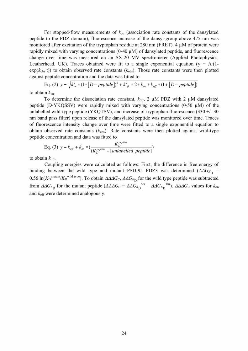
For stopped-flow measurements of kon (association rate constants of the dansylated peptide to the PDZ domain), fluorescence increase of the dansyl-group above 475 nm was monitored after excitation of the tryptophan residue at 280 nm (FRET). 4 µM of protein were rapidly mixed with varying concentrations (0-40 µM) of dansylated peptide, and fluorescence change over time was measured on an SX-20 MV spectrometer (Applied Photophysics, Leatherhead, UK). Traces obtained were fit to a single exponential equation (y = A·(1-exp(kobs·t)) to obtain observed rate constants (kobs). Those rate constants were then plotted against peptide concentration and the data was fitted to
Eq. (2)
!
y = kon2" (1" D# peptide[ ])2 + koff
2 + 2" kon " koff " (1+ D# peptide[ ]) to obtain kon.
To determine the dissociation rate constant, koff, 2 µM PDZ with 2 µM dansylated peptide (D-YKQSSV) were rapidly mixed with varying concentrations (0-50 µM) of the unlabelled wild-type peptide (YKQTSV), and increase of tryptophan fluorescence (330 +/- 30 nm band pass filter) upon release of the dansylated peptide was monitored over time. Traces of fluorescence intensity change over time were fitted to a single exponential equation to obtain observed rate constants (kobs). Rate constants were then plotted against wild-type peptide concentration and data was fitted to
Eq. (3)
!
y = koff + kon'" (
KD
peptide
(KD
peptide+ [unlabelled peptide]
)
to obtain koff. Coupling energies were calculated as follows: First, the difference in free energy of binding between the wild type and mutant PSD-95 PDZ3 was determined (ΔΔGKD = 0.56·ln(KD
mutant/KDwild type). To obtain ΔΔΔGC, ΔΔGKD for the wild type peptide was subtracted
from ΔΔGKD for the mutant peptide (ΔΔΔGC = ΔΔGKDSer – ΔΔGKD
Thr). ΔΔΔGC values for kon and koff were determined analogously.

25
6. Acknowledgements
First of all, jättestort TACK till Per för att jag fick göra ex-jobbet hos dig. Tack för allt jag lärde mig här – på labbet och alla icke-vetenskapliga grejer (svensk grammatik och ordkunskap borde jag väl jobba på lite till ) som intervallträning, orientering och lite orsamål. Danke också för att du ”prov”läste allt jag skrev, svarade på en hel massa frågor och synd att jag inte får ”klonera” längre och dessutom är du en av de snällaste människor som finns. Det var jättekul här och jag kommer att sakna det mycket!
Tack också till Åke för allt hjälp med EnHD-mass-spec!
A big thanks to Chi for helping out with a lot of things and answering several questions, for discussions, and morning-fika (I really appreciated your help and company). Thanks to Raza and Aziz for teaching me some urdu and tack to Andreas for all the information on Swedish music, culture, and history. Ieva, thank you for your female support! And of course, thank you to all the other lab-people.
I would also like to thank all the people outside of the lab (=friends ) for all the fikas, girls meetings, training sessions and many other things. Special thanks go to Claire, Jenna, Kristina, Luba, and Marius, and Jana, Melanie, and Monika.
DANKE Mama, Papa und Peter (und Jimi) für eure Unterstützung, wann immer sie nötig war! Ich hab euch lieb!
26
7. References
Andersson, M., van der Spoel, D., Davidsson, J., and Neutze, R. (2008). A proposed time-resolved X-ray scattering approach to track local and global conformational changes in membrane transport proteins. Structure 16, 21-28.
Anfinsen, C.B. (1973). Principles that govern the folding of protein chains. Science 181, 223-230.
Boehr, D.D., Nussinov, R., and Wright, P.E. (2009). The role of dynamic conformational ensembles in biomolecular recognition. Nature Chemical Biology 5, 789-796.
Bryngelson, J.D., Onuchic, J.N., Socci, N.D., and Wolynes, P.G. (1995). Funnels, Pathways, and the Energy Landscape of Protein-Folding - A Synthesis. Proteins-Structure Function and Genetics 21, 167-195.
Cámara-Artigas, A., Murciano-Calles, J., Gavira, J.A., Cobos, E.S., and Martínez, J.C. (2010). Novel conformational aspects of the third PDZ domain of the neuronal post-synaptic density-95 protein revealed from two 1.4 Å X-ray structures. Journal of Structural Biology.
Chi, C.N., Elfstrom, L., Shi, Y., Snall, T., Engstrom, A., and Jemth, P. (2008). Reassessing a sparse energetic network within a single protein domain. Proceedings of the National Academy of Sciences of the United States of America 105, 4679-4684.
Chi, C.N., Gianni, S., Calosci, N., Travaglini-Allocatelli, C., Engstrom, A., and Jemth, P. (2007). A conserved folding mechanism for PDZ domains. Febs Letters 581, 1109-1113.
Clarke, N.D., Kissinger, C.R., Desjarlais, J., Gilliland, G.L., and Pabo, C.O. (1994). Structural studies of the engrailed homeodomain. Protein Science 3, 1779-1787.
Cooper, A., and Dryden, D.T.F. (1984). Allostery without conformational change - a plausible model. European Biophysics Journal with Biophysics Letters 11, 103-109.
Cui, Q., and Karplus, M. (2008). Allostery and cooperativity revisited. Protein Sci 17, 1295-1307.
Daggett, V., and Fersht, A. (2003a). The present view of the mechanism of protein folding. Nature Reviews Molecular Cell Biology 4, 497-502.
Daggett, V., and Fersht, A.R. (2003b). Is there a unifying mechanism for protein folding? Trends in Biochemical Sciences 28, 18-25.
DeLano, W.L. (2002). The PyMOL Molecular Graphics System (San Carlos, CA, USA, DeLano Scientific).
27
Doyle, D.A., Lee, A., Lewis, J., Kim, E., Sheng, M., and MacKinnon, R. (1996). Crystal structures of a complexed and peptide-free membrane protein-binding domain: Molecular basis of peptide recognition by PDZ. Cell 85, 1067-1076.
Dyson, H.J., and Wright, P.E. (2005). Intrinsically unstructured proteins and their functions. Nature Reviews Molecular Cell Biology 6, 197-208.
Elkins, J.M., Gileadi, C., Shrestha, L., Phillips, C., Wang, J., Muniz, J.R.C., and Doyle, D.A. (2010). Unusual binding interactions in PDZ domain crystal structures help explain binding mechanisms. Protein Science 19, 731-741.
Ferguson, N., Capaldi, A.P., James, R., Kleanthous, C., and Radford, S.E. (1999). Rapid folding with and without populated intermediates in the homologous four-helix proteins Im7 and Im9. Journal of Molecular Biology 286, 1597-1608.
Fersht, A. (1999). Structure and Mechanism in Protein Science: A Guide to Enzyme Catalysis and Protein Folding (New York, Freeman).
Fersht, A.R. (1995). Characterizing transition states in protein folding: an essential step in the puzzle. Curr Opin Struct Biol 5, 79-84.
Fersht, A.R. (2008). From the first protein structures to our current knowledge of protein folding: delights and scepticisms. Nature Reviews Molecular Cell Biology 9, 650-654.
Fersht, A.R., Matouschek, A., and Serrano, L. (1992). The folding of an enzyme. I. Theory of protein engineering analysis of stability and pathway of protein folding. J Mol Biol 224, 771-782.
Fuentes, E.J., Der, C.J., and Lee, A.L. (2004). Ligand-dependent dynamics and intramolecular signaling in a PDZ domain. Journal of Molecular Biology 335, 1105-1115.
Fuentes, E.J., Gilmore, S.A., Mauldin, R.V., and Lee, A.L. (2006). Evaluation of energetic and dynamic coupling networks in a PDZ domain protein. Journal of Molecular Biology 364, 337-351.
Fuentes, E.J., Gilmore, S.A., Mauldin, R.V., and Lee, A.L. (2008). Evaluation of energetic and dynamic coupling networks in a PDZ domain protein (vol 364, pg 337, 2006). Journal of Molecular Biology 375, 1489-1490.
Gasteiger, E., Hoogland, C., Gattiker, A., Duvaud, S., Wilkins, M.R., Appel, R.D., and Bairoch, A. (2005). Protein Indentification and Analysis Tools on the ExPASy Server. In The Proteomics Protocols Handbook, J.M. Walker, ed. (Humana Press), pp. 571-607.
Gianni, S., Guydosh, N.R., Khan, F., Caldas, T.D., Mayor, U., White, G.W.N., DeMarco, M.L., Daggett, V., and Fersht, A.R. (2003). Unifying features in protein-folding mechanisms. Proceedings of the National Academy of Sciences of the United States of America 100, 13286-13291.

28
Gianni, S., Walma, T., Arcovito, A., Calosci, N., Bellelli, A., Engstrom, A., Travaglini-Allocatelli, C., Brunori, M., Jemth, P., and Vuister, G.W. (2006). Demonstration of long-range interactions in a PDZ domain by NMR, kinetics, and protein engineering. Structure 14, 1801-1809.
Ho, B.K., and Agard, D.A. (2010). Conserved tertiary couplings stabilize elements in the PDZ fold, leading to characteristic patterns of conformational flexibility. Protein Science 19, 398-411.
Itzhaki, L.S., Otzen, D.E., and Fersht, A.R. (1995). The Structure of the Transition-State for Folding of Chymotrypsin Inhibitor-2 Analyzed by Protein Engineering Methods - Evidence for a Nucleation-Condensation Mechanism for Protein-Folding. Journal of Molecular Biology 254, 260-288.
Jacques, D.A., and Trewhella, J. (2010). Small-angle scattering for structural biology-Expanding the frontier while avoiding the pitfalls. Protein Science 19, 642-657.
Jemth, P., and Gianni, S. (2007). PDZ domains: Folding and binding. Biochemistry 46, 8701-8708.
Kim, E., Niethammer, M., Rothschild, A., Jan, Y.N., and Sheng, M. (1995). Clustering of Shaker-Type K+ Channels by Interaction with a Family of Membrane-associated Guanylate Kinases. Nature 378, 85-88.
Kim, E.J., and Sheng, M. (2004). PDZ domain proteins of synapses. Nature Reviews Neuroscience 5, 771-781.
Kong, Y.F., and Karplus, M. (2009). Signaling pathways of PDZ2 domain: A molecular dynamics interaction correlation analysis. Proteins-Structure Function and Bioinformatics 74, 145-154.
Kornau, H.C., Schenker, L.T., Kennedy, M.B., and Seeburg, P.H. (1995). Domain Interaction Between NMDA Receptor Subunits and the Postsynaptic Density Protein PSD-95. Science 269, 1737-1740.
Korzhnev, D.M., and Kay, L.E. (2008). Probing invisible, low-populated states of protein molecules by relaxation dispersion NMR spectroscopy: An application to protein folding. Accounts of Chemical Research 41, 442-451.
Levinthal, C. (1968). Are there pathways for protein folding. Journal De Chimie Physique Et De Physico-Chimie Biologique 65, 44-&.
Lockless, S.W., and Ranganathan, R. (1999). Evolutionarily conserved pathways of energetic connectivity in protein families. Science 286, 295-299.
Matouschek, A., Kellis, J.T., Jr., Serrano, L., and Fersht, A.R. (1989). Mapping the transition state and pathway of protein folding by protein engineering. Nature 340, 122-126.

29
Mayor, U., Guydosh, N.R., Johnson, C.M., Grossmann, J.G., Sato, S., Jas, G.S., Freund, S.M.V., Alonso, D.O.V., Daggett, V., and Fersht, A.R. (2003). The complete folding pathway of a protein from nanoseconds to microseconds. Nature 421, 863-867.
Nourry, C., Grant, S.G., and Borg, J.P. (2003). PDZ domain proteins: plug and play! Sci STKE 2003, re7.
Onuchic, J.N., LutheySchulten, Z., and Wolynes, P.G. (1997). Theory of protein folding: The energy landscape perspective. Annual Review of Physical Chemistry 48, 545-600.
Petit, C.M., Law, A.B., Zhang, J., Fuentes, E.J., and Lee, A.L. (2009). Side-Chain Dynamics in PDZ Domain Structure and Function. Journal of Biomolecular Structure & Dynamics 26, 124.
Religa, T.L., Johnson, C.M., Vu, D.M., Brewer, S.H., Dyer, R.B., and Fersht, A.R. (2007). The helix-turn-helix motif as an ultrafast independently folding domain: The pathway of folding of Engrailed homeodomain. Proceedings of the National Academy of Sciences of the United States of America 104, 9272-9277.
Religa, T.L., Markson, J.S., Mayor, U., Freund, S.M.V., and Fersht, A.R. (2005). Solution structure of a protein denatured state and folding intermediate. Nature 437, 1053-1056.
Schreiber, G., and Fersht, A.R. (1995). Energetics of Protein-Protein Interactions - Analysis of the Barnase-Barstar Interface by Single Mutations and Double Mutant Cycles. Journal of Molecular Biology 248, 478-486.
Serrano, L., Kellis, J.T., Jr., Cann, P., Matouschek, A., and Fersht, A.R. (1992a). The folding of an enzyme. II. Substructure of barnase and the contribution of different interactions to protein stability. J Mol Biol 224, 783-804.
Serrano, L., Matouschek, A., and Fersht, A.R. (1992b). The folding of an enzyme. III. Structure of the transition state for unfolding of barnase analysed by a protein engineering procedure. J Mol Biol 224, 805-818.
Serrano, L., Matouschek, A., and Fersht, A.R. (1992c). The folding of an enzyme. VI. The folding pathway of barnase: comparison with theoretical models. J Mol Biol 224, 847-859.
Sheng, M., and Sala, C. (2001). PDZ domains and the organization of supramolecular complexes. Annual Review of Neuroscience 24, 1-29.
Stiffler, M.A., Chen, J.R., Grantcharova, V.P., Lei, Y., Fuchs, D., Allen, J.E., Zaslavskaia, L.A., and MacBeath, G. (2007). PDZ domain binding selectivity is optimized across the mouse proteome. Science 317, 364-369.
Wright, P.E., and Dyson, H.J. (1999). Intrinsically unstructured proteins: Re-assessing the protein structure-function paradigm. Journal of Molecular Biology 293, 321-331.