Protein Crystallography (3)Protein Crystallography (3) Key ... · Protein Crystallography...
46
Protein Crystallography (3) Protein Crystallography (3) Key crystallographic concepts: Theory of diffraction. (Crystallography without tears, Part 1) Cele Abad-Zapatero University of Illinois at Chicago C t f Ph ti lBi t h l Center for Pharmaceutical Biotechnology. Lecture no. 3 This material copyrighted by C. Abad- Zapatero PX_CBMSO_2011
Transcript of Protein Crystallography (3)Protein Crystallography (3) Key ... · Protein Crystallography...

Protein Crystallography (3)Protein Crystallography (3)Key crystallographic concepts:Theory of diffraction.(Crystallography without tears, Part 1)( y g p y )
Cele Abad-ZapateroUniversity of Illinois at ChicagoC t f Ph ti l Bi t h lCenter for Pharmaceutical Biotechnology.Lecture no. 3This material copyrighted by C. Abad-Zapaterop
PX_CBMSO_2011
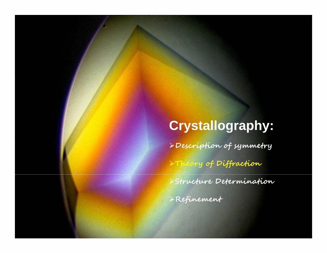
Crystallography:i i fDescription of symmetry
Theory of Diffraction
Structure Determination
Refinement
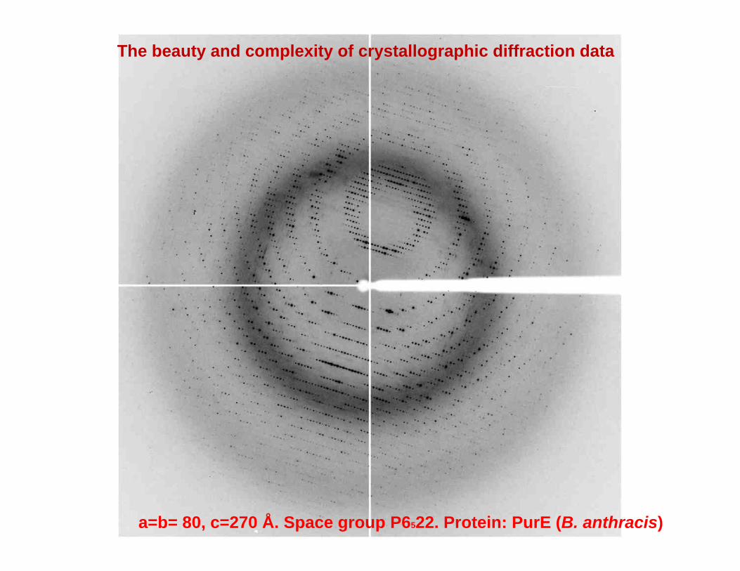
The beauty and complexity of crystallographic diffraction data
a=b= 80, c=270 Å. Space group P6522. Protein: PurE (B. anthracis)
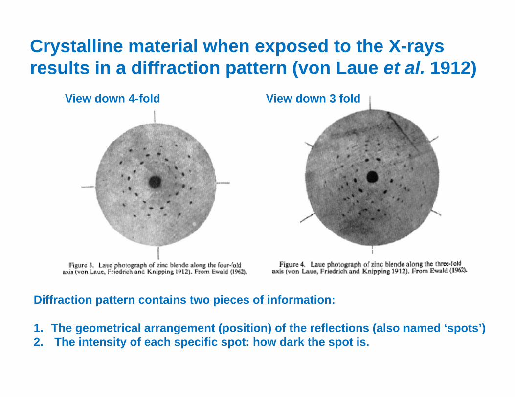
Crystalline material when exposed to the X-rays results in a diffraction pattern (von Laue et al. 1912)results in a diffraction pattern (von Laue et al. 1912)
View down 4-fold View down 3 fold
Diffraction pattern contains two pieces of information:
1 Th t i l t ( iti ) f th fl ti ( l d ‘ t ’)1. The geometrical arrangement (position) of the reflections (also named ‘spots’)2. The intensity of each specific spot: how dark the spot is.
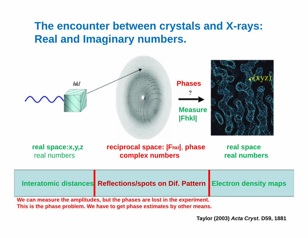
The encounter between crystals and X-rays:Real and Imaginary numbers. g y
ρPhases
Measure|Fhkl|
real space:x,y,z reciprocal space: |Fhkl|, phase real spacereal numbers complex numbers real numbers
In the diffraction experiment: measure intensity of reflection at position h,k,l (or reflection from planes h,k,l)Amplitude of each wave (|Fhkl|) is proportional to the square-root of the intensity measured on the detector.Calculation of the electron density ρ at any position (xyz) in the unit cell of a crystal requires us to perform a summation of the amplitudes from all the (hkl) planes each amplitude with its own phase
Interatomic distances Reflections/spots on Dif. Pattern Electron density maps summation of the amplitudes from all the (hkl) planes, each amplitude with its own phase. We can measure the amplitudes, but the phases are lost in the experiment. This is the phase problem. We have to get phase estimates by other means.
Taylor (2003) Acta Cryst. D59, 1881
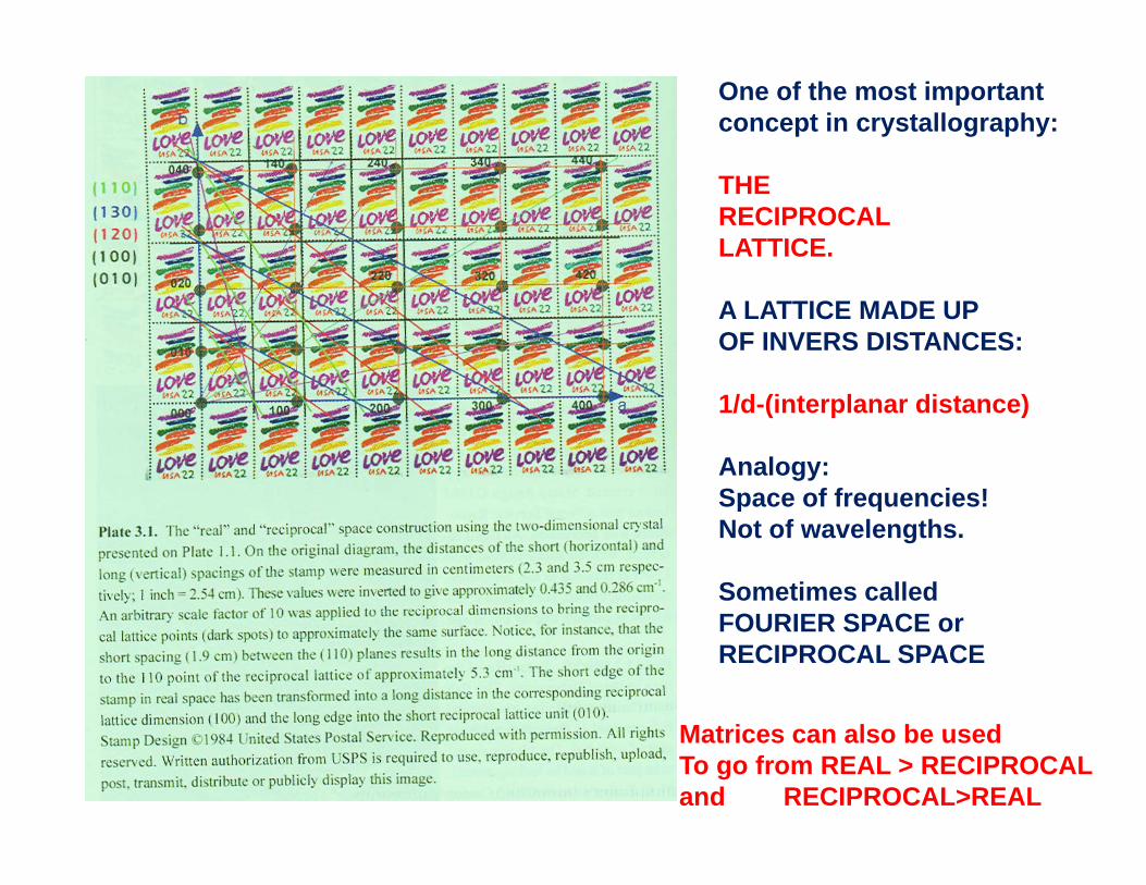
One of the most importantconcept in crystallography:
THERECIPROCALLATTICE.
A LATTICE MADE UPOF INVERS DISTANCES:
1/d-(interplanar distance)
Analogy:Space of frequencies!Space of frequencies!Not of wavelengths.
Sometimes calledFOURIER SPACE orRECIPROCAL SPACE
Matrices can also be usedTo go from REAL > RECIPROCALand RECIPROCAL>REAL
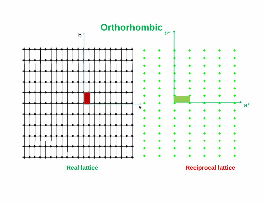
b b*Orthorhombic
a a*
R l l tti R i l l ttiReal lattice Reciprocal lattice
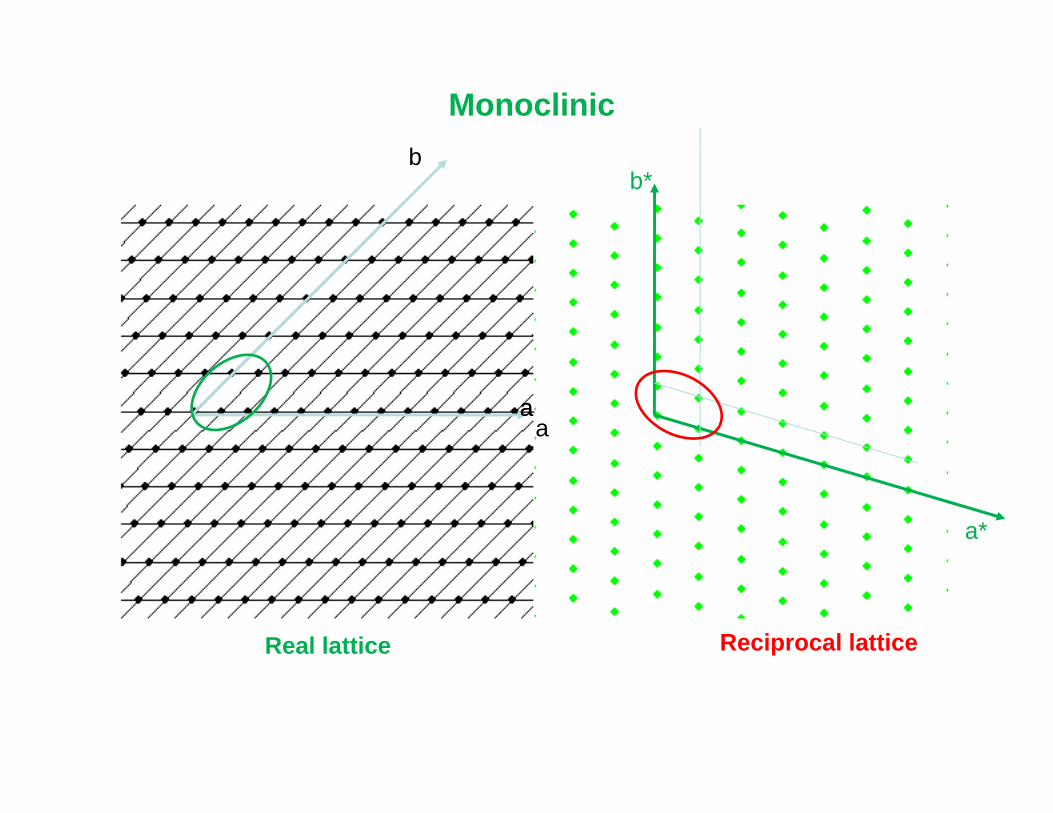
b
Monoclinicb
b*
aa
a
a*
Real lattice Reciprocal lattice
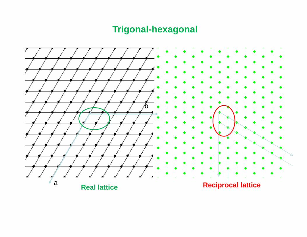
Trigonal-hexagonal
bb
Real lattice Reciprocal latticea
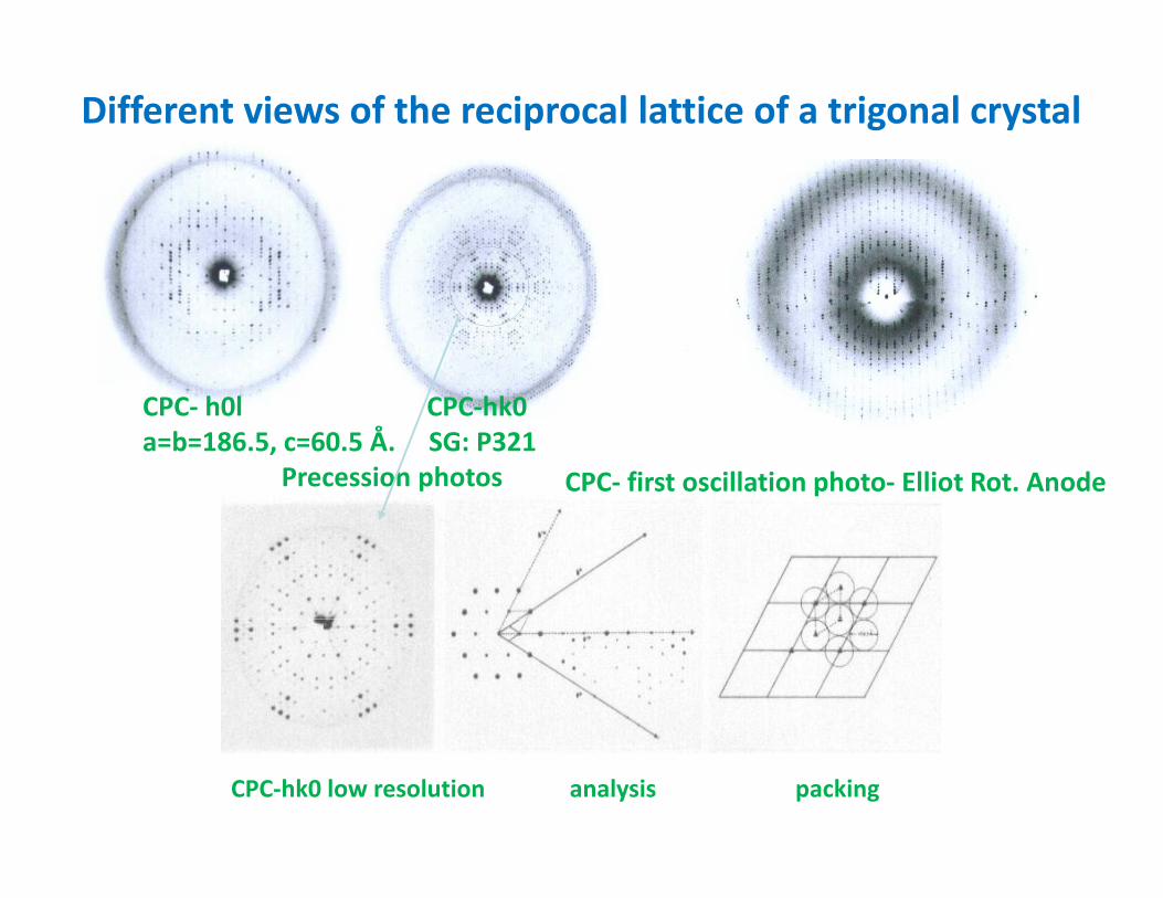
Different views of the reciprocal lattice of a trigonal crystal
CPC‐ h0l CPC‐hk0a=b=186.5, c=60.5 Å. SG: P321
Precession photos CPC‐ first oscillation photo‐ Elliot Rot. Anode
CPC‐hk0 low resolution analysis packing
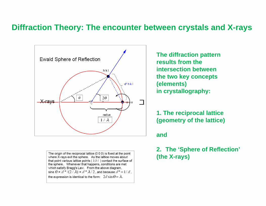
Diffraction Theory: The encounter between crystals and X-rays
The diffraction pattern results from theintersection betweenthe two key concepts (elements)in crystallography:in crystallography:
1. The reciprocal latticep(geometry of the lattice)
and
2. The ‘Sphere of Reflection’(the X-rays)
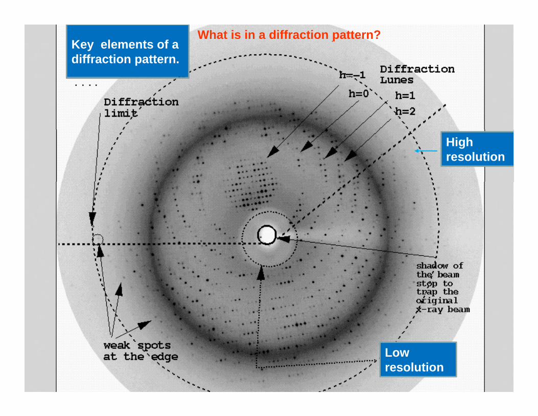
What is in a diffraction pattern?Key elements of a diffraction pattern.
Highresolution
Low resolution
Principles: why is the way it is! • Why X-rays?
– MoKα wavelength, 0.7107Å, CuKα wavelength, 1 5418Å1.5418Å
– similar to distance between bonded carbon atoms (1.54Å); i.e. wavelength commensurate to the distancesdistances
• Why electron density?– X-rays are scattered by the electrons of the atoms– The results are electron density maps, which give the
positions of the atoms• Why crystals?Why crystals?
– One molecule would lead to far too weak a signal– Huge number of molecules in same orientation lead to
an amplified signalan amplified signal– Single molecule diffraction may come in the future!
Theory of diffraction tries to address the following questions:
1. When is there a diffraction spot (reflection) on the detector?detector?
2. Under which conditions?
3. What does the geometric pattern of the diffraction tells us about the crystal?
4 What are the factors that determine the quality and4. What are the factors that determine the quality andextend of the diffraction (or diffraction pattern) from a crystal?
5. What does the position of the reflection depend on?
6. What does the intensity of the reflection depend on?
Try to understand conceptually the answers to all these questions

Some portions of this material have b t t d f L t 4 fbeen extracted from Lecture 4 of the educational materials donated by Prof. JR Helliwell (Univ. Of Manchester UK) to the IUCr forManchester, UK) to the IUCr for educational purposes
Theory of diffraction tries to address the following questions:
1. When is there a diffraction (reflection) on the detector?
2. Under which conditions?
3. What does the geometric pattern of the diffraction tells us about the crystal?
4. What are the factors that determine the quality andextend of the diffraction (or diffraction pattern) from aextend of the diffraction (or diffraction pattern) from a crystal?
5. What does the position of the reflection depend on?
6. What does the intensity of the reflection depend on?
Try to understand conceptually the answers to all these questions
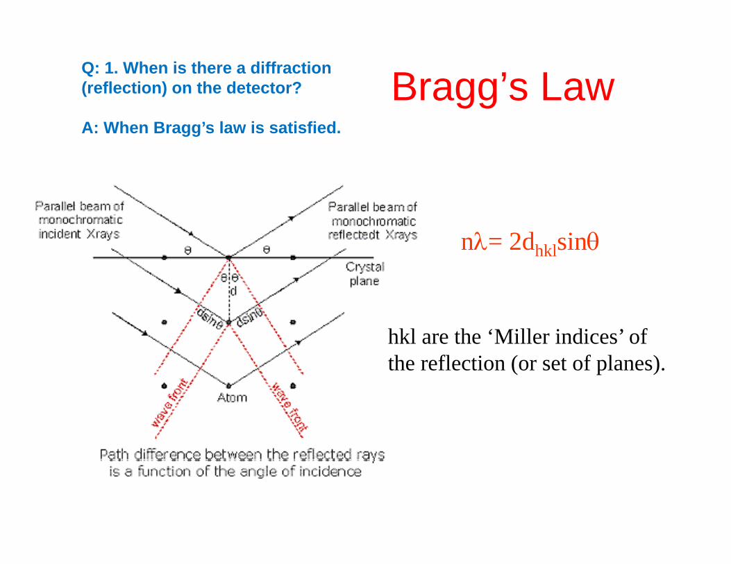
Bragg’s LawQ: 1. When is there a diffraction (reflection) on the detector? ggA: When Bragg’s law is satisfied.
nλ= 2dhklsinθnλ 2dhklsinθ
hkl are the ‘Miller indices’ of the reflection (or set of planes).
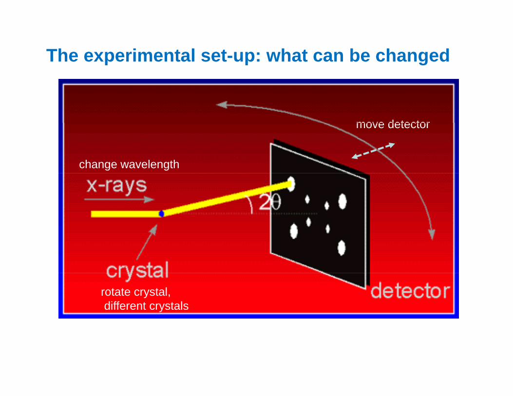
The experimental set-up: what can be changed
move detector
change wavelength
move detector
rotate crystal,different crystals
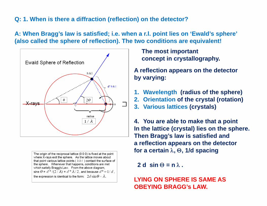
Q: 1. When is there a diffraction (reflection) on the detector?
A: When Bragg’s law is satisfied; i.e. when a r.l. point lies on ‘Ewald’s sphere’ (also called the sphere of reflection). The two conditions are equivalent!
The most importantconcept in crystallography.
A reflection appears on the detectorby varying:
1 W l th ( di f th h )1. Wavelength (radius of the sphere)2. Orientation of the crystal (rotation)3. Various lattices (crystals)
4. You are able to make that a pointIn the lattice (crystal) lies on the sphere.Then Bragg’s law is satisfied anda reflection appears on the detectora reflection appears on the detectorfor a certain λ, Θ, 1/d spacing
2 d sin Θ = n λ .
LYING ON SPHERE IS SAME ASOBEYING BRAGG’s LAW.
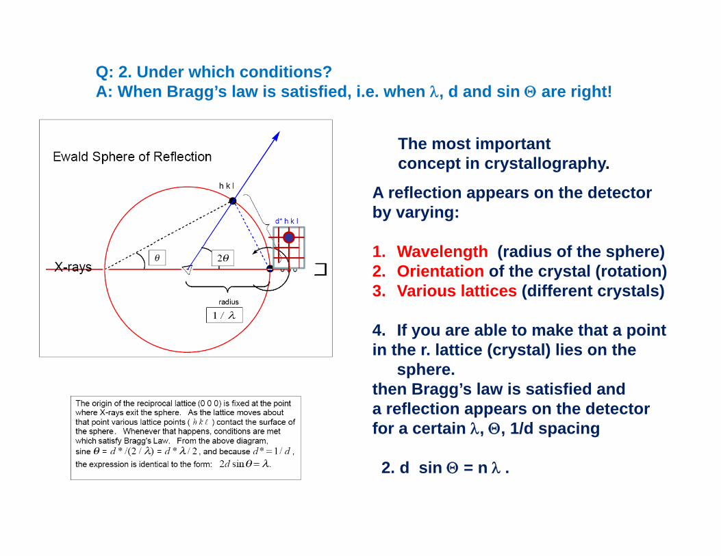
Q: 2. Under which conditions? A: When Bragg’s law is satisfied, i.e. when λ, d and sin Θ are right!
The most importantconcept in crystallography.
A reflection appears on the detectorby varying:
1. Wavelength (radius of the sphere)2. Orientation of the crystal (rotation)3. Various lattices (different crystals)
4. If you are able to make that a pointin the r. lattice (crystal) lies on the
sphere.th B ’ l i ti fi d dthen Bragg’s law is satisfied anda reflection appears on the detectorfor a certain λ, Θ, 1/d spacing
2. d sin Θ = n λ .

What is in a diffraction pattern?Key elements of a diffraction pattern.
Highresolution
Low resolution
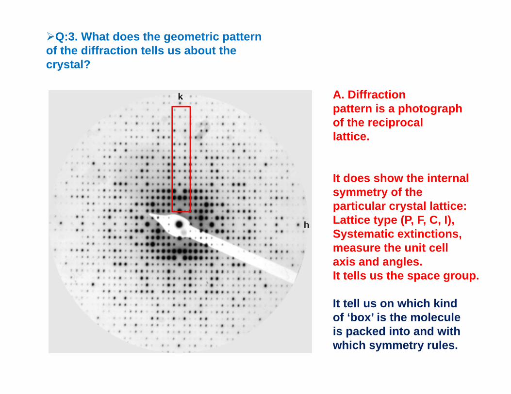
Q:3. What does the geometric pattern of the diffraction tells us about the crystal?
A. Diffractionpattern is a photographof the reciprocalof the reciprocallattice.
It does show the internalsymmetry of theparticular crystal lattice:Lattice type (P F C I)Lattice type (P, F, C, I),Systematic extinctions,measure the unit cellaxis and angles.It t ll thIt tells us the space group.
It tell us on which kindof ‘box’ is the moleculeis packed into and withwhich symmetry rules.
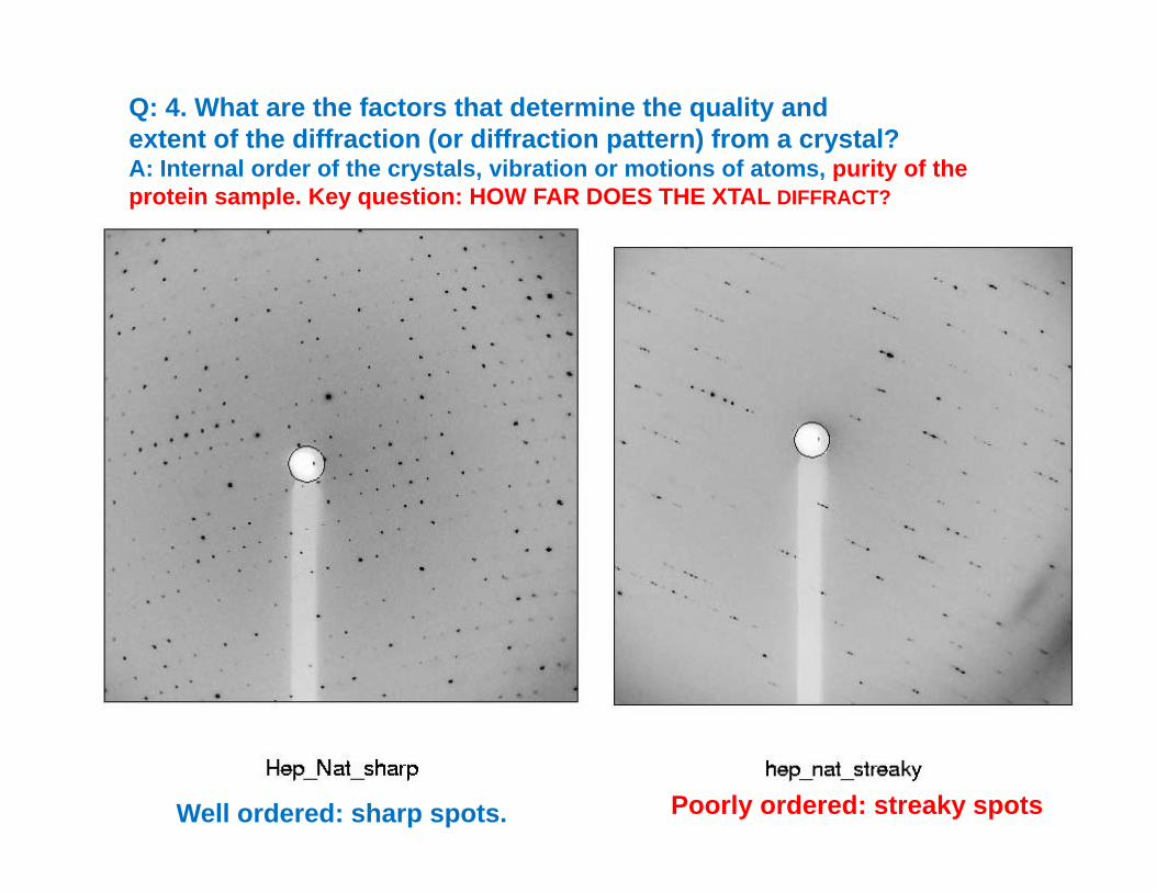
Q: 4. What are the factors that determine the quality andextent of the diffraction (or diffraction pattern) from a crystal?A: Internal order of the crystals vibration or motions of atoms purity of theA: Internal order of the crystals, vibration or motions of atoms, purity of the protein sample. Key question: HOW FAR DOES THE XTAL DIFFRACT?
Well ordered: sharp spots. Poorly ordered: streaky spots
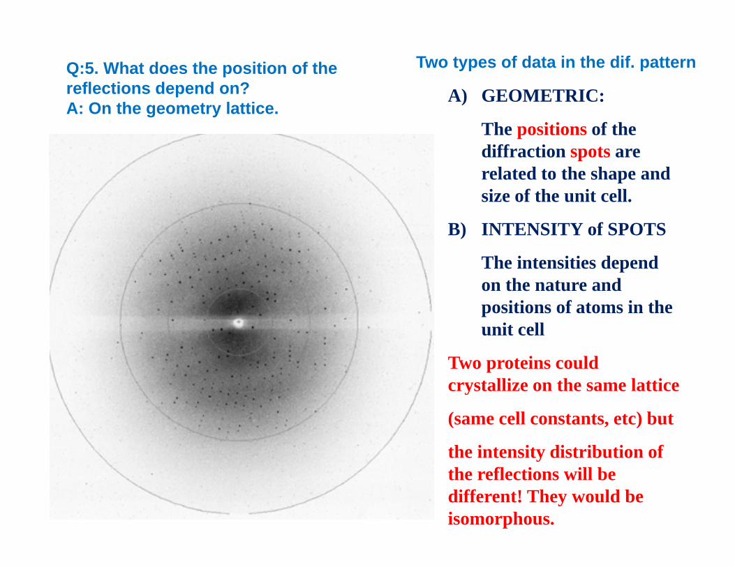
A) GEOMETRIC:Q:5. What does the position of the reflections depend on? A O th t l tti
Two types of data in the dif. pattern
•
)
The positions of the diffraction spots are related to the shape and
A: On the geometry lattice.
related to the shape and size of the unit cell.
B) INTENSITY of SPOTS
The intensities depend on the nature and positions of atoms in the unit cell
Two proteins could crystallize on the same latticecrystallize on the same lattice
(same cell constants, etc) but
the intensity distribution of the reflections will be different! They would be isomorphous.
A) GEOMETRIC:
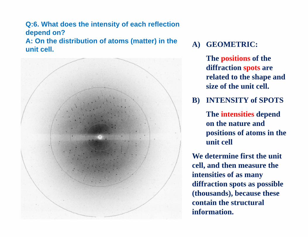
Q:6. What does the intensity of each reflection depend on? A: On the distribution of atoms (matter) in the
•
A) GEOMETRIC:
The positions of the diffraction spots are
( )unit cell.
related to the shape and size of the unit cell.
B) INTENSITY of SPOTS)
The intensities depend on the nature and positions of atoms in thepositions of atoms in the unit cell
We determine first the unit ll d th thcell, and then measure the
intensities of as many diffraction spots as possible (thousands) because these(thousands), because these contain the structural information.
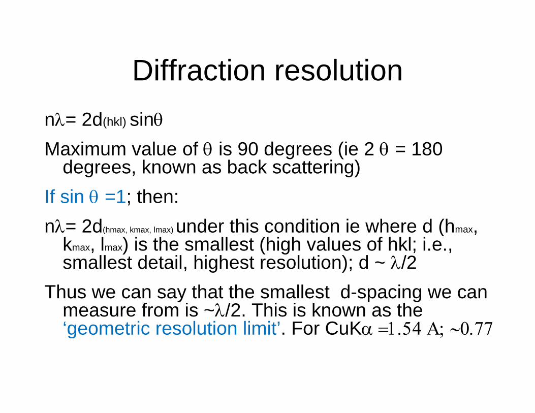
Diffraction resolutionDiffraction resolution nλ= 2d(hkl) sinθ
Maximum value of θ is 90 degrees (ie 2 θ = 180 degrees, known as back scattering)
If sin θ =1; then:nλ= 2d(hmax, kmax, lmax) under this condition ie where d (hmax, d( , , ) u de s co d o e e e d ( ,
kmax, lmax) is the smallest (high values of hkl; i.e., smallest detail, highest resolution); d ~ λ/2
Th h h ll d iThus we can say that the smallest d-spacing we can measure from is ~λ/2. This is known as the ‘geometric resolution limit’. For CuKα =1.54 Α; ∼0.77g ;
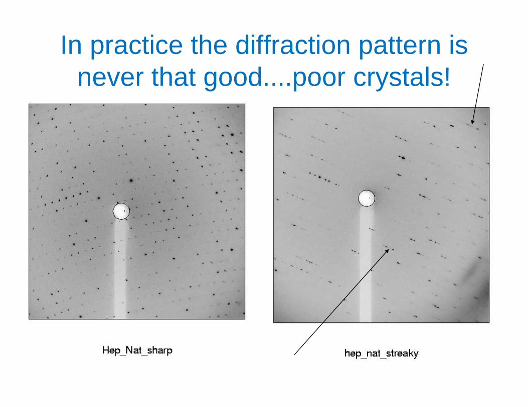
In practice the diffraction pattern is never that good poor crystals!never that good....poor crystals!
The real resolution limitThe real resolution limit• This is usually set by the crystalline order
f th l it lf i th f di t f thof the sample itself ie the fading out of the diffraction pattern as one goes to a higher
d hi h diff ti l θ d iand higher diffraction angle θ and is usually well before θ reaches 180 degrees.
• In practical terms ‘resolution’ means the smallest Angstrom spacing (highest h,k,l values) included in the Fourier series withreasonable (>60-70%) completeness. ( ) p
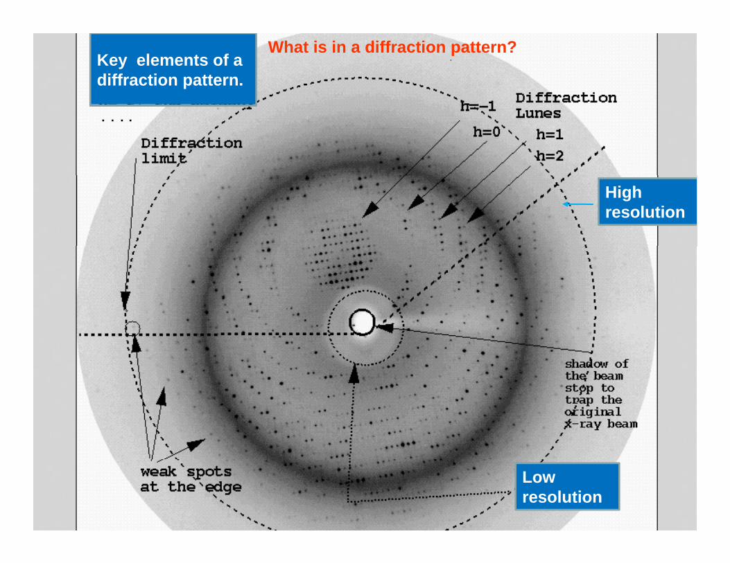
What is in a diffraction pattern?Key elements of a diffraction pattern.
Highresolution
Low resolution
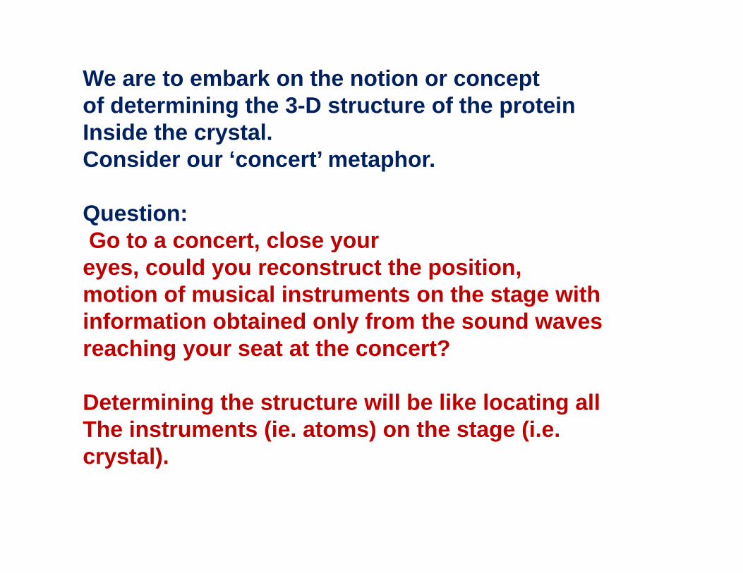
We are to embark on the notion or conceptof determining the 3-D structure of the proteinof determining the 3-D structure of the proteinInside the crystal. Consider our ‘concert’ metaphor.
Question:Go to a concert, close your, yeyes, could you reconstruct the position,motion of musical instruments on the stage with information obtained only from the sound wavesinformation obtained only from the sound waves reaching your seat at the concert?
D t i i th t t ill b lik l ti llDetermining the structure will be like locating allThe instruments (ie. atoms) on the stage (i.e. crystal). y )
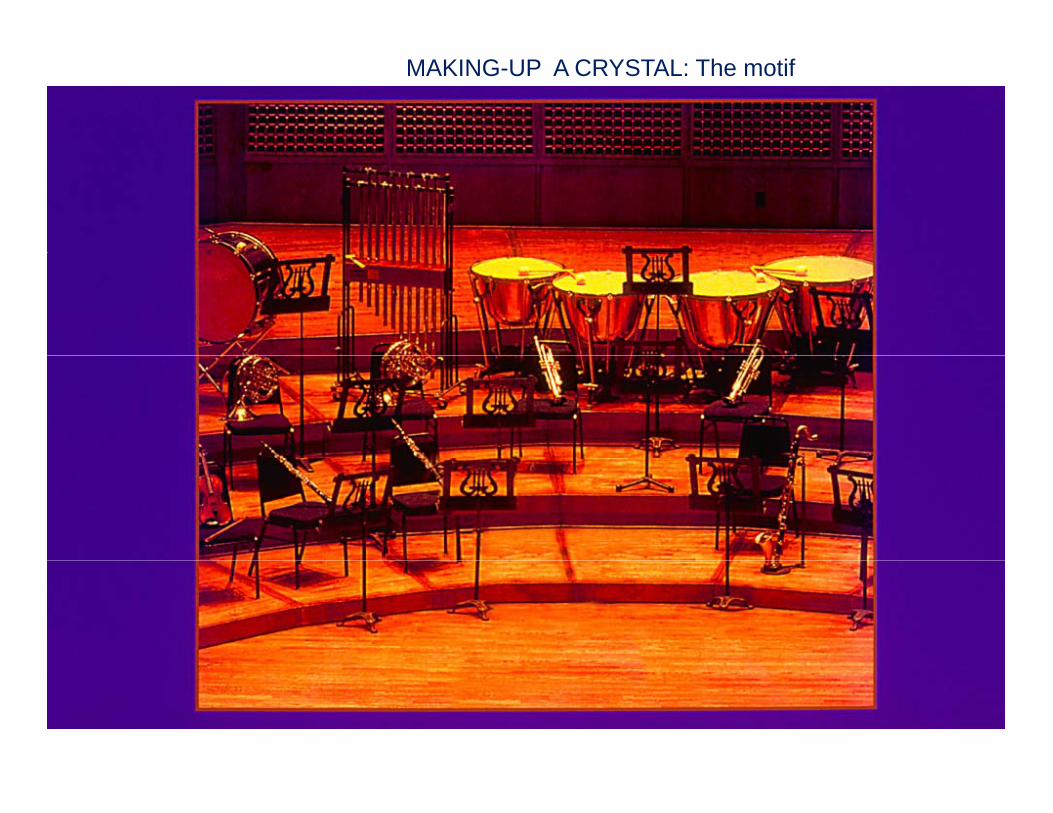
MAKING-UP A CRYSTAL: The motif
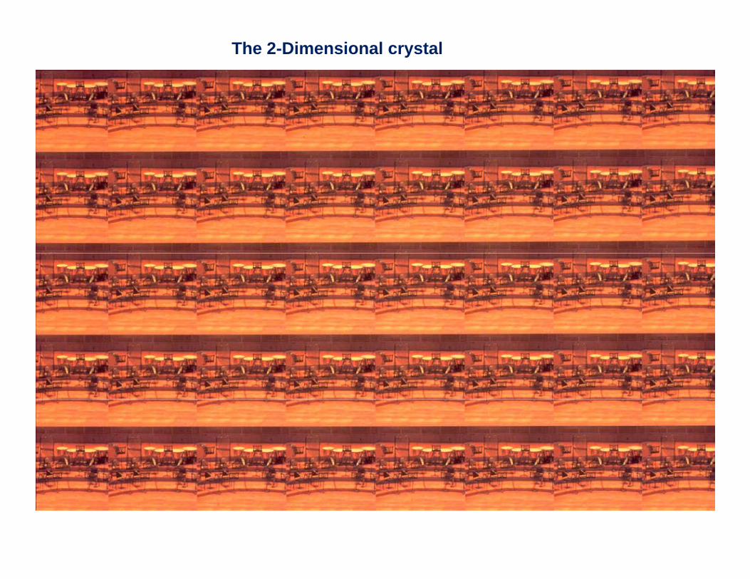
The 2-Dimensional crystal

Metaphor of a Diffraction Experiment as a Symphonic Concert
Audience soundwaves l r s; S
Wi d
waves l, r, s; S
Wind
Orchestrastage crystal: h k l; Fstage crystal: h, k, l; Fo
Detector surface
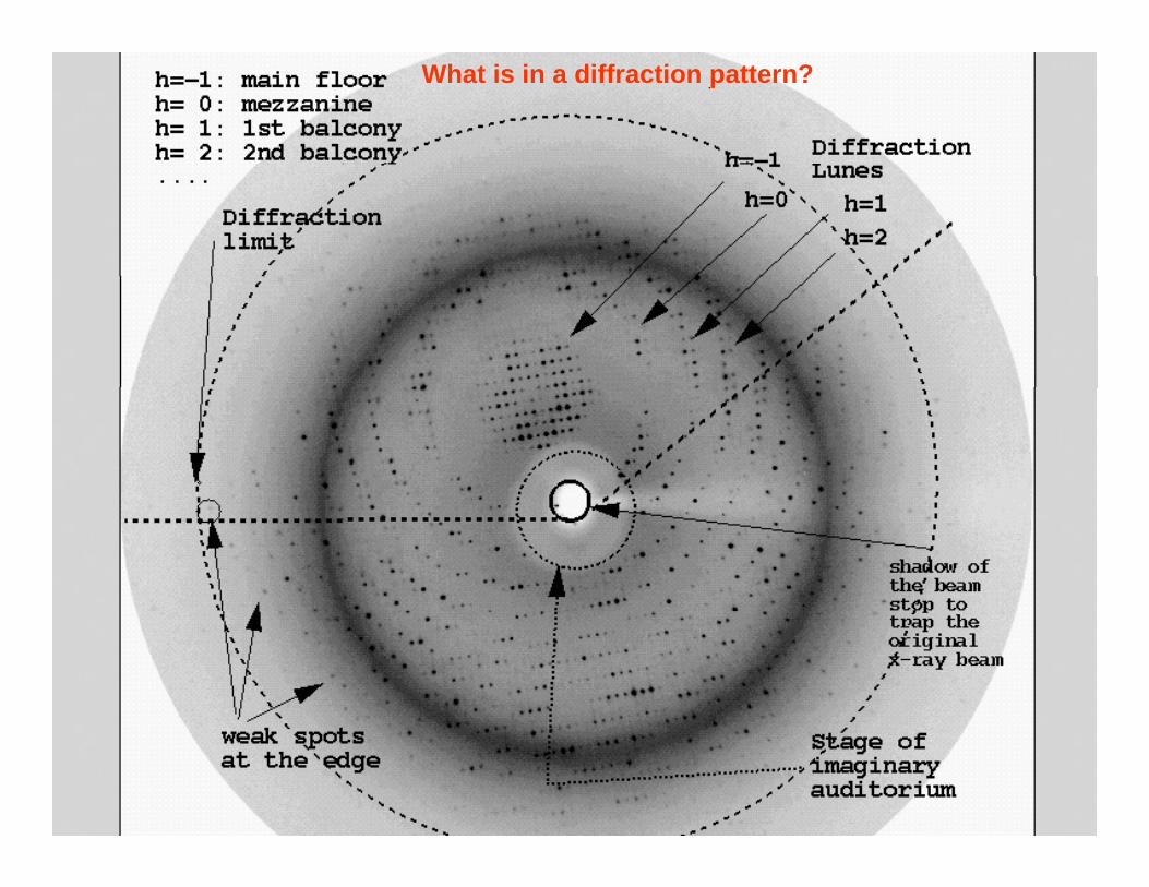
What is in a diffraction pattern?
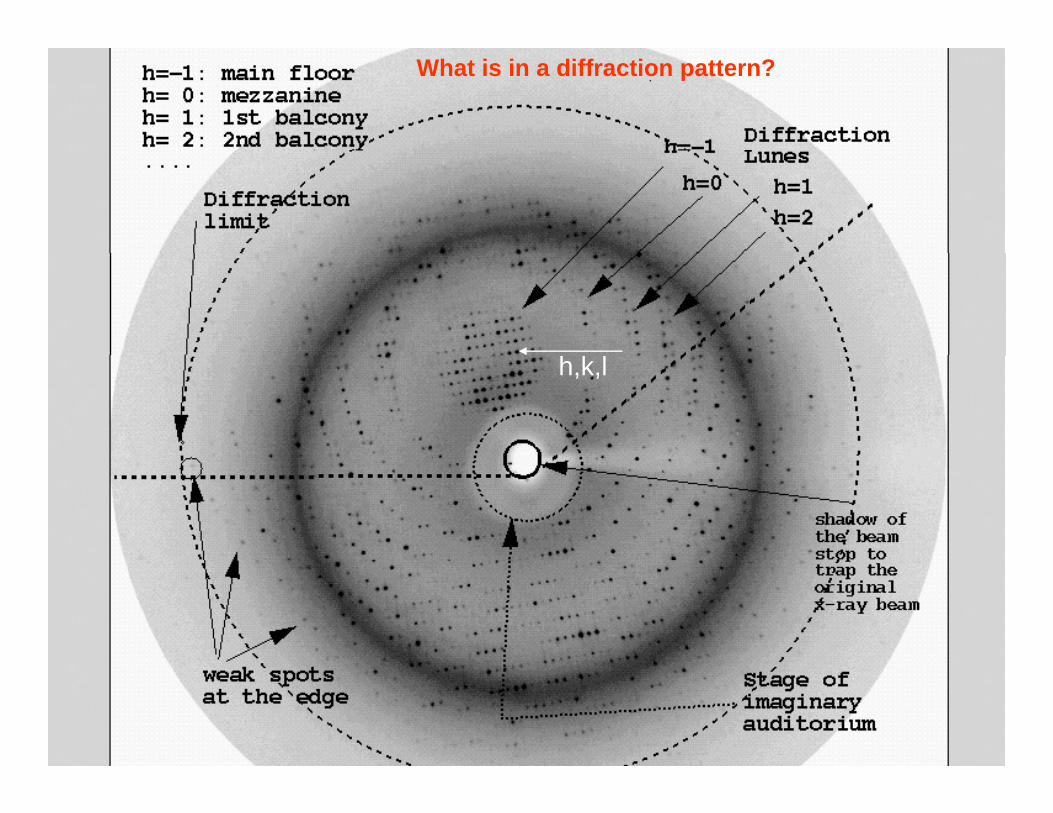
What is in a diffraction pattern?
h,k,l
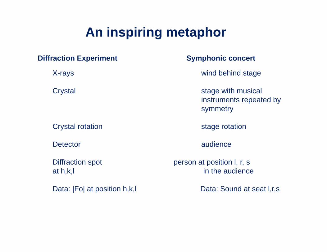
An inspiring metaphor
Diffraction Experiment Symphonic concert
X-rays wind behind stage
Crystal stage with musical instruments repeated by symmetrysymmetry
Crystal rotation stage rotation
D t t diDetector audience
Diffraction spot person at position l, r, sat h,k,l in the audience, ,
Data: |Fo| at position h,k,l Data: Sound at seat l,r,s
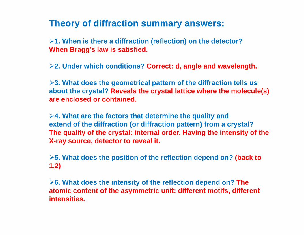
Theory of diffraction summary answers:
1. When is there a diffraction (reflection) on the detector?1. When is there a diffraction (reflection) on the detector?When Bragg’s law is satisfied.
2. Under which conditions? Correct: d, angle and wavelength.
3. What does the geometrical pattern of the diffraction tells us about the crystal? Reveals the crystal lattice where the molecule(s) are enclosed or contained.
4. What are the factors that determine the quality andextend of the diffraction (or diffraction pattern) from a crystal?The quality of the crystal: internal order Having the intensity of theThe quality of the crystal: internal order. Having the intensity of the X-ray source, detector to reveal it.
5. What does the position of the reflection depend on? (back to 1,2)
6. What does the intensity of the reflection depend on? The atomic content of the asymmetric unit: different motifs differentatomic content of the asymmetric unit: different motifs, different intensities.
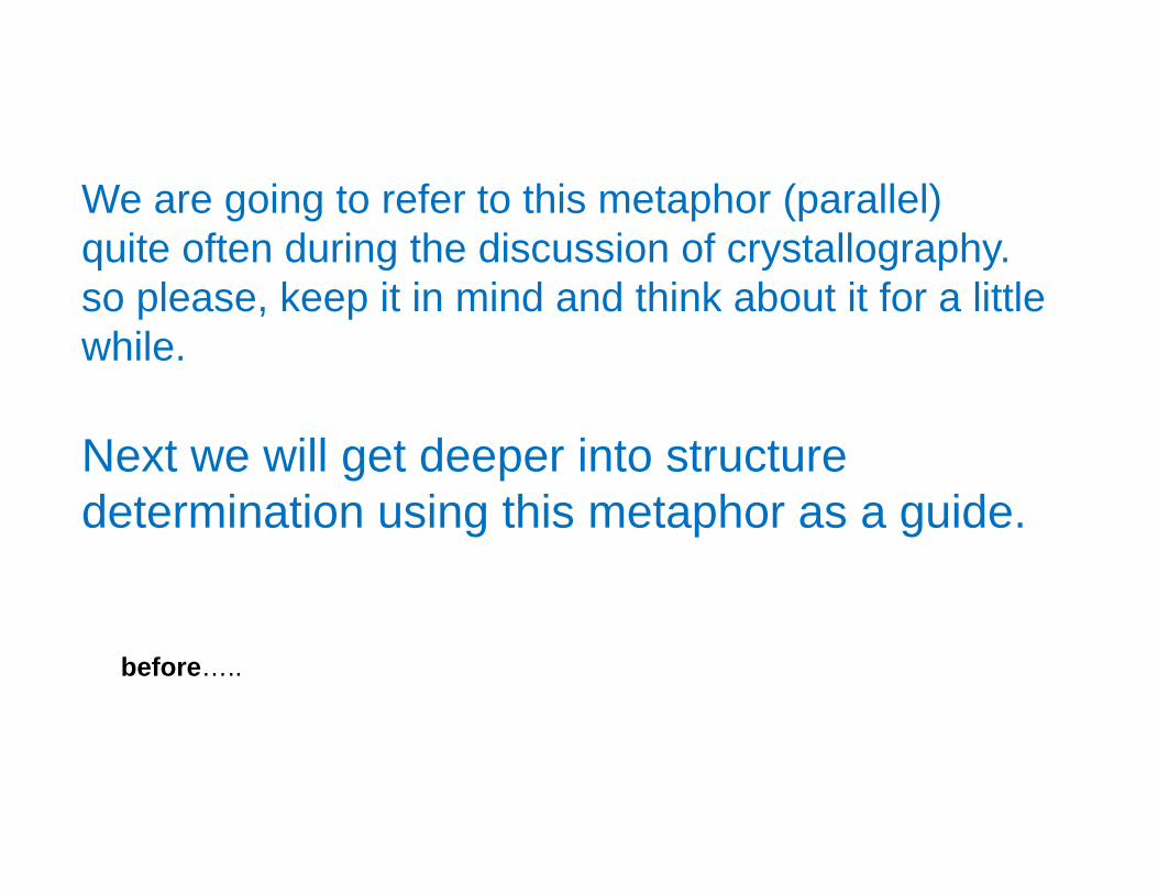
We are going to refer to this metaphor (parallel)quite often during the discussion of crystallography.so please, keep it in mind and think about it for a littlewhile.
Next we will get deeper into structure determination using this metaphor as a guidedetermination using this metaphor as a guide.
before…..

A f thi d bit f iA few more things and a bit of review…
Read as many chapters as you canRead as many chapters as you canof PARTS I-IV of ‘Crystals and Life’.It will help you!It will help you!
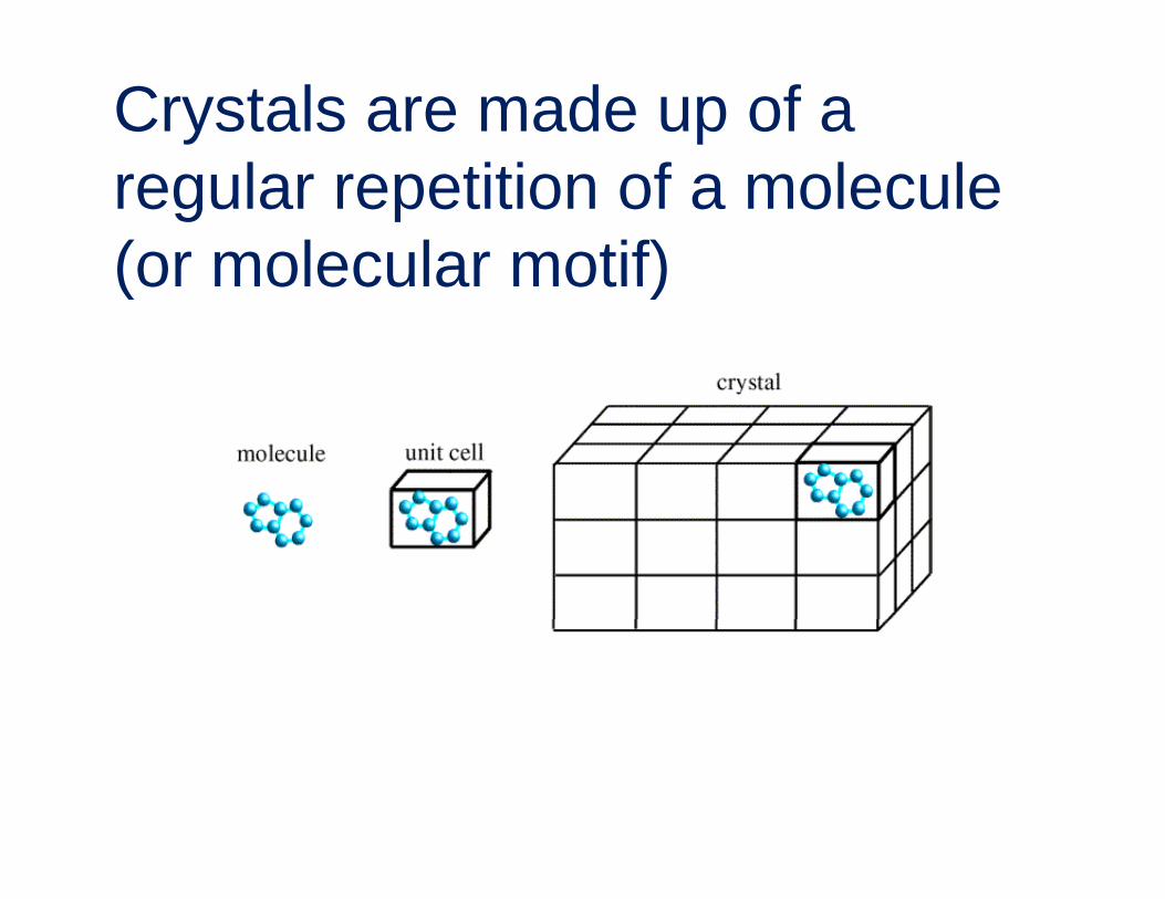
Crystals are made up of a l titi f l lregular repetition of a molecule
(or molecular motif)(or molecular motif)
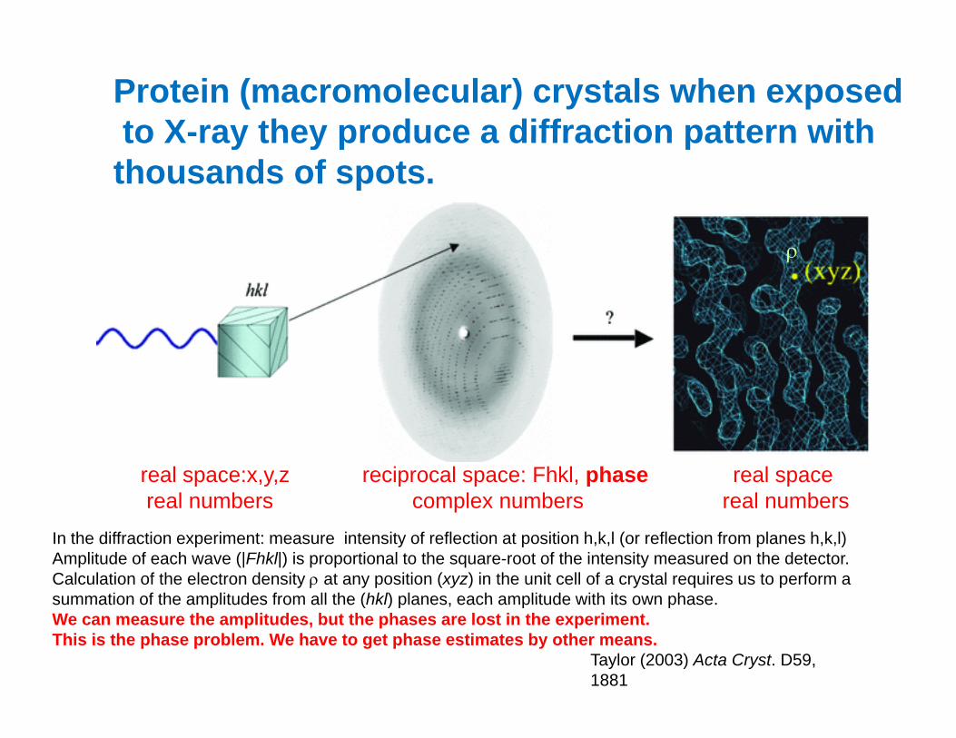
Protein (macromolecular) crystals when exposedto X-ray they produce a diffraction pattern withto X ray they produce a diffraction pattern withthousands of spots.
ρ
real space:x,y,z reciprocal space: Fhkl, phase real spacereal numbers complex numbers real numbers
In the diffraction experiment: measure intensity of reflection at position h,k,l (or reflection from planes h,k,l)Amplitude of each wave (|Fhkl|) is proportional to the square-root of the intensity measured on the detector.Calculation of the electron density ρ at any position (xyz) in the unit cell of a crystal requires us to perform a summation of the amplitudes from all the (hkl) planes each amplitude with its own phase
p
summation of the amplitudes from all the (hkl) planes, each amplitude with its own phase. We can measure the amplitudes, but the phases are lost in the experiment. This is the phase problem. We have to get phase estimates by other means.
Taylor (2003) Acta Cryst. D59, 1881
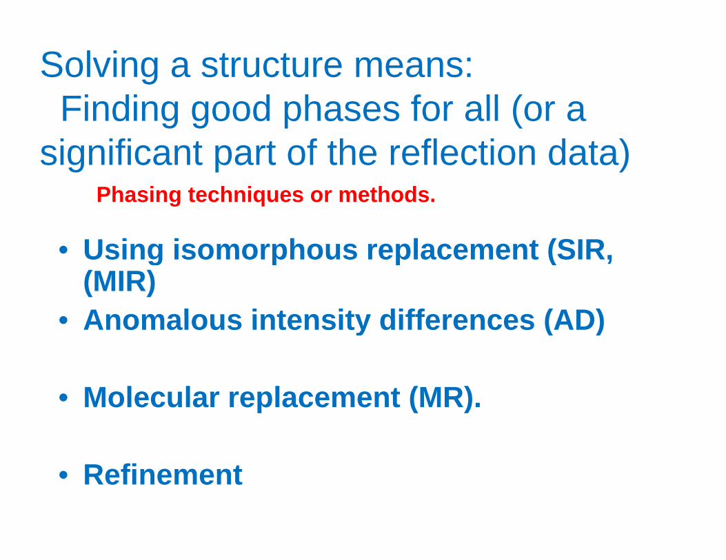
Solving a structure means:Finding good phases for all (or aFinding good phases for all (or a
significant part of the reflection data)
Using isomorphous replacement (SIR
Phasing techniques or methods.
• Using isomorphous replacement (SIR, (MIR) Anomalous intensity differences (AD)• Anomalous intensity differences (AD)
( )• Molecular replacement (MR).
• Refinement
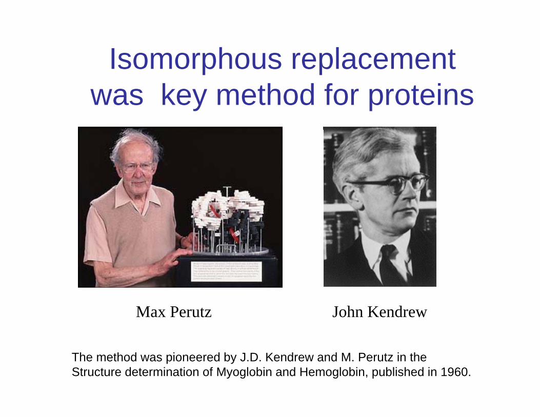
Isomorphous replacementwas key method for proteins
Max Perutz John Kendrew
The method was pioneered by J.D. Kendrew and M. Perutz in theStructure determination of Myoglobin and Hemoglobin, published in 1960.

This completes the outline of diffraction theoryand limitations.
Next we will discuss the concept of ‘structureDetermination’.
Questions?