Protein-carbohydrate ingestion alters Vps34 cellular ... · Protein-carbohydrate ingestion alters...
29
Protein-carbohydrate ingestion alters Vps34 cellular localization independent of changes in kinase activity in human skeletal muscle Nathan Hodson 1# , Jessica R. Dent 1# , Zhe Song 1 , Mary F. O’Leary 1 , Thomas Nicholson 2 , Simon W. Jones 2 , James T. Murray 3 , Stewart Jeromson 4 , D. Lee Hamilton 5 , Leigh Breen 1 , Andrew Philp 1,6,7 . 1. School of Sport, Exercise and Rehabilitation Sciences, University of Birmingham, UK. 2. Institute of Inflammation and Ageing, University of Birmingham, UK. 3. Trinity Biomedical Sciences Institute, Trinity College, Dublin, Ireland. 4. Physiology, Exercise and Nutrition Research Group, School of Sport, Faculty of Health, Stirling University, Stirling, UK. 5. Institute for Physical Activity and Nutrition (IPAN), School of Exercise & Nutrition Sciences, Deakin University, Geelong 3216, Australia. 6. Garvan Institute of Medical Research, NSW, Australia. 7. St Vincent’s Clinical School, UNSW Medicine, UNSW Sydney, NSW, Australia. * Corresponding author: Andrew Philp, Ph.D. Mitochondrial Metabolism and Ageing Laboratory Healthy Ageing Theme Garvan Institute of Medical Research 384 Victoria Street, Darlinghurst, NSW, 2010, Australia Phone: +61 (0) 292958249 Email: [email protected] # Authors contributed equally . CC-BY-NC-ND 4.0 International license (which was not certified by peer review) is the author/funder. It is made available under a The copyright holder for this preprint this version posted February 11, 2020. . https://doi.org/10.1101/2020.02.10.941054 doi: bioRxiv preprint
Transcript of Protein-carbohydrate ingestion alters Vps34 cellular ... · Protein-carbohydrate ingestion alters...

Protein-carbohydrate ingestion alters Vps34 cellular localization independent of changes in 1
kinase activity in human skeletal muscle 2
Nathan Hodson1#, Jessica R. Dent1#, Zhe Song1, Mary F. O’Leary1, Thomas Nicholson2, Simon 3
W. Jones2, James T. Murray3, Stewart Jeromson4, D. Lee Hamilton5, Leigh Breen1, Andrew 4
Philp1,6,7. 5
1. School of Sport, Exercise and Rehabilitation Sciences, University of Birmingham, UK. 6
2. Institute of Inflammation and Ageing, University of Birmingham, UK. 7
3. Trinity Biomedical Sciences Institute, Trinity College, Dublin, Ireland. 8
4. Physiology, Exercise and Nutrition Research Group, School of Sport, Faculty of Health, 9
Stirling University, Stirling, UK. 10
5. Institute for Physical Activity and Nutrition (IPAN), School of Exercise & Nutrition 11
Sciences, Deakin University, Geelong 3216, Australia. 12
6. Garvan Institute of Medical Research, NSW, Australia. 13
7. St Vincent’s Clinical School, UNSW Medicine, UNSW Sydney, NSW, Australia. 14
* Corresponding author: 15
Andrew Philp, Ph.D. 16
Mitochondrial Metabolism and Ageing Laboratory 17
Healthy Ageing Theme 18
Garvan Institute of Medical Research 19
384 Victoria Street, Darlinghurst, NSW, 2010, Australia 20
Phone: +61 (0) 292958249 Email: [email protected] 21
#Authors contributed equally 22
23
.CC-BY-NC-ND 4.0 International license(which was not certified by peer review) is the author/funder. It is made available under aThe copyright holder for this preprintthis version posted February 11, 2020. . https://doi.org/10.1101/2020.02.10.941054doi: bioRxiv preprint

Abstract 24
The mechanistic target of rapamycin (mTOR) complex 1 (mTORC1) regulates cell size and 25
growth in response to nutrients, however, the mechanisms by which nutrient levels are sensed by 26
mTORC1 in human skeletal muscle are yet to be fully elucidated. The Class III PI3Kinase Vps34 27
has recently been proposed as a sensor essential for mTORC1 activation following nutrient 28
stimulation. We therefore investigated the effects of increasing nutrient availability through 29
protein-carbohydrate (PRO-CHO) feeding on Vps34 kinase activity and cellular localization in 30
human skeletal muscle. Eight young, healthy males (age – 21 ± 0.5yrs, mean ± SEM) ingested a 31
PRO-CHO beverage containing 20/44/1g PRO/CHO/FAT respectively, with skeletal muscle 32
biopsies obtained at baseline and 1h and 3h post-feeding. PRO-CHO feeding did not alter Vps34 33
kinase activity, but did stimulate Vps34 translocation toward the cell periphery (PRE 34
(mean±SEM) - 0.273±0.021, 1h - 0.347±0.022, Pearson’s Coefficient (r)) where it co-localized 35
with mTOR (PRE – 0.312±0.018, 1h – 0.348±0.024, Pearson’s Coefficient (r))). These 36
alterations occurred in parallel to an increase in S6K1 kinase activity – 941±164% of PRE at 1h 37
post-feeding). Subsequent in vitro experiments in C2C12 and human primary myotubes 38
displayed no effect of the Vps34-specific inhibitor SAR405 on mTORC1 signalling responses to 39
elevated nutrient availability. Therefore, in summary, PRO-CHO ingestion does not increase 40
Vps34 activity in human skeletal muscle, whilst pharmacological inhibition of Vps34 does not 41
prevent nutrient stimulation of mTORC1 in vitro. However, PRO-CHO ingestion promotes 42
Vps34 translocation to the cell periphery, enabling Vps34 to associate with mTOR. Therefore, 43
our data suggests that interaction between Vps34 and mTOR, rather than changes in Vps34 44
activity per se may be involved in PRO-CHO activation of mTORC1 in human skeletal muscle. 45
.CC-BY-NC-ND 4.0 International license(which was not certified by peer review) is the author/funder. It is made available under aThe copyright holder for this preprintthis version posted February 11, 2020. . https://doi.org/10.1101/2020.02.10.941054doi: bioRxiv preprint

Introduction 46
Amino acids (AAs) are critical to skeletal muscle plasticity, acting as both substrates in the 47
process of muscle protein synthesis (MPS) as well as initiating the signaling pathways which 48
activate this cellular process (32, 33). Carbohydrate (CHO) ingestion can also elevate MPS via 49
insulin action (3), and a combination of these nutrients is believed to act synergistically on MPS 50
following exercise (20). In skeletal muscle, it is believed that increases in MPS are governed 51
primarily by the activation of the mechanistic target of rapamycin complex 1 (mTORC1) (5, 6), 52
an evolutionarily conserved serine/threonine kinase complex which stimulates translation 53
initiation and elongation (14, 16, 31) in response to increased nutrient provision. 54
The canonical mechanism by which AAs stimulate mTORC1 activity is thought to be through 55
the elevation of mTORC1 complex co-localization with the lysosome (27) in vitro, or through 56
mTORC1/lysosomal trafficking in vivo/vitro (11, 15, 28). However, how nutrients stimulate 57
mTORC1 activity in human skeletal muscle is still poorly understood. A potential nutrient-58
sensitive activator of mTORC1 is the vacuolar protein sorting 34 (Vps34), a class III PI3Kinase. 59
The primary function of Vps34 is the production of phosphatidylinositol 3-phosphate (PI(3)P) 60
through the phosphorylation of phosphatidylinositol (2), a product responsible for the 61
recruitment of various proteins to phospholipid bilayers (i.e. plasma and lysosomal membranes) 62
(8). A role for Vps34 in nutrient sensing was first proposed by Byfield et al. (4), who reported 63
that overexpression of Vps34 in HEK293 cells elicited a 2-fold increase in S6K1 activity, a 64
common readout of mTORC1 activation. Conversely, siRNA targeting Vps34 abolished insulin-65
stimulated S6K1Thr389 phosphorylation (4). Nobukuni et al. (22) reiterated these findings, 66
displaying that siRNA-mediated reductions in Vps34 expression, in HEK293 cells, dramatically 67
attenuated mTORC1 activation in response to both AA and insulin stimulation. In addition, 68
.CC-BY-NC-ND 4.0 International license(which was not certified by peer review) is the author/funder. It is made available under aThe copyright holder for this preprintthis version posted February 11, 2020. . https://doi.org/10.1101/2020.02.10.941054doi: bioRxiv preprint

recent in vitro evidence suggests that Vps34 colocalises with mTOR, close to cellular 69
membranes, following insulin stimulation (10), and is required for nutrient-stimulated 70
translocation and activation of mTORC1 (10). As such, Vps34 represents a novel candidate as a 71
nutrient-sensitive activator of mTORC1. 72
With regard to skeletal muscle, 3h and 24h exposure to leucine (5mM) and insulin (100nM) 73
elevated Vps34 protein content and mTORSer2448 and S6K1Thr389 phosphorylation in human 74
primary myotubes (9), whilst supra-physiological levels of AA’s increases Vps34 activity in 75
C2C12 myotubes (17). In addition, high frequency electrical contraction has been reported to 76
increase Vps34 activity in rodent Tibialis Anterior muscle (17), whereas sprint exercise and 77
protein ingestion failed to activate Vps34 in human skeletal muscle (26). Overall, such data 78
implicates a possible role for Vps34 in nutrient/contraction sensing within skeletal muscle. 79
However, a more detailed investigation in human skeletal muscle is required. 80
Therefore, our primary aim was to investigate if AA/CHO feeding could affect Vps34 activity 81
and cellular localisation in human skeletal muscle. We hypothesised that Vps34 activity would 82
increase in response to AA/CHO feeding in parallel to increases in mTORC1 signaling. Our 83
secondary aim was to examine whether inhibition of Vps34 kinase activity in vitro with the 84
specific inhibitor SAR405 (24, 25) would attenuate nutrient-activation of mTORC1. 85
Methods 86
Participants 87
Eight young, healthy males (age – 21 ± 0.5yrs, mean ± SEM) volunteered to partake in the 88
current study. All participants were considered healthy (as assessed by a general health 89
questionnaire) and recreationally active (~3 exercise sessions per week) but not involved in a 90
.CC-BY-NC-ND 4.0 International license(which was not certified by peer review) is the author/funder. It is made available under aThe copyright holder for this preprintthis version posted February 11, 2020. . https://doi.org/10.1101/2020.02.10.941054doi: bioRxiv preprint

structured exercise training program. Exclusion criteria encompassed current cigarette smokers, 91
recreational drug users (including anabolic steroids), the presence of neuromuscular disease and 92
any medication/condition that may affect nutrient digestion/absorption i.e. inflammatory bowel 93
disease. Participants provided written informed consent prior to participation and all procedures 94
were approved by the NHS West Midlands Black Country Research Ethics Committee 95
(15/WM/0003) and conformed to the standards set out in the Declaration of Helsinki (7th 96
version). 97
Study Design 98
On the day of the experimental trial, participants reported to the laboratory following an 99
overnight fast (~10h) and having refrained from strenuous exercise and alcohol consumption in 100
the prior 48h. Upon arrival, participants were placed in a supine position and a 21G cannula was 101
inserted into the antecubital vein of one arm to allow for repeated blood sampling. At this point 102
an initial baseline blood sample was obtained from all participants. A skeletal muscle biopsy 103
sample was then taken from the vastus lateralis of a randomised leg using the Bergstrom 104
percutaneous needle technique, modified for suction (30). Participants then consumed a 105
commercially available protein-carbohydrate beverage (Gatorade Recover®, Gatorade, Chicago, 106
IL, USA.) providing 20/44/1g of protein, carbohydrate and fat respectively. Further venous blood 107
samples were taken every 20 minutes for a 3h post-prandial period and subsequent skeletal 108
muscle biopsy samples were obtained at 1h and 3h following beverage ingestion. Muscle 109
samples were blotted free of excess blood and dissected free of any excess adipose and 110
connective tissue, then immediately frozen in liquid nitrogen and stored at -80ºC until analysis. 111
A separate piece of muscle tissue was placed in optimal cutting temperature (OCT) compound 112
(VWR, Lutterworth, UK.) and frozen in liquid nitrogen-cooled isopentane before storage at -113
.CC-BY-NC-ND 4.0 International license(which was not certified by peer review) is the author/funder. It is made available under aThe copyright holder for this preprintthis version posted February 11, 2020. . https://doi.org/10.1101/2020.02.10.941054doi: bioRxiv preprint

80ºC. Blood samples were collected into EDTA-coated vacutainers (BD, Franklin Lakes, NJ, 114
USA.) and then centrifuged at 1000g for 15min to separate plasma. Plasma was then aliquotted 115
into micro-centrifuge tubes and stored at -80ºC until analysis. The experimental design is 116
depicted in Figure 1. 117
Blood analyses 118
Plasma insulin concentrations were quantified using a commercially-available ELISA kit (IBL 119
International, Hamburg, Germany.) as per the manufacturer’s instructions. Plasma leucine 120
concentrations were determined via gas chromatography-mass spectrometry (GC-MS) using an 121
internal standard method, as previously described (19), following the conversion of plasma free 122
amino acids to their N-tert-butyldimethyl-silyl-N-methyltrifluoracetamide (MTBSTFA) 123
derivative. 124
S6K1 and AKT Kinase Activity Assays 125
S6K1 and AKT kinase activity assays were conducted as described previously (18) with the 126
following antibodies; S6K1 – SCBT no.2708 (Santa Cruz Biotechnologies, Dallas, TX, USA.) & 127
AKT (DSTT, Dundee, UK). Briefly, a ~30mg piece of muscle tissue was homogenized on ice in 128
RIPA buffer (50 mmol/l Tris·HCl pH 7.5, 50 mmol/l NaF, 500 mmol/l NaCl, 1 mmol/l sodium 129
vanadate, 1 mmol/l EDTA, 1% (vol/vol) Triton X-100, 5 mmol/l sodium pyrophosphate, 0.27 130
mmol/l sucrose, and 0.1% (vol/vol) 2-mercaptoethanol and Complete protease inhibitor cocktail 131
(Roche)). Cellular debris was then removed via centrifugation at 13000g for 15min (4ºC). 132
Protein concentrations of samples was then determined via bicinchoninic acid (BCA) protein 133
assay. Immunoprecipitation of the target protein was then conducted on 200µg protein for 2h at 134
4ºC in homogenization buffer (50 mM Tris·HCl pH 7.5, 0.1 mM EGTA, 1 mM EDTA, 1% 135
.CC-BY-NC-ND 4.0 International license(which was not certified by peer review) is the author/funder. It is made available under aThe copyright holder for this preprintthis version posted February 11, 2020. . https://doi.org/10.1101/2020.02.10.941054doi: bioRxiv preprint

(vol/vol) Triton X-100, 50 mM NaF, 5 mM NaPPi, 0.27 M sucrose, 0.1% -mercaptoethanol, 1 136
mM Na3(OV)4, and 1 Complete (Roche) protease inhibitor tablet per 10 mL) combined with 137
2.5µL Protein G Sepharose beads and appropriate antibody. Immunoprecipitates were 138
subsequently washed twice in high-salt buffer (homogenization buffer with 0.5M NaCl added) 139
and once in assay buffer (50 mM Tris·HCl pH 7.4, 0.03% Brij35, and 0.1% -mercaptoethanol). 140
Immunoprecipitates were then resuspended in 10µL assay buffer and activity assay commenced 141
every 20 seconds through the addition of a hot assay mix (assay buffer + 100µM ATP + 10mM 142
MgCl2 + 32γATP + synthetic substrate (S6tide - KRRRLASLR at 30 µM & Crosstide - 143
GRPRTSSFAEG at 30µM for S6K1 and AKT assays respectively). Every 20s reactions were 144
stopped through spotting on to chromatography paper, immersion in 75mM phosphoric acid and 145
drying. Chromatography paper was immersed in GoldStar LT Quinta Scintillation fluid 146
(Meridian Biotechnologies, Chestefield, UK) and spots were counted in a Packard 2200CA 147
TriCarb Scintillation Counter (United Technologies) as fmol·min-1·mg-1. 148
Vps34 Kinase Activity Assay 149
Vps34 kinase activity assays were conducted as previously described (17) from 25mg muscle 150
homogenized in Cantley lysis buffer. Vps34 was immunoprecipitated overnight at 4ºC from 151
tissue lysates containing ~1mg total protein using 2µg anti-Vps34 antibody (sheep antibody 152
produced by Dr. James T. Murray, Trinity College Dublin) before immobilisation on Protein G 153
Sepharose beads for 1h. Immunoprecipitates were then washed 3 times in Cantley lysis buffer, 154
once in Tris-LiCl (10 mM Tris, pH 7.5, 5 mM LiCl, 0.1 mM Na2VO4) and twice in TNE (10 155
mM Tris, pH 7.5, 150 mM NaCl, 1 mM EDTA, 0.1 mM Na2VO4) and then resuspended in 60 μl 156
TNE+ (TNE, 0.5 mM EGTA, pH 8.0, 1 : 1000 2-mercaptoethanol). Samples were then incubated 157
with 20µg Vps34 antigen peptide for 10min before 10μl of 30 mM MnCl2 and 10μl of 2 mg 158
.CC-BY-NC-ND 4.0 International license(which was not certified by peer review) is the author/funder. It is made available under aThe copyright holder for this preprintthis version posted February 11, 2020. . https://doi.org/10.1101/2020.02.10.941054doi: bioRxiv preprint

ml−1 phosphoinositol were added to provide substrate for the reaction. Reactions then 159
commenced through the addition of 5µL assay buffer (400 μM unlabelled ATP, 12.5 μCi of 160
32γATP, 4.3 μl water) for 10 minutes at 30ºC. Reactions were terminated by the addition of 161
20µL 8M HCl, phase separated using 1:1 chloroform and methanol and the lower phase spotted 162
onto an aluminium-backed 60 A silica ˚ TLC plate (Merck, Damstadt, Germany). This was then 163
run in a TLC chamber solvent system to determine 32γP transfer to substrate. 164
Immunohistochemistry 165
Immunohistochemical analysis was conducted as described previously (28). In short, 5µm 166
sections of muscle tissue were sectioned at -25ºC using a Bright 5040 Cryostat (Bright 167
Instrument Company Ltd., Huntingdon, UK) and transferred to room temperature (RT) glass 168
slides (VWR international, UK) and allowed to airdry for ~1h. Sections from each time point for 169
each participant were sectioned onto the same slide in duplicate to remove slide-to-slide 170
variation during analysis. Muscle sections were subsequently fixed in a 3:1 solution of acetone 171
and ethanol, washed 3 times in Phosphate Buffered Saline (PBS) before incubation in relevant 172
primary antibodies (antibodies and dilutions in Table 1) diluted in 5%NGS to prevent non-173
specific secondary binding for 2h at RT. Subsequently, sections were again washed in PBS and 174
then incubated in corresponding secondary antibodies (details in Table 1) for 1h at RT. 175
Following further washes, slides were then incubated in Wheat Germ Agglutinin (WGA – 176
conjugated to 350nm fluorophore) for 30min at RT in order to mark the sarcolemmal membrane. 177
After a final wash in PBS, slides were then mounted in Mowiol® 4–88 (Sigma-Aldrich, Poole, 178
UK) to protect fluorophores and a glass coverslip was applied. Slides were then left to dry 179
overnight in a dark cabinet prior to image capture. Pilot stains were also conducted with and 180
.CC-BY-NC-ND 4.0 International license(which was not certified by peer review) is the author/funder. It is made available under aThe copyright holder for this preprintthis version posted February 11, 2020. . https://doi.org/10.1101/2020.02.10.941054doi: bioRxiv preprint

without the presence of the Vps34 primary antibody to ensure no non-specific binding of the 181
secondary antibody. 182
Image Capture and Analysis 183
Stained muscle sections were observed under a Nikon E600 widefield microscope using a 184
40x0.75NA objective under three colour filters achieved by a SPOT RT KE colour three shot 185
CCD camera (Diagnostic Instruments Inc., MI, USA) illuminated by a 170W Xenon light source. 186
In the current study, DAPI UV (340-380nm) excitation filter was utilized to visualize WGA, 187
TxRed (540-580nm) for mTOR visualization and FITC (465-495nm) for LAMP2.Vps34 188
depending on the stain conducted. For each time point, approximately 8 images were taken per 189
section, each consisting of ~8 muscle fibers. As sections were analysed in duplicate, 190
approximately 120 muscle fibres per time point per participant were included in analysis. Image 191
processing and quantification was completed on ImageProPlus 5.1 software (Media Cybernetics, 192
MD, USA.) with all variables kept consistent for all sections on a given slide. Prior to co-193
localisation analysis, all images underwent a no neighbour deconvolution algorithm as a filter. 194
Pearson’s correlation coefficient (Image-Pro software) was used to quantify co-localization of 195
proteins stained in different channels. This method of assessing co-localization was utilized as it 196
measures co-localization on a pixel-by-pixel basis and is relatively free of user bias (7). 197
In vitro experiments 198
C2C12 myoblasts were purchased from American Type Culture Collection (ATCC, Manassas, 199
VA, USA.) and cultured on 150mm culture plates in high glucose Dulbecco’s minimum essential 200
medium (DMEM, ThermoFisher Scientific, Waltham, MA, USA.) supplemented with 10% 201
foetal bovine serum (FBS, Hyclone, VWR, Lutterworth, UK.) and 1% penicillin-streptomycin 202
.CC-BY-NC-ND 4.0 International license(which was not certified by peer review) is the author/funder. It is made available under aThe copyright holder for this preprintthis version posted February 11, 2020. . https://doi.org/10.1101/2020.02.10.941054doi: bioRxiv preprint

(PS, ThermoFisher Scientific). When 80% confluent, cells were trypsinized (0.05% Trypsin-203
EDTA, ThermoFisher Scientific) and seeded onto 6-well plates at a density of 2x10⁵cells/well. 204
Myoblasts were then cultured until ~95% confluency (~36h) at which time media was changed 205
to elicit differentiation of myoblasts to myotubes (DMEM supplemented with 2% horse serum 206
(HS, Hyclone, VWR) and 1% PS). Differentiation was allowed to occur for 5 days, with media 207
replaced every other day, until myotubes were fully formed. 208
At this point, myotubes were nutrient deprived in Earl’s Balanced Salt Solution (EBSS, 209
ThermoFisher Scientific) for ~14h, with a subset of myotubes maintained in DMEM (2%HS, 210
1%PS) to serve as a ‘baseline’ condition. Following nutrient deprivation, a subset of myotubes 211
were collected and the remaining myotubes were split into 2 conditions, serum recovery and 212
serum recovery + Vps34 inhibition. Vps34 inhibition was achieved via the addition of the 213
specific Vps34 inhibitor SAR405 (10µM) for 1h prior to serum recovery, a concentration and 214
incubation time previously shown to fully inhibit Vps34 kinase activity in vitro (25). Serum 215
recovery occurred through the removal of EBSS and addition of DMEM (2%HS) for 30min prior 216
to collection. Before collection, myotubes were washed twice in ice-cold PBS, before being 217
scraped into 150 µL ice-cold sucrose lysis buffer (50 mM Tris, 1 mM EDTA, 1 mM EGTA, 50 218
mM NaF, 5 mM Na4P2O7-10H2O, 270 mM sucrose, 1 M Triton-X, 25 mM β-glycerophosphate, 219
1µM Trichostatin A, 10 mM Nicatinamide, 1 mM 1,4-Dithiothreitol, 1% Phosphatase Inhibitor 220
Cocktail 2 (Sigma), 1% Phosphatase Inhibitor Cocktail 2 (Sigma), 4.8% cOmplete Mini Protease 221
Inhibitor Cocktail; (Roche). Lysates were then immediately frozen in liquid nitrogen and stored 222
at -80ºC until analysis. Experiments were conducted in triplicate at 3 separate passage numbers 223
equalling n=9 for statistical analysis. 224
.CC-BY-NC-ND 4.0 International license(which was not certified by peer review) is the author/funder. It is made available under aThe copyright holder for this preprintthis version posted February 11, 2020. . https://doi.org/10.1101/2020.02.10.941054doi: bioRxiv preprint

Human primary myoblasts were isolated from 4 patients (age 61±6yrs, BMI 28.7±0.65kg/m2, 225
mean±SEM) as previously described (23). Cells were passaged at 60% confluency on 0.2% 226
gelatin-coated 100mm culture plates in Hams F10 media (ThermoFisher Scientific, 227
supplemented with 20% FBS and 1% PS) to prevent spontaneous fusion of myoblasts to 228
myotubes, and at passage 3 were seeded onto 6-well plates at a density of 5x10⁴ cells/well. 229
Myoblasts were then cultured to 80-90% confluency, at which time media was changed to induce 230
differentiation to myotubes (F10 supplemented with 6% HS and 1% PS). Once myotubes were 231
fully formed (6-10days), experiments were conducted in a similar fashion to those described 232
above for C2C12 myotubes with certain alterations. Baseline conditions for human primary 233
myotubes were ~14h incubation in Hams F10 media (20%FBS, 1%PS) and serum recovery 234
experiments were conducted for 30min in Hams F10 (20%FBS, 1%PS) following ~14h EBSS 235
incubation. All other experimental variables were consistent between C2C12 and human primary 236
experiments and cells were collected in an identical fashion. Experiments were run in triplicate 237
for myotubes isolated from each patient and the mean of these results utilized for statistical 238
analysis. 239
Cell lysates were subsequently homogenised by sonication (3x15s at 50% maximal wattage) and 240
centrifuged at 8000g for 10mins at 40ºC to remove insoluble material. Protein content of these 241
lysates was then determined by DC protein assay (BioRad, Hercules, CA, USA.) and samples 242
were diluted to a desired protein concentration in 1x Laemmli sample buffer and boiled at 95ºC 243
for 5 minutes to denature proteins. 244
Immunoblotting 245
.CC-BY-NC-ND 4.0 International license(which was not certified by peer review) is the author/funder. It is made available under aThe copyright holder for this preprintthis version posted February 11, 2020. . https://doi.org/10.1101/2020.02.10.941054doi: bioRxiv preprint

Immunoblotting analysis was conducted as described previously (29). Briefly, equal amounts of 246
protein were loaded into 8-15% polyacrylamide gels and separated by SDS-PAGE. Proteins were 247
then transferred to BioTrace NT nitrocellulose membranes (Pall Life Sciences, Pensacola, FL, 248
USA.) and stained with Ponceau S as a loading control. Membranes were then blocked in 3% 249
skimmed-milk diluted in Tris-buffered Saline with tween (TBST) for 1h at RT. Following 250
washing in TBST, membranes were then incubated overnight in relevant primary antibodies, 251
subsequently washed again and incubated in corresponding HRP-conjugated secondary 252
antibodies (anti-rabbit IgG #7074, Cell Signaling Technologies (CST), Danvers, MA, USA. 253
1:10000). Enhanced chemiluminescence HRP detection kit (Merck-Millipore, Watford, UK.) 254
was used to quantify antibody binding. Each phosphorylated protein visualized was expressed in 255
relation to its total protein content, after each target had been normalized to a loaded control 256
(Ponceau). All primary antibodies utilized for immunoblotting were purchased from CST and 257
diluted at 1:1000 in TBST unless stated otherwise: p70 ribosomal S6 kinase 1 (S6K1, #2708), p-258
S6K1Thr389 (#9205), ribosomal protein S6 (S6, #2217), p-S6Ser235/236 (#4858), p-S6Ser240/244 259
(#5364), eukaryotic translation initiation factor 4E-binding protein 1 (4EBP1, #9452, 1:500) and 260
p-4EBP1Thr37/46 (#9459). 261
Statistical analysis 262
Alterations in enzyme kinase activity, protein-protein colocalization, plasma insulin and plasma 263
leucine concentrations were analysed utilizing a repeated measures analysis of variance 264
(ANOVA) with one within-subject factor (time). Changes in phosphorylation status of proteins 265
in human primary myotubes was also analysed with a repeated measures ANOVA with one 266
within-subject factor (condition). A one-way ANOVA with one between-subject factor 267
(condition) was used to analyse changes in phosphorylation status of proteins in C2C12 268
.CC-BY-NC-ND 4.0 International license(which was not certified by peer review) is the author/funder. It is made available under aThe copyright holder for this preprintthis version posted February 11, 2020. . https://doi.org/10.1101/2020.02.10.941054doi: bioRxiv preprint

myotubes. Greenhouse-Geisser corrections were applied to F values if data did not pass 269
Mauchly’s test of sphericity. If a significant main effect was found, post-hoc analysis was 270
conducted on comparisons determined a priori with the Holm-Bonferroni correction for multiple 271
comparisons. Significance for all variables was set at p<0.05 and data are presented as 272
mean±SEM unless otherwise stated. 273
Results 274
Blood Analyses 275
For plasma insulin analysis, physiological results from 7 out of 8 participants were obtained and, 276
as such, statistical analysis was completed on n=7. A significant time effect was observed for 277
changes in plasma insulin concentrations (p<0.001), however, following post hoc analysis no 278
differences between individual time points were noted (p>0.05, Figure 2A). A significant time 279
effect was also observed for plasma leucine concentrations (p<0.001). Plasma leucine 280
concentrations were elevated above basal levels at 20min post-feeding (0.113±0.006 vs. 281
0.187±0.016mmol/L, p=0.02, Figure 2B) and remained above baseline (all p<0.012) until 282
160min post-feeding (0.113±0.006 vs. 0.118±0.006, p=0.51). 283
Kinase Activity Assays 284
A significant time effect was observed for S6K1 activity (p=0.001), with S6K1 activity 285
significantly higher 1h post-feeding compared to PRE and 3h post-feeding (1h – 941.3±164.6% 286
of PRE, 3h – 149.6±23.2% of PRE, p=0.003 & p=0.004 respectively, Figure 2C). Activity of 287
S6K1 also trended toward being greater at 3h post-feeding compared to PRE (p=0.07, Figure 288
2C). A significant time effect was also apparent for AKT activity (p=0.05, Figure 2D). Following 289
post hoc analysis, however, no differences in AKT activity between individual time points was 290
.CC-BY-NC-ND 4.0 International license(which was not certified by peer review) is the author/funder. It is made available under aThe copyright holder for this preprintthis version posted February 11, 2020. . https://doi.org/10.1101/2020.02.10.941054doi: bioRxiv preprint

apparent (1h – 206.9±46.6% of PRE, 3h – 118.1±15.5% of PRE, p>0.05, Figure 2D). Finally, no 291
differences in Vps34 activity were noted at any time point (p>0.05, Figure 2E). 292
Co-localization 293
No time effect for mTOR co-localization with LAMP2 (lysosomal marker) was found (p=0.347, 294
Figure 3B) suggesting these proteins are co-localized independently of a nutritional stimulus. A 295
significant time effect was observed for mTOR co-localization with WGA (membrane marker, 296
p=0.026). Following feeding, mTOR-WGA co-localization increased by 17% at 1h before 297
returning to basal values by 3h, however, following post hoc analysis, no alterations were 298
significant (PRE – 0.186±0.009, 1h – 0.212±0.014, 3h – 0.184±0.009, PRE vs. 1h p=0.090, 1h 299
vs. 3h p=0.067, Figure 3C). Vps34 co-localization with WGA exhibited a trend toward a time 300
effect (p=0.053) and subsequent post hoc analysis revealed that co-localization was greater 1h 301
post-feeding compared to PRE feeding levels (0.347±0.022 vs. 0.273±0.021, p=0.043, Figure 302
4B), however no other differences were apparent (p>0.05). Finally, there was a significant effect 303
of time observed for mTOR co-localization with Vps34 (p=0.045). Here, following post hoc 304
analysis no differences between individual time points were apparent (p>0.05), although a trend 305
toward a greater mTOR-Vps34 co-localization 1h post-feeding compared to 3h was noted 306
(0.347±0.024 vs. 0.315±0.016, p=0.067, Figure 4C). 307
In vitro experiments 308
In C2C12 myotubes, a significant effect of treatment was found for S6K1Thr389 phosphorylation 309
(p<0.001, Figure 5A). Here, nutrient/serum withdrawal significantly attenuated S6K1Thr389 310
phosphorylation compared to baseline levels (34% reduction, p<0.001), whereas phosphorylation 311
was elevated by 65% and 50% in serum recovery (SR) and SR+SAR405 treatments respectively 312
.CC-BY-NC-ND 4.0 International license(which was not certified by peer review) is the author/funder. It is made available under aThe copyright holder for this preprintthis version posted February 11, 2020. . https://doi.org/10.1101/2020.02.10.941054doi: bioRxiv preprint
(both p<0.001, Figure 5A) with no difference between these two conditions (p=0.26). A 313
treatment effect was also noted for 4EBP1Thr37/46 phosphorylation (p<0.001), however subsequent 314
post hoc analysis revealed nutrient/serum withdrawal only significantly altered phosphorylation 315
compared to baseline (~28% reduction, p=0.015, Figure 5B). Again, no difference between SR 316
and SR+SAR405 was observed (p=0.57). 317
A significant treatment effect was also noted for both RPS6Ser235/236 and RPS6Ser240/244 318
phosphorylation (both p<0.001). RPS6Ser235/236 phosphorylation was significantly reduced by 319
nutrient/serum withdrawal (33%, p<0.001, Figure 5C), whereas SR elicited a significant 320
elevation in RPS6ser235/236 phosphorylation above baseline levels (32.7% increase, p=0.038, 321
Figure 5C). A trend toward SR+SAR405 eliciting an elevation in RPS6Ser235/236 phosphorylation, 322
compared to baseline, was also observed (29.5% increase, p=0.05) with no difference between 323
the response of this treatment compared to SR (p=0.83). Following post hoc analysis of 324
RPS6Ser240/244 phosphorylation, only serum/nutrient withdrawal altered phosphorylation status in 325
relation to baseline (18% reduction, p<0.001, Figure 5D). No difference between SR and 326
SR+SAR405 was observed (p=0.80). Representative immunoblots are displayed in Figure 5E. 327
In human primary myotubes, a significant treatment effect was noted for S6K1Thr389 328
phosphorylation (p<0.001). Here, serum/nutrient withdrawal reduced S6K1Thr389 phosphorylation 329
by ~70% compared to baseline (p=0.026, Figure 6A). SR and SR+SAR405 both elevated 330
S6K1Thr389 phosphorylation above baseline levels (92% & 54%, p=0.026 & 0.035 respectively. 331
Figure 6A). A trend for a greater response in SR, compared to SR+SAR405, was also observed 332
(p=0.069). A treatment effect for 4EBP1Thr37/46 phosphorylation was also observed (p=0.004), 333
however, following post hoc analysis no differences in 4EBP1Thr37/46 phosphorylation between 334
individual treatment conditions was apparent (p>0.05, Figure 6B). Significant treatment effects 335
.CC-BY-NC-ND 4.0 International license(which was not certified by peer review) is the author/funder. It is made available under aThe copyright holder for this preprintthis version posted February 11, 2020. . https://doi.org/10.1101/2020.02.10.941054doi: bioRxiv preprint
were also observed for RPS6Ser235/236 and RPS6Ser240/244 phosphorylation, however post hoc 336
analysis for both these variables did not reveal differences between individual treatments 337
(p>0.05, Figures 6C & 6D respectively). Representative immunoblots are displayed in Figure 338
6E. 339
Discussion 340
The class III PI3Kinase, Vps34, has been proposed as a nutrient/amino acid sensitive regulator of 341
mTORC1 activity (4, 10, 22). To examine Vps34 action in human skeletal muscle, we examined 342
changes in Vps34 activity and cellular localization following PRO-CHO ingestion in vivo and 343
assessed the effect of Vps34 inhibition on anabolic responses to nutrient availability in vitro in 344
C2C12 and human primary myotubes. We observed that PRO-CHO ingestion altered Vps34 345
localization, promoting translocation to the cell periphery and co-localization with mTORC1. Of 346
note, these changes occurred independent of alterations in Vps34 kinase activity. In parallel, our 347
in vitro studies demonstrated that the Vps34 specific inhibitor SAR405 did not affect nutrient 348
stimulated activation of mTORC1. Together, these observations suggest a change in Vps34 349
cellular location, rather than an increase in kinase activity, may contribute to mTORC1 nutrient 350
sensing in human skeletal muscle, however, loss of Vps34 activity does not prevent nutrient 351
stimulation of mTORC1 in vitro. 352
The finding that PRO-CHO ingestion did not increase Vps34 kinase activity was contrary to our 353
hypothesis and contrasts previous studies (4, 22). Previously, it has been shown that high-354
frequency electrical stimulation, a potent stimulator of mTORC1 activity, elevated Vps34 kinase 355
activity in rodent skeletal muscle, a response suggested by the authors to be mediated by 356
contraction-induced elevations in intracellular leucine (17). Given the increase in plasma leucine 357
.CC-BY-NC-ND 4.0 International license(which was not certified by peer review) is the author/funder. It is made available under aThe copyright holder for this preprintthis version posted February 11, 2020. . https://doi.org/10.1101/2020.02.10.941054doi: bioRxiv preprint

reported in the current study, we would expect our feeding protocol to result in similar increases 358
in intramuscular leucine (1). In human skeletal muscle, there is only one previous study to have 359
assessed Vps34 kinase activity (26). Here, sprint exercise combined with PRO-CHO ingestion 360
did not alter kinase activity, whereas exercise in the fasted state elicited a trend toward elevated 361
activity ~1.5h following the final exercise bout. Importantly, in combination with our findings, 362
this suggests that Vps34 kinase is not solely activated by leucine in human skeletal muscle and 363
may suggest that a contraction stimulus is needed to activate this kinase. 364
In an attempt to further clarify the role of Vps34 in mTORC1 activation in skeletal muscle, we 365
completed in vitro experiments in both C2C12 and human primary myotubes, utilising the Vps34 366
specific inhibitor SAR405 (25). In support of our findings in vivo, we observed no effect of 367
SAR405 administration on mTORC1 signaling responses to serum recovery in C2C12 or human 368
primary myotubes, suggesting Vps34 kinase activity is not necessary for mTORC1 activation. 369
Recent work from our lab (11, 28), and others (15) suggests that mTORC1 activation in skeletal 370
muscle involves the translocation of mTORC1-lysosome complexes to peripheral regions of the 371
cell (12). Here, we report a similar process by which mTOR-LAMP2 co-localize in the fasted 372
state, prior to mTOR-LAMP2 complex translocation post PRO-CHO ingestion. Vps34 has 373
previously been implicated in mTOR translocation in vitro, where it is required for the 374
recruitment of mTOR to lamellipodia (cellular projections of motile cells) in response to insulin 375
stimulation, co-localizing with mTOR in these regions (10). In the current study, we also found 376
Vps34 translocation toward the cell periphery following nutrient provision, with a trend toward a 377
time effect noted for Vps34-WGA co-localization (p=0.053). In this context, Vps34-WGA co-378
localization increased significantly above basal fasted levels 1 hour post-feeding (p=0.043) 379
before returning to basal fasted levels 3 hours post PRO-CHO ingestion. Therefore, our 380
.CC-BY-NC-ND 4.0 International license(which was not certified by peer review) is the author/funder. It is made available under aThe copyright holder for this preprintthis version posted February 11, 2020. . https://doi.org/10.1101/2020.02.10.941054doi: bioRxiv preprint

observation that Vps34 translocation, and localization with mTORC1, occurs in human skeletal 381
muscle indicates that Vps34 may act as a scaffold for mTORC1 recruitment toward the cell 382
periphery, with an increase in Vps34 kinase activity not required for this process. 383
From the current data it is not possible to conclude whether Vps34 and mTORC1 translocate in 384
tandem or independently before co-localizing, or the physiological relevance of these events in 385
human skeletal muscle. A potential mechanism as to how Vps34 may regulate mTORC1 386
translocation and activation has recently been proposed by Hong and colleagues (13) who 387
suggested that the product of Vps34 kinase activity, PI(3)P, may regulate lysosomal positioning 388
via its receptor, FYCO1 (13). In this model, AAs increase the association between FYCO1 and 389
lysosomes, whereas the ablation of this protein caused the clustering of mTOR-positive 390
lysosomes to perinuclear regions and attenuated mTORC1 activity irrespective of nutrient 391
availability (13). Other potential mechanisms as to how Vps34 may regulate mTORC1 activity 392
include via Tuberous Sclerosis Complex 2 (TSC2) ubiquitination (21) and leucyl t-RNA 393
synthetase (LRS)-regulated mTORC1 activation (34), however each of these processes require 394
further investigation to determine their relevance for mTORC1 activity in skeletal muscle. 395
In conclusion, we report that PRO-CHO ingestion does not increase Vps34 activity in human 396
skeletal muscle, whilst pharmacological inhibition of Vps34 does not prevent nutrient 397
stimulation of mTORC1. However, PRO-CHO ingestion did promote Vps34 translocation to the 398
cell periphery, where Vps34/mTOR co-localize. Therefore, our data suggests that cellular 399
trafficking of Vps34 may result from increased PRO-CHO availability and occur in order to 400
increase Vps34 association with mTOR. Future research studying the effects of resistance 401
exercise, independently or in combination with AA ingestion may be required to fully understand 402
the role of Vps34 in nutrient sensing and skeletal muscle anabolism. 403
.CC-BY-NC-ND 4.0 International license(which was not certified by peer review) is the author/funder. It is made available under aThe copyright holder for this preprintthis version posted February 11, 2020. . https://doi.org/10.1101/2020.02.10.941054doi: bioRxiv preprint

Authors Contributions 404
N.H. & A.P. conceived the study. J.R.D. Z.S. L.B. & A.P. designed and conducted in vivo 405
experiments. N.H. conducted and completed analysis for all in vitro experiments. N.H. J.R.D. 406
Z.S. S.J. D.L.H. J.T.M. M.F.O. T.N. & S.W.J. performed analysis. N.H. completed data 407
processing and statistical analysis. N.H. J.R.D. & A.P. drafted the manuscript. All authors 408
approved the final version. 409
410
References 411
1. Apro W, Moberg M, Hamilton DL, Ekblom B, Rooyackers O, Holmberg HC, Blomstrand E. 412
Leucine does not affect mechanistic target of rapamycin complex 1 assembly but is required for 413
maximal ribosomal protein s6 kinase 1 activity in human skeletal muscle following resistance 414
exercise. Faseb j (2015). doi: 10.1096/fj.15-273474. 415
2. Backer JM. The intricate regulation and complex functions of the Class III phosphoinositide 3-416
kinase Vps34. Biochem J 473: 2251–2271, 2016. 417
3. Borsheim E, Cree MG, Tipton KD, Elliott TA, Aarsland A, Wolfe RR. Effect of carbohydrate 418
intake on net muscle protein synthesis during recovery from resistance exercise. J Appl Physiol 96: 419
674–678, 2004. 420
4. Byfield MP, Murray JT, Backer JM. hVps34 is a nutrient-regulated lipid kinase required for 421
activation of p70 S6 kinase. J Biol Chem 280: 33076–33082, 2005. 422
5. Dickinson JM, Fry CS, Drummond MJ, Gundermann DM, Walker DK, Glynn EL, 423
Timmerman KL, Dhanani S, Volpi E, Rasmussen BB. Mammalian target of rapamycin 424
complex 1 activation is required for the stimulation of human skeletal muscle protein synthesis by 425
essential amino acids. J Nutr 141: 856–862, 2011. 426
6. Drummond MJ, Fry CS, Glynn EL, Dreyer HC, Dhanani S, Timmerman KL, Volpi E, 427
Rasmussen BB. Rapamycin administration in humans blocks the contraction-induced increase in 428
skeletal muscle protein synthesis. J Physiol 587: 1535–1546, 2009. 429
7. Dunn KW, Kamocka MM, McDonald JH. A practical guide to evaluating colocalization in 430
biological microscopy. Am J Physiol Cell Physiol 300: C723-42, 2011. 431
8. Gillooly DJ, Simonsen A, Stenmark H. Cellular functions of phosphatidylinositol 3-phosphate 432
and FYVE domain proteins [Online]. Biochem J 355: 249–258, 2001. 433
https://www.ncbi.nlm.nih.gov/pmc/articles/PMC1221734/pdf/11284710.pdf. 434
9. Gran P, Cameron-Smith D. The actions of exogenous leucine on mTOR signalling and amino 435
.CC-BY-NC-ND 4.0 International license(which was not certified by peer review) is the author/funder. It is made available under aThe copyright holder for this preprintthis version posted February 11, 2020. . https://doi.org/10.1101/2020.02.10.941054doi: bioRxiv preprint

acid transporters in human myotubes. BMC Physiol 11: 10, 2011. 436
10. Hirsch DS, Shen Y, Dokmanovic M, Yu J, Mohan N, Elzarrad MK, Wu WJ. Insulin 437
activation of vacuolar protein sorting 34 mediates localized phosphatidylinositol 3-phosphate 438
production at lamellipodia and activation of mTOR/S6K1. Cell Signal 26: 1258–1268, 2014. 439
11. Hodson N, McGlory C, Oikawa SY, Jeromson S, Song Z, Ruegg MA, Hamilton DL, Phillips 440
SM, Philp A. Differential localization and anabolic responsiveness of mTOR complexes in human 441
skeletal muscle in response to feeding and exercise. Am J Physiol Cell Physiol 313: C604-c611, 442
2017. 443
12. Hodson N, Philp A. The Importance of mTOR Trafficking for Human Skeletal Muscle 444
Translational Control [Online]. Exerc Sport Sci Rev 47, 2019. https://journals.lww.com/acsm-445
essr/Fulltext/2019/01000/The_Importance_of_mTOR_Trafficking_for_Human.8.aspx. 446
13. Hong Z, Pedersen NM, Wang L, Torgersen ML, Stenmark H, Raiborg C. PtdIns3P controls 447
mTORC1 signaling through lysosomal positioning. J Cell Biol 216: 4217–4233, 2017. 448
14. Kim DH, Sarbassov DD, Ali SM, King JE, Latek RR, Erdjument-Bromage H, Tempst P, 449
Sabatini DM. mTOR interacts with raptor to form a nutrient-sensitive complex that signals to the 450
cell growth machinery [Online]. Cell 110: 163–175, 2002. http://ac.els-451
cdn.com/S0092867402008085/1-s2.0-S0092867402008085-main.pdf?_tid=6375041c-87ed-11e5-452
8ca3-00000aab0f02&acdnat=1447189115_c63136ee9570adfaf0e602858fe83dcb. 453
15. Korolchuk VI, Saiki S, Lichtenberg M, Siddiqi FH, Roberts EA, Imarisio S, Jahreiss L, 454
Sarkar S, Futter M, Menzies FM, O’Kane CJ, Deretic V, Rubinsztein DC. Lysosomal 455
positioning coordinates cellular nutrient responses. Nat Cell Biol 13: 453–460, 2011. 456
16. Ma XM, Blenis J. Molecular mechanisms of mTOR-mediated translational control [Online]. Nat 457
Rev Mol Cell Biol 10: 307–318, 2009. http://dx.doi.org/10.1038/nrm2672. 458
17. MacKenzie MG, Hamilton DL, Murray JT, Taylor PM, Baar K. mVps34 is activated 459
following high-resistance contractions. J Physiol 587: 253–260, 2009. 460
18. McGlory C, White A, Treins C, Drust B, Close GL, Maclaren DP, Campbell IT, Philp A, 461
Schenk S, Morton JP, Hamilton DL. Application of the [gamma-32P] ATP kinase assay to study 462
anabolic signaling in human skeletal muscle. J Appl Physiol 116: 504–513, 2014. 463
19. McKendry J, Perez-Lopez A, McLeod M, Luo D, Dent JR, Smeuninx B, Yu J, Taylor AE, 464
Philp A, Breen L. Short inter-set rest blunts resistance exercise-induced increases in myofibrillar 465
protein synthesis and intracellular signalling in young males. Exp Physiol 101: 866–882, 2016. 466
20. Miller SL, Tipton KD, Chinkes DL, Wolf SE, Wolfe RR. Independent and combined effects of 467
amino acids and glucose after resistance exercise. Med Sci Sports Exerc 35: 449–455, 2003. 468
21. Mohan N, Shen Y, Dokmanovic M, Endo Y, Hirsch DS, Wu WJ. VPS34 regulates TSC1/TSC2 469
heterodimer to mediate RheB and mTORC1/S6K1 activation and cellular transformation. 470
Oncotarget (2016). doi: 10.18632/oncotarget.10469. 471
22. Nobukuni T, Joaquin M, Roccio M, Dann SG, Kim SY, Gulati P, Byfield MP, Backer JM, 472
Natt F, Bos JL, Zwartkruis FJT, Thomas G. Amino acids mediate mTOR/raptor signaling 473
through activation of class 3 phosphatidylinositol 3OH-kinase. Proc Natl Acad Sci U S A 102: 474
14238–14243, 2005. 475
.CC-BY-NC-ND 4.0 International license(which was not certified by peer review) is the author/funder. It is made available under aThe copyright holder for this preprintthis version posted February 11, 2020. . https://doi.org/10.1101/2020.02.10.941054doi: bioRxiv preprint

23. O’Leary MF, Wallace GR, Bennett AJ, Tsintzas K, Jones SW. IL-15 promotes human 476
myogenesis and mitigates the detrimental effects of TNFα on myotube development. Sci Rep 7: 477
12997, 2017. 478
24. Pasquier B. SAR405, a PIK3C3/Vps34 inhibitor that prevents autophagy and synergizes with 479
MTOR inhibition in tumor cells. Autophagy 11: 725–726, 2015. 480
25. Ronan B, Flamand O, Vescovi L, Dureuil C, Durand L, Fassy F, Bachelot MF, Lamberton 481
A, Mathieu M, Bertrand T, Marquette JP, El-Ahmad Y, Filoche-Romme B, Schio L, Garcia-482
Echeverria C, Goulaouic H, Pasquier B. A highly potent and selective Vps34 inhibitor alters 483
vesicle trafficking and autophagy. Nat Chem Biol 10: 1013–1019, 2014. 484
26. Rundqvist HC, Lilja MR, Rooyackers O, Odrzywol K, Murray JT, Esbjornsson M, Jansson 485
E. Nutrient ingestion increased mTOR signaling, but not hVps34 activity in human skeletal 486
muscle after sprint exercise. Physiol Rep 1: e00076, 2013. 487
27. Sancak Y, Bar-Peled L, Zoncu R, Markhard AL, Nada S, Sabatini DM. Ragulator-Rag 488
complex targets mTORC1 to the lysosomal surface and is necessary for its activation by amino 489
acids. Cell 141: 290–303, 2010. 490
28. Song Z, Moore DR, Hodson N, Ward C, Dent JR, O’Leary MF, Shaw AM, Hamilton DL, 491
Sarkar S, Gangloff Y-G, Hornberger TA, Spriet LL, Heigenhauser GJ, Philp A. Resistance 492
exercise initiates mechanistic target of rapamycin (mTOR) translocation and protein complex co-493
localisation in human skeletal muscle. Sci Rep 7: 5028, 2017. 494
29. Stocks B, Dent JR, Ogden HB, Zemp M, Philp A. Postexercise skeletal muscle signaling 495
responses to moderate- to high-intensity steady-state exercise in the fed or fasted state. Am J 496
Physiol Endocrinol Metab 316: E230–E238, 2019. 497
30. Tarnopolsky MA, Pearce E, Smith K, Lach B. Suction-modified Bergstrom muscle biopsy 498
technique: experience with 13,500 procedures. Muscle Nerve 43: 717–725, 2011. 499
31. Wang X, Li W, Williams M, Terada N, Alessi DR, Proud CG. Regulation of elongation factor 500
2 kinase by p90(RSK1) and p70 S6 kinase. Embo j 20: 4370–4379, 2001. 501
32. Witard OC, Jackman SR, Breen L, Smith K, Selby A, Tipton KD. Myofibrillar muscle protein 502
synthesis rates subsequent to a meal in response to increasing doses of whey protein at rest and 503
after resistance exercise. Am J Clin Nutr 99: 86–95, 2014. 504
33. Wolfe RR. Skeletal muscle protein metabolism and resistance exercise [Online]. J Nutr 136: 505
525s–528s, 2006. http://jn.nutrition.org/content/136/2/525S.full.pdf. 506
34. Yoon MS, Son K, Arauz E, Han JM, Kim S, Chen J. Leucyl-tRNA Synthetase Activates Vps34 507
in Amino Acid-Sensing mTORC1 Signaling. Cell Rep 16: 1510–1517, 2016. 508
509
.CC-BY-NC-ND 4.0 International license(which was not certified by peer review) is the author/funder. It is made available under aThe copyright holder for this preprintthis version posted February 11, 2020. . https://doi.org/10.1101/2020.02.10.941054doi: bioRxiv preprint

Table Legends 510
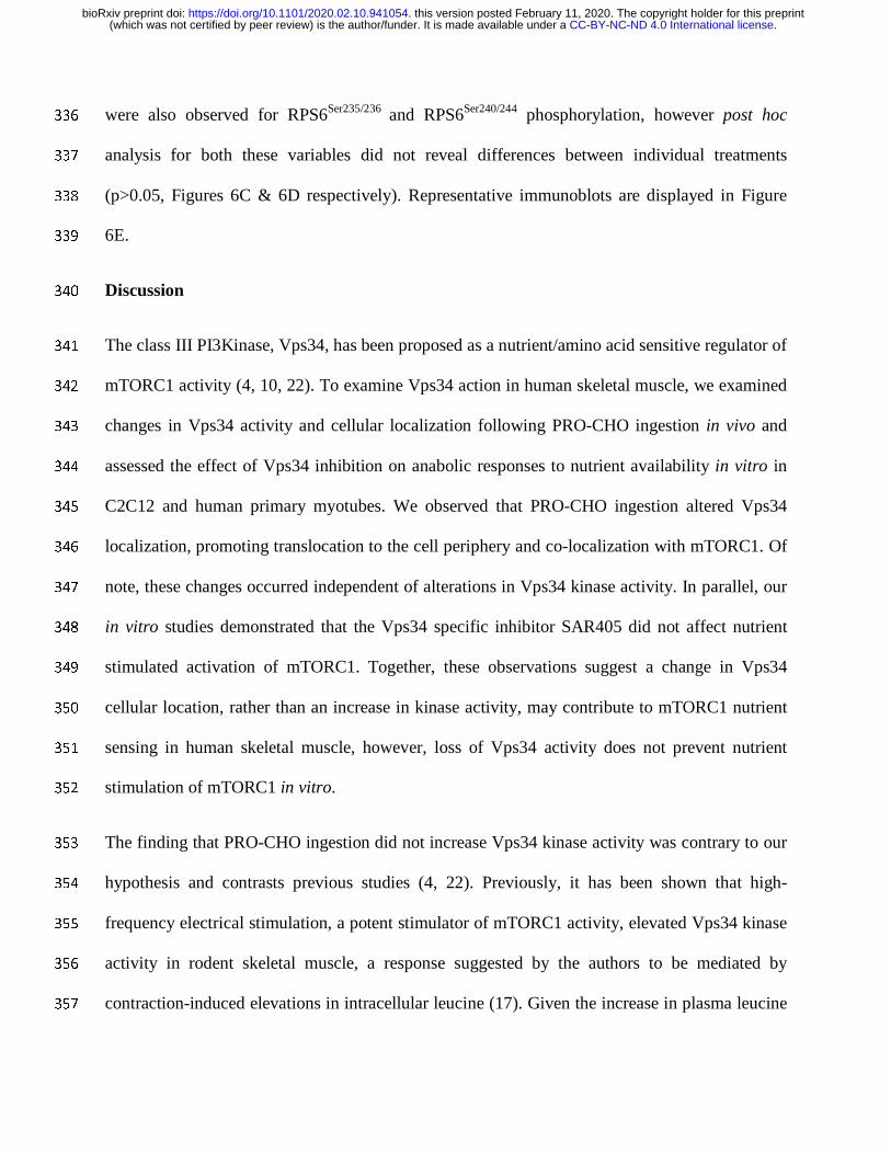
Table 1. Summary of Antibodies Used 511
512
Figure Legends 513
Figure 1. Schematic of Experimental Protocol for Human Trial. 514
515
Figure 2. The effect of protein-carbohydrate feeding on plasma insulin and leucine 516
concentrations and enzyme kinase activities. Insulin concentrations (A) are presented as µU/ml 517
and Leucine concentrations (B) are presented as mM. Kinase activity of S6K1 (C), AKT (D) and 518
Vps34 (E) are presented as % of PRE. For A & B, Ψ denotes a significant effect of time (p<0.05) 519
and *denotes a significant difference at this time point compared to 0 (p<0.05). For C, D & E 520
*denotes a significant difference at this time point compared to PRE (p<0.05), #denotes a 521
significant difference at this time point compared to 3h (p<0.05) and Ψ denotes a significant 522
effect of time (p<0.05). All values are presented as mean±SEM. Data analyzed on SPSS using 523
Repeated Measures ANOVA with Holm-Bonferroni post hoc comparisons conducted on 524
Microsoft Excel. Insulin – n=7, Leucine – n=8. Kinase acitivies – n=8 525
526
Figure 3. The effect of protein-carbohydrate ingestion on mTOR-LAMP2 and mTOR-WGA co-527
localization. Representative images of mTOR (red), LAMP2 (green) and WGA (blue) stains at 528
each time point are provided (A). Quantification of mTOR-LAMP2 (B) and mTOR-WGA (C) 529
co-localization is presented as Pearson’s correlation coefficient. Data in B and C are presented as 530
mean±SEM. Ψ denotes a significant effect of time (p<0.05). Data analyzed on SPSS using 531
Repeated Measures ANOVA with Holm-Bonferroni post hoc comparisons conducted on 532
Microsoft Excel. All analyses – n=8. 533
534
Figure 4. The effect of protein-carbohydrate ingestion on mTOR-VPS34 and mTOR-WGA co-535
localization. Representative images of mTOR (red), VPS34 (green) and WGA (blue) stains at 536
each time point are provided (A). Quantification of VPS34-WGA (B) and mTOR-VPS34 (C) co-537
localization is presented as Pearson’s correlation coefficient. Data in B and C are presented as 538
mean±SEM. Ψ denotes a significant effect of time (p<0.05). *denotes a significant differences at 539
this time point compared to PRE (p<0.05). Data analyzed on SPSS using Repeated Measures 540
ANOVA with Holm-Bonferroni post hoc comparisons conducted on Microsoft Excel. All 541
analyses – n=8. 542
543
Figure 5. The effects of serum/nutrient withdrawal (~14h) and subsequent serum recovery (30 544
min), +/- SAR405, on anabolic signalling in C2C12 myotubes (n=9/group). S6K1Thr389 (A), 545
4EBP1Thr37/46 (B), RPS6Ser235/236 (C) and RPS6Ser240/244 (D) phosphorylation were quantified in 546
relation to their total proteins and ponceau staining was used as a loading control. Representative 547
.CC-BY-NC-ND 4.0 International license(which was not certified by peer review) is the author/funder. It is made available under aThe copyright holder for this preprintthis version posted February 11, 2020. . https://doi.org/10.1101/2020.02.10.941054doi: bioRxiv preprint

images are also provided (E). Data is presented in relation to baseline as Mean±SEM. *denotes a 548
significant difference in this treatment compared to B (p<0.05). Data analyzed on SPSS using 549
One-Way ANOVA with Holm-Bonferroni post hoc comparisons conducted on Microsoft Excel. 550
All analyses – n=9. B = Baseline, SW = Serum Withdrawal & SR = Serum Recovery. 551
552
Figure 6. The effects of serum/nutrient withdrawal (~14h) and subsequent serum recovery (30 553
min), +/- SAR405, on anabolic signalling in human primary myotubes (n=4). S6K1Thr389 (A), 554
4EBP1Thr37/46 (B), RPS6Ser235/236 (C) and RPS6Ser240/244 (D) phosphorylation were quantified in 555
relation to their total proteins and ponceau staining was used as a loading control. Representative 556
images are also provided (E). Data is presented in relation to baseline as Mean±SEM. *denotes a 557
significant difference in this treatment compared to B (p<0.05). Ψ denotes a significant effect of 558
treatment (p<0.05). Data analyzed on SPSS using Repeated Measures ANOVA with Holm-559
Bonferroni post hoc comparisons conducted on Microsoft Excel. All analyses – n=4. B = 560
Baseline, SW = Serum Withdrawal & SR = Serum Recovery. 561
562
.CC-BY-NC-ND 4.0 International license(which was not certified by peer review) is the author/funder. It is made available under aThe copyright holder for this preprintthis version posted February 11, 2020. . https://doi.org/10.1101/2020.02.10.941054doi: bioRxiv preprint
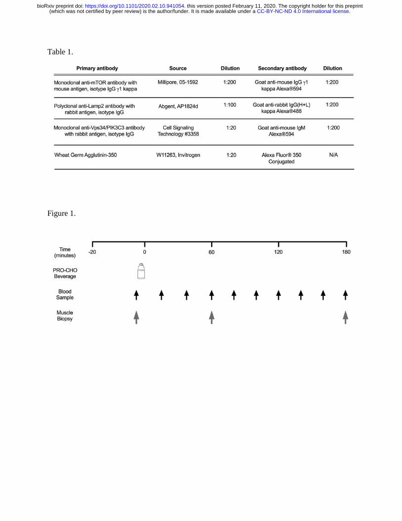
Table 1.
Figure 1.
.CC-BY-NC-ND 4.0 International license(which was not certified by peer review) is the author/funder. It is made available under aThe copyright holder for this preprintthis version posted February 11, 2020. . https://doi.org/10.1101/2020.02.10.941054doi: bioRxiv preprint

Figure 2.
.CC-BY-NC-ND 4.0 International license(which was not certified by peer review) is the author/funder. It is made available under aThe copyright holder for this preprintthis version posted February 11, 2020. . https://doi.org/10.1101/2020.02.10.941054doi: bioRxiv preprint

Figure 3.
.CC-BY-NC-ND 4.0 International license(which was not certified by peer review) is the author/funder. It is made available under aThe copyright holder for this preprintthis version posted February 11, 2020. . https://doi.org/10.1101/2020.02.10.941054doi: bioRxiv preprint

Figure 4.
.CC-BY-NC-ND 4.0 International license(which was not certified by peer review) is the author/funder. It is made available under aThe copyright holder for this preprintthis version posted February 11, 2020. . https://doi.org/10.1101/2020.02.10.941054doi: bioRxiv preprint

Figure 5.
.CC-BY-NC-ND 4.0 International license(which was not certified by peer review) is the author/funder. It is made available under aThe copyright holder for this preprintthis version posted February 11, 2020. . https://doi.org/10.1101/2020.02.10.941054doi: bioRxiv preprint

Figure 6.
.CC-BY-NC-ND 4.0 International license(which was not certified by peer review) is the author/funder. It is made available under aThe copyright holder for this preprintthis version posted February 11, 2020. . https://doi.org/10.1101/2020.02.10.941054doi: bioRxiv preprint