Protective effect of vardenafil on ischemia–reperfusion...
6
684 http://journals.tubitak.gov.tr/medical/ Turkish Journal of Medical Sciences Turk J Med Sci (2013) 43: 684-689 © TÜBİTAK doi:10.3906/sag-1207-108 Protective effect of vardenafil on ischemia–reperfusion injury in rat ovary Hatice BAŞ 1 , Özlem KARA 1 , Mustafa KARA 2 , Dilek PANDIR 1, * 1 Department of Biology, Faculty of Arts and Science, Bozok University, Divanlıyolu, Yozgat, Turkey 2 Department of Obstetrics and Gynecology, Faculty of Medicine, Bozok University, Divanlıyolu, Yozgat, Turkey * Correspondence: [email protected] 1. Introduction Ovarian torsion is a serious gynecologic emergency condition. Although it might affect all women, it is usually seen in women of reproductive age (1). Ovarian ischemia is the result of torsion and leads to cell death because of insufficient perfusion of the tissue (2,3). Ischemic tissues need to recover blood supply for regeneration of cells and disposal of toxic metabolites. However, reperfusion of the ischemic tissue paradoxically leads to much more serious damage to the tissue than the damage caused by ischemia (4). e ischemia and reperfusion injuries are related to the production of reactive oxygen species (ROS) (5). Ischemia– reperfusion (I/R) injury increases the production of free radicals such as ROS and malondialdehyde (MDA). Activities of superoxide dismutase (SOD), glutathione peroxidase (GPx), and catalase (CAT) increase to protect the ischemic tissue from the harmful effects of toxic metabolites (3). Free oxygen species result in oxidative injury via different mechanisms, such as metabolic dysfunction or impairment of calcium intracellular homeostasis. ey may lead to cell injury or cell death by themselves or by deactivating various enzymes (6). Extreme production of ROS causes alterations in subcellular structures. ese changes are modifications of protein and DNA, lipid peroxidation (LPO) of polyunsaturated fatty acids in membranes (7), apoptosis (8), and depression of cellular antioxidant system (9). MDA is the end product of LPO, and thus increased MDA content is an important indicator of LPO (10). Cells have different mechanisms against oxidative damage. ese mechanisms involve enzymatic and nonenzymatic defense systems. SOD, CAT, and GPx are included in enzymatic defense systems (11). If the cells do not have enough antioxidant enzymatic activity, ROS levels increase. Apoptosis protease-activating factor 1 (Apaf-1), an important part of apoptosome, is a protein that plays a central role in apoptosis (12). When apoptosis is stimulated, Apaf-1 binds cytochrome c and procaspase 9, and then caspase-3 and -7 become activated. is complex is called apoptosome and results in the loss of the cell (13). Cellular stresses, such as hypoxia, lead to caspase activation. is process results in apoptotic cell death (12). Aim: To investigate the effect of vardenafil on ischemia–reperfusion (I/R) injury in rat ovary. Materials and methods: A total of 21 Wistar rats were divided into 3 groups. In group 1, ovary torsion was not performed and no drug was administered. In group 2, 1 h of ischemia and 2 h of reperfusion were performed and no drug was given. In group 3, 1 h of ischemia and 2 h of reperfusion were performed and 1 mg/kg vardenafil was administered aſter 30 min of ischemia. Right ovaries were surgically removed in all groups. Malondialdehyde (MDA) levels and activities of superoxide dismutase (SOD), catalase (CAT), and glutathione peroxidase (GPx) were measured. Apoptosis protease-activating factor 1 (Apaf-1) was measured by using immunohistochemistry. Results: MDA level was significantly increased and SOD, CAT, and GPx activities were significantly decreased in group 2 compared to the other groups (P < 0.05). In light microscopic investigations, severe vascular congestion, edema, hemorrhage, and follicular degeneration were assessed in the ovary tissue. Ovarian tissue damage was higher in group 2 than group 3. Apaf-1 expression was stronger in group 2 than group 3. Conclusion: Vardenafil treatment seems to reduce microscopic histopathological changes, decrease Apaf-1 response, and alter oxidative stress in I/R injury in rat ovaries. Key words: Ovarian torsion, ischemia–reperfusion, vardenafil, antioxidant enzymes, apoptosis Received: 29.07.2012 Accepted: 05.12.2012 Published Online: 26.08.2013 Printed: 20.09.2013 Research Article
Transcript of Protective effect of vardenafil on ischemia–reperfusion...
684
http://journals.tubitak.gov.tr/medical/
Turkish Journal of Medical Sciences Turk J Med Sci(2013) 43: 684-689© TÜBİTAKdoi:10.3906/sag-1207-108
Protective effect of vardenafil on ischemia–reperfusion injury in rat ovary
Hatice BAŞ1, Özlem KARA1, Mustafa KARA2, Dilek PANDIR1,*1Department of Biology, Faculty of Arts and Science, Bozok University, Divanlıyolu, Yozgat, Turkey
2Department of Obstetrics and Gynecology, Faculty of Medicine, Bozok University, Divanlıyolu, Yozgat, Turkey
* Correspondence: [email protected]
1. IntroductionOvarian torsion is a serious gynecologic emergency condition. Although it might affect all women, it is usually seen in women of reproductive age (1). Ovarian ischemia is the result of torsion and leads to cell death because of insufficient perfusion of the tissue (2,3). Ischemic tissues need to recover blood supply for regeneration of cells and disposal of toxic metabolites. However, reperfusion of the ischemic tissue paradoxically leads to much more serious damage to the tissue than the damage caused by ischemia (4). The ischemia and reperfusion injuries are related to the production of reactive oxygen species (ROS) (5). Ischemia–reperfusion (I/R) injury increases the production of free radicals such as ROS and malondialdehyde (MDA). Activities of superoxide dismutase (SOD), glutathione peroxidase (GPx), and catalase (CAT) increase to protect the ischemic tissue from the harmful effects of toxic metabolites (3).
Free oxygen species result in oxidative injury via different mechanisms, such as metabolic dysfunction or impairment of calcium intracellular homeostasis. They may lead to cell injury or cell death by themselves or by
deactivating various enzymes (6). Extreme production of ROS causes alterations in subcellular structures. These changes are modifications of protein and DNA, lipid peroxidation (LPO) of polyunsaturated fatty acids in membranes (7), apoptosis (8), and depression of cellular antioxidant system (9). MDA is the end product of LPO, and thus increased MDA content is an important indicator of LPO (10). Cells have different mechanisms against oxidative damage. These mechanisms involve enzymatic and nonenzymatic defense systems. SOD, CAT, and GPx are included in enzymatic defense systems (11). If the cells do not have enough antioxidant enzymatic activity, ROS levels increase.
Apoptosis protease-activating factor 1 (Apaf-1), an important part of apoptosome, is a protein that plays a central role in apoptosis (12). When apoptosis is stimulated, Apaf-1 binds cytochrome c and procaspase 9, and then caspase-3 and -7 become activated. This complex is called apoptosome and results in the loss of the cell (13). Cellular stresses, such as hypoxia, lead to caspase activation. This process results in apoptotic cell death (12).
Aim: To investigate the effect of vardenafil on ischemia–reperfusion (I/R) injury in rat ovary.
Materials and methods: A total of 21 Wistar rats were divided into 3 groups. In group 1, ovary torsion was not performed and no drug was administered. In group 2, 1 h of ischemia and 2 h of reperfusion were performed and no drug was given. In group 3, 1 h of ischemia and 2 h of reperfusion were performed and 1 mg/kg vardenafil was administered after 30 min of ischemia. Right ovaries were surgically removed in all groups. Malondialdehyde (MDA) levels and activities of superoxide dismutase (SOD), catalase (CAT), and glutathione peroxidase (GPx) were measured. Apoptosis protease-activating factor 1 (Apaf-1) was measured by using immunohistochemistry.
Results: MDA level was significantly increased and SOD, CAT, and GPx activities were significantly decreased in group 2 compared to the other groups (P < 0.05). In light microscopic investigations, severe vascular congestion, edema, hemorrhage, and follicular degeneration were assessed in the ovary tissue. Ovarian tissue damage was higher in group 2 than group 3. Apaf-1 expression was stronger in group 2 than group 3.
Conclusion: Vardenafil treatment seems to reduce microscopic histopathological changes, decrease Apaf-1 response, and alter oxidative stress in I/R injury in rat ovaries.
Key words: Ovarian torsion, ischemia–reperfusion, vardenafil, antioxidant enzymes, apoptosis
Received: 29.07.2012 Accepted: 05.12.2012 Published Online: 26.08.2013 Printed: 20.09.2013
Research Article
685
BAŞ et al. / Turk J Med Sci
Vardenafil is a vasoactive drug developed for treatment of erectile dysfunction. It inhibits the phosphodiesterase type-5 (PDE-5) enzyme, which changes cyclical guanosine monophosphate (cGMP) into inactive GMP. The increased levels of cGMP lead to smooth muscle relaxation and vasodilation (14). Furthermore, these drugs are found to be effective in the treatment of injuries in other organs, such as the liver and brain (15). As discussed above, vardenafil would be a useful drug in the short-term treatment of I/R damage of the ovary due to torsion.
Therefore, in this study, we planned to give a drug decreasing oxidative damage and reversing harmful histopathological changes in the early phase of torsion/detorsion. To our best knowledge, there is no information about the effects of vardenafil on I/R damage of the ovary. We aimed to investigate the effect of vardenafil against I/R condition in the ovary of rat.
2. Materials and methods2.1. Animals Çukurova University’s Experimental Animal Laboratory TIBDAM Institute supplied 21 healthy adult female Wistar rats, weighing between 240 and 260 g. The rats were selected according to their estrous cycle. All the rats were acclimated to the environment 10 days before the beginning of treatment. The rats were housed in plastic rat cages at 26 ± 2 °C and they were exposed to 10–12 h of daylight. Animals were fed a standard laboratory diet and tap water ad libitum. All rats were handled in compliance with the guidelines for the use and care of laboratory animals. 2.2. Treatment periodAnimals were randomized into 1 control group and 2 treatment groups (I/R and vardenafil + I/R groups). Each group consisted of 7 animals. All rats were initially randomly numbered. In the control group, right ovaries of the rats were removed without any treatment. Ovary ischemia of the rats in treatment groups were made according to the method described by Hascalik et al. (16). In the I/R group no drug was given and 1 h of ischemia and 2 h of reperfusion were performed. In the vardenafil + I/R group, 1 h of ischemia and 2 h of reperfusion were performed. Vardenafil, in quantities of 1 mg/kg, was administered intraperitoneally 30 min after ischemia. After reperfusion, all the rats were sacrificed and their right ovaries were surgically extirpated. Surgical procedures were performed under general anesthesia 2 times. Rats were anesthetized by an intraperitoneal injection of ketamine hydrochloride (60 mg/kg, Ketalar, Eczacıbaşı, İstanbul, Turkey) and xylazine hydrochloride (10 mg/kg, Rompun, Bayer, Leverkusen, Germany). The operation was performed under sterile conditions by using a 2-cm laparotomic incision. The right
ovaries were exteriorized and atraumatic vascular clamps were used to produce ovarian ischemia. Ischemia was terminated after 1 h, and then the skin closed with 3/0 silk sutures. Relaparotomy was performed in former operation areas. Reperfusion was provided for 2 h and the ovary was resected at the end of this time. The animals were sacrificed via cervical dislocation by forceps. 2.3. Biochemical evaluationThe ovaries were washed in a sodium phosphate buffer (pH 7.2) and samples were stored at –80 °C until the analysis. CAT enzyme activity was measured according to the method described by Aebi (17). Data were expressed as nmol/mg protein. GPx activity was measured according to the method of Paglia and Valentine (18) at absorbance of 340 nm. Activity was given as nmol/mg protein. The method of Marklund and Marklund (19) was used to determine total SOD activity. Data were expressed as U/mg protein. MDA content was assayed by using the thiobarbituric acid test as described by Ohkawa et al. (20). Specific activity was presented as nmol/mg protein. MDA levels and SOD, CAT, and GPx activities were determined by measuring the absorbance of the samples in a spectrophotometer (Shimadzu UV 1800, Kyoto, Japan). Protein concentration was determined according to the method described by Lowry et al. (21). 2.4. ImmunohistochemistryImmunohistochemistry was used to determine the Apaf-1 expression. Samples were fixed in formalin then embedded in paraffin. Tissue sections were cut at 5-µm thickness and then dried at 37 °C. Immunostaining was performed with a Leica Bond-Max automatic immunostainer (Leica, Bannockburn, IL, USA).2.5. Histopathological examinationFor histopathological examinations, ovary samples were fixed for 24 h in a 10% formalin solution. The tissues were then dehydrated and embedded in paraffin. The tissues were cut at thicknesses of 5–6 µm. The sections were stained by utilizing hematoxylin and eosin dye. The sections were analyzed and photographed with a Olympus BX 51 light microscope (Olympus Corp., Tokyo, Japan). Histopathological damage was evaluated by using ovarian tissue damage scores. The degrees of vascular congestion, interstitial edema, follicular degeneration, and hemorrhage were detected. Each ovary slide was examined and the severity of the changes observed was scored by using a scale of no (_), moderate (+), and severe (++) damage. 2.6. Statistical analysisData were expressed using SPSS 11.0 (SPSS Inc., Chicago, IL, USA). The experimental groups were compared using an analysis of variance (ANOVA) test and Tukey’s post hoc comparison. P < 0.05 was assigned as statistically significant.
686
BAŞ et al. / Turk J Med Sci
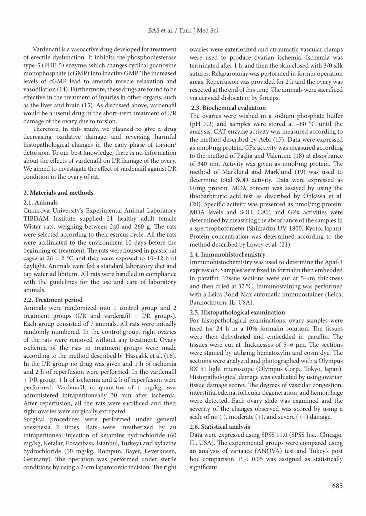
3. ResultsAt the end of the study, MDA levels were significantly higher in the I/R group than in the vardenafil + I/R group (P < 0.05) (Figure 1A). SOD, CAT, and GPx activities were higher in the vardenafil + I/R group than the I/R group and the differences were found to be statistically significant (P < 0.05) (Figures 1B–1D).
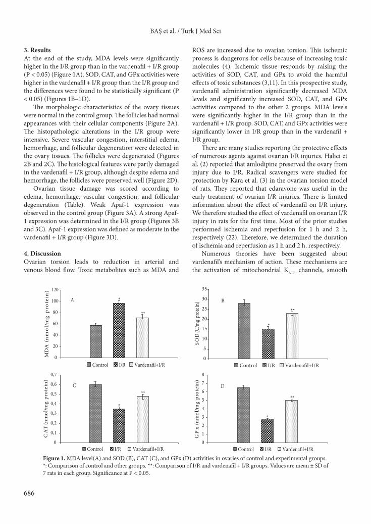
The morphologic characteristics of the ovary tissues were normal in the control group. The follicles had normal appearances with their cellular components (Figure 2A). The histopathologic alterations in the I/R group were intensive. Severe vascular congestion, interstitial edema, hemorrhage, and follicular degeneration were detected in the ovary tissues. The follicles were degenerated (Figures 2B and 2C). The histological features were partly damaged in the vardenafil + I/R group, although despite edema and hemorrhage, the follicles were preserved well (Figure 2D).
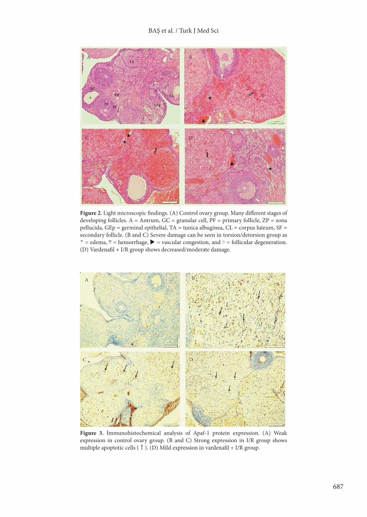
Ovarian tissue damage was scored according to edema, hemorrhage, vascular congestion, and follicular degeneration (Table). Weak Apaf-1 expression was observed in the control group (Figure 3A). A strong Apaf-1 expression was determined in the I/R group (Figures 3B and 3C). Apaf-1 expression was defined as moderate in the vardenafil + I/R group (Figure 3D).
4. DiscussionOvarian torsion leads to reduction in arterial and venous blood flow. Toxic metabolites such as MDA and
ROS are increased due to ovarian torsion. This ischemic process is dangerous for cells because of increasing toxic molecules (4). Ischemic tissue responds by raising the activities of SOD, CAT, and GPx to avoid the harmful effects of toxic substances (3,11). In this prospective study, vardenafil administration significantly decreased MDA levels and significantly increased SOD, CAT, and GPx activities compared to the other 2 groups. MDA levels were significantly higher in the I/R group than in the vardenafil + I/R group. SOD, CAT, and GPx activities were significantly lower in I/R group than in the vardenafil + I/R group.
There are many studies reporting the protective effects of numerous agents against ovarian I/R injuries. Halici et al. (2) reported that amlodipine preserved the ovary from injury due to I/R. Radical scavengers were studied for protection by Kara et al. (3) in the ovarian torsion model of rats. They reported that edaravone was useful in the early treatment of ovarian I/R injuries. There is limited information about the effect of vardenafil on I/R injury. We therefore studied the effect of vardenafil on ovarian I/R injury in rats for the first time. Most of the prior studies performed ischemia and reperfusion for 1 h and 2 h, respectively (22). Therefore, we determined the duration of ischemia and reperfusion as 1 h and 2 h, respectively.
Numerous theories have been suggested about vardenafil’s mechanism of action. These mechanisms are the activation of mitochondrial KATP channels, smooth
0
20
40
60
80
100
120)niet
orp
gm/l
om
n( A
DM
Control I/R Vardenafil+I/R
*
**
A
0
5
10
15
20
25
30
35
)nietorp gm/ U( D
OS
Control I/R Vardenafil+I/R
*
**
B
012345678
)nietorp gm/lo
mn( xP
G
Control I/R Vardenafil+I/R
**
*
D
0
0,1
0,2
0,3
0,4
0,5
0,6
0,7
)nietorp gm/lo
mn( TA
C
Control I/R Vardenafil+I/R
*
**C
Figure 1. MDA level(A) and SOD (B), CAT (C), and GPx (D) activities in ovaries of control and experimental groups.*: Comparison of control and other groups. **: Comparison of I/R and vardenafil + I/R groups. Values are mean ± SD of 7 rats in each group. Significance at P < 0.05.
687
BAŞ et al. / Turk J Med Sci
Figure 2. Light microscopic findings. (A) Control ovary group. Many different stages of developing follicles. A = Antrum, GC = granular cell, PF = primary follicle, ZP = zona pellucida, GEp = germinal epithelial, TA = tunica albuginea, CL = corpus luteum, SF = secondary follicle. (B and C) Severe damage can be seen in torsion/detorsion group as ✶ = edema, ⇈ = hemorrhage, = vascular congestion, and ⇧ = follicular degeneration. (D) Vardenafil + I/R group shows decreased/moderate damage.
Figure 3. Immunohistochemical analysis of Apaf-1 protein expression. (A) Weak expression in control ovary group. (B and C) Strong expression in I/R group shows multiple apoptotic cells (↑). (D) Mild expression in vardenafil + I/R group.

688
BAŞ et al. / Turk J Med Sci
muscle relaxation, and vasodilation (14,15). Vardenafil is also a potent PDE-5 enzyme inhibitor and is mainly used in the treatment of erectile dysfunction (14). Soydan et al. reported that there are many protection mechanisms against I/R-induced injury and they still have not been completely understood (23). The effect of vardenafil (1 mg/kg) was investigated on testicular torsion/detorsion injury. Vardenafil treatment reduced testicular damage due to I/R (24). Because of this, we thought that vardenafil utilization would be useful for the treatment of ovarian I/R damage. As a potent PDE-5 enzyme inhibitor, vardenafil was assessed as a conservative factor in the rat ovary I/R injury. According to our findings, vardenafil seems to reduce the I/R damage due to ovarian torsion. The possible mechanisms for the protective effect of vardenafil were vascular dilatation, PDE-5 enzyme inhibition, and electrolyte alteration such as K. However, more studies are required to evaluate the effect of vardenafil on ovarian I/R injury.
Many studies have shown that the effects of some chemical substances on I/R injury in rat ovaries (25–29). In our study, the histopathological changes such as vascular congestion, edema, hemorrhage, and follicular degeneration were found to be decreased in the vardenafil + I/R group.
Apoptosis is a form of cell death and provides tissue development and homeostasis. There are many signals
in animal cells, such as hypoxia, DNA damage, and oncogene activation. These signals lead to the activation of mitochondrial pathways and generate apoptotic protein (12). Robles et al. showed that apoptosis led to increased Apaf-1 expression in the granulosa cells of mouse ovary (30). Erol et al. evaluated germ cell apoptosis by using the Apaf-1 antibody (24). They reported that vardenafil administration reduced the I/R damage in testis tissue. To date, the expression of the Apaf-1 antibody and vardenafil treatment has not been evaluated in ovarian I/R injury. In our study, the expression of the signal protein of apoptosis, Apaf-1, was lower in the vardenafil+ I/R group than the I/R group.
In this randomized controlled study, vardenafil was studied as a protective agent to avoid I/R injury of the ovary. Our results have shown that MDA levels were increased and SOD, CAT, and GPx activities were decreased in ovarian torsion. Furthermore, ischemic changes such as vascular congestion, edema, hemorrhage, and follicular degeneration were much higher in the I/R group. Vardenafil administration decreased these harmful changes. Vardenafil seems to be useful in reducing the biochemical and histopathological damage.
Acknowledgment We would like to thank Cengiz Çolak for helping us prepare this study.
Table. Grading of the histopathological changes in ovary sections of rats exposed to torsion/detorsion.
Groups Edema Hemorrhage Vascular congestion Follicular degeneration
Control - - - -
I/R ++ ++ ++ ++
Vardenafil + I/R + + + _
The features were scored as follows: no damage (_), moderate damage (+), and severe damage (++).
References
1. Becker JH, Jan de Graaff J, Vosb CM. Torsion of the ovary: a known but frequently missed diagnosis. Eur J Emerg Med 2009; 16: 124–6.
2. Halici Z, Karaca M, Keles ON, Borekci B, Odabasoglu F, Suleyman H et al. Protective effects of amlodipine on ischemia-reperfusion injury of rat ovary: biochemical and histopathologic evaluation. Fertil Steril 2008; 90: 2408–15.
3. Kara M, Daglioglu YK, Kuyucu Y, Tuli A, Tap A. The effect of edaravone on ischemia–reperfusion injury in rat ovary. Eur J Obstet Gynecol Reprod Biol 2012; 162: 197–202.
4. Abramov AY, Scorziello A, Duchen MR. Three distinct mechanisms generate oxygen free radicals in neurons and contribute to cell death during anoxia and reoxygenation. J Neurosci 2007; 27: 1129–38.
5. Demircioğlu Rİ, Usta B, Sert H, Muslu B, Gözdemir M. Taurine is protective against oxidative stress during cold ischemia in the rat kidney. Turk J Med Sci 2011; 41: 843–9.
6. Özdemir B, Kaya A, Söğüt Ö, Kaya H, Gökdemir MT, Celbiş O. Oxidative stress status of individuals involved in traffic accidents. Turk J Med Sci 2012; 42: 507–14.
689
BAŞ et al. / Turk J Med Sci
7. Kalender S, Kalender Y, Durak D, Ogutcu A, Uzunhisarcikli M, Cevrimli BS. Methyl parathion induced nephrotoxicity in male rats and protective role of vitamins C and E. Pestic Biochem Physiol 2007; 88: 213–8.
8. Parlakpinar H, Sahna E, Acet A, Mizrak B, Polat A. Protective effect of caffeic acid phenethyl ester (CAPE) on myocardial ischemia–reperfusion induced apoptotic cell death. Toxicol 2005; 209: 1–14.
9. Kalender Y, Kaya S, Durak D, Uzun FG, Demir F. Protective effects of catechin and quercetin on antioxidant status, lipid peroxidation and testis histoarchitecture induced by chlorpyrifos in male rats. Environ Toxicol Phar 2012; 33: 141–8.
10. Çömelekoğlu Ü, Yalın S, Ballı E, Berköz M. Ovariectomy decreases biomechanical quality of skin via oxidative stress in rat. Turk J Med Sci 2012; 42: 201–9.
11. Sayın O, Arslan N, Altun ZS, Akdoğan G. In vitro effect of resveratrol against oxidative injury of human coronary artery endothelial cells. Turk J Med Sci 2011; 41: 211–8.
12. Hajra KM, Liu JR. Apoptosome dysfunction in human cancer. Apoptosis 2004; 9: 691–704.
13. Watson AJM. Apoptosis and colorectal cancer. Gut 2004; 53: 1701–9.
14. Shabsigh R. Therapy of ED: PDE-5 inhibitors. Endocrine 2004; 23: 135–41.
15. Zhang RL, Zhang Z, Zhang L, Wang Y, Zhang C, Chopp M. Delayed treatment with sildenafil enhances neurogenesis and improves functional recovery in aged rats after focal cerebral ischemia. J Neurosci Res 2006; 15;83: 1213–9.
16. Hascalik S, Celik O, Turkoz Y, Mizrak B. Clip Turcica: a new apparatus for experimental ovarian ischemia and reperfusion model in rats. Fertil Steril 2005; 84: 219–20.
17. Aebi H. Catalase in vitro. Methods Enzymol 1984; 105: 121–6.
18. Paglia DE, Valentine WN. Studies on the quantitative and qualitative characterization of glutathione peroxidase. J Lab Med 1987; 70: 158–65.
19. Marklund S, Marklund G. Involvement of the superoxide anion radical in the autoxidation of pyrogallol and a convenient assay for superoxide dismutase. Eur J Biochem 1974; 47: 469–74.
20. Ohkawa H, Ohishi N, Yagi K. Assay for lipid peroxides in animal tissues by thiobarbituric acid rection. Anal Biochem 1979; 95: 351–8.
21. Lowry OH, Rosebrough NJ, Farr AL, Randall RJ. Protein measurement with the Folin phenol reagent. J Biol Chem 1951; 193: 265–75.
22. Obermaier R, Benz S, Von Dobschuetz E, Drognitz O, Schareck W, Jonas L et al. Characterization of microcirculatory disturbance in a novel model of pancreatic ischemia-reperfusion using intravital fluorescence-microscopy. Pancreas 2002; 25: 142–8.
23. Soydan G, Sokmensuer C, Kilinc K, Tuncer M. The effects of sildenafil on the functional and structural changes of ileum induced by intestinal ischemia-reperfusion in rats. Eur J Pharmacol 2009; 610: 87–92.
24. Erol B, Tokgoz H, Hanci V, Bektas S, Akduman B, Yencilek F. Vardenafil reduces testicular damage following ischemia/reperfusion injury in rats. Kaohsiung J Med Sci 2009; 25: 374–80.
25. Arikan DC, Bakan V, Kurutas EB, Sayar H, Coskun A. Protective effect of tadalafil on ischemia/reperfusion injury of rat ovary. J Pediatr Surg 2010; 45: 2203–9.
26. Oral A, Odabasoglu F, Halici Z, Keles ON, Unal B, Coskun AK et al. Protective effects of montelukast on ischemia-reperfusion injury in rat ovaries subjected to torsion and detorsion: biochemical and histopathologic evaluation. Fertil Steril 2011; 95: 1360–6.
27. Yigiter M, Halici Z, Odabasoglu F, Keles ON, Atalay F, Unal B et al. Growth hormone reduces tissue damage in rat ovaries subjected to torsion and detorsion evaluation. Eur J Obstet Gynecol Reprod Biol 2011; 157: 94–100.
28. Bayir Y, Karagoz Y, Karakus E, Albayrak A, Sengul O, Can I et al. Nigella sativa reduces tissue damage in rat ovaries subjected to torsion and detorsion: oxidative stress, proinflammatory response and histopathological evaluation. Gynecol Obstet Invest 2012; 74: 41–9.
29. Osmanağaoğlu M, Kesim M, Yuluğ E, Menteşe A, Karahan CS. The effect of high dose methylprednisolone on experimental ovarian torsion/reperfusion injury in rats. Geburtsh Frauenheilk 2012; 72: 70–4.
30. Robles R, Tao XJ, Trbovich AM, Maravel DV, Nahum R, Perez GI et al. Localization, regulation and possible consequences of apoptotic protease-activating factor-1 (Apaf-1) expression in granulosa cells of the mouse ovary. Endocrinology 1999; 140: 2641–4.