Prosthetic Rehabilitation of Partial Ear Defect: 2 Case ...
6
CLINICAL REPORT Prosthetic Rehabilitation of Partial Ear Defect: 2 Case Reports Meryem Gu ¨lce Subas ¸ ı • Gamze Alnıac ¸ ık • Abdullah Kalaycı • Serhan Akman • Ercan Durmus ¸ Received: 5 July 2012 / Accepted: 29 December 2012 / Published online: 6 January 2013 Ó Indian Prosthodontic Society 2013 Abstract The loss or absence of an auricle may result from trauma, disease or congenital anomalies and causes a considerable aesthetic problem. If the deformity involves the external auditory canal, it can affect hearing. This case report describes the surgical and prosthetic treatment of two patients with partial defects of their right external ears from different causes. Implant-retained auricular prosthe- ses fabricated from heat-temperature-vulcanised silicone were used in both the cases; they were designed to be harmonious with the remaining tissues. The patients experienced improved retention, aesthetics, hearing and quality of life with these prostheses. During the approxi- mately 3 year follow-up, both the prostheses were re-fab- ricated once; however, problems related to implant stability and peri-implant tissue health were not encountered. Keywords Partial ear defect Á Implant Á Retention Á Implant-retained auricular prosthesis Introduction Ear defects can occur secondarily to congenital malfor- mations, trauma or tumour surgery. The absence of an ear is a considerable aesthetic problem that may affect the patient’s psychology and social behaviour [1]. Correction of ear defects can be accomplished surgically, prostheti- cally or through a combination of these approaches; the choice of treatment depends on the site, size, age and aetiology of the defect as well as the patient’s desires [1, 2]. However, reconstructive surgery is limited by the age and medical conditions of the patient, insufficient residual tis- sue, vascular compromise due to radiation and the patient’s preferences [3, 4]. Further, after a surgical procedure, the reconstructed ear may not resemble the normal one [5]. On the other hand, prosthetic treatment can produce an anatomically accurate and aesthetic device [4, 6]. Before the introduction of osseointegration, auricular prostheses were retained by adhesives or a connection to eyeglasses [4, 6, 7]. Nowadays, craniofacial implants are used to support and retain such prostheses. Studies have shown successful retention and stability of auricular prostheses anchored to the temporal bone with titanium implants [7–9]. Tjelstrom et al. [7] used titanium implants to attach auricular prostheses and obtained successful results after a 5-year follow-up. This case report describes the surgical and prosthetic treatment of two patients with partial defects of their right external ears from different causes who were followed for *3 years. Case Report Case 1 A 58 year-old man with a deformed right ear was referred to the Selcuk University Department of Prosthodontics. His M. G. Subas ¸ ı (&) Department of Prosthodontics, Faculty of Dentistry, Aydin University, Istanbul, Turkey e-mail: [email protected] G. Alnıac ¸ ık Department of Prosthodontics, Faculty of Dentistry, Kocaeli University, Kocaeli, Turkey e-mail: [email protected] A. Kalaycı Á E. Durmus ¸ Department of Oral and Maxillofacial Surgery, Faculty of Dentistry, Selcuk University, Konya, Turkey S. Akman Department of Prosthodontics, Faculty of Dentistry, Selcuk University, Konya, Turkey 123 J Indian Prosthodont Soc (December 2014) 14(Suppl. 1):S196–S201 DOI 10.1007/s13191-012-0251-5
Transcript of Prosthetic Rehabilitation of Partial Ear Defect: 2 Case ...
CLINICAL REPORT
Prosthetic Rehabilitation of Partial Ear Defect: 2 Case Reports
Meryem Gulce Subası • Gamze Alnıacık •
Abdullah Kalaycı • Serhan Akman •
Ercan Durmus
Received: 5 July 2012 / Accepted: 29 December 2012 / Published online: 6 January 2013
� Indian Prosthodontic Society 2013
Abstract The loss or absence of an auricle may result
from trauma, disease or congenital anomalies and causes a
considerable aesthetic problem. If the deformity involves
the external auditory canal, it can affect hearing. This case
report describes the surgical and prosthetic treatment of
two patients with partial defects of their right external ears
from different causes. Implant-retained auricular prosthe-
ses fabricated from heat-temperature-vulcanised silicone
were used in both the cases; they were designed to be
harmonious with the remaining tissues. The patients
experienced improved retention, aesthetics, hearing and
quality of life with these prostheses. During the approxi-
mately 3 year follow-up, both the prostheses were re-fab-
ricated once; however, problems related to implant stability
and peri-implant tissue health were not encountered.
Keywords Partial ear defect � Implant � Retention �Implant-retained auricular prosthesis
Introduction
Ear defects can occur secondarily to congenital malfor-
mations, trauma or tumour surgery. The absence of an ear
is a considerable aesthetic problem that may affect the
patient’s psychology and social behaviour [1]. Correction
of ear defects can be accomplished surgically, prostheti-
cally or through a combination of these approaches; the
choice of treatment depends on the site, size, age and
aetiology of the defect as well as the patient’s desires [1, 2].
However, reconstructive surgery is limited by the age and
medical conditions of the patient, insufficient residual tis-
sue, vascular compromise due to radiation and the patient’s
preferences [3, 4]. Further, after a surgical procedure, the
reconstructed ear may not resemble the normal one [5].
On the other hand, prosthetic treatment can produce an
anatomically accurate and aesthetic device [4, 6]. Before the
introduction of osseointegration, auricular prostheses were
retained by adhesives or a connection to eyeglasses [4, 6, 7].
Nowadays, craniofacial implants are used to support and
retain such prostheses. Studies have shown successful
retention and stability of auricular prostheses anchored to the
temporal bone with titanium implants [7–9]. Tjelstrom et al.
[7] used titanium implants to attach auricular prostheses and
obtained successful results after a 5-year follow-up. This
case report describes the surgical and prosthetic treatment of
two patients with partial defects of their right external ears
from different causes who were followed for *3 years.
Case Report
Case 1
A 58 year-old man with a deformed right ear was referred
to the Selcuk University Department of Prosthodontics. His
M. G. Subası (&)
Department of Prosthodontics, Faculty of Dentistry,
Aydin University, Istanbul, Turkey
e-mail: [email protected]
G. AlnıacıkDepartment of Prosthodontics, Faculty of Dentistry,
Kocaeli University, Kocaeli, Turkey
e-mail: [email protected]
A. Kalaycı � E. Durmus
Department of Oral and Maxillofacial Surgery,
Faculty of Dentistry, Selcuk University, Konya, Turkey
S. Akman
Department of Prosthodontics, Faculty of Dentistry,
Selcuk University, Konya, Turkey
123
J Indian Prosthodont Soc (December 2014) 14(Suppl. 1):S196–S201
DOI 10.1007/s13191-012-0251-5
medical history revealed that he had undergone surgery for
malignant melanoma. After the operation, he had received
6,000 cGy of external beam irradiation in 30 sessions over
6 weeks. Three years after radiotherapy, in 2007, he pre-
sented for prosthetic treatment. Extra-oral examination
revealed that the right lobule, tragus and antitragus were
missing, but the helix, antihelix, crura of the antihelix,
scapha, cymba concha and external auditory canal were
present. The patient was dissatisfied with his appearance.
An implant-retained auricular prosthesis was selected to
correct the defect.
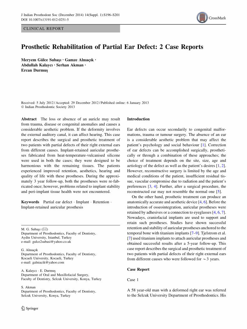
The implant sites were evaluated for bone width and
depth by computerised tomography (CT). In October 2007,
three 5 mm-long extra-oral endosseous implants (042.362S;
Institut Straumann AG, Basel, Switzerland) were placed at
the 9, 10 and 11 o’clock positions in the mastoid region
under local anaesthesia (Fig. 1). The implants were left
unloaded for 3 months to allow osseointegration. They were
exposed during the second stage of surgery, and extra-oral
conical abutments (048.526; Institut Straumann AG) were
screwed onto them under 15 N by using a torque-control
device (Institut Straumann AG).
The peri-implant tissue was allowed to heal for
approximately 2 weeks. Before taking the final impression,
the hair adjacent to the remaining tissues was isolated with
Vaseline, and the external auditory canal was blocked by
using lubricated cotton. Impression cylinders (048.104;
Institut Straumann AG) were screwed to the extra-oral
conical abutments and used with polyvinyl siloxane
impression material (Elite HD; Zhermack, Rovigo, Italy) to
obtain an impression of the defect and abutments. After
controlling the impression, the impression cylinders were
disassembled, screwed to the extra-oral conical implant
analogues (048.136; Institut Straumann AG) and placed in
the obtained impression. Then, a die stone cast was
obtained, and extra-oral gold caps (048.236; Institut
Straumann AG) were screwed onto the conical implant
analogues with an SCS guide screw (048.360V4; Institut
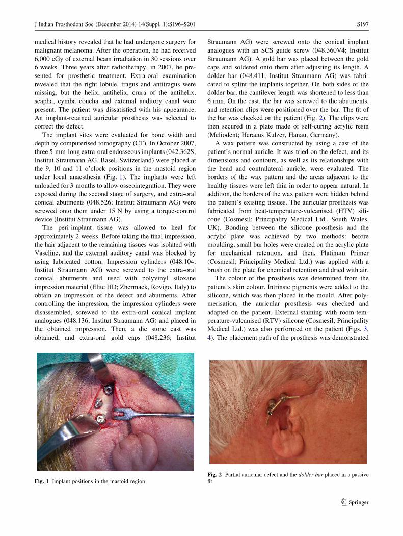
Straumann AG). A gold bar was placed between the gold
caps and soldered onto them after adjusting its length. A
dolder bar (048.411; Institut Straumann AG) was fabri-
cated to splint the implants together. On both sides of the
dolder bar, the cantilever length was shortened to less than
6 mm. On the cast, the bar was screwed to the abutments,
and retention clips were positioned over the bar. The fit of
the bar was checked on the patient (Fig. 2). The clips were
then secured in a plate made of self-curing acrylic resin
(Meliodent; Heraeus Kulzer, Hanau, Germany).
A wax pattern was constructed by using a cast of the
patient’s normal auricle. It was tried on the defect, and its
dimensions and contours, as well as its relationships with
the head and contralateral auricle, were evaluated. The
borders of the wax pattern and the areas adjacent to the
healthy tissues were left thin in order to appear natural. In
addition, the borders of the wax pattern were hidden behind
the patient’s existing tissues. The auricular prosthesis was
fabricated from heat-temperature-vulcanised (HTV) sili-
cone (Cosmesil; Principality Medical Ltd., South Wales,
UK). Bonding between the silicone prosthesis and the
acrylic plate was achieved by two methods: before
moulding, small bur holes were created on the acrylic plate
for mechanical retention, and then, Platinum Primer
(Cosmesil; Principality Medical Ltd.) was applied with a
brush on the plate for chemical retention and dried with air.
The colour of the prosthesis was determined from the
patient’s skin colour. Intrinsic pigments were added to the
silicone, which was then placed in the mould. After poly-
merisation, the auricular prosthesis was checked and
adapted on the patient. External staining with room-tem-
perature-vulcanised (RTV) silicone (Cosmesil; Principality
Medical Ltd.) was also performed on the patient (Figs. 3,
4). The placement path of the prosthesis was demonstrated
Fig. 1 Implant positions in the mastoid region
Fig. 2 Partial auricular defect and the dolder bar placed in a passive
fit
J Indian Prosthodont Soc (December 2014) 14(Suppl. 1):S196–S201 S197
123
to the patient, and hygiene procedures, including how to
clean the silicone prosthesis with a soft toothbrush and
mild soap, were explained. He was also advised to remove
the prosthesis before bathing and sleeping.
The patient was followed both medically and prosthet-
ically for about 3 years. During this period, no tumour
recurrence was observed, but the prosthesis was replaced
once after 2 years because of discolouration. In the follow-
up period, the implant stability was good, and no adverse
skin reactions were noted.
Case 2
A 36 year-old woman with a deformed right ear was
referred to the Selcuk University Department of Prostho-
dontics. The patient’s right helix, antihelix, crura of the
antihelix, scaphoid fossa and external auditory canal were
congenitally missing. The lobule and crus of the helix were
present, and the lobule covered the tragus. The patient had
no systemic disorders, and her chief complaint was her
appearance.
An implant-retained auricular prosthesis was selected
for treatment. Considering the bone width and thickness,
which were determined by CT, two extra-oral implants
(042.362S; Institut Straumann AG) were placed at the 9
and 11 o’clock positions in the mastoid region under local
anaesthesia, in November 2007. The inter-implant distance
was 15 mm. A two-stage procedure was used for this
patient as well. The implants were left unloaded for
approximately 3 months to allow osseointegration.
During the second surgical stage, the implants were
exposed, and extra-oral conical abutments (048.526;
Institut Straumann AG) were screwed to them under 15 N
by using a torque-control device (Institut Straumann AG).
The surgical sites were then closed, and the peri-implant
tissue was allowed to heal for approximately 2 weeks.
Thereafter, a detailed impression of the defect and
abutments was made with polyvinyl siloxane impression
material (Elite HD; Zhermack). Extra-oral conical implant
analogues (048.136; Institut Straumann AG) were con-
nected to the impression cylinders (048.104; Institut
Straumann AG), and a die stone cast was obtained. On the
model, a dolder bar (048.411; Institut Straumann AG) was
fabricated to splint the implants together. A rigid frame-
work was also fabricated. The bar was screwed to the
abutments (Fig. 5), and retention clips were positioned
over the bar. The clips were then secured in a plate made of
self-curing acrylic resin (Meliodent; Heraeus Kulzer).
Meanwhile, an impression of the normal external ear
was obtained and used to sculpt the anatomical contours of
the missing one. A wax pattern was constructed and tried
on the defective external ear, and its size and shape were
modified to fit. The auricular prosthesis was fabricated
from HTV silicone (Cosmesil; Principality Medical Ltd.).
Fig. 3 The placed silicone prosthesis
Fig. 4 Anterior view of the patient
Fig. 5 Partial external-ear defect and the dolder bar placed in a
passive fit
S198 J Indian Prosthodont Soc (December 2014) 14(Suppl. 1):S196–S201
123
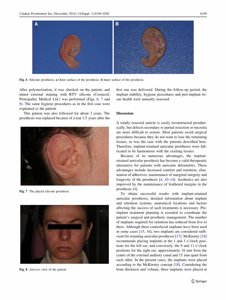
After polymerisation, it was checked on the patient, and
minor external staining with RTV silicone (Cosmesil;
Principality Medical Ltd.) was performed (Figs. 6, 7 and
8). The same hygiene procedures as in the first case were
explained to the patient.
This patient was also followed for about 3 years. The
prosthesis was replaced because of a tear 1.5 years after the
first one was delivered. During the follow-up period, the
implant stability, hygiene procedures and peri-implant tis-
sue health were annually assessed.
Discussion
A totally resected auricle is easily reconstructed prosthet-
ically, but defects secondary to partial resection or microtia
are more difficult to restore. Most patients avoid surgical
procedures because they do not want to lose the remaining
tissues, as was the case with the patients described here.
Therefore, implant-retained auricular prostheses were fab-
ricated to be harmonious with the existing tissues.
Because of its numerous advantages, the implant-
retained auricular prosthesis has become a valid therapeutic
alternative for patients with auricular deformities. These
advantages include increased comfort and retention, elim-
ination of adhesives, maintenance of marginal integrity and
longevity of the prosthesis [4, 10–14]. Aesthetics are also
improved by the maintenance of feathered margins in the
prosthesis [4].
To obtain successful results with implant-retained
auricular prostheses, detailed information about implant
and retention systems, anatomical locations and factors
affecting the success of such treatments is necessary. Pre-
implant treatment planning is essential to coordinate the
patient’s surgical and prosthetic management. The number
of implants required for retention has reduced from five to
three. Although three craniofacial implants have been used
in some cases [15, 16], two implants are considered suffi-
cient for retaining auricular prostheses [17]. McKinstry [18]
recommends placing implants at the 1 and 3 o’clock posi-
tions for the left ear, and conversely, the 9 and 11 o’clock
positions for the right ear, approximately 18 mm from the
centre of the external auditory canal and 15 mm apart from
each other. In the present cases, the implants were placed
according to the McKinstry concept [18]. Considering the
bone thickness and volume, three implants were placed at
Fig. 6 Silicone prosthesis. a Outer surface of the prosthesis. b Inner surface of the prosthesis
Fig. 7 The placed silicone prosthesis
Fig. 8 Anterior view of the patient
J Indian Prosthodont Soc (December 2014) 14(Suppl. 1):S196–S201 S199
123
the 9, 10 and 11 o’clock positions in the mastoid region of
patient 1 and two implants were placed at the 9 and 11
o’clock positions in the mastoid region of patient 2.
Patients with auricular prostheses that are fixed to the
abutments of osseointegrated implants may encounter
some problems, especially during bathing and sleeping [7].
Therefore, these prostheses have removable external parts.
Numerous attachments can be used for implant-retained
prostheses. A bar with clips or a magnetic attachment is
usually used as the main retainer [7, 9, 11–13, 19, 20].
These attachments are used to connect the removable parts
to the implants [7, 9, 20] and aid in the proper placement of
the prosthesis by both the patient and the dentist.
The morphology of the prosthesis is currently repro-
duced by one of the following methods: (1) using a pre-
surgical cast; (2) obtaining a cast of the patient’s normal
external ear by taking a direct impression and sculpting a
mirror-image wax pattern for the missing one; (3) obtaining
a cast of an external ear with compatible morphology and
using it to create the prosthesis or (4) making a wax cast of
the normal external ear, sectioning it into 1 mm slices and
reversing the sections to create a mirror-image wax pattern
[21–23]. In the cases described here, the morphology of the
auricular prostheses was reproduced by sculpting mirror-
image wax patterns for the missing tissues.
Silicones have been used for many years in the field of
maxillofacial prosthetics, because of desirable material
properties such as aesthetics, flexibility, biocompatibility,
ability to accept intrinsic and extrinsic colourants, trans-
lucency, chemical and physical inertness, mouldability,
softness and ease of cleaning [24, 25]. The borders of
prostheses fabricated from these materials were once very
evident; however, with the silicones currently available,
micron-level borders can be produced. Therefore, the
implant-retained auricular prostheses of the patients were
constructed by using HTV silicone.
Conclusions
By using the implant-retained auricular prostheses, the
patients with right partial external-ear defects experienced
improved retention, aesthetics, hearing and quality of life.
In addition, the remaining tissues were protected. During
the follow-up period, although both the prostheses were re-
fabricated once, implant stability, peri-implant soft tissue
health and hygiene procedures were found to be good.
References
1. Henry PJ (1992) Maxillofacial prosthetic considerations. In:
Worthington P, Branemark PI (eds) Advanced osseointegration
surgery: applications in the maxillofacial region, 1st edn. Quin-
tessence, Chicago, pp 313–326
2. Ciorba A, Martini A (2005) Tissue engineering and cartilage
regeneration for auricular reconstruction. Int J Pediatr Otorhino-
laryngol 70:1507–1515. doi:10.1016/j.ijporl.2006.03.013
3. Cognetti DM, Weber RS, Lai SY (2008) Head and neck cancer:
an evolving treatment paradigm. Cancer 113:1911–1932. doi:
10.1002/cncr.23654
4. Hooper SM, Westcott T, Evans PL, Bocca AP et al (2005)
Implant-supported facial prostheses provided by a maxillofacial
unit in a UK regional hospital: longevity and patient opinions.
J Prosthodont 14:32–38. doi:10.1111/j.1532-849X.2005.00004.x
5. Brent B (1981) A personal approach to total auricular construc-
tion: case study. Clin Plast Surg 8:211–221
6. Holgers KM, Tjellstrom A, Bjursten LM, Erlandsson BE (1987)
Soft tissue reactions around percutaneous implants: a clinical
study on skin-penetrating titanium implants used for bone-
anchored auricular prostheses. Int J Oral Maxillofac Implants
2:35–39
7. Tjellstrom A, Yountchev E, Lindstrom J, Branemark PI (1985)
Five years’ experience with bone-anchored auricular prostheses.
Otolaryngol Head Neck Surg 93:366–372
8. Nishimura RD, Roumanas E, Sugai T, Moy PK (1995) Auricular
prostheses and osseointegrated implants: UCLA experience.
J Prosthet Dent 73:553–558. doi:10.1016/S0022-3913(05)80115-5
9. Wazen JJ, Wright R, Hatfield RB, Asher ES (1999) Auricular
rehabilitation with bone-anchored titanium implants. Laryngo-
scope 109:523–527. doi:10.1097/00005537-199904000-00001
10. Tjellstrom A (1990) Osseointegrated implants for replacement of
absent or defective ears. Clin Plast Surg 17:355–366
11. Wang RR, Andres CJ (1999) Hemifacial microsomia and treatment
options for auricular replacement: a review of the literature.
J Prosthet Dent 82:197–204. doi:10.1016/S0022-3913(99)70156-3
12. Wright RF, Wazen JJ, Asher ES, Evans JH (1999) Multidisci-
plinary treatment for an implant retained auricular prosthesis
rehabilitation. N Y State Dent J 65:26–31
13. Lemon JC, Chambers MS (2002) Locking retentive attachment
for an implant-retained auricular prosthesis. J Prosthet Dent
87:336–338. doi:10.1067/mpr.2002.122016
14. Raghoebar GM, Van Oort RP, Roodenburg JL, Reintsema H et al
(1994) Fixation of auricular prostheses by osseointegrated implants.
J Invest Surg 7:283–290. doi:10.3109/08941939409051146
15. Ciocca L, Mingucci R, Gassino G, Scotti R (2007) CAD/CAM
ear model and virtual construction of the mold. J Prosthet Dent
98:339–343. doi:10.1016/S0022-3913(07)60116-4
16. Aydin C, Karakoca S, Yilmaz H, Yilmaz C (2008) Implant-
retained auricular prosthesis: an assessment of implant success
and prosthetic complications. Int J Prosthodont 21:241–244
17. Alberktsson T (1998) Surgical and prosthetic considerations. In:
Branemark PI, Tolman DE (eds) Osseointegration in craniofacial
reconstruction, 1st edn. Illinosis, Quintessence, pp 213–223
18. McKinstry RE (1995) Fundamentals of Facial Prosthetics. ABI
Professional Publications, Arlington, pp 25–29
19. Cheng AC, Morrison D, Cho RS, Archibald D (1998) Vacuum-
formed matrix as a guide for the fabrication of craniofacial
implant tissue bar-retained auricular prostheses. J Prosthet Dent
79:711–714. doi:10.1016/S0022-3913(98)70081-2
20. McCartney JW (1991) Osseointegrated implant-supported and
magnetically retained ear prosthesis: a clinical report. J Prosthet
Dent 66:6–9. doi:10.1016/0022-3913(91)90342-T
21. Andres CJ, Haug SP (2000) Facial prosthesis fabrication: tech-
nical aspects. In: Taylor TD (ed) Clinical maxillofacial pros-
thetics, 1st edn. Quintessence, Chicago, pp 237–240
22. Nusinov NS, Gay WD (1980) A method for obtaining the reverse
image of an ear. J Prosthet Dent 44:68–71. doi:10.1016/0022-
3913(80)90049-9
S200 J Indian Prosthodont Soc (December 2014) 14(Suppl. 1):S196–S201
123

23. Lemon JC, Chambers MS, Wesley PJ, Martin JW (1996) Tech-
nique for fabricating a mirror-image prosthetic ear. J Prosthet
Dent 75:292–293. doi:10.1016/S0022-3913(96)90487-4
24. Kiat-amnuay S, Johnston DA, Powers JM, Jacob RF (2005) Color
stability of dry earth pigmented maxillofacial silicone A-2186
subjected to microwave energy exposure. J Prosthodont
14:91–96. doi:10.1111/j.1532-849X.2005.00017.x
25. Jani RM, Schaaf NG (1978) An evaluation of facial prostheses.
J Prosthet Dent 39:546–550. doi:10.1016/S0022-3913(78)80191-7
J Indian Prosthodont Soc (December 2014) 14(Suppl. 1):S196–S201 S201
123