Prosthetic rehabilitation of a partially amputated finger ...
4
82 © 2018 The Journal of Indian Prosthodontic Society | Published by Wolters Kluwer - Medknow Prosthetic rehabilitation of a partially amputated finger using a customized ring‑wire substructure Siddharth Mehta, B. Leela, Abha Karanjkar 1 , Arpit J. Halani Departments of Prosthodoncs and 1 Periodoncs, Bapuji Dental College and Hospital, Davangere, Karnataka, India Case Report INTRODUCTION It is rightly said, “we never realize how important some things are until we lose it.” Moreover, the realization is hard‑hitting when that something turns out to be a body part, limb, or a digit. According to the information from National Centre for Health Statistics, trauma alone accounts for >75% of the reason for limb amputation. [1] It was also reported that the most common among such amputations is partial hand amputation with loss of one or more fingers. [1] Hand injuries count for a 1/3 of all injuries at work, 1/3 of chronic injuries, 1/4 of lost working time, and 1/5 of permanent disability. [1] Apart from these statistics, the physical and the psychological trauma endured makes the individual have a lower self‑esteem and less social interaction. Silicone prosthesis is the second best option available for rehabilitating such defects, the first one being surgical reconstruction. Ideal finger prosthesis should be lifelike with realistic surface detailing, closest skin shade matching, thin margins merging with the skin, incorporation of characterization like fingernails, and reproducing wrinkles and pigmentations. [2,3] The anatomy of the residual stump is of extreme importance and will dictate the mode of retention to be used, and the level of esthetics one can expect. An ideal residual stump should have adequate length, compressibility, and contours that will provide excellent esthetics and good retention. [4] However, this may not be encountered in all cases that we come across. This case report describes a cost‑effective and simple approach of rehabilitation of a partially amputated Defects of fingers or hands due to congenital reasons or trauma can be a catastrophic setback to an individual physically, emotionally, and psychologically. An artificial finger prosthesis is a lucrative option to camouflage such defects. The anatomy of the residual stump of the defect is of extreme importance and will dictate the mode of retention to be used, and the level of esthetics one can expect. Despite the availability of the advanced skills, best of materials, and laboratory support, sometimes, the anatomy of the defect may be a hindrance in furnishing a better prosthesis. This case report describes a cost-effective and simple approach of rehabilitation of a partially amputated finger with bulbous distal anatomy using a custom-made ring-wire substructure and maxillofacial silicone, thereby striking a balance between adequate retention and optimal esthetics. Keywords: Finger prosthesis, ring-wire substructure, silicone prosthesis Abstract Address for correspondence: Dr. Siddharth Mehta, Door No 538, Behind Nutan College, LIC Colony, Davangere ‑ 577 004, Karnataka, India. E-mail: [email protected] Received: 21 st August, 2017, Accepted: 26 th December, 2017 Access this article online Quick Response Code: Website: www.j-ips.org DOI: 10.4103/jips.jips_221_17 How to cite this article: Mehta S, Leela B, Karanjkar A, Halani AJ. Prosthetic rehabilitation of a partially amputated finger using a customized ring-wire substructure. J Indian Prosthodont Soc 2018;18:82-85. This is an open access arcle distributed under the terms of the Creave Commons Aribuon‑NonCommercial‑ShareAlike 3.0 License, which allows others to remix, tweak, and build upon the work non‑commercially, as long as the author is credited and the new creaons are licensed under the idencal terms. For reprints contact: [email protected] [Downloaded free from http://www.j-ips.org on Thursday, November 1, 2018, IP: 183.82.145.117]
Transcript of Prosthetic rehabilitation of a partially amputated finger ...
82 © 2018 The Journal of Indian Prosthodontic Society | Published by Wolters Kluwer - Medknow
Prosthetic rehabilitation of a partially amputated finger using a customized ring‑wire substructure
Siddharth Mehta, B. Leela, Abha Karanjkar1, Arpit J. HalaniDepartments of Prosthodontics and 1Periodontics, Bapuji Dental College and Hospital, Davangere, Karnataka, India
Case Report
INTRODUCTION
It is rightly said, “we never realize how important some things are until we lose it.” Moreover, the realization is hard‑hitting when that something turns out to be a body part, limb, or a digit. According to the information from National Centre for Health Statistics, trauma alone accounts for >75% of the reason for limb amputation.[1] It was also reported that the most common among such amputations is partial hand amputation with loss of one or more fingers.[1] Hand injuries count for a 1/3 of all injuries at work, 1/3 of chronic injuries, 1/4 of lost working time, and 1/5 of permanent disability.[1] Apart from these statistics, the physical and the psychological trauma endured makes the individual have a lower self‑esteem and less social interaction.
Silicone prosthesis is the second best option available for rehabilitating such defects, the first one being surgical reconstruction. Ideal finger prosthesis should be lifelike with realistic surface detailing, closest skin shade matching, thin margins merging with the skin, incorporation of characterization like fingernails, and reproducing wrinkles and pigmentations.[2,3] The anatomy of the residual stump is of extreme importance and will dictate the mode of retention to be used, and the level of esthetics one can expect. An ideal residual stump should have adequate length, compressibility, and contours that will provide excellent esthetics and good retention.[4] However, this may not be encountered in all cases that we come across.
This case report describes a cost‑effective and simple approach of rehabilitation of a partially amputated
Defects of fingers or hands due to congenital reasons or trauma can be a catastrophic setback to an individual physically, emotionally, and psychologically. An artificial finger prosthesis is a lucrative option to camouflage such defects. The anatomy of the residual stump of the defect is of extreme importance and will dictate the mode of retention to be used, and the level of esthetics one can expect. Despite the availability of the advanced skills, best of materials, and laboratory support, sometimes, the anatomy of the defect may be a hindrance in furnishing a better prosthesis. This case report describes a cost-effective and simple approach of rehabilitation of a partially amputated finger with bulbous distal anatomy using a custom-made ring-wire substructure and maxillofacial silicone, thereby striking a balance between adequate retention and optimal esthetics.
Keywords: Finger prosthesis, ring-wire substructure, silicone prosthesis
Abstract
Address for correspondence: Dr. Siddharth Mehta, Door No 538, Behind Nutan College, LIC Colony, Davangere ‑ 577 004, Karnataka, India. E-mail: [email protected] Received: 21st August, 2017, Accepted: 26th December, 2017
Access this article onlineQuick Response Code:
Website:
www.j-ips.org
DOI:
10.4103/jips.jips_221_17How to cite this article: Mehta S, Leela B, Karanjkar A, Halani AJ. Prosthetic rehabilitation of a partially amputated finger using a customized ring-wire substructure. J Indian Prosthodont Soc 2018;18:82-85.
This is an open access article distributed under the terms of the Creative Commons Attribution‑NonCommercial‑ShareAlike 3.0 License, which allows others to remix, tweak, and build upon the work non‑commercially, as long as the author is credited and the new creations are licensed under the identical terms.
For reprints contact: [email protected]
[Downloaded free from http://www.j-ips.org on Thursday, November 1, 2018, IP: 183.82.145.117]
Mehta, et al.: Finger prosthesis using custom‑made ring‑wire substructure
The Journal of Indian Prosthodontic Society | Volume 18 | Issue 1 | January-March 2018 83
finger with bulbous distal anatomy using a custom‑made ring‑wire substructure and maxillofacial silicone, thereby striking a balance between adequate retention and optimal esthetics.
CASE REPORT
A 12‑year‑old girl came with her mother to the Department of Prosthodontics with a chief complaint of amputated index finger of the right hand. The mother gave a history of her daughter putting her finger into a running motor when she was just 3 years old. It was also noted that the patient was very eager to get the amputated finger rehabilitated due to the constant bullying and ridiculing from her peers.
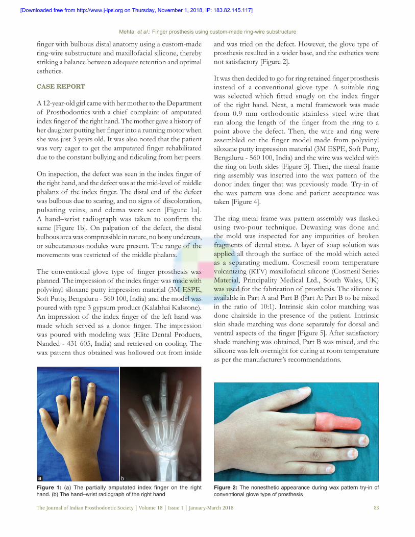
On inspection, the defect was seen in the index finger of the right hand, and the defect was at the mid‑level of middle phalanx of the index finger. The distal end of the defect was bulbous due to scaring, and no signs of discoloration, pulsating veins, and edema were seen [Figure 1a]. A hand–wrist radiograph was taken to confirm the same [Figure 1b]. On palpation of the defect, the distal bulbous area was compressible in nature, no bony undercuts, or subcutaneous nodules were present. The range of the movements was restricted of the middle phalanx.
The conventional glove type of finger prosthesis was planned. The impression of the index finger was made with polyvinyl siloxane putty impression material (3M ESPE, Soft Putty, Bengaluru ‑ 560 100, India) and the model was poured with type 3 gypsum product (Kalabhai Kalstone). An impression of the index finger of the left hand was made which served as a donor finger. The impression was poured with modeling wax (Elite Dental Products, Nanded ‑ 431 605, India) and retrieved on cooling. The wax pattern thus obtained was hollowed out from inside
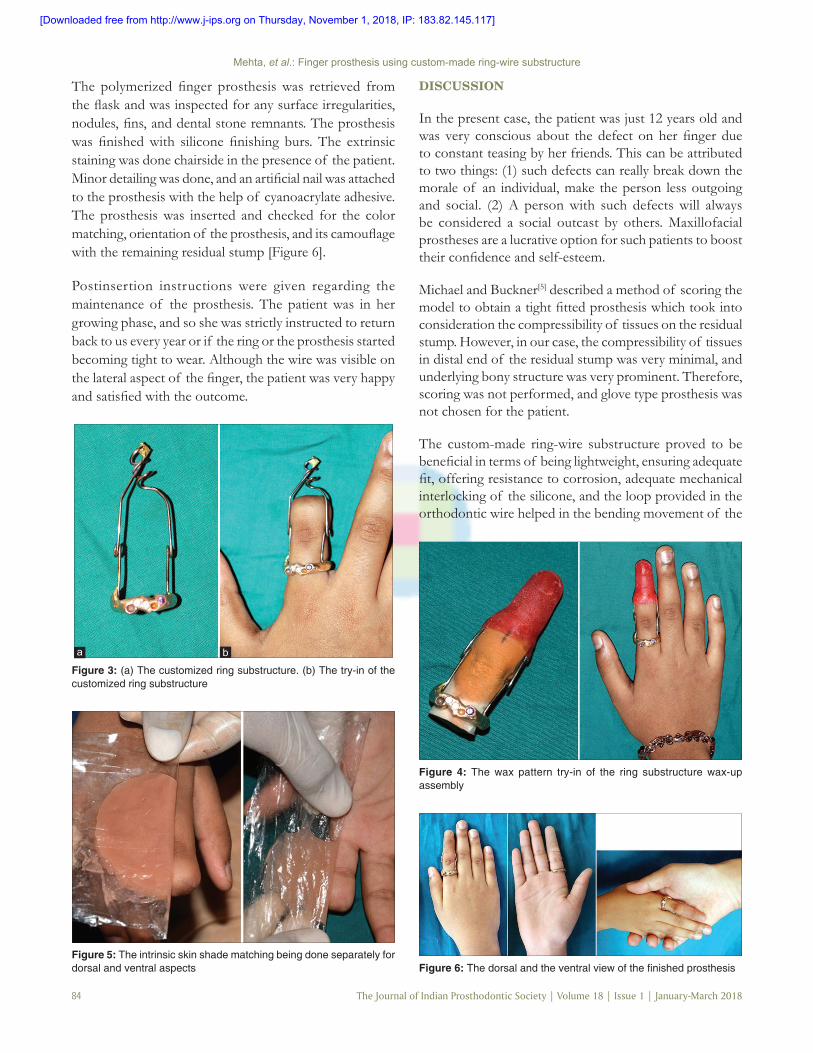
and was tried on the defect. However, the glove type of prosthesis resulted in a wider base, and the esthetics were not satisfactory [Figure 2].
It was then decided to go for ring retained finger prosthesis instead of a conventional glove type. A suitable ring was selected which fitted snugly on the index finger of the right hand. Next, a metal framework was made from 0.9 mm orthodontic stainless steel wire that ran along the length of the finger from the ring to a point above the defect. Then, the wire and ring were assembled on the finger model made from polyvinyl siloxane putty impression material (3M ESPE, Soft Putty, Bengaluru ‑ 560 100, India) and the wire was welded with the ring on both sides [Figure 3]. Then, the metal frame ring assembly was inserted into the wax pattern of the donor index finger that was previously made. Try‑in of the wax pattern was done and patient acceptance was taken [Figure 4].
The ring metal frame wax pattern assembly was flasked using two‑pour technique. Dewaxing was done and the mold was inspected for any impurities of broken fragments of dental stone. A layer of soap solution was applied all through the surface of the mold which acted as a separating medium. Cosmesil room temperature vulcanizing (RTV) maxillofacial silicone (Cosmesil Series Material, Principality Medical Ltd., South Wales, UK) was used for the fabrication of prosthesis. The silicone is available in Part A and Part B (Part A: Part B to be mixed in the ratio of 10:1). Intrinsic skin color matching was done chairside in the presence of the patient. Intrinsic skin shade matching was done separately for dorsal and ventral aspects of the finger [Figure 5]. After satisfactory shade matching was obtained, Part B was mixed, and the silicone was left overnight for curing at room temperature as per the manufacturer’s recommendations.
Figure 1: (a) The partially amputated index finger on the right hand. (b) The hand–wrist radiograph of the right hand
Figure 2: The nonesthetic appearance during wax pattern try-in of conventional glove type of prosthesis
ba
[Downloaded free from http://www.j-ips.org on Thursday, November 1, 2018, IP: 183.82.145.117]
Mehta, et al.: Finger prosthesis using custom‑made ring‑wire substructure
84 The Journal of Indian Prosthodontic Society | Volume 18 | Issue 1 | January-March 2018
The polymerized finger prosthesis was retrieved from the flask and was inspected for any surface irregularities, nodules, fins, and dental stone remnants. The prosthesis was finished with silicone finishing burs. The extrinsic staining was done chairside in the presence of the patient. Minor detailing was done, and an artificial nail was attached to the prosthesis with the help of cyanoacrylate adhesive. The prosthesis was inserted and checked for the color matching, orientation of the prosthesis, and its camouflage with the remaining residual stump [Figure 6].
Postinsertion instructions were given regarding the maintenance of the prosthesis. The patient was in her growing phase, and so she was strictly instructed to return back to us every year or if the ring or the prosthesis started becoming tight to wear. Although the wire was visible on the lateral aspect of the finger, the patient was very happy and satisfied with the outcome.
DISCUSSION
In the present case, the patient was just 12 years old and was very conscious about the defect on her finger due to constant teasing by her friends. This can be attributed to two things: (1) such defects can really break down the morale of an individual, make the person less outgoing and social. (2) A person with such defects will always be considered a social outcast by others. Maxillofacial prostheses are a lucrative option for such patients to boost their confidence and self‑esteem.
Michael and Buckner[5] described a method of scoring the model to obtain a tight fitted prosthesis which took into consideration the compressibility of tissues on the residual stump. However, in our case, the compressibility of tissues in distal end of the residual stump was very minimal, and underlying bony structure was very prominent. Therefore, scoring was not performed, and glove type prosthesis was not chosen for the patient.
The custom‑made ring‑wire substructure proved to be beneficial in terms of being lightweight, ensuring adequate fit, offering resistance to corrosion, adequate mechanical interlocking of the silicone, and the loop provided in the orthodontic wire helped in the bending movement of the
Figure 3: (a) The customized ring substructure. (b) The try-in of the customized ring substructure
Figure 4: The wax pattern try-in of the ring substructure wax-up assembly
Figure 5: The intrinsic skin shade matching being done separately for dorsal and ventral aspects Figure 6: The dorsal and the ventral view of the finished prosthesis
ba
[Downloaded free from http://www.j-ips.org on Thursday, November 1, 2018, IP: 183.82.145.117]
Mehta, et al.: Finger prosthesis using custom‑made ring‑wire substructure
The Journal of Indian Prosthodontic Society | Volume 18 | Issue 1 | January-March 2018 85
finger. A similar technique where custom‑made retentive aids were fabricated was followed by Saxena et al.[6] and Ahmad et al.[7]
Cosmesil brand medical grade silicone was used for the fabrication of the prosthesis. It has high flexibility, tear strength, edge strength, and color stability and remains dimensionally stable for a longer period of time. Cosmesil contains condensation RTV silicone and polymerizes at room temperature without the requirement of any special equipment for processing.[8,9]
The main problem with this case was that the patient was in her growing phase. This was an important reason why implant‑supported prosthesis was not considered in this case. It could hamper the growth of the finger since the patient was only 12 years old. Strict instructions were given to the patient to come for recall checkup every year. The patient was also instructed to discontinue the use of the finger prosthesis if it becomes tight or difficult to insert or remove.
CONCLUSION
It is the God‑given right of every human to appear human. Rehabilitating the defects of the hand or limbs with an artificial prosthesis can be rewarding and satisfying for a maxillofacial prosthodontist. Despite the availability of the advanced skills, best of materials, and laboratory support, sometimes, the anatomy of the defect may be a hindrance in furnishing a better prosthesis. The finger prosthesis with custom‑made ring‑wire substructure was functionally adequate and esthetically acceptable by the patient. Even though there was a display of the wire on the lateral aspect of the index finger, the patient was highly satisfied with the appearance of the prosthesis.
Declaration of patient consentThe authors certify that they have obtained all appropriate patient consent forms. In the form the patient(s) has/have given his/her/their consent for his/her/their images and other clinical information to be reported in the journal. The patients understand that their names and initials will not be published and due efforts will be made to conceal their identity, but anonymity cannot be guaranteed.
Financial support and sponsorshipNil.
Conflicts of interestThere are no conflicts of interest.
REFERENCES
1. Marty J, Porcher B, Autissier R. Hand injuries and occupational accidents. Statistics and prevention. Ann Chir Main 1983;2:368‑70.
2. Pattanaik B, Pattanaik S. Fabrication of a functional finger prosthesis with simple attachment. J Indian Prosthodont Soc 2013;13:631‑4.
3. Tripathi S, Singh RD, Chand P, Mishra N, Yadav LK, Singh SV, et al. A modified approach of impression technique for fabrication of finger prostheses. Prosthet Orthot Int 2012;36:121‑4.
4. Ozkan A, Senel B, Durmaz CE, Uyar HA, Evinc R. Use of dental implants to retain finger prostheses: A case report. Oral Health Dent Manag 2012;11:11‑5.
5. Michael JW, Buckner H. Options for finger prosthesis. J Prosthet Orthot 1994;6:10‑9.
6. Saxena K, Sharma A, Hussain MA, Thombare RU, Bhasin SS. A hollow silicone finger prosthesis with modified metal‑mesh conformer. J Indian Prosthodont Soc 2014;14:301‑4.
7. Ahmad M, Balakrishnan D, Narayan A, Naim H. Comprehensive rehabilitation of partially amputated index finger with silicone prosthesis: A Case report with 3 years of follow up. J Indian Prosthodont Soc 2014;14:222‑6.
8. Hatamleh MM, Watts DC. Mechanical properties and bonding of maxillofacial silicone elastomers. Dent Mater 2010;26:185‑91.
9. Leow ME, Pho RW. RTV silicone elastomers in hand prosthetics: Properties, applications and techniques. Prosthet Orthot Int 1999;23:169‑73.
[Downloaded free from http://www.j-ips.org on Thursday, November 1, 2018, IP: 183.82.145.117]