Prosthetic ball free in the left ventricle in a patient ...
3
Prosthetic ball free in the left ventricle in a patient with an aortic Starr-Edwards prosthesis Daniel D. Schräder, MD Miguel A. Sellanes, MD Norberto Kelijman Laschover, MD Roberto J. Macchi, MD, FACC Marcos Eserequis, MD Jorge I. Mizraji, MD Carlos A. Nojek, MD Ball-valve intermittent poppet dislodgment into the left ventricle is a rare complication, having been re- ported previously only in patients with the Braunwald- Cutter prosthesis. This type of variance is reported in a patient with an aortic Starr-Edwards prosthesis and minimal symptoms. Index term: Heart valve prosthesiss Cleve Clin J Med 1988; 55:175-177 Several cases have been reported with pros- thetic valve dysfunction including dislodgment of the ball from the cage and sudden death. We here describe a patient with intermittent dislodg- ment of the ball of the Starr-Edwards valve into the ventricle with no hemodynamic jeopardy and minimal symptoms. Case report This 51-year-old patient was transferred to our hospital (Hospital Español de Buenos Aires) on March 26, 1984, for surgical treatment of prosthetic valve dysfunction. In 1972, he had bacterial endocarditis, and, in 1973, he underwent aortic valve replacement for severe aortic regurgitation. A Starr-Edwards prosthesis was implanted. The patient had an uncomplicated postoperative course and was very Cardiology Institute, Hospital Espanol de Buenos Aires (D.D. S., M.A.S., N.K.L., R.J.M., M.E., J.I.M.) and Cardiology Depart- ment, Policlinico Ferroviario Central (C.A.N.), Buenos Aires, Ar- gentina. Submitted Dec. 1986; accepted Oct.1987. active until two months before this admission, when he noticed a change in the prosthetic sounds, as well as palpi- tations. He had no other associated symptoms. Physical findings Physical examination revealed a well-built man in no acute distress. The blood pressure was 150/90 mm Hg, the pulse was regular at 70 beats per min. Examination of the neck showed no jugular venous distension. The lungs were clear. Cardiovascular examination revealed a point of max- imal impulse at the fifth intercostal space and an intermittent pounding that could be felt 2 cm lateral from the midcla- vicular line. The first heart sound was normal, and the opening click of the aortic prosthesis also sounded normal, except for intermittent beats in which it could not be heard; after these pauses its intensity increased. The second heart sound had a normal pulmonary component and a normal- sounding closing click, which disappeared intermittently, as did the opening click. There was a 2/6 systolic ejection murmur in aortic area, as well as an intermittent 3/6 dia- stolic blowing murmur, which coincided with the pulse and point of maximal impulse findings (Fig. 1). Laboratory findings The electrocardiogram revealed a sinus rhythm and non- diagnostic STT changes. The chest radiograph showed mild cardiomegaly and clear lung fields. Blood studies were within normal limits except for prolonged prothrombin time, due to anticoagulants. The echocardiogram revealed mildly dilated left atrium. The left ventricle was moderately dilated, but with an increased percentage shortening. At the mitral level, we detected an intermittent fine tremor of the anterior mitral leaflet as well as an early closure. Also notice the ball artifact (Fig. 2). On the bidimensional echocardi- ogram, the prosthetic ball could be seen at the apex of the 175 on November 19, 2021. For personal use only. All other uses require permission. www.ccjm.org Downloaded from
Transcript of Prosthetic ball free in the left ventricle in a patient ...
Prosthetic ball free in the left ventricle in a patient with an aortic Starr-Edwards prosthesis Daniel D. Schräder, MD Miguel A. Sellanes, MD Norberto Kelijman Laschover, MD Roberto J. Macchi, MD, FACC Marcos Eserequis, MD Jorge I. Mizraji, MD Carlos A. Nojek, MD
Ball-valve intermittent poppet dislodgment into the left ventricle is a rare complication, having been re-ported previously only in patients with the Braunwald-Cutter prosthesis. This type of variance is reported in a patient with an aortic Starr-Edwards prosthesis and minimal symptoms.
Index term: Heart valve prosthesiss
Cleve Clin J Med 1988; 55:175-177
Several cases have been reported with pros-thetic valve dysfunction including dislodgment of the ball from the cage and sudden death. We here describe a patient with intermittent dislodg-ment of the ball of the Starr-Edwards valve into the ventricle with no hemodynamic jeopardy and minimal symptoms.
Case report This 51-year-old patient was transferred to our hospital
(Hospital Español de Buenos Aires) on March 26, 1984, for surgical treatment of prosthetic valve dysfunction. In 1972, he had bacterial endocarditis, and, in 1973, he underwent aortic valve replacement for severe aortic regurgitation.
A Starr-Edwards prosthesis was implanted. The patient had an uncomplicated postoperative course and was very
Cardiology Institute, Hospital Espanol de Buenos Aires (D.D. S., M.A.S., N.K.L., R.J.M., M.E., J.I.M.) and Cardiology Depart-ment, Policlinico Ferroviario Central (C.A.N.), Buenos Aires, Ar-gentina. Submitted Dec. 1986; accepted Oct.1987.
active until two months before this admission, when he noticed a change in the prosthetic sounds, as well as palpi-tations. He had no other associated symptoms.
Physical findings Physical examination revealed a well-built man in no
acute distress. The blood pressure was 150/90 mm Hg, the pulse was regular at 70 beats per min. Examination of the neck showed no jugular venous distension. The lungs were clear. Cardiovascular examination revealed a point of max-imal impulse at the fifth intercostal space and an intermittent pounding that could be felt 2 cm lateral from the midcla-vicular line. The first heart sound was normal, and the opening click of the aortic prosthesis also sounded normal, except for intermittent beats in which it could not be heard; after these pauses its intensity increased. The second heart sound had a normal pulmonary component and a normal-sounding closing click, which disappeared intermittently, as did the opening click. There was a 2/6 systolic ejection murmur in aortic area, as well as an intermittent 3/6 dia-stolic blowing murmur, which coincided with the pulse and point of maximal impulse findings (Fig. 1).
Laboratory findings The electrocardiogram revealed a sinus rhythm and non-
diagnostic STT changes. The chest radiograph showed mild cardiomegaly and clear lung fields. Blood studies were within normal limits except for prolonged prothrombin time, due to anticoagulants. The echocardiogram revealed mildly dilated left atrium. The left ventricle was moderately dilated, but with an increased percentage shortening. At the mitral level, we detected an intermittent fine tremor of the anterior mitral leaflet as well as an early closure. Also notice the ball artifact (Fig. 2). On the bidimensional echocardi-ogram, the prosthetic ball could be seen at the apex of the
175
on November 19, 2021. For personal use only. All other uses require permission.www.ccjm.orgDownloaded from
176 Cleveland Clinic J o u r n a l of Medicine Vo i . 55, No . 2
Fig. 1. P h o n o c a r d i o g r a m in aor t ic a rea and carot id pulse trac-ing. T h e first beat shows a n o r m a l o p e n i n g click with no diastolic m u r m u r s ; the carot id pulse is no rmal . In the second beat , the open ing click is unchanged and the closing click is slightly wider , followed by a diastolic m u r m u r coincident with a d r o p (arrows) in the pulse d u r i n g diastole. T h e th i rd beat has a very diminished first hea r t sound , a midsystolic ball sound , a n d diastolic m u r m u r . BS = ball sound; CC = closing click; DM = diastolic m u r m u r ; MC = mitral closure; OC = o p e n i n g click; PHI—5 = p h o n o c a r d i o g r a m ; SI = first hea r t sound.
'•"".»•»v* "*WY". """./—̂A
Fig. 2. Mitral valve echoca rd iog ram. T h e first beat shows a no rma l mitral valve, fol lowed by t h r e e beats (pr in ted with arrows) with early c losure and diastolic t r e m o r of the an te r io r leaflet . T h e sixth beat shows a ball a r t i fac t in mid-diastole. BA — ball ar t i fact ; MV = mitral valve.
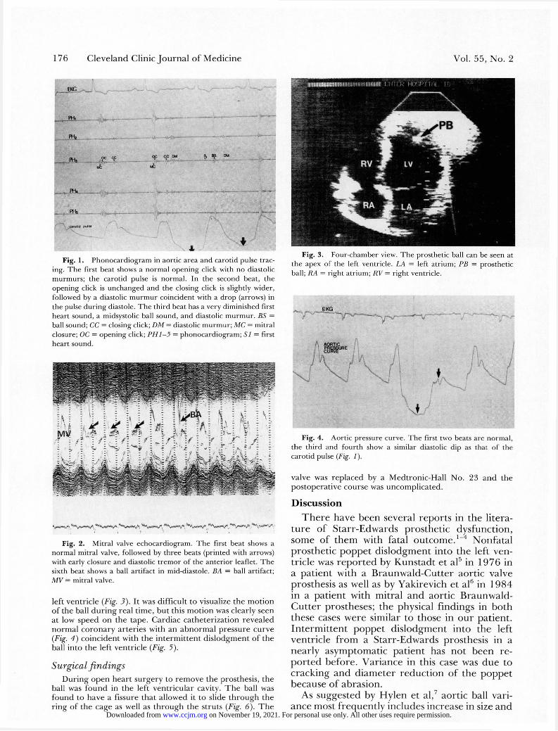
left ventricle (Fig. 3). It was difficult to visualize the motion of the ball during real time, but this motion was clearly seen at low speed on the tape. Cardiac catheterization revealed normal coronary arteries with an abnormal pressure curve (Fig. 4) coincident with the intermittent dislodgment of the ball into the left ventricle (Fig. 5).
Surgical findings During open heart surgery to remove the prosthesis, the
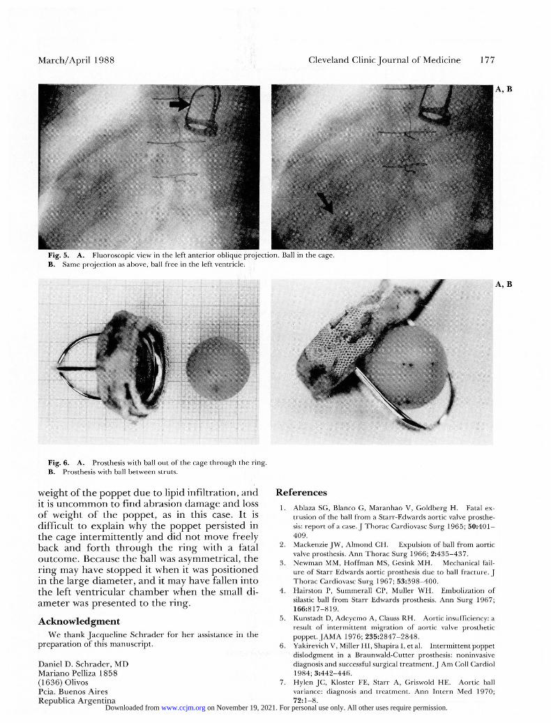
ball was found in the left ventricular cavity. The ball was found to have a fissure that allowed it to slide through the ring of the cage as well as through the struts (Fig. 6). The
Fig. 3. Fou r - chamber view. T h e prosthet ic ball can be seen at t h e apex of the left ventricle. LA = left a t r ium; PB = pros thet ic ball; RA = r ight a t r ium; RV = r ight ventricle.
Fig. 4. Aor t ic pressure curve. T h e first two beats a re n o r m a l , t h e th i rd and f o u r t h show a similar diastolic dip as that of the carot id pulse (Fig. 1).
valve was replaced by a Medtronic-Hall No. 23 and the postoperative course was uncomplicated.
Discussion T h e r e have been several reports in the litera-
tu re of Starr-Edwards prosthetic dysfunction, some of them with fatal outcome. 1 - 4 Nonfatal prosthetic poppet dislodgment into the left ven-tricle was repor ted by Kunstadt et al5 in 1976 in a patient with a Braunwald-Cutter aortic valve prosthesis as well as by Yakirevich et al6 in 1984 in a patient with mitral and aortic Braunwald-Cut ter prostheses; the physical findings in both these cases were similar to those in our patient. In termit tent poppet dislodgment into the left ventricle f rom a Starr-Edwards prosthesis in a nearly asymptomatic patient has not been re-por ted before. Variance in this case was due to cracking and diameter reduction of the poppet because of abrasion.
As suggested by Hylen et al,7 aortic ball vari-ance most frequently includes increase in size and
on November 19, 2021. For personal use only. All other uses require permission.www.ccjm.orgDownloaded from
M a r c h / Apri l 1988 Cleveland Clinic J o u r n a l of Medic ine 177
A, B
Fig. 5. A. Fluoroscopic view in the left an te r io r oblique project ion. Ball in the cage. B. Same projec t ion as above, ball f r ee in the left ventricle.
Fig. 6. A. Prosthesis with ball ou t of the cage th rough the r ing. B. Prosthesis with ball between struts.
weight of the poppet due to lipid infiltration, and it is uncommon to find abrasion damage and loss of weight of the poppet , as in this case. It is difficult to explain why the poppet persisted in the cage intermittently and did not move freely back and fo r th th rough the r ing with a fatal outcome. Because the ball was asymmetrical, the r ing may have stopped it when it was positioned in the large diameter , and it may have fallen into the left ventricular chamber when the small di-ameter was presented to the ring.
Acknowledgment We thank Jacqueline Schrader for her assistance in the
preparation of this manuscript.
Daniel D. Schrader, MD Mariano Pelliza 1858 (1636) Olivos Pcia. Buenos Aires Republica Argentina
References 1. Ablaza SG, Blanco G, Maranhao V, Goldberg H. Fatal ex-
trusion of the ball f r o m a Starr -Edwards aortic valve prosthe-sis: r epor t of a case. J T h o r a c Cardiovasc Surg 1965; 5 0 : 4 0 1 -409.
2. Mackenzie J W , A lmond CH. Expulsion of ball f r o m aort ic valve prosthesis. Ann T h o r a c Surg 1966; 2 : 4 3 5 - 4 3 7 .
3. N e w m a n MM, H o f f m a n MS, Gesink MH. Mechanical fail-u re of Starr Edwards aortic prosthesis d u e to ball f rac ture . J T h o r a c Cardiovasc Surg 1967; 5 3 : 3 9 8 - 4 0 0 .
4. Hairs ton P, Summeral l CP, Muller W H . Embolizat ion of silastic ball f r o m Starr Edwards prosthesis. Ann Surg 1967; 1 6 6 : 8 1 7 - 8 1 9 .
5. Kunstadt D, Adeyemo A, Clauss R H . Aortic insufficiency: a result of in termi t tent migrat ion of aortic valve prosthet ic poppet . J A M A 1976; 2 3 5 : 2 8 4 7 - 2 8 4 8 .
6. Yakirevich V, Miller HI , Shapira I, et al. In te rmi t ten t poppe t dis lodgment in a Braunwald-Cut ter prosthesis: noninvasive diagnosis and successful surgical t r ea tment . J Am Coll Cardiol 1984; 3 : 4 4 2 - 4 4 6 .
7. Hylen JC , Kloster FE, Starr A, Griswold HE. Aort ic ball variance: diagnosis and t rea tment . A n n Intern Med 1970; 72 :1 -8 .
on November 19, 2021. For personal use only. All other uses require permission.www.ccjm.orgDownloaded from