Prostaglandin pathway gene expression in human placenta, amnion ...
14
RESEARCH ARTICLE Open Access Prostaglandin pathway gene expression in human placenta, amnion and choriodecidua is differentially affected by preterm and term labour and by uterine inflammation Robert J Phillips 1 , Michel A Fortier 2 and Andrés López Bernal 1,3* Abstract Background: Elucidation of the biochemical pathways involved in activation of preterm and term human labour would facilitate the development of effective management and inform judgements regarding the necessity for preterm tocolysis and post-term induction. Prostaglandins act at all stages of human reproduction, and are potentially activators of labour. Methods: Expression of 15 genes involved in prostaglandin synthesis, transport and degradation was measured by qPCR using tissue samples from human placenta, amnion and choriodecidua at preterm and full-term vaginal and caesarean delivery. Cellular localisation of eight prostaglandin pathway proteins was determined by immunohistochemistry. Results: Expression of prostaglandin pathway genes was differentially affected by factors including gestational age at delivery, and the incidence and duration of labour. Chorioamnionitis/deciduitis was associated with upregulation of PTGS2 (prostaglandin-endoperoxide synthase 2 (prostaglandin G/H synthase and cyclooxygenase)), along with the inflammatory genes IL8 (interleukin 8), S100A8 (S100 calcium binding protein A8) and TLR2 (toll-like receptor 2), in amnion and choriodecidua, and with downregulation of CBR1 (carbonyl reductase 1) and HPGD (hydroxyprostaglandin dehydrogenase 15-(NAD)) in choriodecidua. Protein localisation differed greatly between the various maternal and fetal cell types. Conclusions: Preterm and term labour are associated with distinct prostaglandin pathway expression profiles; inflammation provokes specific changes, unrelated to the presence of labour; spontaneous and induced term labour are indistinguishable. Keywords: Parturition, Inflammation, Pregnancy, Uterus Background Human labour requires a dramatic transition from a state of uterine quiescence and immune tolerance of the fetus—that prevails throughout pregnancy—to a brief period of intense uterine activation involving connective tissue remodelling and coordinated smooth muscle ac- tivity. The signals that initiate this process are not yet known, but among the candidates are the prostaglandins, which are known regulators of many aspects of reproduct- ive physiology [1,2]. Evidence suggests that, during uterine activation there is positive feedback between prostaglan- dins and inflammatory cytokines that are released by infil- trating leukocytes [3]. Our early studies demonstrated that there is a relationship between inflammatory infiltration of the placenta, fetal membranes and decidua and increased prostaglandin and leukotriene release [4,5]. Inflammation has been associated with initiation of term and preterm labour both in the presence and absence of observable in- fection [6-12]. It is therefore possible that prostaglandins * Correspondence: [email protected] 1 Henry Wellcome Laboratories for Integrative Neuroscience and Endocrinology, School of Clinical Sciences, University of Bristol, Dorothy Hodgkin Building, Bristol BS1 3NY, UK 3 St Michael’s Hospital, Southwell Street, Bristol BS2 8EG, UK Full list of author information is available at the end of the article © 2014 Phillips et al.; licensee BioMed Central Ltd. This is an Open Access article distributed under the terms of the Creative Commons Attribution License (http://creativecommons.org/licenses/by/2.0), which permits unrestricted use, distribution, and reproduction in any medium, provided the original work is properly credited. The Creative Commons Public Domain Dedication waiver (http://creativecommons.org/publicdomain/zero/1.0/) applies to the data made available in this article, unless otherwise stated. Phillips et al. BMC Pregnancy and Childbirth 2014, 14:241 http://www.biomedcentral.com/1471-2393/14/241
Transcript of Prostaglandin pathway gene expression in human placenta, amnion ...
Phillips et al. BMC Pregnancy and Childbirth 2014, 14:241http://www.biomedcentral.com/1471-2393/14/241
RESEARCH ARTICLE Open Access
Prostaglandin pathway gene expression in humanplacenta, amnion and choriodecidua is differentiallyaffected by preterm and term labour and byuterine inflammationRobert J Phillips1, Michel A Fortier2 and Andrés López Bernal1,3*
Abstract
Background: Elucidation of the biochemical pathways involved in activation of preterm and term human labourwould facilitate the development of effective management and inform judgements regarding the necessity forpreterm tocolysis and post-term induction. Prostaglandins act at all stages of human reproduction, and arepotentially activators of labour.
Methods: Expression of 15 genes involved in prostaglandin synthesis, transport and degradation was measured byqPCR using tissue samples from human placenta, amnion and choriodecidua at preterm and full-term vaginaland caesarean delivery. Cellular localisation of eight prostaglandin pathway proteins was determined byimmunohistochemistry.
Results: Expression of prostaglandin pathway genes was differentially affected by factors including gestationalage at delivery, and the incidence and duration of labour. Chorioamnionitis/deciduitis was associated withupregulation of PTGS2 (prostaglandin-endoperoxide synthase 2 (prostaglandin G/H synthase and cyclooxygenase)),along with the inflammatory genes IL8 (interleukin 8), S100A8 (S100 calcium binding protein A8) and TLR2(toll-like receptor 2), in amnion and choriodecidua, and with downregulation of CBR1 (carbonyl reductase 1)and HPGD (hydroxyprostaglandin dehydrogenase 15-(NAD)) in choriodecidua. Protein localisation differedgreatly between the various maternal and fetal cell types.
Conclusions: Preterm and term labour are associated with distinct prostaglandin pathway expression profiles;inflammation provokes specific changes, unrelated to the presence of labour; spontaneous and induced termlabour are indistinguishable.
Keywords: Parturition, Inflammation, Pregnancy, Uterus
BackgroundHuman labour requires a dramatic transition from astate of uterine quiescence and immune tolerance of thefetus—that prevails throughout pregnancy—to a briefperiod of intense uterine activation involving connectivetissue remodelling and coordinated smooth muscle ac-tivity. The signals that initiate this process are not yet
* Correspondence: [email protected] Wellcome Laboratories for Integrative Neuroscience andEndocrinology, School of Clinical Sciences, University of Bristol, DorothyHodgkin Building, Bristol BS1 3NY, UK3St Michael’s Hospital, Southwell Street, Bristol BS2 8EG, UKFull list of author information is available at the end of the article
© 2014 Phillips et al.; licensee BioMed CentralCommons Attribution License (http://creativecreproduction in any medium, provided the orDedication waiver (http://creativecommons.orunless otherwise stated.
known, but among the candidates are the prostaglandins,which are known regulators of many aspects of reproduct-ive physiology [1,2]. Evidence suggests that, during uterineactivation there is positive feedback between prostaglan-dins and inflammatory cytokines that are released by infil-trating leukocytes [3]. Our early studies demonstrated thatthere is a relationship between inflammatory infiltration ofthe placenta, fetal membranes and decidua and increasedprostaglandin and leukotriene release [4,5]. Inflammationhas been associated with initiation of term and pretermlabour both in the presence and absence of observable in-fection [6-12]. It is therefore possible that prostaglandins
Ltd. This is an Open Access article distributed under the terms of the Creativeommons.org/licenses/by/2.0), which permits unrestricted use, distribution, andiginal work is properly credited. The Creative Commons Public Domaing/publicdomain/zero/1.0/) applies to the data made available in this article,
Phillips et al. BMC Pregnancy and Childbirth 2014, 14:241 Page 2 of 14http://www.biomedcentral.com/1471-2393/14/241
and inflammatory pathways are involved in uterine acti-vation. It is important to establish the interactions be-tween these pathways, both for women at risk of pretermbirth who may be treated with anti-inflammatory drugsand prostaglandin synthesis inhibitors, and for womenfacing post-term induction of labour involving prosta-glandin treatment.We previously compared the relative levels of expres-
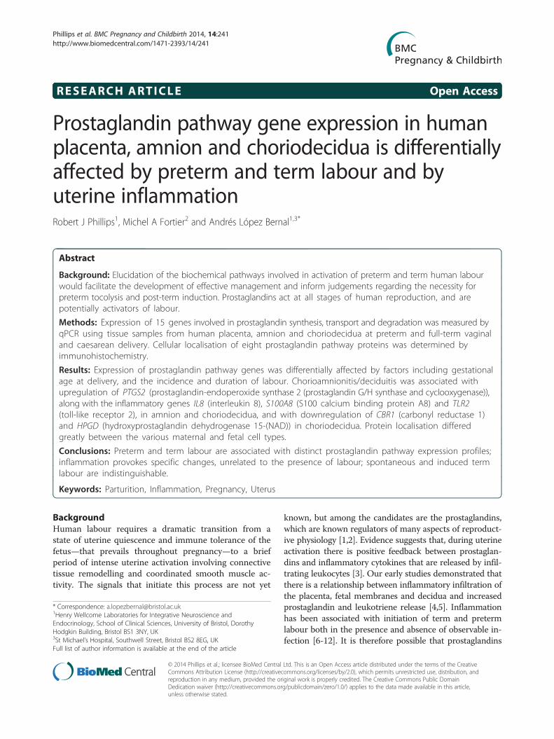
sion of 15 genes acting in all stages of prostaglandin me-tabolism (their relationships are illustrated in Figure 1) inhuman uterine tissues [13], demonstrating specific capaci-ties for synthesis and catabolism of PGD2, PGE2, PGF2and PGI2 in each tissue. We have now made a detailedexamination of these genes in samples of placenta, chorio-decidua and amnion, demonstrating that factors such asgestational age and the incidence and duration of labourare associated with significant changes in expression pat-terns. We have also characterised the distribution of pros-taglandin pathway proteins throughout the constituentcells of the uterus using immunohistochemistry.We have found distinct uterine prostaglandin gene ex-
pression and immunolocalisation in the presence of inflam-mation, suggesting uterine activation occurring through
Figure 1 Cellular pathways of prostaglandin (PG) metabolism. A cell isin precursor prostaglandin synthesis, terminal prostaglandin synthesis, prosarrows) and products (open circles).
increased PTGS2 expression in the fetal membranes anddecreased degradative HPGD in the choriodecidua. Ex-pression patterns in spontaneous preterm and term labourwithout inflammation differed from each other and fromthose with inflammatory changes. There were no differ-ences between spontaneous and induced labour at term.
MethodsCollection of tissueAll women gave written informed consent accordingto the requirements of the North Somerset and SouthBristol Research Ethics Committee. Placenta and gesta-tional membranes were collected immediately post-partumfrom the following groups of women: preterm (25–36weeks gestation) not-in-labour (PNIL), delivery by caesar-ean section for maternal or fetal complications; spon-taneous preterm labour (SPL), with vaginal delivery;term (≥ 37 weeks gestation) not-in-labour (TNIL), de-livery by elective caesarean section indicated by previoussection and/or breech presentation; spontaneous termlabour (STL), with vaginal delivery; term following induc-tion of labour (IOL) with intravaginal PGE2 pessary and/or intravenous oxytocin infusion, with delivery vaginally
depicted, showing enzymatic components (coloured boxes) involvedtaglandin transport and prostaglandin inactivation, with reactions (thin
Phillips et al. BMC Pregnancy and Childbirth 2014, 14:241 Page 3 of 14http://www.biomedcentral.com/1471-2393/14/241
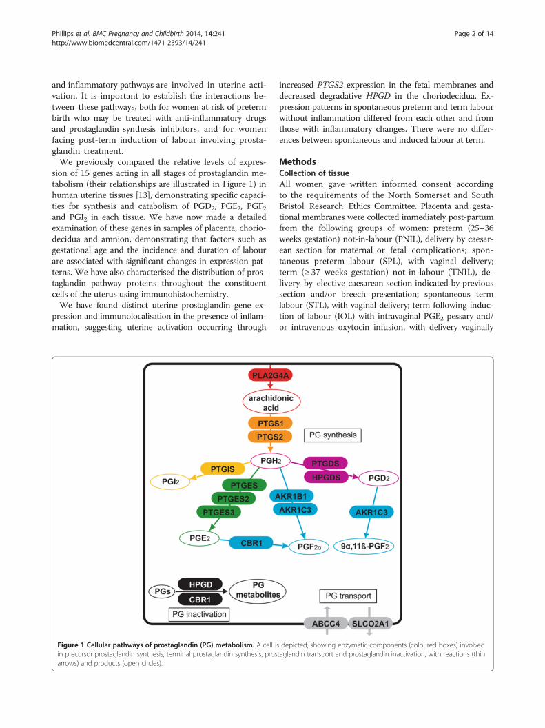
or by emergency caesarean section (failure to progress).The women were of mixed parity and all delivered live sin-gletons. None of the women in preterm labour receivedsteroid treatment. Tissues were also collected from a groupof women (INF) with evidence of inflammation, as sug-gested by clinical features of the women (pyrexia or uterinetenderness) and gross pathology of the delivered placentas,and confirmed histologically by the presence of leucocyteinfiltration in the fetal membranes (chorioamnionitis), de-cidua (deciduitis) or placenta (intervillositis), with or with-out maternal pyrexia or uterine tenderness [4]. Clinicalinformation for the women providing uterine tissues forthis study is given in Table 1. Tissues from 36 women wereused in this study; tissues from 31 of these women werepreviously among those used to study overall levels ofprostaglandin pathway gene expression in placenta andgestational membranes [13]. Myometrial tissues used inthe previous study were taken from a separate group ofwomen. Gestational membranes were dissected from be-tween 1 cm and 4 cm from the placental border. Placentaltissue was dissected from > 5 mm beneath the maternalsurface of the placenta. Tissue samples were dissected im-mediately after delivery (amnion and choriodecidua wereseparated by blunt dissection), washed in sterile phosphate-buffered saline (PBS), snap-frozen and stored in liquid ni-trogen. Tissues were also fixed and paraffin-embeddedfollowing standard procedures for immunohistochemistry.
Quantitative real-time PCR (qPCR)Total RNA was extracted from 100 mg tissue samples bythe guanidine isothiocyanate/phenol method using 1 mlTRIzol (Invitrogen, Carlsbad, CA, US), giving yields of10–150 μg. RNA was quantified using a GeneQuant IIspectrophotometer (GE Healthcare, Little Chalfont, UK).2 μg total RNA was used as a template for cDNA synthe-sis primed by random primers using the High CapacitycDNA Reverse Transcription Kit (Applied Biosystems,Foster City, CA, US). cDNA was diluted fourfold and 2 μlused as template for qPCR with the Power SYBR GreenPCR Master Mix (Applied Biosystems), with reaction
Table 1 Clinical information
Mode ofdelivery
Numberof women
Maternalage (years)
Gestational ageat birth (weeks)
Duration oflabour (hours)
Birt(kg)
PNIL 8 29 ± 9 33 ± 4 n/a 1.7 ±
SPL 5 27 ± 5 33 ± 1 4 2.0 ±
TNIL 7 31 ± 3 39 ± 2 n/a 4.0 ±
STL 6 31 ± 3 40 ± 1 4 3.6 ±
IOL 5 32 ± 9 40 ± 2 8 3.6 ±
INF 5 36 ± 7 32 ± 6 6 2.0 ±
Values are mean, mean ± standard deviation, or relative numbers in two groups. Abinduction of labour, PNIL preterm not-in-labour, SPL spontaneous preterm labour, Sterm not-in-labour.
volume of 20 μl, forward and reverse primer concentra-tions of 75 nM, and 45 cycles of 95 C for 15 s and 60 Cfor 60 s, followed by a dissociation stage, using a 7500Real-Time PCR System (Applied Biosystems). Two geneswith least Ct variability, POLR2A (polymerase (RNA) II(DNA directed) polypeptide A, 220 kDa) and ARHGDIA(Rho GDP dissociation inhibitor (GDI) alpha), werechosen from five candidates for use as endogenous con-trols. PCR reaction efficiencies for all primer pairs weretested by serial template dilution, and were between 90%and 110%. The ‘sample maximization’ method was used,with reactions for each gene run on the minimum numberof plates. A standard set of inter-run calibrators was in-cluded on each plate. Analysis was as previously described[13]. Sequences for all primers used in this study are givenin Table 2.
ImmunohistochemistrySlide-mounted, paraffin-embedded tissue sections weredewaxed in Histoclear (National Diagnostics, Atlanta, GA),hydrated in a graded ethanol series (absolute, 90%, 70%ethanol) and incubated in 1% (w/w) aqueous hydrogen per-oxide solution for 15 min to block endogenous peroxidaseactivity. Antigen retrieval was achieved by incubation incitrate buffer (10 mM sodium citrate, pH6.0, 0.05% (v/v)Tween-20) at 95°C for 20 min. Slides were incubated for20 min with 2% (v/v) blocking serum, washed with PBSand incubated overnight with primary antibody at thefollowing dilutions: PTGS1 (prostaglandin-endoperoxidesynthase 1 (prostaglandin G/H synthase and cyclooxygen-ase)) 1:60 (sc-1752, Santa Cruz Biotechnology, Santa Cruz,CA); PTGS2 1:60 (sc-1745); AKR1B1 (aldo-keto reductasefamily 1, member B1 (aldose reductase)) 1:200 (in house,Fortier MA); AKR1C3 (aldo-keto reductase family 1,member C3) 1:200 (ab27491, Abcam, Cambridge, UK);CBR1 1:300 (ab4148); PTGES (prostaglandin E synthase)1:200 (160140, Cayman Europe, Tallinn, Estonia); HPGD1:300 (in house, Fortier MA); SLCO2A1 (solute carrier or-ganic anion transporter family, member 2A1) 1:3500 (inhouse, Fortier MA); VIM (vimentin) 1:200/1:1000 (V4630,
hweight Emergency: ElectiveCaesarean section
Membrane rupture(SRM:ARM)
Neonatal gender(male:female)
0.7 2:6 n/a 2:6
0.3 0:0 4:1 3:2
0.4 0:7 n/a 4:3
0.4 0:0 5:1 4:2
0.5 1:0 3:2 5:0
1.3 2:0 3:2 4:1
breviations: ARM assisted rupture of the membranes, INF inflammation, IOLRM spontaneous rupture of the membranes, STL spontaneous term labour, TNIL
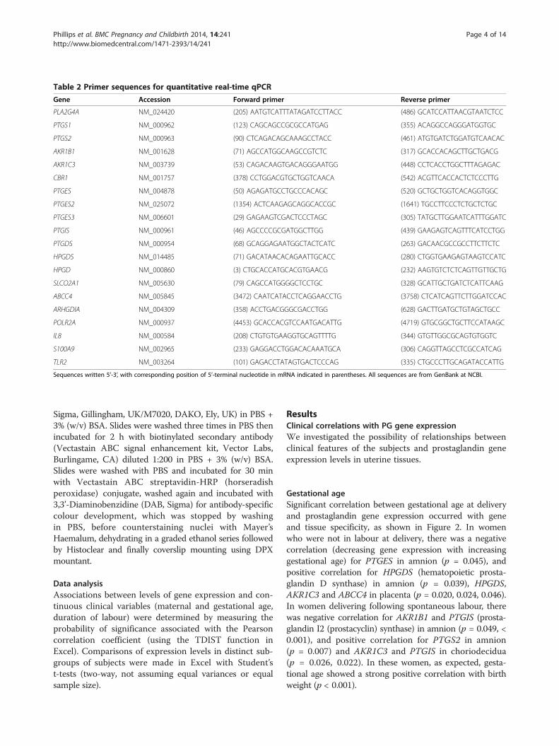
Table 2 Primer sequences for quantitative real-time qPCR
Gene Accession Forward primer Reverse primer
PLA2G4A NM_024420 (205) AATGTCATTTATAGATCCTTACC (486) GCATCCATTAACGTAATCTCC
PTGS1 NM_000962 (123) CAGCAGCCGCGCCATGAG (355) ACAGGCCAGGGATGGTGC
PTGS2 NM_000963 (90) CTCAGACAGCAAAGCCTACC (461) ATGTGATCTGGATGTCAACAC
AKR1B1 NM_001628 (71) AGCCATGGCAAGCCGTCTC (317) GCACCACAGCTTGCTGACG
AKR1C3 NM_003739 (53) CAGACAAGTGACAGGGAATGG (448) CCTCACCTGGCTTTAGAGAC
CBR1 NM_001757 (378) CCTGGACGTGCTGGTCAACA (542) ACGTTCACCACTCTCCCTTG
PTGES NM_004878 (50) AGAGATGCCTGCCCACAGC (520) GCTGCTGGTCACAGGTGGC
PTGES2 NM_025072 (1354) ACTCAAGAGCAGGCACCGC (1641) TGCCTTCCCTCTGCTCTGC
PTGES3 NM_006601 (29) GAGAAGTCGACTCCCTAGC (305) TATGCTTGGAATCATTTGGATC
PTGIS NM_000961 (46) AGCCCCGCGATGGCTTGG (439) GAAGAGTCAGTTTCATCCTGG
PTGDS NM_000954 (68) GCAGGAGAATGGCTACTCATC (263) GACAACGCCGCCTTCTTCTC
HPGDS NM_014485 (71) GACATAACACAGAATTGCACC (280) CTGGTGAAGAGTAAGTCCATC
HPGD NM_000860 (3) CTGCACCATGCACGTGAACG (232) AAGTGTCTCTCAGTTGTTGCTG
SLCO2A1 NM_005630 (79) CAGCCATGGGGCTCCTGC (328) GCATTGCTGATCTCATTCAAG
ABCC4 NM_005845 (3472) CAATCATACCTCAGGAACCTG (3758) CTCATCAGTTCTTGGATCCAC
ARHGDIA NM_004309 (358) ACCTGACGGGCGACCTGG (628) GACTTGATGCTGTAGCTGCC
POLR2A NM_000937 (4453) GCACCACGTCCAATGACATTG (4719) GTGCGGCTGCTTCCATAAGC
IL8 NM_000584 (208) CTGTGTGAAGGTGCAGTTTTG (344) GTGTTGGCGCAGTGTGGTC
S100A9 NM_002965 (233) GAGGACCTGGACACAAATGCA (306) CAGGTTAGCCTCGCCATCAG
TLR2 NM_003264 (101) GAGACCTATAGTGACTCCCAG (335) CTGCCCTTGCAGATACCATTG
Sequences written 5’-3’, with corresponding position of 5’-terminal nucleotide in mRNA indicated in parentheses. All sequences are from GenBank at NCBI.
Phillips et al. BMC Pregnancy and Childbirth 2014, 14:241 Page 4 of 14http://www.biomedcentral.com/1471-2393/14/241
Sigma, Gillingham, UK/M7020, DAKO, Ely, UK) in PBS +3% (w/v) BSA. Slides were washed three times in PBS thenincubated for 2 h with biotinylated secondary antibody(Vectastain ABC signal enhancement kit, Vector Labs,Burlingame, CA) diluted 1:200 in PBS + 3% (w/v) BSA.Slides were washed with PBS and incubated for 30 minwith Vectastain ABC streptavidin-HRP (horseradishperoxidase) conjugate, washed again and incubated with3,3’-Diaminobenzidine (DAB, Sigma) for antibody-specificcolour development, which was stopped by washingin PBS, before counterstaining nuclei with Mayer’sHaemalum, dehydrating in a graded ethanol series followedby Histoclear and finally coverslip mounting using DPXmountant.
Data analysisAssociations between levels of gene expression and con-tinuous clinical variables (maternal and gestational age,duration of labour) were determined by measuring theprobability of significance associated with the Pearsoncorrelation coefficient (using the TDIST function inExcel). Comparisons of expression levels in distinct sub-groups of subjects were made in Excel with Student’st-tests (two-way, not assuming equal variances or equalsample size).
ResultsClinical correlations with PG gene expressionWe investigated the possibility of relationships betweenclinical features of the subjects and prostaglandin geneexpression levels in uterine tissues.
Gestational ageSignificant correlation between gestational age at deliveryand prostaglandin gene expression occurred with geneand tissue specificity, as shown in Figure 2. In womenwho were not in labour at delivery, there was a negativecorrelation (decreasing gene expression with increasinggestational age) for PTGES in amnion (p = 0.045), andpositive correlation for HPGDS (hematopoietic prosta-glandin D synthase) in amnion (p = 0.039), HPGDS,AKR1C3 and ABCC4 in placenta (p = 0.020, 0.024, 0.046).In women delivering following spontaneous labour, therewas negative correlation for AKR1B1 and PTGIS (prosta-glandin I2 (prostacyclin) synthase) in amnion (p = 0.049, <0.001), and positive correlation for PTGS2 in amnion(p = 0.007) and AKR1C3 and PTGIS in choriodecidua(p = 0.026, 0.022). In these women, as expected, gesta-tional age showed a strong positive correlation with birthweight (p < 0.001).
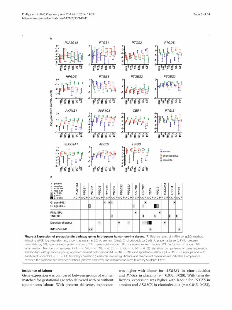
Figure 2 Expression of prostaglandin pathway genes in pregnant human uterine tissues. (A) Relative levels of mRNA by ΔΔCt methodfollowing qPCR, log10-transformed, shown as mean ± SD. A, amnion (blue); C, choriodecidua (red); P, placenta (green). PNIL, pretermnot-in-labour; SPL, spontaneous preterm labour; TNIL, term not-in-labour; STL, spontaneous term labour; IOL, induction of labour; INF,inflammation. Numbers of samples: PNIL = 4; SPL = 4; TNIL = 6; STL = 5; IOL = 5; INF = 4. (B) Statistical comparisons of gene expression.Relationships with gestational age (g. age) in combined not-in-labour (NIL = PNIL + TNIL) and spontaneous labour (SL = SPL + STL) groups, and withduration of labour (SPL + STL + IOL) tested by correlation (Pearson’s); level of significance and direction of correlation are indicated. Comparisonsbetween the presence and absence of labour (preterm and term) and inflammation were tested by Student’s t-tests.
Phillips et al. BMC Pregnancy and Childbirth 2014, 14:241 Page 5 of 14http://www.biomedcentral.com/1471-2393/14/241
Incidence of labourGene expression was compared between groups of womenmatched for gestational age who delivered with or withoutspontaneous labour. With preterm deliveries, expression
was higher with labour for AKR1B1 in choriodeciduaand PTGIS in placenta (p = 0.032, 0.028). With term de-liveries, expression was higher with labour for PTGES inamnion and AKR1C3 in choriodecidua (p = 0.045, 0.033),

Phillips et al. BMC Pregnancy and Childbirth 2014, 14:241 Page 6 of 14http://www.biomedcentral.com/1471-2393/14/241
while levels of PTGIS, ABCC4 and HPGD in amnionwere higher in deliveries without labour (p = 0.043,0.049, 0.038).
Duration of labourDuration of labour in spontaneous and induced labourdeliveries ranged from 33 minutes to 17 hours. Pearsoncorrelation coefficients were calculated to determine theassociation between duration of labour and gene expres-sion. Negative correlation, indicating decreasing expres-sion with increasing duration, was seen with expressionof CBR1 in amnion (p = 0.006), PTGDS (prostaglandinD2 synthase 21 kDa (brain)), PTGES3 (prostaglandin Esynthase 3 (cytosolic)), AKR1C3 and CBR1 in choriodeci-dua (p = 0.049, 0.011, 0.013, <0.001) and AKR1C3 in pla-centa (p = 0.031). Positive correlation was seen for PTGES2(prostaglandin E synthase 2) in amnion (p = 0.022) andSLCO2A1 in choriodecidua (p = 0.010).
Presence of inflammationPlacenta and gestational membranes were collected fromwomen with uterine inflammation, and PG gene expres-sion in this group was compared by t-test with expres-sion in a subgroup of women with no inflammation thatwas matched for gestational age and mode of delivery(Figure 2). Effects of inflammation were limited to up-regulation of PTGS2 in amnion and choriodecidua (p =0.022, 0.038), and downregulation of CBR1 and HPGDin choriodecidua (p = 0.018, 0.011).Women were assigned to the inflammation group on
the basis of established histological criteria [4], and we
Figure 3 Expression of inflammatory genes in pregnant human uterinqPCR, log10-transformed, shown as mean ± SD. PNIL, preterm not-in-labouspontaneous term labour; IOL, induction of labour; INF, inflammation. Num(B) Statistical comparisons of gene expression. No significant relationshipslabour groups, between preterm and term not-in-labour or with duration oexpression in the presence and absence of labour at term and of inflammaof differential comparison are indicated. A, amnion; C, choriodecidua; P, pla
further characterised the inflammatory status of all tissuesamples by measurement of the expression of three genesknown to be involved in inflammatory responses: IL8,S100A8 and TLR2 (Figure 3). All three genes were signifi-cantly upregulated in both amnion (p = 0.021, < 0.001,0.012) and choriodecidua (p = 0.002, <0.001, 0.002) fromwomen assigned to the inflammation (INF) group. In pla-centa, the only change was an increase in S100A8 (p =0.037) with inflammation. Both S100A8 and TLR2 wereexpressed at significantly higher levels in choriodeciduafrom women in the STL compared to the TNIL group(p = 0.014, 0.010) confirming a degree of inflammatoryactivity in term labour. Levels of both genes also ap-peared to be higher in SPL rather than PNIL choriodeci-dua, but these differences were of borderline significance(p = 0.061, 0.057).
Immunolocalisation of PG pathway proteins in placentaLow magnification images of H&E-stained placental sec-tions in Figure 4A show (i) the fetal trophoblastic villiand intervillous space, which make up the great majorityof the placenta, and (ii) the basal plate, which lies adja-cent to the uterine wall. Figure 4B-I show placentalimmunolocalisation of eight of the PG pathway proteins,while Figure 4J shows the localisation of vimentin in vil-lous fibroblasts, vascular cells, macrophages and decid-ual cells, but not trophoblasts.In the chorionic plate (the surface of the placenta adja-
cent to the amniotic cavity), the amnion epithelium showedstaining for PTGS2 and PTGES (not shown). Extravillouscytotrophoblasts, which form an incomplete layer at the
e tissues. (A) Relative levels of mRNA by ΔΔCt method followingr; SPL, spontaneous preterm labour; TNIL, term not-in-labour; STL,bers of samples: PNIL = 4; SPL = 4; TNIL = 6; STL = 5; IOL = 5; INF = 4.were observed with gestational age in not-in-labour or spontaneousf labour, so these comparisons are not shown. Comparisons of genetion were tested by Student’s t-tests. Level of significance and directioncenta.

Figure 4 Immunohistochemical localisation of PG pathway proteins in the placenta. (A) H&E-stained control indicating structure of (i)placental villi, interspersed with maternal blood (MB), (ii) basal plate, containing extravillous trophoblasts (EVT) and decidual cells (DC).(B-K) Higher magnification images of (i) placental villi, indicating syncytiotrophoblasts (ST), vascular cells (VC) and villous macrophages(VM), (ii) basal plate. (K) Negative control without addition of primary antibody. Scale bar = 50 μm.
Phillips et al. BMC Pregnancy and Childbirth 2014, 14:241 Page 7 of 14http://www.biomedcentral.com/1471-2393/14/241
inner border of the chorionic plate, showed staining forHPGD, PTGES, SLCO2A1, AKR1B1, AKR1C3 and CBR1.In the placental villi (Figure 4A-K(i)), syncytiotrophoblasts
displayed staining for AKR1B1, HPGD PTGS2, SLCO2A1,CBR1, AKR1C3, and PTGES. Villous fibroblasts showed
PTGS2 and SLCO2A1 staining and heterogeneous AKR1B1staining. Villous macrophages were positive for PTGS1and PTGES.The basal plate of the placenta (Figure 4A-K(ii)) consists of
maternal decidual cells and fetal extravillous cytotrophoblasts,
Phillips et al. BMC Pregnancy and Childbirth 2014, 14:241 Page 8 of 14http://www.biomedcentral.com/1471-2393/14/241
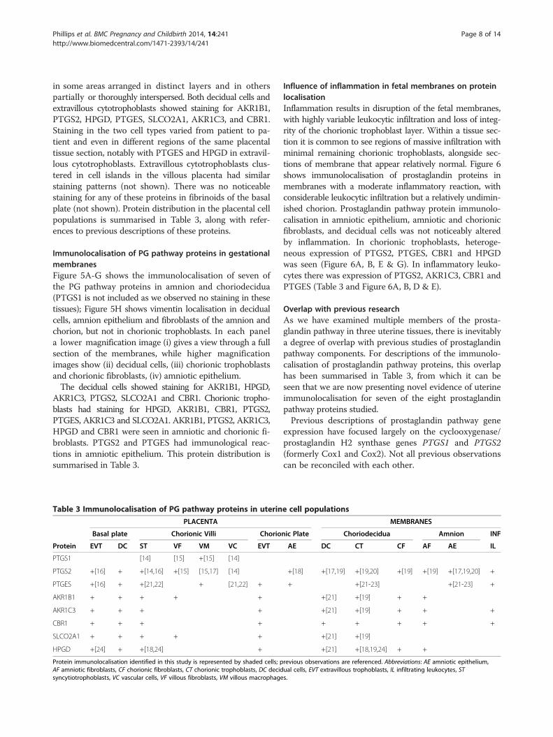
in some areas arranged in distinct layers and in otherspartially or thoroughly interspersed. Both decidual cells andextravillous cytotrophoblasts showed staining for AKR1B1,PTGS2, HPGD, PTGES, SLCO2A1, AKR1C3, and CBR1.Staining in the two cell types varied from patient to pa-tient and even in different regions of the same placentaltissue section, notably with PTGES and HPGD in extravil-lous cytotrophoblasts. Extravillous cytotrophoblasts clus-tered in cell islands in the villous placenta had similarstaining patterns (not shown). There was no noticeablestaining for any of these proteins in fibrinoids of the basalplate (not shown). Protein distribution in the placental cellpopulations is summarised in Table 3, along with refer-ences to previous descriptions of these proteins.
Immunolocalisation of PG pathway proteins in gestationalmembranesFigure 5A-G shows the immunolocalisation of seven ofthe PG pathway proteins in amnion and choriodecidua(PTGS1 is not included as we observed no staining in thesetissues); Figure 5H shows vimentin localisation in decidualcells, amnion epithelium and fibroblasts of the amnion andchorion, but not in chorionic trophoblasts. In each panela lower magnification image (i) gives a view through a fullsection of the membranes, while higher magnificationimages show (ii) decidual cells, (iii) chorionic trophoblastsand chorionic fibroblasts, (iv) amniotic epithelium.The decidual cells showed staining for AKR1B1, HPGD,
AKR1C3, PTGS2, SLCO2A1 and CBR1. Chorionic tropho-blasts had staining for HPGD, AKR1B1, CBR1, PTGS2,PTGES, AKR1C3 and SLCO2A1. AKR1B1, PTGS2, AKR1C3,HPGD and CBR1 were seen in amniotic and chorionic fi-broblasts. PTGS2 and PTGES had immunological reac-tions in amniotic epithelium. This protein distribution issummarised in Table 3.
Table 3 Immunolocalisation of PG pathway proteins in uterin
Protein
PLACENTA
Basal plate Chorionic Villi Chorio
EVT DC ST VF VM VC EVT
PTGS1 [14] [15] +[15] [14]
PTGS2 +[16] + +[14,16] +[15] [15,17] [14]
PTGES +[16] + +[21,22] + [21,22] +
AKR1B1 + + + + +
AKR1C3 + + + +
CBR1 + + + +
SLCO2A1 + + + + +
HPGD +[24] + +[18,24] +
Protein immunolocalisation identified in this study is represented by shaded cells; pAF amniotic fibroblasts, CF chorionic fibroblasts, CT chorionic trophoblasts, DC decidsyncytiotrophoblasts, VC vascular cells, VF villous fibroblasts, VM villous macrophage
Influence of inflammation in fetal membranes on proteinlocalisationInflammation results in disruption of the fetal membranes,with highly variable leukocytic infiltration and loss of integ-rity of the chorionic trophoblast layer. Within a tissue sec-tion it is common to see regions of massive infiltration withminimal remaining chorionic trophoblasts, alongside sec-tions of membrane that appear relatively normal. Figure 6shows immunolocalisation of prostaglandin proteins inmembranes with a moderate inflammatory reaction, withconsiderable leukocytic infiltration but a relatively undimin-ished chorion. Prostaglandin pathway protein immunolo-calisation in amniotic epithelium, amniotic and chorionicfibroblasts, and decidual cells was not noticeably alteredby inflammation. In chorionic trophoblasts, heteroge-neous expression of PTGS2, PTGES, CBR1 and HPGDwas seen (Figure 6A, B, E & G). In inflammatory leuko-cytes there was expression of PTGS2, AKR1C3, CBR1 andPTGES (Table 3 and Figure 6A, B, D & E).
Overlap with previous researchAs we have examined multiple members of the prosta-glandin pathway in three uterine tissues, there is inevitablya degree of overlap with previous studies of prostaglandinpathway components. For descriptions of the immunolo-calisation of prostaglandin pathway proteins, this overlaphas been summarised in Table 3, from which it can beseen that we are now presenting novel evidence of uterineimmunolocalisation for seven of the eight prostaglandinpathway proteins studied.Previous descriptions of prostaglandin pathway gene
expression have focused largely on the cyclooxygenase/prostaglandin H2 synthase genes PTGS1 and PTGS2(formerly Cox1 and Cox2). Not all previous observationscan be reconciled with each other.
e cell populations
MEMBRANES
nic Plate Choriodecidua Amnion INF
AE DC CT CF AF AE IL
+[18] +[17,19] +[19,20] +[19] +[19] +[17,19,20] +
+ +[21-23] +[21-23] +
+[21] +[19] + +
+[21] +[19] + + +
+ + + + +
+[21] +[19]
+[21] +[18,19,24] + +
revious observations are referenced. Abbreviations: AE amniotic epithelium,ual cells, EVT extravillous trophoblasts, IL infiltrating leukocytes, STs.
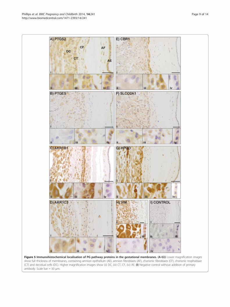
Figure 5 Immunohistochemical localisation of PG pathway proteins in the gestational membranes. (A-I(i)) Lower magnification imagesshow full thickness of membranes, containing amnion epithelium (AE), amnion fibroblasts (AF), chorionic fibroblasts (CF), chorionic trophoblast(CT) and decidual cells (DC). Higher magnification images show (ii) DC, (iii) CT, CF, (iv) AE. (I) Negative control without addition of primaryantibody. Scale bar = 50 μm.
Phillips et al. BMC Pregnancy and Childbirth 2014, 14:241 Page 9 of 14http://www.biomedcentral.com/1471-2393/14/241
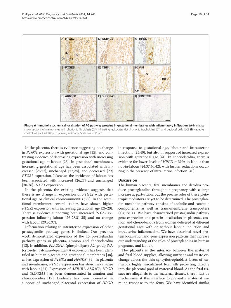
Figure 6 Immunohistochemical localisation of PG pathway proteins in gestational membranes with inflammatory infiltration. (A-I) Imagesshow sections of membranes with chorionic fibroblasts (CF), infiltrating leukocytes (IL), chorionic trophoblast (CT) and decidual cells (DC). (I) Negativecontrol without addition of primary antibody. Scale bar = 50 μm.
Phillips et al. BMC Pregnancy and Childbirth 2014, 14:241 Page 10 of 14http://www.biomedcentral.com/1471-2393/14/241
In the placenta, there is evidence suggesting no changein PTGS1 expression with gestational age [15], and con-trasting evidence of decreasing expression with increasinggestational age at labour [25]. In gestational membranes,increasing gestational age has been associated with in-creased [26,27], unchanged [27,28], and decreased [29]PTGS1 expression. Likewise, the incidence of labour hasbeen associated with increased [26,27] and unchanged[30-36] PTGS1 expression.In the placenta, the existing evidence suggests that
there is no change in expression of PTGS2 with gesta-tional age or clinical chorioamnionitis [25]. In the gesta-tional membranes, several studies have shown higherPTGS2 expression with increasing gestational age [26-29].There is evidence supporting both increased PTGS2 ex-pression following labour [26-28,31-35] and no changewith labour [20,36,37].Information relating to intrauterine expression of other
prostaglandin pathway genes is limited. Our previouswork demonstrated expression of the 15 prostaglandinpathway genes in placenta, amnion and choriodecidua[13]. In addition, PLA2G4A (phospholipase A2, group IVA(cytosolic, calcium-dependent)) expression has been iden-tified in human placenta and gestational membranes [38],as has expression of PTGDS and HPGDS [39]. In placentaand membranes, PTGES expression has shown no changewith labour [21]. Expression of AKR1B1, AKR1C3, HPGDand SLCO2A1 has been demonstrated in amnion andchoriodecidua [19]. Evidence has been presented insupport of unchanged placental expression of HPGD
in response to gestational age, labour and intrauterineinfection [25,40], but also in support of increased expres-sion with gestational age [41]. In choriodecidua, there isevidence for lower levels of HPGD mRNA in labour thannot-in-labour [24,37,40,42], with further reductions occur-ring in the presence of intrauterine infection [40].
DiscussionThe human placenta, fetal membranes and decidua pro-duce prostaglandins throughout pregnancy with a largeincrease at parturition, but the precise roles of these pleio-tropic mediators are yet to be determined. The prostaglan-din metabolic pathway consists of anabolic and cataboliccomponents, as well as trans-membrane transporters(Figure 1). We have characterised prostaglandin pathwaygene expression and protein localisation in placenta, am-nion and choriodecidua from women delivered at differentgestational ages with or without labour, induction andintrauterine inflammation. We have described novel pro-tein localisation and gene expression patterns that increaseour understanding of the roles of prostaglandins in humanpregnancy and labour.The placenta is the interface between the maternal
and fetal blood supplies, allowing nutrient and waste ex-change across the thin syncytiotrophoblast layers of nu-merous highly vascularised fetal villi projecting directlyinto the placental pool of maternal blood. As the fetal tis-sues are allogeneic to the maternal tissues, there must bemechanisms at this interface to prevent a maternal im-mune response to the fetus. We have identified similar
Phillips et al. BMC Pregnancy and Childbirth 2014, 14:241 Page 11 of 14http://www.biomedcentral.com/1471-2393/14/241
patterns of protein localisation in decidual cells and extra-villous trophoblasts of the placental bed and syncytio-trophoblasts of placental villi. These cells all expressAKR1B1, PTGS2, HPGD, PTGES, SLCO2A1, AKR1C3and CBR1, thus having the capacity for PGF2 and PGE2synthesis and PG uptake and degradation. Gene expres-sion patterns described here and in our previous work[13] support these observations and we now describe thepresence of PGD2, PGE2 and PGI2 synthases in the pla-centa. Comparisons of placental gene expression in differ-ent groups of women identified increasing HPGDS,AKR1C3 and ABCC4 with gestational age in the absenceof labour, and higher PTGIS in labour than not-in-labourpreterm.The fetal membranes consist of the fetal amnion and
chorion and the attached maternal decidua, which to-gether comprise a major structural element of the uter-ine tissues and have endocrine functions in pregnancyand parturition not yet fully elucidated [43]. As in theplacenta, the trophoblast and decidua are the interfacebetween maternal and fetal tissues. Immunolocalisationof prostaglandin pathway proteins in chorionic tropho-blast cells and adjacent decidua are similar to each other,and to some extent resemble placental patterns, withHPGD, AKR1B1, AKR1C3, CBR1, PTGS2 and SLCO2A1expressed in choriodecidua. Unlike in placental cells,variable protein expression is evident in choriodecidua,with the immunolocalisation of PTGES in chorionictrophoblast but not decidua, and higher chorionic levelsof CBR1, and decidual levels of AKR1C3. Prostaglandingene expression changes in choriodecidua include increasedAKR1C3 and PTGIS with gestational age and labour, withhigher AKR1B1 in labour preterm, and higher AKR1C3 inlabour at term compared with not-in-labour.In the region between the chorionic trophoblast and
amniotic epithelium, fibroblasts express PTGS2, PGF2synthases and HPGD, while the amniotic epithelium it-self, which is known to be a source of PGE2 synthesis[43,44], expresses PTGS2 and PTGES proteins, and alsohigh levels of PTGS2, PTGES and PTGES3 mRNA. BothPTGS2 and PTGES are differentially expressed in am-nion, with PTGS2 increasing with gestational age in thepresence of labour, and PTGES decreasing as gestationalage rises in the absence of labour, and displaying higherexpression in labour than not-in-labour at term. Despiteprevious observations of increased levels of prostaglan-dins and their metabolites in amniotic fluid with labour[39,45,46], we did not observe a significant alteration inPTGS2 in amnion and choriodecidua with either pre-term or term labour.Taken together, these expression patterns suggest dis-
tinct roles for prostaglandin metabolism in tissues at thematernal:fetal interface and in tissues within the fetal com-partment. At the interface there is the ability to synthesise
PGD2, PGE2, and PGF2, but these prostaglandins might belimited to autocrine or paracrine function by the co-expressed degradative complex of SLCO2A1 and HPGD,which is considered to be a barrier between the maternaland fetal prostaglandin systems [24,47,48]. These prosta-glandins could participate in the immunomodulation ofmaternal leukocytes present in decidua, placental bed andmaternal blood, to prevent rejection of the fetal tissues.PGE2 synthesised in the amnion and released into the
amniotic fluid could influence fetal physiology, for ex-ample by inhibiting fetal breathing [49]. The reductionin amniotic PTGES expression and amniotic fluid PGE2[8] with increasing gestational age might then allow lungmovements to develop in sync with fetal maturation. Itshould, of course, be noted that PTGES is the only one ofthe three PGE2 synthases that displays this dependence ongestational age for amniotic expression. PTGES is also theonly PGE2 synthase that shows higher expression in theamnion than in the other tissues. Furthermore, as amnioticexpression of both SLCO2A1 and HPGD are some ordersof magnitude lower than in placenta and choriodecidua, itsuggests that there is sufficient degradation of the PGE2that is released into the amniotic cavity in fetal tissues,such as the lung, to prevent accumulation in the amnioticfluid.In addition to gestational age and the incidence of labour,
we investigated the correlation of prostaglandin gene ex-pression with other characteristics. Duration of labour wasassociated with different expression changes in each of thetissues, with both upregulation and downregulation ofprostaglandin genes. The only gene to be affected by bothduration of labour and the presence or absence of labourwas AKR1C3 in the choriodecidua. This suggests thatregulation of some genes is associated with the process oflabour, regardless of its duration, whereas others are af-fected by exposure to the prolonged stressful effects oflabour. As we could not follow gene expression through-out labour, we cannot rule out that the differential regula-tion of these genes is a cause rather than an effect of theduration of labour. In a rarely quoted study involving >200deliveries, Keski-Nisula et al. demonstrated that decidualinflammation is significantly more common in women inadvanced labour compared to early labour, and concludedthat the inflammatory changes are more likely to be a con-sequence of labour rather than its cause [50]. Given thetraumatic effects of labour on both mother and child, elu-cidating the true nature of this relationship could providevaluable information.We were very interested in evaluating the presence or
absence of intrauterine inflammation. There has been agreat deal of effort expended on establishing the causativerelationship between intrauterine infection, inflammationand labour, particularly preterm labour. The premature ac-tivation of inflammatory pathways by intrauterine infection

Phillips et al. BMC Pregnancy and Childbirth 2014, 14:241 Page 12 of 14http://www.biomedcentral.com/1471-2393/14/241
has been proposed as a major contributor to pretermlabour [51,52]. Amniotic fluid metabolomic profiles differin women delivering preterm in the presence and absenceof intra-amniotic infection and inflammation [53].We compared gene expression in a group of women with
histological signs of inflammation with expression in agroup of women matched for gestational age at delivery,and without substantial differences in other recorded vari-ables, but with no signs of inflammation. To confirm thehistological observations of inflammation, we measured theexpression of three known inflammatory genes, finding sig-nificant upregulation of all three in amnion and choriodeci-dua samples from the INF group. Among the prostaglandinpathway genes, PTGS2 was upregulated with inflammationin both amnion and choriodecidua, whereas CBR1 andHPGD were downregulated in choriodecidua. In the pla-centa only one of the inflammatory control genes wasupregulated, and none of the prostaglandin genes was af-fected by inflammation, but as the intrauterine inflamma-tion was largely limited to chorioamnionitis/deciduitis, wecannot rule out that placentas affected by villitis, whichshow altered leukotriene synthesis [5], would also showprostaglandin pathway expression changes. The uniqueexpression patterns of prostaglandin pathway and inflam-matory control genes that we have observed suggestthat in cases of uncomplicated spontaneous preterm labour,there is no underlying inflammatory expression profile.There must be an alternative mechanism for uterine ac-tivation in SPL in the absence of inflammation. In thisregard it is worth mentioning that oxytocin, a strong utero-tonic agent, stimulates PTGS2 expression in human myo-metrial cells through previously undescribed pathways suchas NFAT (nuclear factor of activated T cells) [54].Although these results support the idea that labour
usually occurs in the absence of inflammation, there isevidence that the presence of inflammation can be atrigger for labour, with [8,12] or without [10,12] signs ofinfection. This delivery mechanism can provide a re-sponse to intrauterine infections that can threaten thelives of mother and fetus. Tocolysis is not always an ap-propriate treatment, even for very early preterm labour,as the uterus can become a hostile environment. How-ever, when infections can be overcome, and in instancesof premature labour without infection and/or inflamma-tion, there are great potential benefits to effective tocoly-sis. Our observation of different prostaglandin pathwayexpression profiles in preterm labour and inflammationcould have implications for the choice of tocolytics usedin different situations. Although elevation of PTGS2 inplacenta and membranes affected by inflammation couldbe countered by selective PTGS2 inhibitors, PTGS2 isnot upregulated with preterm labour in these tissues, al-though it is in myometrium [13]. Better understandingof the roles of PTGS2 in the different uterine tissues in
preterm and term labour with and without inflammationcould clarify when PTGS2 inhibitors are most likely tobe effective.We observed an increase in PTGS2 expression in the
amnion with term versus preterm labour that has alsobeen seen previously [31,32,55]. An increase in amnioticfluid IL1 (interleukin 1) with labour at term has been de-scribed [56], and could be responsible for the PTGS2 up-regulation, although as with other observations in thisfield, there is contradictory evidence suggesting lowerIL1 at term [8]. Increased PTGS2 expression induced bycytokines, would explain the upregulation of PTGS2 inthe inflamed membranes of chorioamnionitis.Limitations of this study include the numbers of sam-
ples in each of the groups; there is no enough data tocorrelate with previous preterm deliveries, hypertension,BMI, asthma, smoking and socioeconomic status of thewomen. Immunohistochemistry was used as a qualitativeassay for only a subset of the prostaglandin pathway pro-teins, so that no quantitative data on protein levels wereobtained. Another potential limitation is the lack of stat-istical correction for multiple comparisons, which couldlead to type I errors of false positive identification ofstatistical significance. However, in order to avoid type IIerrors of rejection of true significance, we have pre-sented the results of our statistical tests uncorrected,with the caveat that further studies are required beforethe changes that we have identified can be unequivocallyconfirmed.
ConclusionsThe principal aim of our research is to identify the causesof preterm labour, to enable reliable prediction of its oc-currence and to facilitate its prevention by identifying bio-chemical pathways suitable for intervention. In light ofconsiderable evidence linking prostaglandin function withuterine activation, we have undertaken a detailed analysisof prostaglandin pathway gene expression in human pla-centa, amnion and choriodecidua, identifying changes inassociation with gestational age, labour, inflammation andduration of labour, although there were no significant dif-ferences between spontaneous and induced labour at term.Inflammation provokes specific changes, unrelated to thepresence of labour. The use of tocolytics should take intoaccount these differences, in particular between uncom-plicated spontaneous preterm labour and chorioam-nionitis. Greater understanding of the different PG pathwaychanges in idiopathic and inflammation-associated pre-term labour should facilitate the targeting of appropriatepharmacological intervention to these very differentgroups of women.
Competing interestsThe authors declare that they have no competing interest that could beperceived as prejudicing the impartiality of the research reported. MAF has a
Phillips et al. BMC Pregnancy and Childbirth 2014, 14:241 Page 13 of 14http://www.biomedcentral.com/1471-2393/14/241
patent for methods for the regulation of the prostaglandin F synthase (PGFS)activity of AKR1B1 and uses thereof.
Authors’ contributionsRJP: experimentation, analysis and manuscript preparation; MAF providedreagents helped with the preparation of manuscript; ALB: design of studyand preparation of manuscript.
AcknowledgementsWe are grateful to research midwives Anne Duffner and Alison Kirby forobtaining consent from women at St Michael’s Hospital and organising thecollection of samples. Dr Hana Al-Zamil also contributed to sample collectionand processing.
FundingThis work was supported by Wellbeing of Women [grant RG825].
Author details1Henry Wellcome Laboratories for Integrative Neuroscience andEndocrinology, School of Clinical Sciences, University of Bristol, DorothyHodgkin Building, Bristol BS1 3NY, UK. 2Axe Reproduction, santé Périnatale etpédiatrie, Centre Hospitalier Universitaire de Québec (CHUL), Université Laval,2705 boulevard Laurier, Ste-Foy, QC G1V 4G2, Canada. 3St Michael’s Hospital,Southwell Street, Bristol BS2 8EG, UK.
Received: 29 November 2013 Accepted: 15 July 2014Published: 22 July 2014
References1. Challis JR, Sloboda DM, Alfaidy N, Lye SJ, Gibb W, Patel FA, Whittle WL,
Newnham JP: Prostaglandins and mechanisms of preterm birth.Reproduction 2002, 124:1–17.
2. Fortier MA, Krishnaswamy K, Danyod G, Boucher-Kovalik S, Chapdelaine JA:A postgenomic integrated view of prostaglandins in reproduction:implications for other body systems. J Phys Pharm 2008, 59(Suppl 1):65–89.
3. Christiaens I, Zaragoza DB, Guilbert L, Robertson SA, Mitchell BF, Olson DM:Inflammatory processes in preterm and term parturition. J ReprodImmunol 2008, 79:50–57.
4. López Bernal A, Hansell DJ, Khong TY, Keeling JW, Turnbull AC:Prostaglandin E production by the fetal membranes in unexplainedpreterm labour and preterm labour associated with chorioamnionitis.Br J Obstet Gynaecol 1989, 96:1133–1139.
5. López Bernal A, Hansell DJ, Khong TY, Keeling JW, Turnbull AC: Placentalleukotriene B4 release in early pregnancy and in term and pretermlabour. Early Hum Dev 1990, 23:93–99.
6. López Bernal A, Hansell DJ, Cañete Soler R, Keeling JW, Turnbull AC:Prostaglandins, chorioamnionitis and preterm labour. Br J ObstetGynaecol 1987, 94:1156–1158.
7. Mueller-Heubach E, Rubinstein DN, Schwarz SS: Histologic chorioamnionitisand preterm delivery in different patient populations. Obstet Gynecol1990, 75:622–626.
8. Hillier SL, Witkin SS, Krohn MA, Watts DH, Kiviat NB, Eschenbach DA: Therelationship of amniotic fluid cytokines and preterm delivery, amnioticfluid infection, histologic chorioamnionitis, and chorioamnion infection.Obstet Gynecol 1993, 81:941–948.
9. Opsjøn SL, Wathen NC, Tingulstad S, Wiedswang G, Sundan A, Waage A,Austgulen R: Tumor necrosis factor, interleukin-1 and interleukin-6 innormal human pregnancy. Am J Obstet Gynecol 1993, 169:397–404.
10. Steinborn A, Günes H, Röddiger S, Halberstadt E: Elevated placentalcytokine release, a process associated with preterm labor in the absenceof intrauterine infection. Obstet Gynecol 1996, 88:534–539.
11. Leitich H, Bodner-Adler B, Brunbauer M, Kaider A, Egarter C, Husslein P:Bacterial vaginosis as a risk factor for preterm delivery: a meta-analysis.Am J Obstet Gynecol 2003, 189:139–147.
12. Shim SS, Romero R, Hong JS, Park CW, Jun JK, Kim BI, Yoon BH:Clinical significance of intra-amniotic inflammation in patients withpreterm premature rupture of membranes. Am J Obstet Gynecol 2004,191:1339–1345.
13. Phillips RJ, Al-Zamil H, Hunt LP, Fortier MA, López Bernal A: Genes forprostaglandin synthesis, transport and inactivation are differentiallyexpressed in human uterine tissues, and the prostaglandin F
synthase AKR1B1 is induced in myometrial cells by inflammatorycytokines. Mol Hum Reprod 2011, 17:1–13.
14. Xu Y, Knipp GT, Cook TJ: Expression of cyclooxygenase isoforms indeveloping rat placenta, human term placenta, and BeWo humantrophoblast model. Mol Pharm 2005, 2:481–490.
15. Wetzka B, Nüsing R, Charnock-Jones DS, Schäfer W, Zahradnik HP, Smith SK:Cyclooxygenase-1 and −2 in human placenta and placental bed afternormal and pre-eclamptic pregnancies. Hum Reprod 1997, 12:2313–2320.
16. Meadows JW, Pitzer B, Brockman DE, Myatt L: Differential localization ofprostaglandin E synthase isoforms in human placental cell types.Placenta 2004, 25:259–265.
17. Dunn-Albanese LR, Ackerman WE 4th, Xie Y, Iams JD, Kniss DA: Reciprocalexpression of peroxisome proliferator-activated receptor-gamma andcyclooxygenase-2 in human term parturition. Am J Obstet Gynecol 2004,190:809–816.
18. Cheung PY, Walton JC, Tai HH, Riley SC, Challis JR: Immunocytochemicaldistribution and localization of 15-hydroxyprostaglandin dehydrogenasein human fetal membranes, decidua and placenta. Am J Obstet Gynecol1990, 163:1445–1449.
19. Breuiller-Fouché M, Leroy MJ, Dubois O, Reinaud P, Chissey A, Qi H,Germain G, Fortier MA, Charpigny G: Differential expression of theenzymatic system controlling synthesis, metabolism, and transport ofPGF2 alpha in human fetal membranes. Biol Reprod 2010, 83:155–162.
20. Gibb W, Sun M: Localization of prostaglandin H synthase type 2 proteinand mRNA in term human fetal membranes and decidua. J Endocrinol1996, 150:497–503.
21. Alfaidy N, Sun M, Challis JR, Gibb W: Expression of membraneprostaglandin E synthase in human placenta and fetal membranes andeffect of labor. Endocrine 2003, 20:219–225.
22. Premyslova M, Li W, Alfaidy N, Bocking AD, Campbell K, Gibb W, Challis JR:Differential expression and regulation of microsomal prostaglandin E(2)synthase in human fetal membranes and placenta with infection and incultured trophoblast cells. J Clin Endocrinol Metab 2003, 88:6040–6047.
23. Meadows JW, Eis AL, Brockman DE, Myatt L: Expression and localization ofprostaglandin E synthase isoforms in human fetal membranes in termand preterm labor. J Clin Endocrinol Metab 2003, 88:433–439.
24. Sangha RK, Walton JC, Ensor CM, Tai HH, Challis JR: Immunohistochemicallocalization, messenger ribonucleic acid abundance, and activity of15-hydroxyprostaglandin dehydrogenase in placenta and fetalmembranes during term and preterm labor. J Clin Endocrinol Metab1994, 78:982–989.
25. Hirsch E, Goldstein M, Filipovich Y, Wang H: Placental expression ofenzymes regulating prostaglandin synthesis and degradation. Am JObstet Gynecol 2005, 192:1836–1842.
26. Mijovic JE, Zakar T, Nairn TK, Olson DM: Prostaglandin endoperoxide Hsynthase (PGHS) activity and PGHS-1 and −2 messenger ribonucleic acidabundance in human chorion throughout gestation and with pretermlabor. J Clin Endocrinol Metab 1998, 83:1358–1367.
27. Mijovic JE, Zakar T, Angelova J, Olson DM: Prostaglandin endoperoxide Hsynthase mRNA expression in the human amnion and decidua duringpregnancy and in the amnion at preterm labour. Mol Hum Reprod 1999,5:182–187.
28. Slater DM, Dennes WJ, Campa JS, Poston L, Bennett PR: Expression ofcyclo-oxygenase types-1 and −2 in human myometrium throughoutpregnancy. Mol Hum Reprod 1999, 5:880–884.
29. Johnson RF, Mitchell CM, Giles WB, Bisits A, Zakar T: Mechanisms regulatingprostaglandin H2 synthase-2 mRNA level in the amnion and chorionduring pregnancy. J Endocrinol 2006, 188:603–610.
30. Freed KA, Aitken MA, Brennecke SP, Rice GE: Prostaglandin G/H synthase-1messenger RNA relative abundance in human amnion, choriodeciduaand placenta before, during and after spontaneous-onset labour at term.Gynecol Obstet Invest 1995, 39:73–78.
31. Hirst JJ, Teixeira FJ, Zakar T, Olson DM: Prostaglandin endoperoxide-Hsynthase-1 and −2 messenger ribonucleic acid levels in human amnionwith spontaneous labor onset. J Clin Endocrinol Metab 1995, 80:517–523.
32. Slater DM, Berger LC, Newton R, Moore GE, Bennett PR: Expression ofcyclooxygenase types 1 and 2 in human fetal membranes at term. Am JObstet Gynecol 1995, 172:77–82.
33. Slater D, Allport V, Bennett P: Changes in the expression of the type-2 butnot the type-1 cyclo-oxygenase enzyme in chorion-decidua with theonset of labour. Br J Obstet Gynaecol 1998, 105:745–748.

Phillips et al. BMC Pregnancy and Childbirth 2014, 14:241 Page 14 of 14http://www.biomedcentral.com/1471-2393/14/241
34. Zakar T, Olson DM, Teixeira FJ, Hirst JJ: Regulation of prostaglandinendoperoxide H2 synthase in term human gestational tissues.Acta Physiol Hung 1996, 84:109–118.
35. Mijovic JE, Zakar T, Nairn TK, Olso DM: Prostaglandin-endoperoxide Hsynthase-2 expression and activity increases with term labor in humanchorion. Am J Physiol 1997, 272:E832–840.
36. Hirst JJ, Mijovic JE, Zakar T, Olson DM: Prostaglandin endoperoxide Hsynthase-1 and −2 mRNA levels and enzyme activity in human deciduaat term labor. J Soc Gynecol Investig 1998, 5:13–20.
37. Makino S, Zaragoza DB, Mitchell BF, Yonemoto H, Olson DM: Decidualactivation: abundance and localization of prostaglandin F2alphareceptor (FP) mRNA and protein and uterine activation proteins inhuman decidua at preterm birth and term birth. Placenta 2007,28:557–565.
38. Freed KA, Moses EK, Brennecke SP, Rice GE: Differential expression of typeII, IV and cytosolic PLA2 messenger RNA in human intrauterine tissues atterm. Mol Hum Reprod 1997, 3:493–499.
39. Helliwell RJ, Keelan JA, Marvin KW, Adams L, Chang MC, Anand A, Sato TA,O’Carroll S, Chaiworapongsa T, Romero RJ, Mitchell MD: Gestationalage-dependent up-regulation of prostaglandin D synthase (PGDS)and production of PGDS-derived antiinflammatory prostaglandins inhuman placenta. J Clin Endocrinol Metab 2006, 91:597–606.
40. van Meir CA, Matthews SG, Keirse MJ, Ramirez MM, Bocking A, Challis JR:15-hydroxyprostaglandin dehydrogenase: implications in preterm laborwith and without ascending infection. J Clin Endocrinal Metab 1997,82:969–976.
41. Schoof E, Girstl M, Frobenius W, Kirschbaum M, Repp R, Knerr I, Rascher W,Dötsch J: Course of placental 11beta-hydroxysteroid dehydrogenase type2 and 15-hydroxyprostaglandin dehydrogenase mRNA expression duringhuman gestation. Eur J Endocrinol 2001, 145:187–192.
42. Pomini F, Patel FA, Mancuso S, Challis JR: Activity and expression of15-hydroxyprostaglandin dehydrogenase in cultured chorionictrophoblast and villous trophoblast cells and in chorionic explants atterm with and without spontaneous labor. Am J Obstet Gynecol 2000,182:221–226.
43. Myatt L, Sun K: Role of fetal membranes in signaling of fetal maturationand parturition. Int J Dev Biol 2010, 54:545–553.
44. López Bernal A, Newman GE, Phizackerley PJR, Turnbull AC: Surfactantstimulates prostaglandin E production in human amnion. Br J ObstetGynaecol 1988, 95:1013–1017.
45. Mitchell MD, Keirse MJ, Brunt JD, Anderson AB, Turnbull AC: Concentrationsof the prostacyclin metabolite, 6-keto-prostaglandin F1 alpha, inamniotic fluid during late pregnancy and labour. Br J Obstet Gynaecol1979, 86:350–353.
46. Lee SE, Romero R, Park IS, Seong HS, Park CW, Yoon BH: Amniotic fluidprostaglandin concentrations increase before the onset of spontaneouslabor at term. J Matern Fetal Neonatal Med 2008, 21:89–94.
47. Keirse MJ, Turnbull AC: Metabolism of prostaglandins within the pregnantuterus. Br J Obstet Gynaecol 1975, 82:887–893.
48. Nomura T, Lu R, Pucci ML, Schuster VL: The two-step model of prostaglandinsignal termination: in vitro reconstitution with the prostaglandintransporter and prostaglandin 15 dehydrogenase. Mol Pharmacol 2004,65:973–978.
49. Sorokin Y, Hallak M, Klein O, Kalderon I, Abramovici H: Effects of inductionof labor with prostaglandin E2 on fetal breathing and bodymovements: controlled, randomized, double-blind study. Obstet Gynecol1992, 80:788–791.
50. Keski-Nisula L, Aalto ML, Katila ML, Kirkinen P: Intrauterine inflammation atterm: a histopathologic study. Hum Pathol 2000, 31:841–846.
51. Goldenberg RL, Hauth JC, Andrews WW: Intrauterine infection andpreterm delivery. N Engl J Med 2000, 342:1500–1507.
52. Romero R, Espinoza J, Kusanovic JP, Gotsch F, Hassan S, Erez O,Chaiworapongsa T, Mazor M: The preterm parturition syndrome. Br JObstet Gynaecol 2006, 113(Suppl 3):17–42.
53. Romero R, Mazaki-Tovi S, Vaisbuch E, Kusanovic JP, Chaiworapongsa T,Gomez R, Nien JK, Yoon BH, Mazor M, Luo J, Banks D, Ryals J, Beecher C:Metabolomics in premature labor: a novel approach to identify patientsat risk for preterm delivery. J Matern Fetal Neonatal Med 2010, 23:1344–1359.
54. Pont JN, McArdle CA, López Bernal A: Oxytocin-stimulated NFATtranscriptional activation in human myometrial cells. Mol Endocrinol2012, 26:1743–1756.
55. Fuentes A, Spaziani EP, O’Brien WF: The expression of cyclooxygenase-2(COX-2) in amnion and decidua following spontaneous labor. Prostaglandins1996, 52:261–267.
56. Romero R, Parvizi ST, Oyarzun E, Mazor M, Wu YK, Avila C, Athanassiadis AP,Mitchell MD: Amniotic fluid interleukin-1 in spontaneous labor at term.J Reprod Med 1990, 35:235–238.
doi:10.1186/1471-2393-14-241Cite this article as: Phillips et al.: Prostaglandin pathway gene expressionin human placenta, amnion and choriodecidua is differentially affected bypreterm and term labour and by uterine inflammation. BMC Pregnancy andChildbirth 2014 14:241.
Submit your next manuscript to BioMed Centraland take full advantage of:
• Convenient online submission
• Thorough peer review
• No space constraints or color figure charges
• Immediate publication on acceptance
• Inclusion in PubMed, CAS, Scopus and Google Scholar
• Research which is freely available for redistribution
Submit your manuscript at www.biomedcentral.com/submit